
MRI compatibility for accurate diagnostic
examinations of ‘former’ pacemaker & ICD technology
Pierpaolo LUPO , Hussam ALI, Guido DE AMBROGGI,
Sara FORESTI, Gianluca EPICOCO and Riccardo CAPPATO*
Arrhythmia & Electrophysiology II Center Humanitas-Gavazzeni Hospital Bergamo, Italy
8th RHYTHM Congress 2015 Marsellie, France, May 28-30
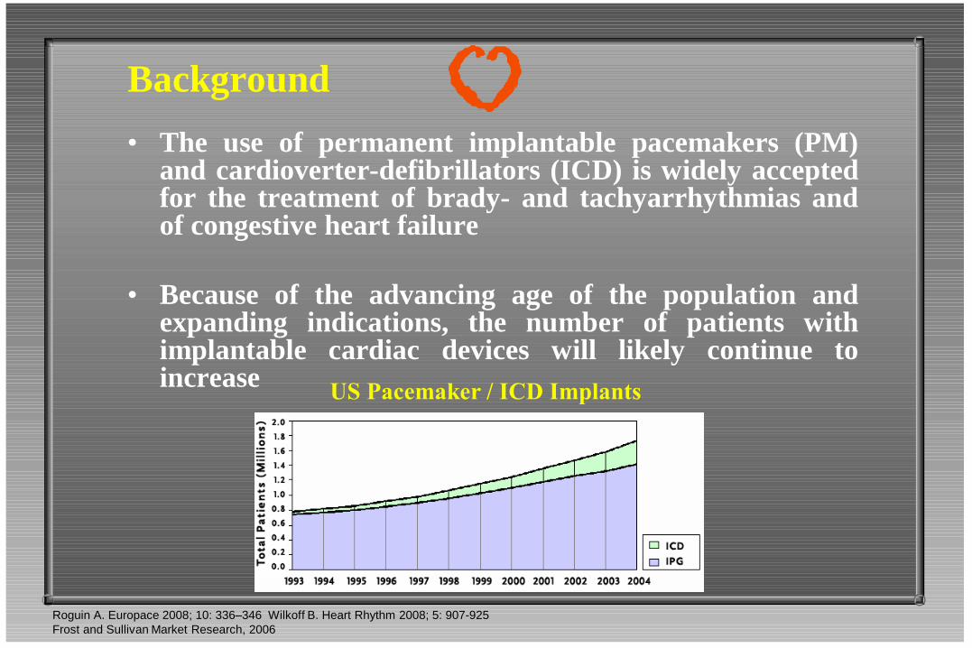
Background
• The use of permanent implantable pacemakers (PM) and cardioverter-defibrillators (ICD) is widely accepted for the treatment of brady- and tachyarrhythmias and of congestive heart failure
• Because of the advancing age of the population and expanding indications, the number of patients with implantable cardiac devices will likely continue to increase
Roguin A. Europace 2008; 10: 336–346 Wilkoff B. Heart Rhythm 2008; 5: 907-925
Frost and Sullivan Market Research, 2006
US Pacemaker / ICD Implants

• Magnetic resonance imaging (MRI) is an important diagnostic tool playing an increasing role in the diagnosis and management of both cardiac and extra-cardiac diseases (over 35 millions MRI studies are performed annually, with an annual growth rate of 10%)
• It is estimated that 50% to 75% of patients with implantable cardiac device will require an MRI at some point after implantation (17% within 12 months of implant)
US MRI procedures
Roguin A. Europace 2008; 10: 336–346 Wilkoff B. Heart Rhythm 2008; 5: 907-925
Frost and Sullivan Market Research, 2006

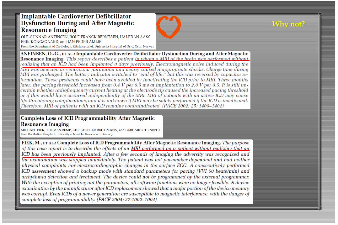
However, the increasing PM/ICD population has been
routinely denied access to MRI due to safety reasons and both
medical community and manufactures considered MRI an
absolute contrindication in these patients.

Cardiac Devices and MRI
Why not?
• Patients with cardiac devices (PM/ICD) are restricted from MRI because the static magnetic field and the variable electromagnetic fields (RF pulses and gradient system) are generally believed to be potentially harmful to the patient/device.
• There are some reports of deaths in patients with PM/ICD undergoing MRI studies in uncontrolled conditions

Adverse Interactions between MRI and PM/ICD
• Movement of the device (translational attraction, torque) and lead dislodgement
• Excessive heating
• Inappropriate (asynchronous) pacing (risk of VF) or inhibition of pacing
• Activation of tachyarrhythmia therapies (ICD)
• MRI-induced arrhythmias associated with current induction in the leads
• Functional alterations (programming changes, battery depletion)
• Artifacts (pulse generator, leads)
Why not?

Why not?
Why not?
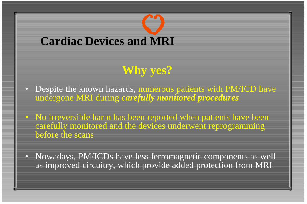
Cardiac Devices and MRI
Why yes?
• Despite the known hazards, numerous patients with PM/ICD have undergone MRI during carefully monitored procedures
• No irreversible harm has been reported when patients have been carefully monitored and the devices underwent reprogramming before the scans
• Nowadays, PM/ICDs have less ferromagnetic components as well as improved circuitry, which provide added protection from MRI
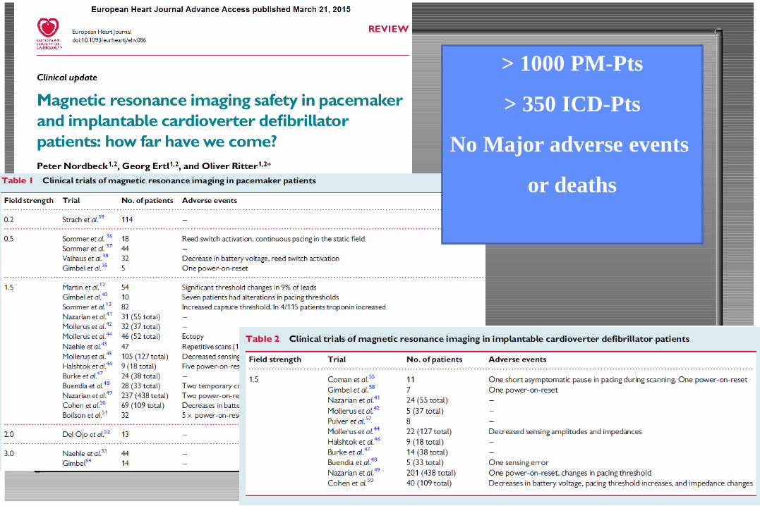
> 1000 PM-Pts
> 350 ICD-Pts
No Major adverse events
or deaths

Our experience (2009-2013, Policlinico S.Donato)
• 120 pts with conventional PM/ICD implanted after 2000 underwent (142) MRI scans based on clinical indications.
• Local /instituitional scientific/ethical comitte approval
• All pacing systems were considered elegible for inclusion
• Pediatic (<16 y) and PM-dependent patients; recent implants (<2 months), abandoned/fractured/epicardial leads were excluded
• All MR studies were performed with a Siemens SONATA 1.5 T (64 MHz) equipment.
• No restrictions were placed on the body segment to be studied
• Continuous pulse oximetry + ECG monitoring and verbal comunication-patient were used during the MR scans.
• An electrophysiologist with full resuscitation equipment was present during each MRI for the entire examination.
• Each device was fully interrogated immediately before and after MRI scanning
Why Yes?
• to assess the immediate and mid-term safety
of MRI in patients with PM or ICD
• to assess the diagnostic yield (efficacy) of
MRI in this setting
Aim of the study
Why yes?

120 pts (91 M, 29 F)
mean age 62 y (± 17)
142 MRI
50% ICDs
50% PMs
Mean time from implant to MRI = 33 m (± 28)
MRI segments
THORACIC 65 (58 Cardiac)
SPINE = 20
BRAIN = 40
ABDOMEN = 7
LOWER EXTREMITIES = 8
BREAST = 2
Follow-up 12 m (± 5)

12
58
1
21
34
16
Device Type PM
ICD
VVI DDD BIV VVI DDD BIV

65
40
20
8 7
2
MRI type
THORACIC BRAIN SPINE EXTREMITIES ABDOMEN BREAST

0
100
200
300
400
500
600
700
800
ATRIAL LEAD VENTRICULAR LEAD CS LEAD COIL
650 664
710
53
645 657
692
54
666 646
706
55
IMPEDANCE (ohms)
IMP PRE
IMP POST
IMP F-UP
p=0,28 p=0,2
p=0,06 p=0,12
p=0,1
p=0,13 p=0,89

0
2
4
6
8
10
12
14
ATRIAL LEAD VENTRICULAR LEAD CS LEAD
3,1
10,6
12,4
3,2
10,6
12
2,8
10,2
11,3
SENSING (mV)
SENSING PRE
SENSING POST
SENSING F-UP
p=0,24
p=0,43
p=0,64 p=0,004
p=0,9

0
0,2
0,4
0,6
0,8
1
1,2
1,4
1,6
1,8
2
ATRIAL LEAD VENTRICULAR LEAD CS LEAD
0,8
1
1,9
0,7
1
1,8
0,7
1
1,3
THRESHOLD (Volts)
THRESHOLD PRE
THRESHOLD POST
THRESHOLD F-UP
p=0,62 p=0,31
p=0,62 p=0,43
p=0,3

IMPEDANCE (ohms) MAGNETIC FREQUENCY (bpm) VOLTAGE (volts)
881
97
3,36
897
97
3,35
996
96
3.30
BATTERY PARAMETERS
PRE
POST
F-UP
p=0,07
p=0,0002
p=0,5 p=0,67
p=0,13 p=0,002
0
10
20
30
40
50
60
70
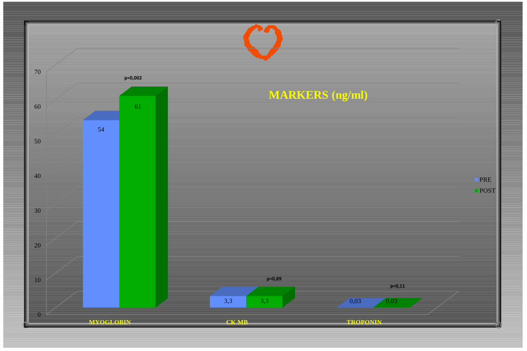
MYOGLOBIN CK MB TROPONIN
54
3,3 0,03
61
3,3 0,03
MARKERS (ng/ml)
PRE
POST
p=0,002
p=0,89
p=0,11

Results
• Post-MRI interrogation and telemetry of each device proceeded without difficulty and the programmed settings remained unchanged
• There were no significant differences comparing PM/ICD parameters before/after MRI exposure
• No patient reported significant symptoms during or immediately after the MRI scan
• No rapid activation of pacing was observed during MRI
all devices were functioning appropriately after MRI
Why Yes?

0%
10%
20%
30%
40%
50%
60%
70%
80%
90%
100%
TOTAL EXTRA THORACIC THORACIC
76%
100%
46%
18%
40%
6%
14%
EFFICACY
FULLY DIAGN
PART DIAGN
NON DIAGN

0%
10%
20%
30%
40%
50%
60%
70%
80%
90%
100%
THORACIC TOTAL THORACIC PM THORACIC ICD
46%
95%
26%
40%
5%
54%
14%
20%
EFFICACY THORACIC MRI
FULLY DIAGN
PART DIAGN
NON DIAGN
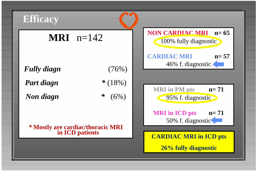
Efficacy
MRI n=142
Fully diagn (76%)
Part diagn * (18%)
Non diagn * (6%)
* Mostly are cardiac/thoracic MRI in ICD patients
NON CARDIAC MRI n= 65
100% fully diagnostic
CARDIAC MRI n= 57
46% f. diagnostic
MRI in PM pts n= 71
95% f. diagnostic
MRI in ICD pts n= 71
50% f. diagnostic
CARDIAC MRI in ICD pts
26% fully diagnostic
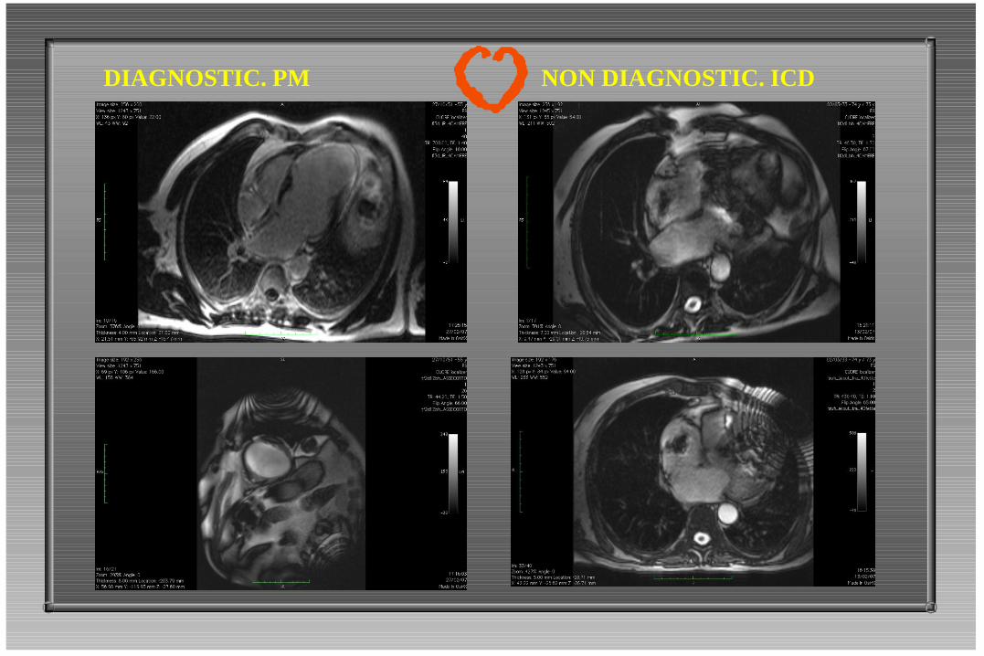
DIAGNOSTIC. PM NON DIAGNOSTIC. ICD

PVs MRI in a patient with ICD Brain MRI in a patient with ICD

Study conclusions
• Under controlled conditions, 1.5-T MRI can be performed in non-PM-dependent patients with a good risk/benefit profile
• Artifacts determined significant diagnostic issues mainly in ICD patients who underwent cardiac/thoracic MRI
Why yes?

Novel Technology
MRI-conditional devices:
overcomes technical challenges and legal issues

• Should we implant all pts with MRI-
conditional/compatible devices?
They should be used in selected pts in whom MRI Follow-
up is warranted, and young Pts
- Longer follow-up is required to confirm this new
technology performance,
- Its diagnostic efficacy in cardiac MRI is still questionable
- Costs!

• Should we replace older devices and leads with
MRI-safe devices?
MRI in PM/ICD pts might be safer than leads
extraction procedures!
Heart Rhythm, Vol 6, No 7, July 2009

ESC Guidelines 2013

Cardiac Devices and MRI
Why not? Why yes?
“…failing to identify an adverse event is
not equivalent to demonstrating
safety…”
JR Gimbel, E Kanal JACC,43,7;2004

“It might be useful to recall that perhaps a mere 1500
or so scans have been reported on device patients in
the medical literature. Surely, not enough safe scans
have been done to declare all our previous
concerns ‘hysterical’ “

…Thank you for your attention


