Download - Multiple Paths to Multispecificity
Immunity 24, 359–368, April 2006 ª2006 Elsevier Inc. DOI 10.1016/j.immuni.2006.03.009
Previews
Multiple Paths to Multispecificity
The limited primary antibody repertoire uses multiplemechanisms to account for the large number of poten-
tial antigens. In this issue of Immunity, Sethi et al.(2006) describe a new means for expanding the anti-
body repertoire, whereby a single antibody isomerbinds diverse antigens at different regions of the bind-
ing site.
A hallmark of the adaptive immune system is the capac-ity to recognize virtually any antigen without havingbeen previously exposed to it. Given the very large num-ber of clonotypically unique antibody or T cell receptor(TCR) that can be generated by gene rearrangementand N region substitution, it was initially believed thatadaptive immunity might be capable of generatinga unique antibody or TCR for every antigen or antigenicpeptide. However, subsequent estimates of the size ofthe foreign peptide repertoire recognized by the TCRsrevealed that there are far more potentially immuno-genic peptides in the environment of a mouse, or otheranimals, than the animal has T cells (Mason, 1998). Sim-ilar considerations probably apply to antibodies, forwhich the size of the germline repertoire and the numberof circulating B cells appear insufficient to cover the al-most infinite diversity of antigens to which an animal isexposed. Multispecificity, or the ability of a single re-ceptor molecule to engage multiple ligands, offersa means for expanding the effective size of the antibodyor TCR repertoire, thereby providing comprehensivecoverage of the antigenic universe. Although multispec-ificity (alternately referred to in the immunological litera-ture as crossreactivity, promiscuity, or degeneracy) ismost often associated with antibodies and TCRs, it isalso an important property of other immune system re-ceptors, such as natural killer (NK) cell receptors thatbind MHC or MHC-like molecules.
Two general mechanisms may be considered to ex-plain multispecific ligand recognition by antibodies orother receptors. In one case, an essentially rigid recep-tor binding site may make different interactions withstructurally distinct ligand surfaces, without significantconformational changes in the receptor (Figure 1A),a mechanism termed ‘‘rigid adaptation’’ (McFarlandand Strong, 2003). Alternatively, the receptor may pos-sess a degree of conformational flexibility that allowsit to reconfigure its binding site to accommodate di-verse ligands (‘‘induced fit’’) (Figure 1B). X-ray crystallo-graphic and thermodynamic studies have providedample evidence for both mechanisms in immune recog-nition. For example, the lysozyme antibody D1.3 bindsboth lysozyme and the idiotypic antibody E5.2 by usingessentially the same set of binding site residues, withminimal structural adjustments in D1.3 or its ligands(Fields et al., 1995). Atoms of E5.2 that contact D1.3are positioned close to those of lysozyme that contactD1.3, and most hydrogen bonds in the D1.3-lysozyme
interface are structurally equivalent to ones in theD1.3-E5.2 interface. In another case of rigid adaptation,the multispecific NK receptor NKG2D has evolved torecognize, through a single binding site, the distantly re-lated MHC-like ligands MICA, RAE-1b, and ULBP3,again without recourse to conformational change inthe receptor (McFarland et al., 2003). Mutagenesis ofthe corresponding complexes showed that the bind-ing-free energy is unevenly distributed across theNKG2D-ligand interfaces, such that energetic hot spotsare associated with structurally conserved receptor ele-ments that interact with relatively conserved residues ofthe ligands.
With respect to multispecificity mediated throughconformational flexibility of the receptor binding site,binding of the germline antibody 7G12 to two structur-ally unrelated ligands (porphyrin and jeffamine) wasfound to involve two distinct conformational states of7G12 (Yin et al., 2003). As a result of combining site re-organization, 7G12 uses two partially overlapping setsof residues to interact with these haptens. Similarly,structural plasticity allows the Fc region of IgG to bindligands as diverse as protein G and rheumatoid factorat a common site between its CH2 and CH3 domains(DeLano et al., 2000). A comprehensive analysis of thedinitrophenyl antibody SPE7 by X-ray crystallographyand presteady-state kinetics revealed a dynamic equi-librium between different preexisting conformations,each conferring a different antigen specificity (Figure 1B)(James et al., 2003). One of these conformers displayeda deep and narrow binding site for aromatic ligands,such as the immunizing hapten, whereas another con-former had a broad, shallow binding site that recog-nized a protein ligand. In this view, proteins are an en-semble of preexisting isomers, whereby one aminoacid sequence adopts multiple structures and, conse-quently, multiple functions.
Based on these, and related, studies, one might rea-sonably have supposed that the essential mechanismsfor achieving multispecificity had been discovered.However, Salunke and colleagues (Sethi et al., 2006)now report a new mechanism for expanding the primaryantibody repertoire, which they term ‘‘differential ligandpositioning.’’ The essential feature of this mechanism isthat a single antibody conformer may bind diverse anti-gens at spatially distinct regions of the binding site(Figure 1C). In this study, the authors first determinedthe crystal structure of the germline antibody 36-65,which was raised against the hapten azophenylarso-nate, in unbound form. In all, four antigen-free antibodystructures were examined, which revealed a degree ofstructural diversity consistent with previous thermody-namic evidence that 36-65 is conformationally flexible(Manivel et al., 2000), as expected for a germline anti-body. No two molecules exhibited similar combiningsite topologies, with differences attributable to varia-tions in both backbone and side chain conformation.The authors then determined the structure of 36-65 incomplex with three independent 12-mer peptides, iso-lated by screening a phage-displayed random peptide
Immunity360
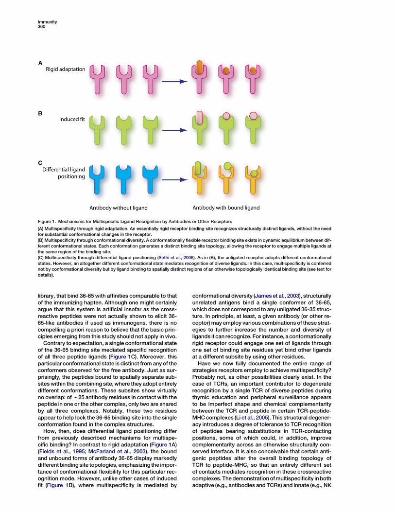
Figure 1. Mechanisms for Multispecific Ligand Recognition by Antibodies or Other Receptors
(A) Multispecificity through rigid adaptation. An essentially rigid receptor binding site recognizes structurally distinct ligands, without the need
for substantial conformational changes in the receptor.
(B) Multispecificity through conformational diversity. A conformationally flexible receptor binding site exists in dynamic equilibrium between dif-
ferent conformational states. Each conformation generates a distinct binding site topology, allowing the receptor to engage multiple ligands at
the same region of the binding site.
(C) Multispecificity through differential ligand positioning (Sethi et al., 2006). As in (B), the unligated receptor adopts different conformational
states. However, an altogether different conformational state mediates recognition of diverse ligands. In this case, multispecificity is conferred
not by conformational diversity but by ligand binding to spatially distinct regions of an otherwise topologically identical binding site (see text for
details).
library, that bind 36-65 with affinities comparable to thatof the immunizing hapten. Although one might certainlyargue that this system is artificial insofar as the cross-reactive peptides were not actually shown to elicit 36-65-like antibodies if used as immunogens, there is nocompelling a priori reason to believe that the basic prin-ciples emerging from this study should not apply in vivo.
Contrary to expectation, a single conformational stateof the 36-65 binding site mediated specific recognitionof all three peptide ligands (Figure 1C). Moreover, thisparticular conformational state is distinct from any of theconformers observed for the free antibody. Just as sur-prisingly, the peptides bound to spatially separate sub-sites within the combining site, where they adopt entirelydifferent conformations. These subsites show virtuallyno overlap: of w25 antibody residues in contact with thepeptide in one or the other complex, only two are sharedby all three complexes. Notably, these two residuesappear to help lock the 36-65 binding site into the singleconformation found in the complex structures.
How, then, does differential ligand positioning differfrom previously described mechanisms for multispe-cific binding? In contrast to rigid adaptation (Figure 1A)(Fields et al., 1995; McFarland et al., 2003), the boundand unbound forms of antibody 36-65 display markedlydifferent binding site topologies, emphasizing the impor-tance of conformational flexibility for this particular rec-ognition mode. However, unlike other cases of inducedfit (Figure 1B), where multispecificity is mediated by
conformational diversity (James et al., 2003), structurallyunrelated antigens bind a single conformer of 36-65,which does not correspond to any unligated 36-35 struc-ture. In principle, at least, a given antibody (or other re-ceptor) may employ various combinations of these strat-egies to further increase the number and diversity ofligands it can recognize. For instance, a conformationallyrigid receptor could engage one set of ligands throughone set of binding site residues yet bind other ligandsat a different subsite by using other residues.
Have we now fully documented the entire range ofstrategies receptors employ to achieve multispecificity?Probably not, as other possibilities clearly exist. In thecase of TCRs, an important contributor to degeneraterecognition by a single TCR of diverse peptides duringthymic education and peripheral surveillance appearsto be imperfect shape and chemical complementaritybetween the TCR and peptide in certain TCR-peptide-MHC complexes (Li et al., 2005). This structural degener-acy introduces a degree of tolerance to TCR recognitionof peptides bearing substitutions in TCR-contactingpositions, some of which could, in addition, improvecomplementarity across an otherwise structurally con-served interface. It is also conceivable that certain anti-genic peptides alter the overall binding topology ofTCR to peptide-MHC, so that an entirely different setof contacts mediates recognition in these crossreactivecomplexes. The demonstration of multispecificity in bothadaptive (e.g., antibodies and TCRs) and innate (e.g., NK
Previews361
receptors) immune systems underscores its critical rolein expanding immune surveillance as well as the impor-tance of understanding this phenomenon at the molecu-lar level.
Roy A. Mariuzza1
1Center for Advanced Research in BiotechnologyW.M. Keck Laboratory for Structural BiologyUniversity of Maryland Biotechnology InstituteRockville, Maryland 20850
Selected Reading
DeLano, W.L., Ultsch, M.H., de Vos, A.M., and Wells, J.A. (2000).
Science 287, 1279–1283.
Immunity 24, April 2006 ª2006 Elsevier Inc. DOI 10.1016/j.immuni.2006.04.0
Some Nuts Are Tougherto Crack than Others
In this issue of Immunity, Oestreich et al. (2006) show
that, during V(D)J recombination, RSSs may have dis-tinct accessibility requirements. Some rely on an en-
hancer-intrinsic, general chromatin opening function,whereas others require enhancer-promoter interac-
tions that direct local chromatin remodeling.
The antigen receptor repertoires of T and B lympho-cytes are generated by the process of V(D)J recombina-tion, which assembles mature T cell receptor (TCR) andimmunoglobulin (Ig) genes from variable (V), diversity(D), and joining (J) gene segments during the earlystages of lymphocyte development. Individual TCRand Ig loci are subject to distinct programs of V(D)Jrecombination events, which vary as a function of lym-phocyte lineage and developmental stage. Understand-ing the basis for this regulation has been a longstandinggoal of molecular immunologists.
V(D)J recombination is initiated by the recombinase-activating gene (RAG) proteins, which recognize recom-bination signal sequences (RSSs) that flank TCR and Igcoding gene segments. It has long been appreciatedthat the developmental regulation of V(D)J recombina-tion occurs primarily through changes in the accessibil-ity of RSS substrates to the RAG proteins (Stanhope-Baker et al., 1996). RSSs are facile substrates for RAGproteins when present on naked DNA in vitro. However,in lymphocyte nuclei in vivo, RSSs are embedded withinchromatin, a highly organized and highly compacted nu-cleoprotein complex. Chromatin-embedded RSSs areintrinsically inaccessible to RAG and require changesin chromatin structure so that they can participate inthe V(D)J recombination reaction in vivo. Many lines ofevidence implicate cis-acting transcriptional regulatory
Fields, B.A., Goldbaum, F.A., Ysern, X., Poljak, R.J., and Mariuzza,
R.A. (1995). Nature 374, 739–742.
James, L.C., Roversi, P., and Tawfik, D.S. (2003). Science 299, 1362–
1367.
Li, Y., Huang, Y., Lue, J., Quandt, J.A., Martin, R., and Mariuzza, R.A.
(2005). EMBO J. 24, 2968–2979.
Manivel, V., Sahoo, N.C., Salunke, D.M., and Rao, K.V. (2000). Immu-
nity 13, 611–620.
Mason, D. (1998). Immunol. Today 19, 395–404.
McFarland, B.J., and Strong, R.K. (2003). Immunity 19, 803–812.
McFarland, B.J., Kortemme, T., Yu, S.F., Baker, D., and Strong, R.K.
(2003). Structure 11, 411–422.
Sethi, D.K., Agarwal, A., Manivel, V., Rao, K.V.S., and Salunke, D.M.
(2006). Immunity 24, this issue, 429–438.
Yin, J., Beuscher, A.E., Andryski, S.E., Stevens, R.C., and Schultz,
P.G. (2003). J. Mol. Biol. 330, 651–656.
02
elements of antigen receptor loci, including enhancersand promoters, as developmental regulators of bothV(D)J recombination and chromatin structure. However,in molecular terms, what does it really take to make anRSS accessible? How do enhancer and promoter ele-ments accomplish this? The study by Oestreich et al.(2006) in this issue of Immunity moves us several stepscloser to understanding these issues.
The initial step in TCRb recombination involves Db andJb segments situated within the 30 portion of the TCRb lo-cus (Figure 1). Db1 can rearrange to any of six Jb1 genesegments, and Db2 can rearrange to any of six functionalJb2 gene segments. Previous gene targeting experi-ments had shown that the TCRb enhancer (Eb), situated30 of Cb2, was required for substantial levels of eitherDb1-Jb1 or Db2-Jb2 rearrangement (Bouvier et al.,1996). A promoter tightly juxtaposed with Db1 (PDb1)was found to be essential for Db1-Jb1 rearrangement butirrelevant for Db2-Jb2 rearrangement (Whitehurst et al.,2000). An analogous promoter associated with Db2 islikely critical for Db2-Jb2 rearrangement, but this hasyet to be demonstrated experimentally. Nevertheless itis clear from the above that Db1-Jb1 rearrangement de-pends on cooperation between Eb, which must act overa distance of 15 kb, and PDb1, which acts more locally.
Oestreich et al. (2006) began their study with a detailedanalysis of chromatin structure across the Db and Jbsegments in mice carrrying wild-type TCRb alleles oralleles lacking either PDb1 or Eb. As a surrogate forRSS accessibility to RAG, they measured the accessibil-ity of defined restriction sites to digestion with restrictionenzymes introduced into permeabilized nuclei of imma-ture thymocytes. As expected, they found that a HinF1site in the Db1 RSS could only be cleaved efficiently inthe presence of both PDb1 and Eb. However, at a seriesof other restriction sites extending from a point onlya few nucleosomes downstream of the Db1 RSS to theJb1.6 RSS, accessibility was highly dependent on Eb
but minimally dependent on PDb1. These conclusions


