
Muscles
HBS3B

The knee jointLabel the diagrams

The knee joint
1 – tendon2 – patella3 – bursa4 – synovial membrane5 – bursa6 – menisci (articular discs) 7 – cruciate ligaments 8 – articular cartilage 9 - synovial capsule10 – fibula 11 – femur 12 – ligament 13 – tibia

Types of musclesCardiac muscle found only in the heart Smooth muscle or involuntary
muscle found in the walls of the digestive system, the bladder, the lungs and blood vessels
Skeletal (or voluntary) muscle found in body muscles

Movement
Muscles have three properties that allow them to work –1. Contractibility = ability to contract or shorten2. Extensibility = ability to be stretched3. Elasticity = ability to return to the original length after stretching
Movement occurs due to muscles pulling on bones either side of a joint. When a muscle works, it slides its filaments together (contraction), thus becoming shorter and thicker, and pulling the ends of the bones together, and bending (or straightening) a joint.
Muscles can not push bones, so in order for the bones to move in the opposite direction, another set of muscles must contract, while the first set relaxes. Because muscles are elastic, they can stretch when relaxed, and shorten when contracted. These pairs of muscles are called agonist-antagonist pairs. The agonist is the muscle that is contracting or doing the work, while the antagonist is the muscle that is relaxing.
Synergists are muscles that help by steadying a jointFixators are synergists that fixate a joint (stop it moving)
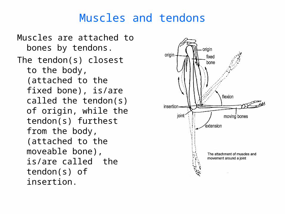
Muscles and tendons
Muscles are attached to bones by tendons.
The tendon(s) closest to the body, (attached to the fixed bone), is/are called the tendon(s) of origin, while the tendon(s) furthest from the body, (attached to the moveable bone), is/are called the tendon(s) of insertion.

Muscle structure

Muscle structure
The sarcolemma is a thin transparent membrane surrounding each muscle cell
Muscle fibres are cylindrical muscle cells
They are made up of many thread-like myofibrils
These are made up of many smaller protein filaments called myofilaments

Microscopic structure of musclesThere are two types of myofilaments
– thick and thin
Actin makes up the thinner myofilaments
Myosin makes up the thicker myofilaments
A sarcomere is a unit containing overlapping bands of actin and myosin
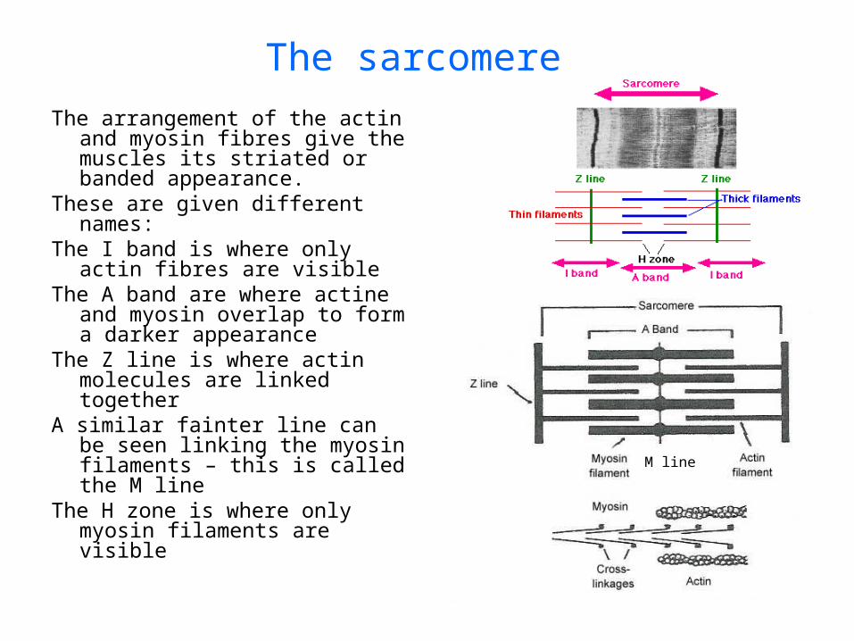
The sarcomere
The arrangement of the actin and myosin fibres give the muscles its striated or banded appearance.
These are given different names:The I band is where only actin
fibres are visibleThe A band are where actine and
myosin overlap to form a darker appearance
The Z line is where actin molecules are linked together
A similar fainter line can be seen linking the myosin filaments – this is called the M line
The H zone is where only myosin filaments are visible
M line

Sliding filament model
When the muscle contracts, the actin and myosin filaments slide over each other, pulling the z-lines closer and shortening the muscle
This requires the use of energy in the form of Adenosine triphosphateWhen the muscle relaxes the actin and myosin filaments are pulled past
each other and return to their previous locations, so the muscle returns to its previous length
Actin and myosin don’t change in length – they just slide closer or further apart.
Myofibrils comprised of actin and myosin myofilaments

Control of muscle movementVoluntary muscle contraction is initiated
by nerve impulses, starting in the brain or spinal cord and travelling through the somatic division of the efferent tract of the peripheral nervous system.
One motor neuron will control several muscle fibres.
A motor unit is the motor neuron and all the fibres it controls

The neuromuscular junction
The neuromuscular junction is the point where the message is passed from neuron to muscle
The synaptic knob is the enlarged area at the end of the axonThe motor end plate is depression in the surface of the muscle fibreThe neurotransmitter used is acetylcholine

Roles of cerebrum and cerebellum
The primary motor cortex initiates voluntary muscle contraction and at the same time sends messages to the cerebellum
The cerebellum sends messages to co-ordinate different muscles so movement is smooth
Balance receptors send messages to the cerebellum so it can track the position and movement occurring in the head
Stretch receptors detect muscle and joint activity and send messages to the cerebellum so it can track movements of extremities
Upper motor neurons have cell bodies in the cerebrum (and some in spinal cord)
Lower motor neurons have cell bodies in the spinal cord

Roles of cerebrum and cerebellumThe diagram shows the nerve pathways from the cerebral cortexShow the pathways• from sensory receptors• to and from cerebellum

Cerebellum involvement
Taken from Newton and Joyce p218

Sample questions
The neuron illustrated above would be classified correctly as
(a) a sensory neuron.
(b) a motor neuron.
(c) a connector neuron.
(d) there is not enough information to say.

Sample exam questions1. Which of the following is NOT true of structure A?
(a) It is surrounded by three layers of meninges. (b) Information from the body terminates in structure A's white matter. (c) Its surface is convoluted to provide greater surface area. (d) It is connected to, and able to influence, the cerebellum.
2. This question refers to the list of features below. (i) regulation of osmotic balance (ii) regulation of the heart rate (iii) coordination of posture and movement- (iv) temperature control
Which of the above features are roles played by structure B? (a) (i), (ii) and (iii) only (b) (i), (ii) and (iv) only (c) (i), (ii), (iii) and (iv) only (d) (ii) and (iv) only
3. Which of the following statements is true of the fluid contained in structure C? (a) It is produced by the meninges. (b) It has an important role in protecting the brain from infection. (c) It assists with nourishment of the cerebral cortex. (d) It assists with temperature regulation in the brain.
Sample questionsUsing a diagram, explain the structures that
comprise a synovial joint and their functions.

Synovial joints• Synovial capsule surrounds the
joint and helps stabilise it and hold it all together
• Synovial membrane is thin and smooth, to reduce friction, and secretes synovial fluid
• Synovial fluid is thick and sticky, and acts as a lubricant for the joint
• Articular cartilage provides a smooth surface to reduce friction as the bones move across each other
• Articular disc are cartilaginous discs which act as shock absorbers
• Bursae are fluid filled sacs which act as shock absorbers
• Accessory ligaments join the bones and keep them together

Sample questionsUsing diagrams, explain the main differences between
compact bone, spongy bone and hyaline cartilage.Relate these differences to the functions of these tissues.

Comparing bone and cartilage
Bone has a few cells but also lots of mineral matrix to make it strong, and a good blood supply, which makes capable of repair
Compact bone is highly organised into osteons to make columnar Haversian systems – this gives it strength.
Spongy bone is less organised, with more spaces – this makes it lighter. The spaces are filled with red bone marrow which make blood cells
Cartilage has relatively few cells and lots of matrix. The matrix has more protein than minerals so it is strong and slightly elastic. It has less of a blood supply, so is slower to heal.
Compact bone Spongy boneCartilage


