Musculoskeletal UltrasoundPocket Guide
2
© Step 4 Medical Arts© Sonosite, Inc., a subsidiary of FUJIFILM
All rights reserved. No part of this publication may be reproduced, distributed, or transmitted in any form or by any means, including photocopying, recording, or other electronic or mechanical methods, without the prior written permission of the publisher, except in the case of brief quotations embodied in critical reviews and certain other noncommercial uses permitted by copyright law. MKT02673_UK
AuthorsDr. Jose Antonio BouffardSports Medicine Ultrasound - DMC Sports, Detroit Medical Center
Dr. Ami MacPhysical Medicine & Rehabilitation
Dr. Brian M. SmileyRadiology PGY-4
Dr. Shivani GuptaResident Physician PGY-1
Jean ParkMedical Student Year 4
Daniel Shelton RT(R)MSK Development (SonoSite)

3
Introduction
Musculoskeletal

4 Introduction
Objective of this guideThe Musculoskeletal Ultrasound Pocket Guide is a portable and easy-to-use reference for students, residents and physicians. It is a beginner’s guide for the most common diagnostic musculoskeletal ultrasound studies. The images provided are those most frequently obtained during the sonographic evaluation of each respective joint.
Each chapter covers the examination of a joint. The examinations covered for the upper extremity are the shoulder, elbow, hand and wrist. The examinations covered for the lower extremity are the hip, knee, foot and ankle. In addition each chapter concludes with some of the more common ultrasound-guided injection techniques.

5Introduction
Each section is organised using the following system:
Anatomy of the region
Patient position
Transducer placement and position
1
2
3
4
5
Important areas to assess during the study
Mistakes to avoid during the study
6 Common pathology of the region
MRI images and corresponding sonograms with identifying landmarks
7
6 Introduction
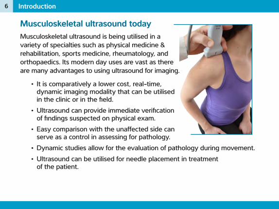
Musculoskeletal ultrasound todayMusculoskeletal ultrasound is being utilised in a variety of specialties such as physical medicine & rehabilitation, sports medicine, rheumatology, and orthopaedics. Its modern day uses are vast as there are many advantages to using ultrasound for imaging.
• It is comparatively a lower cost, real-time, dynamic imaging modality that can be utilised in the clinic or in the field.
• Ultrasound can provide immediate verification of findings suspected on physical exam.
• Easy comparison with the unaffected side can serve as a control in assessing for pathology.
• Dynamic studies allow for the evaluation of pathology during movement. • Ultrasound can be utilised for needle placement in treatment
of the patient.

7Introduction
As a diagnostic tool, ultrasound is often used to assess for:
• Joint effusions • Bony irregularities (osteophytes, loose bodies, fractures, erosions,
degenerative changes, lytic changes)• Tendon pathology (tears, ruptures, dislocations, tendinitis, tendinosis, tenosynovitis)• Muscle pathology (tears, atrophy, impingement, herniations)• Bursal pathology (thickening, enlargement)• Nerve pathology (entrapments, subluxations)
Tips for using this guideWhen utilising this guide, remember that the patient’s right side is used by convention. All images, MRI, ultrasound, anatomic drawings and patient positioning correlate with the patient’s right side. Remember to flip or rotate the images in your mind to better understand the relationships between each of them. From this guide, you will better understand the spatial relationships between structures.

8
9
Sonographic EvaluSonographic Evaluation of the Shoulder1

10 Sonographic Evaluation of the Shoulder
The ShoulderIn the hands of a skilled operator, ultrasound is more sensitive than even MRI for the diagnosis of rotator cuff tears.
Rotator Cuff Tears Ultrasound MRIFull-thickness tearSensitivity 94.3% 91.2%Specificity 95.3% 94.2%Partial-thickness tearSensitivity 79.1% 63.1%Specificity 94.6% 93.7%OverallSensitivity 91.0% 79.6%Specificity 87.0% 90.6%
Data from Diagnostic Imaging of Rotator Cuff Tears: A Meta-Analysis of the Accuracy of US and MR. Department of Radiology, Thomas Jefferson University Hospital, Philadelphia, Pennsylvania.
11Sonographic Evaluation of the Shoulder
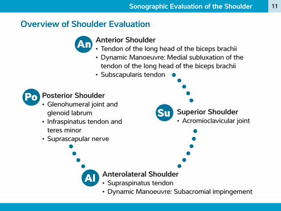
Overview of Shoulder Evaluation
Superior Shoulder• Acromioclavicular joint
Anterolateral Shoulder• Supraspinatus tendon• Dynamic Manoeuvre: Subacromial impingement
Posterior Shoulder• Glenohumeral joint and
glenoid labrum• Infraspinatus tendon and
teres minor• Suprascapular nerve
Anterior Shoulder• Tendon of the long head of the biceps brachii• Dynamic Manoeuvre: Medial subluxation of the
tendon of the long head of the biceps brachii• Subscapularis tendon
An
Su
Al
Po

12 Sonographic Evaluation of the Shoulder
Anterior ShoulderTendon of the long head of the biceps brachii
Anatomy: Biceps BrachiiOrigin (Proximal Attachment)
• Short head: Coracoid process of scapula• Long head: Supraglenoid tubercle
of scapula
Insertion (Distal Attachment)• Radial tuberosity
Action• Flexion and supination of the forearm at
the elbow• Flexion of the arm at the shoulder
Innervation• Nerve root: C5-C6 (C6)• Peripheral nerve: Musculocutaneous
Radha Sampat
An

13Sonographic Evaluation of the Shoulder
Transducer PositionThe long head of the biceps brachii is located lateral to that of the short head. On initial placement of the transducer, start lateral to the region where the short head can be palpated.
Transducer TipsOn short axis, angle the transducer superiorly to eliminate echogenicity of the tendon. The degree of external rotation of the forearm will directly influence the position of the long head of the biceps brachii tendon relative to the head of the humerus. Adjust the patient’s forearm such that the long head appears centered over the humerus.
Patient PositionSeat your patient with the shoulder adducted and elbow flexed to approximately 90°. Supinate the forearm and rest it on the thigh.
Short Axis
Long Axis
Neutral

14 Sonographic Evaluation of the Shoulder
9 Assess the following• Integrity of the cortical surface and depth of the bicipital groove• Thickness and echogenicity of the tendon• Assess for fluid within the tendon sheath• Assess for neovascularisation• Dynamic Manoeuvre: Medial dislocation of the tendon upon external rotation of the shoulder
´ Mistakes to avoid• Do not confuse anisotropy in the proximal tendon with pathologic
changes• Do not confuse tenosynovitis with fluid that originates from within
the joint• Do not assume that fluid in the tendon sheath is limited to biceps tendon pathology (rotator cuff estimate)• Do not confuse flow within the anterior circumflex artery with neovascularisation

15Sonographic Evaluation of the Shoulder
Pathology• Biceps tendon joint effusion• Biceps tenosynovitis• Biceps ganglion cyst• Biceps brachii tenodesis• Biceps brachii subluxation
Notes

16 Sonographic Evaluation of the Shoulder
D
H
B
T
t
Tendon of the Long Head of the Biceps Brachii | Axial Plane
MRI
D
T B
t
H
Radha Sampat
Deltoid muscle
Humerus
Bicipital groove
Tendon of the long head of the biceps brachii (short axis)
Greater tuberosity
Lesser tuberosity
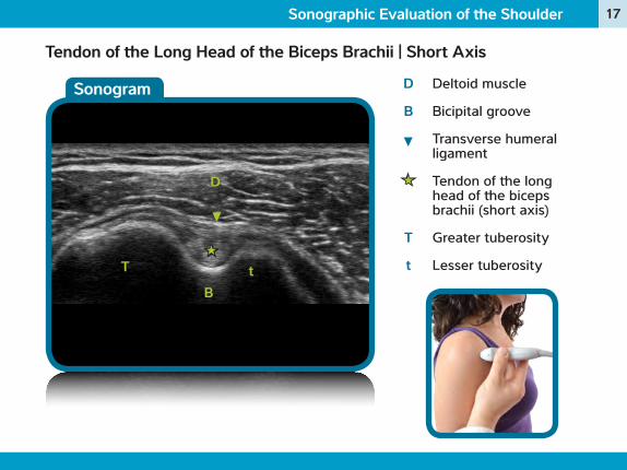
17Sonographic Evaluation of the Shoulder
Deltoid muscle
Bicipital groove
Transverse humeral ligament
Tendon of the long head of the biceps brachii (short axis)
Greater tuberosity
Lesser tuberosity
D
B
T
t
D
tT
B
Sonogram
Tendon of the Long Head of the Biceps Brachii | Short Axis

18 Sonographic Evaluation of the Shoulder
MRI
Tendon of the Long Head of the Biceps Brachii | Axial Plane
Deltoid muscle
Humeral head
Tendon of the long head of the biceps brachii
Greater tuberosity
Lesser tuberosity
D
H
T
t
H
D
►
Radha Sampat

19Sonographic Evaluation of the Shoulder
Deltoid muscle
Tendon of the long head of the biceps brachii (long axis)
Humerus
D
H
D
H
Sonogram
Tendon of the Long Head of the Biceps Brachii | Long Axis
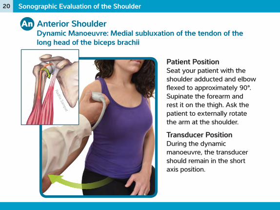
20 Sonographic Evaluation of the Shoulder
Anterior ShoulderDynamic Manoeuvre: Medial subluxation of the tendon of the long head of the biceps brachii
Patient PositionSeat your patient with the shoulder adducted and elbow flexed to approximately 90°. Supinate the forearm and rest it on the thigh. Ask the patient to externally rotate the arm at the shoulder.
Transducer PositionDuring the dynamic manoeuvre, the transducer should remain in the short axis position.
Radha Sampat
An
21Sonographic Evaluation of the Shoulder
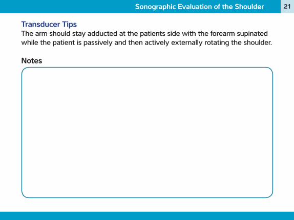
Transducer TipsThe arm should stay adducted at the patients side with the forearm supinated while the patient is passively and then actively externally rotating the shoulder.
Notes
22 Sonographic Evaluation of the Shoulder
Radha Sampat
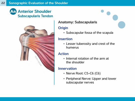
Anterior ShoulderSubscapularis Tendon
Anatomy: SubscapularisOrigin
• Subscapular fossa of the scapula
Insertion• Lesser tuberosity and crest of the
humerus
Action• Internal rotation of the arm at
the shoulder
Innervation• Nerve Root: C5-C6 (C6)• Peripheral Nerve: Upper and lower
subscapular nerves
An
23Sonographic Evaluation of the Shoulder
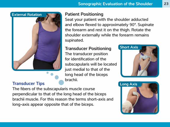
Patient PositioningSeat your patient with the shoulder adducted and elbow flexed to approximately 90°. Supinate the forearm and rest it on the thigh. Rotate the shoulder externally while the forearm remains supinated.
Short Axis
Long Axis
Transducer PositioningThe transducer position for identification of the subscapularis will be located just medial to that of the long head of the biceps brachii.
Transducer TipsThe fibers of the subscapularis muscle course perpendicular to that of the long head of the biceps brachii muscle. For this reason the terms short-axis and long-axis appear opposite that of the biceps.
External Rotation

24 Sonographic Evaluation of the Shoulder
9 Assess the following• Cortex of the lesser tuberosity
• Insertion of the subscapularis tendon• Note: The insertion of the tendon is large, requiring movement of
the probe from proximal to distal. Also, notice the multipennate architecture of the muscle.
• Thickness and echogenicity of the tendon in short and long-axis• Assess for a tear:
• If complete, note the distance that the tendon has retracted• If partial, note whether it is a bursal vs. articular-sided tear• Assess for fluid in the subacromial-subdeltoid bursa
´ Mistakes to avoid• When evaluating the subscapularis tendon in short-axis, do not confuse
the fascicles of the tendon a tear. Recognize that the mulipennate structure is normal.
25Sonographic Evaluation of the Shoulder
Pathology• Subscapularis Tear • Subscapularis Tendinosis• Subscapularis Tendon Avulsion
Notes

26 Sonographic Evaluation of the Shoulder
MRI
Subscapularis Tendon | Axial Plane
Deltoid muscle
Subscapularis tendon
Lesser tuberosity of humerus
Glenoid
Infraspinatus muscle
D
t
G
I
D
t
G
I Radha Sampat
27Sonographic Evaluation of the Shoulder
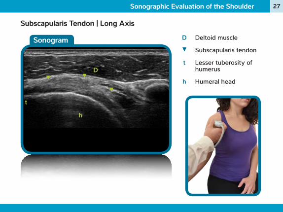
Subscapularis Tendon | Long Axis
Sonogram Deltoid muscle
Subscapularis tendon
Lesser tuberosity of humerus
Humeral head
D
t
h
D
h
t

28 Sonographic Evaluation of the Shoulder
MRI
Subscapularis Tendon | Coronal Plane
Subscapularis tendon
Acromion
Humeral head
Deltoid muscle
S
A
H
D
A
HS
D
Radha Sampat

29Sonographic Evaluation of the Shoulder
Sonogram
Subscapularis Tendon | Short Axis
Subscapularis tendon
Humerus
S
H
SS S
H

30 Sonographic Evaluation of the Shoulder
Superior ShoulderAcromioclavicular Joint
Anatomy: Acromioclavicular JointDescription
• Attaches the acromion of the scapula to the clavicle
• Consists of the superior and inferior acromioclavicular ligaments
Function• Is a synovial gliding joint• Provides mobility and support during
• Protraction/retraction (punching)
• Rotation (raising arm above shoulder)
• Elevation/depression
Su
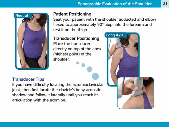
31Sonographic Evaluation of the Shoulder
Patient PositioningSeat your patient with the shoulder adducted and elbow flexed to approximately 90°. Supinate the forearm and rest it on the thigh.
Long Axis
Neutral
Transducer TipsIf you have difficulty locating the acromioclavicular joint, then first locate the clavicle’s bony acoustic shadow and follow it laterally until you reach its articulation with the acomion.
Transducer PositioningPlace the transducer directly on top of the apex (highest point) of the shoulder.

32 Sonographic Evaluation of the Shoulder
9 Assess the following• The bony cortices of both the proximal acromion and the distal clavicle• Evaluate for capsular dilatation• Look for the “geyser sign” (pathognomonic for complete rupture of the
supraspinatus)
´ Mistakes to avoid• Do not confuse “Os Acromiale” with calcification of the joint• Do not confuse the visualisation of a meniscus with fibrocartilage
pathology in young people
Pathology• Os Acromiale• AC Joint Osteoarthritis and cysts• AC Joint infection

33Sonographic Evaluation of the Shoulder
Notes

34 Sonographic Evaluation of the Shoulder
MRI
Acromioclavicular Joint | Coronal Plane
Acromion
Clavicle
Humeral head
Supraspinatus muscle
A
C
H
SA
H
S
C

35Sonographic Evaluation of the Shoulder
Sonogram
Acromioclavicular Joint | Long Axis
Edge of the clavicle
Edge of the acromion
Acromioclavicular capsule
C
A
A C

36 Sonographic Evaluation of the Shoulder
Anterolateral ShoulderSupraspinatus Tendon
Anatomy: SupraspinatusOrigin
• Supraspinatus fossa of scapula
Insertion• Greater tubercle of humerus
Action• Abduction of the arm
Innervation• Nerve Root: C5, C6• Peripheral Nerve: Suprascapular
nerve
Radh
a Sam
pat
Al

37Sonographic Evaluation of the Shoulder
Patient PositioningSeat your patient and ask him or her to perform the Crass Manoeuvre or the Modified Crass Manoeuvre. Either position can be used depending on the patient’s comfort. These positions move the supraspinatus anteriorly and out from underneath the acromion. This allows for better visualisation of the supraspinatus fibers.
Crass ManoeuvreThe shoulder is adducted, in extension, and internally rotated (reaching towards the contralateral scapula).
Modified Crass ManoeuvreThe shoulder is extended with hand in supination and placed on ipsilateral buttock (as if in the patient's back pocket). This manoeuvre tends to be more comfortable in the painful shoulder. Proper positioning requires the elbow to remain in the posterior position.
Crass
Modified Crass
38 Sonographic Evaluation of the Shoulder
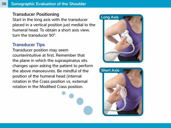
Transducer Positioning Start in the long axis with the transducer placed in a vertical position just medial to the humeral head. To obtain a short axis view, turn the transducer 90°.
Transducer TipsTransducer position may seem counterintuitive at first. Remember that the plane in which the supraspinatus sits changes upon asking the patient to perform the above manoeuvres. Be mindful of the position of the humeral head (internal rotation in the Crass position vs. external rotation in the Modified Crass position.
Long Axis
Short Axis

39Sonographic Evaluation of the Shoulder
9 Assess the following• Cortex of the greater tuberosity• Thickness and echogenicity of the tendon in long and short-axis• Assess for a tear:
• If complete, note the distance the tendon has retracted• If partial, note whether there is a bursal vs. articular tear
• Dynamic Manoeuvre: Abduction of the shoulder to 90° to assess for bursal ± tendinous impingement beneath the acromion.
´ Mistakes to avoid• Do not confuse anisotropy of the tendinous insertion with a partial tear/rupture
Pathology:• Supraspinatus Tear (partial vs. full-thickness)• Supraspinatus Tendinosis
40 Sonographic Evaluation of the Shoulder
MRI
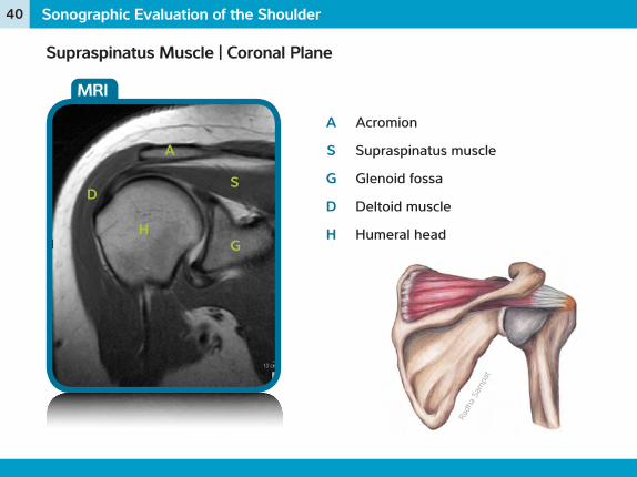
Supraspinatus Muscle | Coronal Plane
Acromion
Supraspinatus muscle
Glenoid fossa
Deltoid muscle
Humeral head
A
S
G
D
H
A
S
G
D
H
Radh
a Sam
pat

41Sonographic Evaluation of the Shoulder
Sonogram
Supraspinatus Muscle | Long Axis
Deltoid
Bursa
Supraspinatus
Hyaline articular cartilage
Greater tuberosity
Humerus
D
S
*
T
H
D
H
T
*
S
*

42 Sonographic Evaluation of the Shoulder
MRI
Supraspinatus Tendon | Saggital View
Deltoid muscle
Supraspinatus muscle
Humeral head
D
S
HS
D
H
Radh
a Sam
pat

43Sonographic Evaluation of the Shoulder
Sonogram
Supraspinatus Tendon | Short Axis
Supraspinatus tendon
Hyaline articular cartilage
Subacromial-subdeltoid bursa
S
*
S
**
44 Sonographic Evaluation of the Shoulder
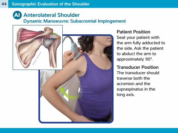
Anterolateral ShoulderDynamic Manoeuvre: Subacromial Impingement
Patient PositionSeat your patient with the arm fully adducted to the side. Ask the patient to abduct the arm to approximately 90°.
Transducer PositionThe transducer should traverse both the acromion and the supraspinatus in the long axis.
Radh
a Sam
pat
Al

45Sonographic Evaluation of the Shoulder
9 Assess the following• Look for impingement of the superior fibers of the supraspinatus beneath the acromion• Bursal thickening
Notes

46 Sonographic Evaluation of the Shoulder
Radh
a Sam
pat
MRI
Dynamic Manoeuvre: Subacromial Impingement | Coronal Plane
Acromion
Supraspinatus muscle
Humeral head
A
S
HS
H
A

47Sonographic Evaluation of the Shoulder
Sonogram
Dynamic Manoeuvre: Subacromial Impingement | Long Axis
A
S
Acromion
Supraspinatus muscle
Subacromial- subdeltoid bursa
A
S
48 Sonographic Evaluation of the Shoulder
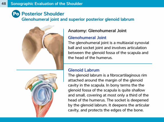
Posterior ShoulderGlenohumeral joint and superior posterior glenoid labrum
Anatomy: Glenohumeral JointGlenohumeral JointThe glenohumeral joint is a multiaxial synovial ball and socket joint and involves articulation between the glenoid fossa of the scapula and the head of the humerus.
Glenoid LabrumThe glenoid labrum is a fibrocartilaginous rim attached around the margin of the glenoid cavity in the scapula. In bony terms the the glenoid fossa of the scapula is quite shallow and small, covering at most only a third of the head of the humerus. The socket is deepened by the glenoid labrum. It deepens the articular cavity, and protects the edges of the bone.
Po

49Sonographic Evaluation of the Shoulder
Neutral Patient PositioningSeat your patient with the shoulder adducted and elbow flexed to approximately 90°. Supinate the forearm and rest it on the thigh. You will be seated behind your patient facing the posterior shoulder.
Long AxisTransducer PositioningImagine a line drawn from the apex of the shoulder to the superior aspect of the axilary fold. Place the transducer at the point 1/3 of the distance from the apex.
1/3
2/3

50 Sonographic Evaluation of the Shoulder
9 Assess the following• Cortex of the greater tuberosity• Integrity of the posterior superior glenoid labrum
• The existence of posterior glenohumeral joint fluid
Pathology• Posterior glenohumeral recess joint effusion
Notes

51Sonographic Evaluation of the Shoulder
Notes

52 Sonographic Evaluation of the Shoulder
MRIAcromion
Glenoid fossa
Humeral head
Deltoid muscle
Posterior labrum and glenohumeral joint
A
G
H
D
A
GH
D
Glenohumeral Joint | Coronal View

53Sonographic Evaluation of the Shoulder
SonogramDeltoid
Infraspinatus muscle
Glenoid
Spinoglenoid notch
Humeral head
Posterior labrum and posterior Glenohumeral joint
D
I
G
N
H
D
GH
1/3
2/3
Glenohumeral Joint | Long Axis
I
N

54 Sonographic Evaluation of the Shoulder
Anatomy: InfraspinatusOrigin
• Infraspinatus fossa of scapula
Insertion• Greater tubercle of humerus
below supraspinatus
Action• Lateral rotation of the arm
Innervation• Nerve Root: C5, C6• Peripheral Nerve: Suprascapular
nerve
Posterior ShoulderInfrspinatus Tendon and Teres Minor
Radha
Sampa
t
Po

55Sonographic Evaluation of the Shoulder
Long Axis Short Axis
Patient PositioningSeat your patient with the shoulder adducted and elbow flexed to approximately 90°. Supinate the forearm and rest it on the thigh. You will be seated behind your patient facing the posterior shoulder.
Neutral
Transducer PositioningPlace the transducer just inferior to the scapular spine at an angle parallel to it. Rotate the transducer to visualise the central tendon of the infraspinatus. This can be followed laterally to the insertion of the tendon onto the middle facet of the greater tuberosity.

56 Sonographic Evaluation of the Shoulder
9 Assess the following• Thickness and echogenicity of both tendons in long-axis• Assess for a tear:
• If complete, note the distance the tendon has retracted• If partial, note whether there is a bursal vs. articular tear
´ Mistakes to avoid• Do not confuse the insertion of the infraspinatus with that of
the teres minor
Pathology• Infraspinatus tear• Infraspinatus tendinosis• Infraspinatus atrophy
57Sonographic Evaluation of the Shoulder
Notes

58 Sonographic Evaluation of the Shoulder
MRI
I
Infraspinatus muscle
Glenoid
Humeral head
I
G
H
G
H
Infraspinatus Tendon | Coronal View
Radha
Sampa
t

59Sonographic Evaluation of the Shoulder
SonogramDeltoid
Infraspinatus tendon (long axis)
Greater tuberosity (middle facet)
D
I
TD
I
T
Infraspinatus Tendon | Long Axis

60 Sonographic Evaluation of the Shoulder
Posterior ShoulderSuprascapular Nerve
Anatomy: Suprascapular NerveCourse
• C5, C6 upper trunk of the brachial plexus deep to the omohyoid & trapezius muscle traverses the suprascapular notch beneath the transverse scapular ligament enters the supraspinatus fossa:
9 Motor branches supply the supraspinatus
9 Sensory branches receive information from the glenohumeral and acromioclavicular joints, rotator cuff, and posterior 2/3 of the capsule
nerve then courses laterally around the scapular spine through the spinoglenoid notch enters the infraspinatus fossa:
9 Pure motor nerve to the infraspinatus
Po
61Sonographic Evaluation of the Shoulder
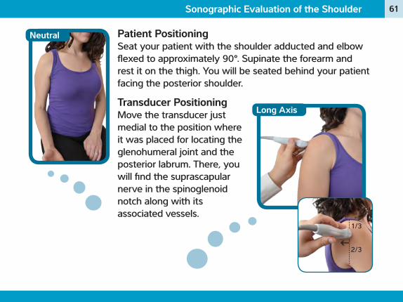
Patient PositioningSeat your patient with the shoulder adducted and elbow flexed to approximately 90°. Supinate the forearm and rest it on the thigh. You will be seated behind your patient facing the posterior shoulder.
Neutral
Transducer PositioningMove the transducer just medial to the position where it was placed for locating the glenohumeral joint and the posterior labrum. There, you will find the suprascapular nerve in the spinoglenoid notch along with its associated vessels.
Long Axis
1/3
2/3

62 Sonographic Evaluation of the Shoulder
Pathology• Suprascapular Nerve Compression • Suprascapular Nerve Hypertrophy• Paralabral Cyst
Notes

63Sonographic Evaluation of the Shoulder
Suprascapular Nerve | Long Axis
SonogramGlenoid
Humeral head
Infraspinatus tendon
Posterior labrum
Suprascapular nerve (in the spinoglenoid notch)
G
H
I
G H
I


