
C H A P T E R
17
Myelin-Associated Glycoprotein Gene
John Georgiou, Michael B. Tropak, and John C. Roder
MAG INTRODUCTION
Specialized glial cells, oligodendrocytes in the central nervous system (CNS), and Schwann
cells in the peripheral nervous system (PNS) elaborate cytoplasmic wrappings known as
myelin around axons. The myelination process requires a complex series of interactions
between the glial cells and axons, which remain poorly understood. Myelin functions to
insulate neurons and facilitates the rapid signal conduction required in organisms with
complex nervous systems. Myelin-associated glycoprotein (MAG) is a relatively minor
constituent of both CNS and PNS myelin that has been implicated in the formation and
maintenance of myelin. However, it is also a cell recognition molecule involved in neuron-
glial interactions, including regulation of axonal outgrowth and nerve regeneration.
Discovery of Myelin-Associated Glycoprotein, MAG
Prior to the discovery of MAG in 1973, the major proteins in compact myelin such as
myelin basic protein (MBP) and proteolipid protein (PLP) were known. During the early
1970s it became clear that proteins on the surface which mediate adhesion are generally
glycosylated. Consequently, Quarles and colleagues used radiolabeled fucose to identify
MAG (Quarles et al., 1973), a myelin glycoprotein that might mediate adhesive inter-
actions between glial and neuronal cells that are important for the formation of the myelin
sheath. MAG was cloned in 1987 (Arquint et al., 1987), and DNA sequence analysis
revealed that the MAG cDNA that was isolated was derived from the same mRNA as
clone p1B236, a randomly selected, brain-speciWc, partial cDNA isolated previously in
1983 (SutcliVe et al., 1983). Originally, 1B236 was thought to be a neuronal protein;
however, subsequent studies conWrmed that MAG and 1B236 are one and the same
(Arquint et al., 1987; Lai et al., 1987a; Noronha et al., 1989). Subsequent Wndings,
described here, have yielded insight into the structure and function of MAG, the major
CNS myelin glycoprotein.
Nomenclature
While antibodies (Ab) consist of domains that belong to the immunoglobulin (Ig) super-
family (IgSF) and are involved in protein-protein interactions, there are several Ig-like
proteins that recognize carbohydrates and that are also known as I-type lectins (see the
review by Kelm, 2001). Cell surface receptors that bind sialic-acid were distinguished as a
subfamily of I-lectins soon after the discovery of sialoadhesin, a macrophage adhesion
molecule that recognizes sialylated glycans (Crocker et al., 1994). Sialoadhesin was found
to share sequence similarity with the CD22 B-lymphocyte surface receptor and also with
Myelin Biology and Disorders, Volume 1 421Copyright 2004, Elsevier Science (USA).
All rights reserved.
MAG. All of these IgSF members bind sialic acid (Freeman et al., 1995; Kelm et al., 1994),
and it was subsequently suggested they be referred to as the ‘‘siglec’’ family, derived from
sialic acid-binding/immunoglobulin-like/lectin (Crocker et al., 1998). There are now 11
members, which are grouped according to their structural and functional similarities (Fig.
17.1). MAG, which is designated siglec-4a, and the related siglec-4b, which is known as
Schwann cell myelin protein (SMP) and found in birds, are two of only three siglecs
expressed by cells outside of the haematopoietic system. A third siglec with marked
sequence similarity to siglec-10, named siglec-11, was recently cloned and found to be
expressed on brain microglia (Angata et al., 2002). However, there is no mouse ortholog of
siglec-11, and hence it is believed to have evolved after the split of primate and rodent
lineages. Most of the scientiWc community continues to use the term MAG instead of
siglec-4a, and hence for convenience we do the same here.
MAG Localization
MAG expression is almost exclusively associated with glial cells that will form myelin.
Immunolocalization studies at the light and electron microscope levels demonstrate that
MAG in the CNS and PNS is found on the Schwann cell and oligodendrocyte membrane
in the periaxonal space (Bartsch et al., 1989; Martini and Schachner, 1986; Sternberger
et al., 1979; Trapp and Quarles, 1982). The periaxonal space is the 12 to 14 nm interface
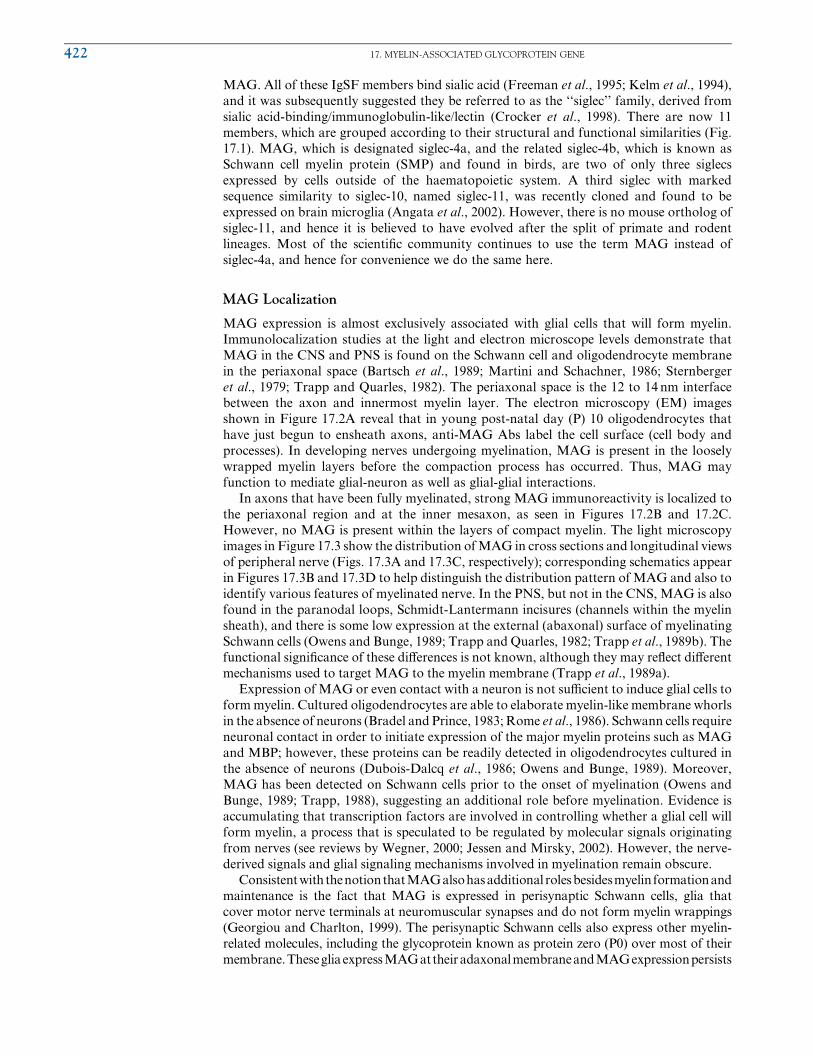
between the axon and innermost myelin layer. The electron microscopy (EM) images
shown in Figure 17.2A reveal that in young post-natal day (P) 10 oligodendrocytes that
have just begun to ensheath axons, anti-MAG Abs label the cell surface (cell body and
processes). In developing nerves undergoing myelination, MAG is present in the loosely
wrapped myelin layers before the compaction process has occurred. Thus, MAG may
function to mediate glial-neuron as well as glial-glial interactions.
In axons that have been fully myelinated, strong MAG immunoreactivity is localized to
the periaxonal region and at the inner mesaxon, as seen in Figures 17.2B and 17.2C.
However, no MAG is present within the layers of compact myelin. The light microscopy
images in Figure 17.3 show the distribution ofMAG in cross sections and longitudinal views
of peripheral nerve (Figs. 17.3A and 17.3C, respectively); corresponding schematics appear
in Figures 17.3B and 17.3D to help distinguish the distribution pattern of MAG and also to
identify various features of myelinated nerve. In the PNS, but not in the CNS, MAG is also
found in the paranodal loops, Schmidt-Lantermann incisures (channels within the myelin
sheath), and there is some low expression at the external (abaxonal) surface of myelinating
Schwann cells (Owens and Bunge, 1989; Trapp and Quarles, 1982; Trapp et al., 1989b). The
functional signiWcance of these diVerences is not known, although they may reXect diVerent
mechanisms used to target MAG to the myelin membrane (Trapp et al., 1989a).
Expression of MAG or even contact with a neuron is not suYcient to induce glial cells to
formmyelin. Cultured oligodendrocytes are able to elaborate myelin-like membrane whorls
in the absence of neurons (Bradel and Prince, 1983; Rome et al., 1986). Schwann cells require
neuronal contact in order to initiate expression of the major myelin proteins such as MAG
and MBP; however, these proteins can be readily detected in oligodendrocytes cultured in
the absence of neurons (Dubois-Dalcq et al., 1986; Owens and Bunge, 1989). Moreover,
MAG has been detected on Schwann cells prior to the onset of myelination (Owens and
Bunge, 1989; Trapp, 1988), suggesting an additional role before myelination. Evidence is
accumulating that transcription factors are involved in controlling whether a glial cell will
form myelin, a process that is speculated to be regulated by molecular signals originating
from nerves (see reviews by Wegner, 2000; Jessen and Mirsky, 2002). However, the nerve-
derived signals and glial signaling mechanisms involved in myelination remain obscure.
Consistentwith thenotionthatMAGalsohasadditional rolesbesidesmyelin formationand
maintenance is the fact that MAG is expressed in perisynaptic Schwann cells, glia that
cover motor nerve terminals at neuromuscular synapses and do not form myelin wrappings
(Georgiou and Charlton, 1999). The perisynaptic Schwann cells also express other myelin-
related molecules, including the glycoprotein known as protein zero (P0) over most of their
membrane.ThesegliaexpressMAGattheiradaxonalmembraneandMAGexpressionpersists
422 17. MYELIN-ASSOCIATED GLYCOPROTEIN GENE

FIGURE 17.1Structure and comparison of siglecs. Schematic showing Ig-like lectins that bind to sialic acid (siglecs). MAG belongs to the siglec-4a group,
together with the related Schwann cell myelin protein (SMP, siglec-4b) found in birds. Not shown is the recently cloned human siglec-11, which has no
rodent homolog. (A) Siglecs are type-I membrane proteins with an extracellular region containing a homologous V-set Ig-like domain and a varying
number of C2-set Ig-like domains at the N-terminus. The cytoplasmic tails of all siglecs apart from sialoadhesin contain tyrosine residues (Y) within
potential signaling motifs. Those motifs that Wt the consensus sequence for an immunoreceptor tyrosine-based inhibition motif (ITIM) are shown in
pink. The membrane-distal tyrosine-based motifs that are highly conserved in CD33-related siglecs are shown in green. Several siglecs undergo
alternative splicing, but only the known full-length forms are illustrated. Potential N-linked glycans are indicated in ball-and-stick form. (B)
Alignment of the C-terminal portions of the cytoplasmic tails of CD33-related siglecs reveals two conserved tyrosine-containing motifs. The
sequences for siglec-4a and -4b, shown at the top, reveal that the distal motif is similar to the CD33-related siglecs, however, the proximal motif is
not conserved. Residues that are identical are boxed in black and residues that are conserved are boxed in gray. The membrane-proximal motif
conforms to the consensus ITIM sequence, whereas the distal motif does not. (C) Positions of key residues in sialoadhesin that bind the
N-acetylneuraminic acid (Neu5Ac) portion of 3’ sialyllactose, as revealed in a ligand-bound crystal structure of the sialoadhesin N-terminal domain.
An essential arginine (Arg97) on the F strand (conserved in the other siglecs) forms a salt bridge with the carboxylate of sialic acid (Neu5Ac) and two
tryptophans (Trp2 and Trp106) on the A and G strands form hydrophobic contacts with the N-acetyl and glycerol side groups of Neu5Ac,
respectively. (D) Schematic diagram of the V- and C-type domains from immunoglobulins, showing the topology of the b-strands in the two b-sheets.Ig domains belonging to the C2 set have a topology similar to the C-type domain. In domain 1 of sialoadhesin, the inter-sheet disulphide bridge
connecting strands B and F is replaced by an intra-sheet dishulphide bridge connecting strands B and E; the C’’ strand is replaced by a coiled structure
and G-strand is split in two. Parts A through C have been modiWed with permission from Crocker and Varki, 2001, TRENDS in Immunology 22,
337-342, 2001, Elsevier Science Ltd.
au10 au11
au12
MAG INTRODUCTION 423
after denervation including at newly formed glial cell extensions. It is possible that factors
present at synapses may prevent myelination, or alternatively, MAG may mediate adhesion
between axons and surrounding glia. Regardless, it is clear that expression of myelin-related
proteins, includingMAG, does not obligate glial cells to formmyelin wrappings.
Various observations, including MAG adaxonal localization, have suggested that it
mediates interactions between the axon and myelin sheath. Expression of MAG on
oligodendrocyte tips prior to contact with axons (Bartsch et al., 1989), and the fact that
MAG is the Wrst myelin protein exported to the tips, further supported a role for MAG in
the initial stages of myelin formation. The myelination process begins when the glial cell
process (mesaxon) begins to spiral around the axon, and initial observations in the PNS
indicate MAG can be Wrst detected in the periaxonal space after 1.5 turns of the mesaxon
around the axon (Martini and Schachner, 1986). This suggests that MAG is involved in
some aspect of the wrapping process, and as discussed later in the section titled ‘‘Multiple
Gene Knockouts That Include Deletion of MAG,’’ MAG’s involvement in the spiraling of
myelinated Schwann cells has also been implicated from studies of double mutant mice
lacking P0 and MAG. Finally, MAG has an important role in the control of axonal
outgrowth, and this feature is discussed later in the section titled ‘‘Control of Axonal
Growth and Regeneration by MAG.’’
Here we introduce that MAG exists primarily as two isoforms, and that the expression
of each is regulated temporally and spatially. The isoforms result from alternative splicing
to yield a relatively shorter protein known as small MAG (S-MAG) and a longer protein
called large MAG (L-MAG). L-MAG predominates during CNS development, including
the myelination process, whereas S-MAG accumulates in later stages (Inuzuka et al., 1991;
Lai et al., 1987a; Pedraza et al., 1991; Tropak et al., 1988). In the peripheral nervous
system, L-MAG is always a minor constituent. MAG isoforms have unique signaling
capacities, for instance, L-MAG can activate Fyn kinase, and thus the diVerential expres-
sion of MAG isoforms likely has important functional consequences. More information on
the unique roles of each MAG isoform will be revealed throughout this chapter.
FIGURE 17.2Electron microscopy reveals MAG’s periaxonal localization. Detection of MAG by immuno-EM from mouse
optic nerve at P10 (A-B) and P14 (C). (A–B) MAG immunoreactivity on the cell surface (A) and processes (B) of
an oligodendrocyte prior or during the time of axon (Ax) ensheathment. Preembedding staining procedures were
used in association with anti-MAG Abs and visualized by peroxidase-coupled protein A. (C) When compact
myelin surrounds the axon, MAG is conWned to the periaxonal region and noncompacted myelin (arrows). Post-
embedding staining technique is necessary to demonstrate the periaxonal localization of MAG due to presence of
compact myelin (M). MAG was detected with secondary Abs absorbed to colloidal gold (gold particles appear as
black dots). Scale bars: A ¼ 1 mm; B, C ¼ 0.2 mm. Reprinted with permission from Bartsch, KirchhoV, and
Schachner, 1989, Journal of Comparative Neurology 284, 451–462, 1989, Wiley-Liss, Inc.
au13 au14
424 17. MYELIN-ASSOCIATED GLYCOPROTEIN GENE

MAG Gene Structure
MAG Promoter and Gene Regulation
Similar to myelin genes such as P0 and P2 basic protein (Peirano et al., 2000), the MAG
promoter lacks a TATA box and instead consists of a GC-rich region (Ye et al., 1994).
IdentiWcation of consensus sequence sites for SP1 and AP2 transcription factors commonly
found at GC-rich promoters is consistent with foot-printing experiments using the MAG
promoter transiently expressed in an immortalized glial cell line (Laszkiewicz et al., 1997).
The regulatory region ofMAG extends to either side of a 152 bp promoter core from�1.6 to
þ0.6 kb. A promoter proximal segment that contains sites for strong transcriptional activa-
tors was identiWed; however, there is a region downstream containing inhibitory cis-elements
(Grubinska et al., 1994). The fact that CpG islands in the MAG regulatory region become
FIGURE 17.3MAG distribution in myelinated nerve sections. (A) Confocal image from a transverse section of rat sciatic nerve,
double-labeled with a mouse monoclonal Ab against MAG (red, detected by TRITC Xuorescence) and also with
rabbit antiserum against b4 integrin (green, detected by FITC Xuorescence). MAG is localized on the inner/
adaxonal membrane, and b4 integrin is localized around the entire circumference of the outer/abaxonal mem-
brane. Compact myelin is not stained and thus appears black. (B) The circumferential organization of a
myelinated axon is shown schematically. (C) Image on the left shows MAG immunoXuorescence from a longitu-
dinal section of sciatic nerve. Image on the right shows MAG distribution in teased single Wbers. (D) Schematic
showing myelinated axon longitudinal organization and MAG localization. Parts A and B were reprinted with
permission from Scherer and Arroyo, 2002, Journal of the Peripheral Nervous System 7, 1–12, 2002 Peripheral
Nerve Society, Inc. Part C was modiWed with permission from Altevogt, Kleopa, Postma, Scherer, and Paul, 2002,
Journal of Neuroscience 22, 6458–6470, 2002, by the Society for Neuroscience.
MAG INTRODUCTION 425
more demethylated as oligodendrocytes progress along the diVerentiation pathway under-
scores the importance of methylation in regulation ofMAG expression. Transcription from
the MAG promoter in Schwann cells does not appear to be aVected by glial transcription
factors Krox-20 (Topilko et al., 1994) or Sox-10 (Stolt et al., 2002). Although MAG
expression is increased in oligodendrocytes in the presence of both transcription factors
Oct and Myc, it is not clear whether this is a direct eVect (Jensen et al., 1999).
MAG Exon Structure
The MAG gene has been localized to human chromosome 19 (Barton et al., 1987), and
mouse chromosome 7 (Barton et al., 1987; D’Eustachio et al., 1988), followed by high-
resolution mapping to 19q13.1 (Spagnol et al., 1989; Trask et al., 1993). Elucidation of the
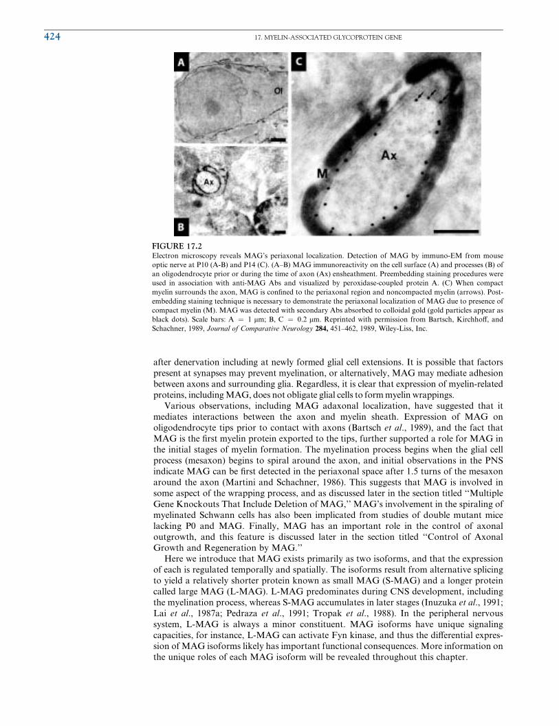
intron/exon structure of rat and mouse MAG genes revealed 13 exons spanning approxi-
mately 16 kb where each Ig domain is encoded by a single exon (Lai et al., 1987a; Nakano
et al., 1991). Exon 12 can be alternatively spliced in, or skipped, to produce S-MAG or
L-MAG, respectively. The schematic shown in Figure 17.4 illustrates the structure and
products of the MAG gene. Exon 2 from the 5’ noncoding region can also be spliced
(Fujita et al., 1989; Lai et al., 1987a; Tropak et al., 1988). Splicing of exon 2 and exon 12
occur independently. One form of MAG lacking exon 2 is predominant in the PNS,
whereas mRNA containing exon 2 predominates in the CNS. In contrast to the known
diVerences in signaling capabilities associated with L-MAG versus S-MAG, the signiW-
cance, if any, of exon 2 splicing is unknown.
Alternative Splicing Produces Two Main MAG Isoforms
Initially, the S- and L-MAG isoforms detected in the CNS were attributed to diVerential
glycosylation (Quarles et al., 1973). Subsequently, the two isoforms were shown to be
derived from diVerent mRNAs (Frail and Braun, 1984; Salzer et al., 1987; Tropak et al.,
1988) by alternative splicing of exons from a primary RNA transcript (Lai et al., 1987a).
The two isoforms of 67 and 72 kDa (sizes of proteins after deglycosylation) share a
common region in their cytoplasmic domain but diVer in the length and sequence of the
amino acids at the C-terminus. The cytoplasmic domain of MAG is encoded by exons 11,
12, and 13. When exon 12 is included, a premature in-frame stop codon results in the
shorter S-MAG isoform with an intracellular sequence of 90 residues that contains a
unique 10 amino acid C-terminal end. When exon 12 is excluded, the larger L-MAG is
produced, which has an additional 54 amino acids derived from exon 13.
DiVerential expression of the two MAG isoforms during development in the PNS and
CNS of rat, mouse, and human was veriWed at the RNA (Frail and Braun, 1984; Frail et al.,
FIGURE 17.4Schematic of MAG gene locus. The MAG gene contains 13 exons spanning 16 kb and each of its Ig domain is
encoded by a single exon (exons 5 to 9). Exons (rectangles), introns (line) and the exons that undergo alternative
splicing are indicated (see legend). Exon 12 can be alternatively spliced in, or skipped, to produce S-MAG or
L-MAG mRNA transcripts, respectively. There is also alternative splicing of exon 2, which occurs independently
of exon 12 splicing (not all combinations are shown); however, it does not contribute to the coding region and its
signiWcance is unknown. Alternative polyadenylation cleavage site is depicted as a vertical dashed line in exon 13.
426 17. MYELIN-ASSOCIATED GLYCOPROTEIN GENE

1985; Lai et al., 1987b; Miescher et al., 1997; Tropak et al., 1988) and protein levels
(Inuzuka et al., 1991; Pedraza et al., 1991). Developmental regulation may be related to
neuronal contact or the local environment because oligodendrocytes in culture express
both MAG isoforms (Tropak et al., 1988). Interestingly, when L-MAG is constitutively
expressed at high levels in Schwann cells, independent of axonal contact, a greater number
of axons are segregated compared to control cells, which express MAG only upon axonal
contact (Owens et al., 1990). This observation strongly supports the adhesive role of MAG
prior to the start of myelination. However, the biological relevance of these observations is
not clear, since only low levels of L-MAG can be detected in control Schwann cells and
during PNS development (Pedraza et al., 1991; Tropak et al., 1988).
Although myelin formation by oligodendrocytes and Schwann cells appears outwardly
similar, the mechanistic details are distinct. However, oligodendrocytes diVer from
Schwann cells in several respects. Unlike Schwann cells, which can only myelinate a single
segment of an axon, oligodendrocytes are able to simultaneously myelinate multiple
segments of diVerent axons. Interestingly, there appears to be a correlation between the
relative levels of L-MAG and S-MAG expression and the four morphological types of
oligodendrocytes (Butt et al., 1998). Thus, it is possible that L-MAG may function in the
early events of myelination related to the ability of oligodendrocytes to myelinate multiple
axonal segments.
Alternative splicing of MAG is regulated by RNA binding proteins known as QKI. The
regulation of MAG isoform expression by QKI was discovered from studies of a naturally
occurring mouse mutant known as ‘‘quaking’’ (qk) that has an altered qkI gene regulatory
region (noncoding). Reduced QKI levels in qk mice alters the developmental expression of
the twoMAG isoforms in the CNS and results in S-MAG as the major mRNA throughout
development, while L-MAG is scarcely expressed (Frail and Braun, 1985; Fujita et al.,
1990). The phenotype of qk mice is described later in the section titled ‘‘Naturally
Occurring Mutations AVecting MAG.’’ QKI contains an RNA-binding domain and
belongs to the signal transduction and activator of RNA (STAR) family (Ebersole et al.,
1996; Vernet and Artzt, 1997). Of the many QKI isoforms that exist, the nuclear localized
isoform QKI-5 has been shown to regulate alternative splicing of a MAG minigene as well
as the myelin genes PLP and MBP (Wu et al., 2002). In the proposed model, binding of
QKI-5 to the QASE consensus sequence downstream of the 5’ splice site may interfere with
recognition of the splice site or the downstream intronic enhancer, thereby resulting in
skipping of exon 12 and concomitant production of the L-MAG mRNA.
MAG BIOCHEMISTRY, STRUCTURE, AND ADHESIVE PROPERTIES
Following translation MAG undergoes several modiWcations including phosphorylation,
remodeling of oligosaccharides by sulfotransferases and sialyltransferases, and palmityla-
tion. With the exception of palmitylation, each of these modiWcations will be discussed in
greater detail. Palmitylation has been shown to be important in membrane targeting and
activity of nonreceptor tyrosine kinases (reviewed in Resh, 1994). Similarly, palmitylation
of haemagglutinin is important for the infectivity of inXuenza virus, although the mechan-
ism is unclear (Fischer et al., 1998). Pedraza and colleagues experimentally veriWed the
initial prediction that MAG is palmitylated by showing that Cys531 residue in the trans-
membrane domain is the site of palmitylation (Pedraza et al., 1990). Based on other
systems, palmitylation of MAG may be important for some aspect of membrane targeting.
Post-Translational Regulation of MAG
MAG Phosphorylation
The primary structure of MAG revealed potential Ser, Thr, and Tyr phosphorylation sites
within its cytoplasmic domain (Arquint et al., 1987; Salzer et al., 1987). In CNS myelin and
oligodendrocytes, L-MAG is the predominant isoform that is phosphorylated, whereas
MAG BIOCHEMISTRY, STRUCTURE, AND ADHESIVE PROPERTIES 427
S-MAG is the major phosphorylated isoform in PNS myelin and Schwann cells (Afar et al.,
1990; Agrawal et al., 1990; Bambrick and Braun, 1991; Edwards et al., 1988, 1989;
Umemori et al., 1994; Yim et al., 1995). L-MAG is phosphorylated in vivo and in vitro
primarily on Ser, but also on Thr and Tyr residues. S-MAG is phosphorylated consti-
tutively only on Ser. In cultured oligodendrocytes, both S- and L-MAG isoforms are
phosphorylated on Ser, while in transformed Schwann cells only S-MAG is present and
phosphorylated (KirchhoV et al., 1993). Phorbol ester enhances phosphorylation two- to
three-fold, suggesting PKC is involved. Recently it was demonstrated that L-MAG is a
PKA substrate (Kursula et al., 2000).
S-MAG and L-MAG contain potential Tyr phosphorylation sites, and tyrosine kinases
can bind and phosphorylate MAG. Phosphorylation of MAG by v-fps and v-src protein-
tyrosine kinases has been demonstrated in vitro (Afar et al., 1990; Edwards et al., 1988).
Fyn kinase, a member of the Src family tyrosine kinases, interacts with MAG and
phosphorylates the Tyr at position 620 found in L-MAG (Jaramillo et al., 1994; Umemori
et al., 1994). In transfected cells, Fyn only associates with L-MAG, not with S-MAG. In
brain lysates however, Fyn was co-immunoprecipitated with both forms of MAG, and
conversely, each of L- and S-MAG co-immunoprecipitated with Fyn. The lack of Fyn
interaction with S-MAG in the expression system was suggested to be due to the lack of
additional molecules that normally mediate this interaction in the brain. Another inter-
pretation is that Fyn does not associate with S-MAG in the brain and that the interaction
is seen because S-MAG dimerizes with L-MAG.
MAG Glycosylation
Approximately 30% of the mass of MAG is due to carbohydrate (Quarles, 1983), which is
consistent with the eight N-linked oligosaccharide addition sites identiWed in the predicted
amino acid sequence of rat MAG (Arquint et al., 1987; Lai et al., 1987b; Salzer et al.,
1987). Mutagenesis experiments of the conserved Asn or Ser residues in the glycosylation
consensus sequence have shown that each of the predicted glycosylation sites are utilized
when expressed in CHO or COS-1 cell lines (Sgroi et al., 1996; Tropak and Roder, 1997).
The majority of oligosaccharides on MAG are of the complex type; about two-thirds are
tri- or tetra-antennary, one-third are biantennary, and few or none are the high-mannose
type (Noronha et al., 1989). A high proportion of the oligosaccharides are sialylated and
sulphated (Matthieu et al., 1975; Quarles et al., 1983). Although some experimental
evidence suggested that O-linked oligosaccharides may be present in MAG (Pedraza
et al., 1990), these results have been attributed to the presence of N-glycosidases in the
batch of O-glycosidase used to test for the presence of O-linked glycans (Salzer, personal
communication).
The carbohydrate epitope recognized by the L2/human natural killer (HNK)-1 Ab
(Noronha et al., 1986) is expressed on MAG (McGarry et al., 1983). It is also found on
a number of IgSF member cell adhesion molecules (CAMs) in the nervous system such as
the neural cell adhesion molecule (N-CAM), L1, P0 (Bollensen et al., 1988; Kruse et al.,
1984), as well as unrelated adhesive molecules such as peripheral myelin-protein-22
(PMP22) and proteoglycans (Snipes et al., 1993). The conservation of the epitope through-
out phylogeny (Bajt et al., 1990) is suggestive of the importance of HNK-1 in adhesion.
Abs against the L2/HNK-1 epitope have been shown to perturb astrocyte-neuron cell
adhesion, neurite outgrowth and attachment of cells to laminin (Hall et al., 1993; Kune-
mund et al., 1988; Riopelle et al., 1986). Oligosaccharides expressing the HNK-1 epitope
have been used to block P0-mediated cell adhesion (GriYth et al., 1992) and cell-cell and
cell-substrate interactions (Kunemund et al., 1988). Currently, the gp120 receptor on the
AIDS virus is the only protein capable of interacting with the HNK-1 carbohydrate on
MAG (van den Berg et al., 1992a). In the case of rat MAG, the HNK-1 epitope has been
localized to oligosaccharides in either domain 4 or domain 5 (Pedraza et al., 1995), whereas
all oligosaccharides on human MAG have been suggested to express the HNK-1 epitope
(Burger et al., 1991). The HNK-1 epitope consists of a 3’ sulphated glucuronyl residue
found on glycolipids (Chou et al., 1986), N-linked oligosaccharides (Voshol et al., 1996),
and to a limited extent on O-linked oligosaccharides (Ong et al., 2002). Sulphation of the
428 17. MYELIN-ASSOCIATED GLYCOPROTEIN GENE
glucuronic acid is critical for binding of the Ab. However, sulphation alone is not suYcient
for binding of Abs recognizing the HNK-1 epitope, since most oligosaccharides on rat
MAG are sulphated, yet the protein is poorly recognized by the Ab (O’Shannessy et al.,
1985). The species-dependent expression (O’Shannessy et al., 1985) of the epitope, as well
as the fact that not all MAGmolecules express the epitope (Burger et al., 1992; Kruse et al.,
1984), suggests that HNK-1 may not play a major role in MAG function. Alternatively,
one component of the HNK-1 epitope, such as the sulphate group, may be important in
MAG function.
MAG Structure
On the basis of the amino acid sequence derived from the cDNA for rat MAG, the
protein was predicted to be a type I membrane glycoprotein consisting of an N-terminal
extracellular domain, a single transmembrane domain and a short cytoplasmic domain
(Arquint et al., 1987; Lai et al., 1987b; Salzer et al., 1987). These predictions were later
experimentally veriWed (Johnson et al., 1989; Pedraza et al., 1990). The extracellular
portion of MAG was predicted to consist of Wve Ig-like domains, based on the presence of
amino acid sequences inMAG,which are conserved among allmembers of the IgSF.Human
and mouse MAG have 98% and 95% amino acid sequence identity, respectively, with rat
MAG (Fujita et al., 1989; Sato et al., 1989; Spagnol et al., 1989). SMP, which overall shares
45% amino acid sequence identity with rat MAG, is very likely a quail ortholog of MAG
(Dulac et al., 1992).
IgSF
The IgSF consists of a large number of closely related proteins containing one or more
domains that are similar in sequence to domains found in Igs (Williams and Barclay, 1988).
The proto-typical Ig domain, found in circulating Igs, consists of two anti-parallel b-sheetsheld together by an inter-sheet disulphide bridge (see Fig. 17.1D and Amzel and Poljak,
1979). Based on the sequences of Ig-like molecules available at that time, the members of
the IgSF were divided into three sets based on the size of the linker between the conserved
cysteines and the presence of conserved residues characteristic of the variable (V-) domain
or constant (C-) domain from Igs (Williams and Barclay, 1988). The variable domain
consists of disulphide-linked anti-parallel sheets with four beta strands (referred to as D, E,
B, and A), in one face, and Wve strands (referred to as C, C’, C’’, F, and G) in the other (see
Figure 17.1D). In contrast, the C-domain consists of two anti-parallel disulphide-linked
sheets with four b-strands (referred to as D, E, B, and A) in one face and three strands
(referred to as C, F, and G) in the other. Domains with greatest similarity to the V-domain
or C-domain were placed in the V- or C1-sets, respectively. Domains which were similar in
size to the shorter C-domain and contained conserved residues found in b-strands D and E
from the V-domain were placed in the C2-set. Recently, a fourth set, the I-set, has been
deWned on the basis of the structure of telokin and its close similarity to other members
of the IgSF (Harpaz and Chothia, 1994). Members of the I-set have the same number of
b-strands found in the C1- and C2-sets and conserved sequences characteristic of V- and
C1-sets.
Disulphide Linkage
Domain 1 of MAG is most similar to Ig domains in the V-set (Williams and Barclay, 1988),
whereas the other four domains are most similar to Ig domains in the C2-set (Arquint et al.,
1987; Lai et al., 1987a, 1987b; Salzer et al., 1987). The V-like domain 1 of MAG is unique
in that the Cys pair (Cys42 and Cys100) may form an intra-sheet disulphide bridge
(Williams and Barclay, 1988) as opposed to the inter-sheet disulphide bridge encountered
in most Igs. A similar intra-sheet disulphide bridge has been identiWed in domain 2 of the
crystallographically determined structure of CD2, another member of the IgSF (Jones
et al., 1992). Domains 1 and 2 of MAG each contain an additional Cys (Cys37 and Cys159)
near the N-termini of the domains. Experimental evidence suggests that the additional Cys
form an interdomain disulphide bridge between domains 1 and 2 (Pedraza et al., 1990).
MAG BIOCHEMISTRY, STRUCTURE, AND ADHESIVE PROPERTIES 429

Modeling studies suggest that Cys37 and Cys159 are suYciently close to form an inter-
domain disulphide bridge (Kursula, 2001).
MAG Is a Member of the Sialic-Acid Binding Family, Siglec
The unique arrangement of conserved Cys residues in domains 1 and 2 in all MAG
orthologues is also a distinguishing feature shared by all IgSF members belonging to the
siglec family of I-type lectins. The siglec family of proteins including sialoadhesin, CD22,
CD33 family all recognize sialylated glycans. Experimental demonstration of MAG’s
ability to recognize sialylated glycans on cells was Wrst shown by Kelm and colleagues
(1994). In addition to the conserved Cys residues, all functional siglecs contain an invariant
Arg residue (Arg118 in MAG, Arg97 in sialoadhesin).
The 3D structure of domain 1 from sialoadhesin complexed with sialyllactose has
enabled domain modeling studies of MAG and other siglecs (May et al., 1998). Although
siglec-1 domain 1 does indeed belong to the V-set of Ig domains, it does, however, diVer in
several respects. First, it possesses, as predicted, an intra-sheet disulphide bridge between
strands B to E rather than B to F. Second, the spacing between the two sheets is increased
from the usual 5.6–7.4A range to 8.6A. Third, the C’’ beta strand, which corresponds to a
region of high diversity in the aligned siglec sequences, is replaced with a quasi alpha-helix
like coiling strand. Lastly, the G-b strand is split in two and the other region of siglec
diversity corresponds to extended BC loop of unknown function.
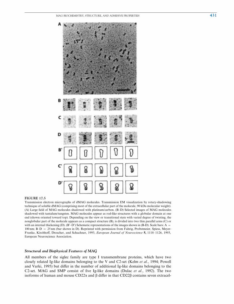
The overall structure of a complete siglec has yet to be determined. However, based on
EM pictures of a proteolytically generated soluble form of MAG (dMAG) (Sato et al.,
1984), the molecule appears to be folded back on itself with domains 1 and 2 folded back
on domains 5 and 4, respectivel y (Fig. 17.5, and Fahrig et al., 1 993). The higher or derstructure of the domains 1 through 5 is also suggested by biophysical studies using a
recombinant soluble derivative of rat MAG (Attia et al., 1993). Furthermore, this type of
arrangement, where the IgSF domains are folded back in a hairpin/horseshoe conWgura-
tion have been shown in the 3D structures of hemolin and TAG-1/axonin derived by X-ray
crystallography (Freigang et al., 2000; Su et al., 1998) and EM pictures of L1 (Schurmann
et al., 2001).
Although domain 1 alone from sialoadhesin retains the ability to bind sialylated glycans
(Nath et al., 1995), the minimum MAG deletion mutant displaying lectin activity consists
of domains 1, 2, and 3 (Kelm et al., 1994; Meyer-Franke et al., 1995). The sialic acid
binding site on MAG has been indirectly mapped to domain 1 using a MAG mutant in
which the invariant Arg in domain 1 found in all siglecs has been mutated to Ala/Asp
(Tang et al., 1997a). Deletion mapping studies of the lectin activity and of a conforma-
tional epitope recognized by the 513 monoclonal Ab suggest that the three N-terminal
domains of MAG are conformationally linked (Meyer-Franke et al., 1995). Similarly, do-
mains 1 and 2 of CD22 appear to be conformationally linked (Nath et al., 1995). Thus, the
N-terminal domains in some siglecs may not fold independently, but may require the
presence of additional domains to attain the mature conformation. The fact that domains
1, 2, and 3 appear to be conformationally linked is consistent with EM and biophysical
studies, which suggest that the domains in MAG adopt a higher-order structure with the
domains folded back on each other (Attia et al., 1993; Fahrig et al., 1987).
Two of the diVerences that contribute to binding of the 3’ sialyllactose lies to the GFCC’
face of the Ig domain. The conserved residues Trp2 and Trp106 are directed away from the
GFCC b-sheet enabling them to hydrophobically interact with the sialyllactose and
resulting in the observed increase in inter-sheet distance. Trp2 interacts with the 5’aminoa-
cyl group, whereas Trp106 contacts the glycerol tail of sialic acid. In most Ig domains these
two residues are directed between the b-sheets where they contribute to formation of the
hydrophobic core. The split G-strand may enable backbone mediated H-bonds with the
hydroxyl groups of the sialic acid glycerol tail. The invariant Arg97, which has been shown
by mutagenesis to be critically important for sialic acid recognition, forms a salt bridge
with the carboxylate of sialic acid. A similar interaction is seen in the 3D structure of an
unrelated sialic acid binding lectin (VP1 from polyoma virus) and possibly other bacterial
sialic acid binding lectins (May and Jones, 2001).
430 17. MYELIN-ASSOCIATED GLYCOPROTEIN GENE
Structural and Biophysical Features of MAG
All members of the siglec family are type I transmembrane proteins, which have two
closely related Ig-like domains belonging to the V and C2-set (Kelm et al., 1994; Powell
and Varki, 1995) but diVer in the number of additional Ig-like domains belonging to the
C2-set. MAG and SMP consist of Wve Ig-like domains (Dulac et al., 1992). The two
isoforms of human and mouse CD22a and b diVer in that CD22b contains seven extracel-
FIGURE 17.5Transmission electron micrographs of dMAG molecules. Transmission EM visualization by rotary-shadowing
technique of soluble dMAG (comprising most of the extracellular part of the molecule, 90 kDa molecular weight).
(A) Large Weld of MAG molecules shadowed with platinum/carbon. (B–D) Selected images of MAG molecules
shadowed with tantalum/tungsten. MAG molecules appear as rod-like structures with a globular domain at one
end (shown oriented toward top). Depending on the view or transitional state with varied degree of twisting, the
nonglobular part of the molecule appears as a compact structure (B), is divided into two thin parallel arms (C) or
with an internal thickening (D). (B’–D’) Schematic representations of the images shown in (B-D). Scale bars: A ¼100 nm; B–D ¼ 25 nm (bar shown in D). Reprinted with permission from Fahrig, Probstmeier, Spiess, Meyer-
Franke, KirchhoV, Drescher, and Schachner, 1993, European Journal of Neuroscience 5, 1118–1126, 1993,
European Neuroscience Association.
au1
MAG BIOCHEMISTRY, STRUCTURE, AND ADHESIVE PROPERTIES 431

lular Ig domains, whereas CD22a lacks Ig domains 3 and 4 (Engel et al., 1993; Stamen-
kovic and Seed, 1990; Stamenkovic et al., 1991; Wilson et al., 1991). Sialoadhesin has 17
Ig-like domains (Crocker et al., 1994). However, cDNAs have been isolated, which suggest
the existence of soluble isoforms which are missing C-terminal domains and consist of
either 3 or 16 Ig domains. Human and mouse CD33 consist solely of the V and C2-like
domains, which deWne the I-type sialyl lectins (Simmons and Seed, 1988; Tchilian et al.,
1994). Two isof orms of mou se CD33 have been isolated, whi ch di V er in the size and amino
acid sequence of the C-terminal tail in the cytoplasmic domain.
In addition to the close similarity of the individual members of the I-type lectins, the
localization of CD22 (Wilson et al., 1993), CD33 (Peiper et al., 1988), and MAG (Barton
et al., 1987) to a syntenic region of human chromosome 19 suggests that the members have
evolved from a common ancestor by gene duplication (Mucklow et al., 1995). This
prediction is consistent with the fact that the exon boundaries of the CD22, CD33 and
MAG (Lai et al., 1987b) genes fall within the same regions of the two N-terminal Ig-like
domains , and that co dons at the bounda ries of the exo ns are in the same pha se. Althou gh
sialoadhesion is located on a diVerent chromosome (Mucklow et al., 1995), this does not
necessa rily exclude the possibi lity that sialoadhesin arose from a n an cestor common to
CD22, CD33, and MAG.
Each member of the siglec family is express ed in a rest ricted cell lineag e and bind
preferentially to certain cell types. SMP, which is most similar to MAG (Dulac et al.,
1992), is expressed on quail Schwann cells (Dulac et al., 1988) and oligodendrocytes
(Cameron-Curry and Le Douarin, 1995) and is presumed to bind to neurons. CD22,
which is expressed on the B-cell lineage (Dorken et al., 1986; Engel et al., 1993; Stamen-
kovic and Seed, 1990; Stamenkovic et al., 1991), binds to B- and T -lymphocytes and
monocytes (Crocker et al., 1995; Engel et al., 1993; Stamenkovic and Seed, 1990; Stamen-
kovic et al., 1991). In contrast, sialoadhesin is a nonphagocytic receptor, which is expressed
on tissue macrophages (Stamenkovic and Seed, 1990; Stamenkovic et al., 1991) and binds
selectively to neutrophils, although it will also bind to other cells from the granulocyte
lineage such as lymphocytes (Crocker et al., 1995; van den Berg et al., 1992b). CD33 is
expressed on the myelomonocytic lineage, such as monocytes and tissue macrophages
(Pierelli et al., 1993), and selectively binds to cell lines from the myeloid lineage (Freeman
et al., 1995).
Sialic acids are a family of 9-carbon carboxylic acids usually added to the terminal
positions of oligosaccharides by sialyltransferases in the Golgi apparatus (Schauer, 1982;
Varki, 1992). Sialyltransferases form a family of proteins, which transfer sialic acid from
the donor, CMP-sialic acid, to a speciWc oligosaccharide acceptor on glycolipids or
glycoproteins (CorWeld et al., 1982; Datta and Paulson, 1995; Paulson and Colley, 1989).
Each sialyltransferase catalyzes the attachment of sialic acid via an a-ketosidic linkage todiVerent positions on a monosaccharide unit of a speciWc disaccharide acceptor. Individual
members of the siglec family preferentially recognize sialylated glycans produced by a
speciWc sialyltransferase. CD22 preferentially recognizes sialylated glycans produced by
Galb1-4GlcNAc a2,6 sialyltransferase (2,6N ST), which catalyzes the addition of sialic
acids linked via an a2,6 linkage to Gal(b1,4)GlcNAc (2,6N), a disaccharide acceptor
commonly found on N-linked oligosaccharides (Kelm et al., 1994; Stamenkovic et al.,
1991). In contrast MAG (Kelm et al., 1994), sialoadhesin (Crocker et al., 1991; Kelm et al.,
1994) and CD33 (Freeman et al., 1995) recognize sialylated glycans produced by either the
Ga1b1-3GalNac a2,3 sialyltransferase (2,3-N ST) or Galb1-3(4)GlcNAc a2,3 sialyltrans-
ferase (2,3-O ST) which catalyze the addition of sialic acid via an a2,3 linkage to Galb1-3GalNAc (2,3O) commonly found on O-linked oligosaccharides or Galb1-3(4)GlcNAc
(2,3N) commonly found on N-linked oligosaccharides, respectively. MAG preferentially
binds sialic acid containing proteins and glycans, especially the NeuAc a3 Gal b3 GalNAc
structure, which is commonly found on gangliosides (Collins et al., 1997b; Crocker et al.,
1996; Kelm et al., 1994).
The ability of MAG and siglecs to bind sialylated glycans on the surface of cells
represents not only a mechanism for cell adhesion but also a means for regulating adhe-
sion. Thus, the adhesive functions of MAG (Tropak and Roder, 1997), CD22 (Sgroi et al.,
au2
432 17. MYELIN-ASSOCIATED GLYCOPROTEIN GENE
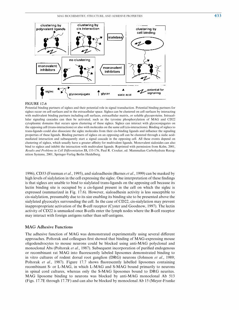
1996), CD33 (Freeman et al., 1995), and sialoadhesin (Barnes et al., 1999) can be masked by
high levels of sialylation in the cell expressing the siglec. One interpretation of these Wndings
is that siglecs are unable to bind to sialylated trans-ligands on the apposing cell because its
lectin binding site is occupied by a cis-ligand present in the cell on which the siglec is
expressed (summarized in Fig. 17.6). However, sialoadhesin activity is less susceptible to
cis-sialylation, presumably due to its size enabling its binding site to be presented above the
sialylated glycocalyx surrounding the cell. In the case of CD22, cis-sialylation may prevent
inappropropriate activation of the B-cell receptor (Cyster and Goodnow, 1997). The lectin
activity of CD22 is unmasked once B-cells enter the lymph nodes where the B-cell receptor
may interact with foreign antigens rather than self-antigens.
MAG Adhesive Functions
The adhesive function of MAG was demonstrated experimentally using several diVerent
approaches. Poltorak and colleagues Wrst showed that binding of MAG-expressing mouse
oligodendrocytes to mouse neurons could be blocked using anti-MAG polyclonal and
monoclonal Abs (Poltorak et al., 1987). Subsequent incorporation of puriWed endogenous
or recombinant rat MAG into Xuorescently labeled liposomes demonstrated binding to
in vitro cultures of rodent dorsal root ganglion (DRG) neurons (Johnson et al., 1989;
Poltorak et al., 1987). Figure 17.7 shows Xuorescently labelled liposomes containing
recombinant S- or L-MAG, in which L-MAG and S-MAG bound primarily to neurons
in spinal cord cultures, whereas only the S-MAG liposomes bound to DRG neurites.
MAG liposome binding to neurons was blocked by anti-MAG monoclonal Ab 513
(Figs . 17.7E throu gh 17.7F) a nd can also be blocked by monoclonal Ab 15 (Meyer -Franke
FIGURE 17.6Potential binding partners of siglecs and their potential role in signal transduction. Potential binding partners for
siglecs occur on cell surfaces and in the extracellular space. Siglecs can be clustered on cell surfaces by interacting
with multivalent binding partners including cell surfaces, extracellular matrix, or soluble glycoproteins. Intracel-
lular signaling cascades can then be activated, such as the tyrosine phosphorylation of MAG and CD22
cytoplasmic domains that occurs upon clustering of these siglecs. Siglecs can interact with glycoconjugates on
the opposing cell (trans-interactions) or also with molecules on the same cell (cis-interactions). Binding of siglecs to
trans-ligands could also dissociate the siglec molecules from their cis-binding ligands and inXuence the signaling
properties of these ligands. Binding partners of siglecs on an opposing cell can be clustered through a sialic acid-
mediated interaction and subsequently start a signal cascade in the opposing cell. All these events depend on
clustering of siglecs, which usually have a greater aYnity for multivalent ligands. Monovalent sialosides can also
bind to siglecs and inhibit the interaction with multivalent ligands. Reprinted with permission from Kelm, 2001,
Results and Problems in Cell DiVerentiation 33, 153-176, Paul R. Crocker, ed. Mammalian Carbohydrate Recog-
nition Systems, 2001, Springer-Verlag Berlin Heidelberg.
au3
MAG BIOCHEMISTRY, STRUCTURE, AND ADHESIVE PROPERTIES 433

and Barres, 1994). MAb 513 was raised against glycoproteins immunopuriWed from
chicken brain membranes using the L2/HNK-1 Ab (Poltorak et al., 1987). In contrast,
the IgM rat MAb 15 was raised against immunopuriWed mouse MAG (Meyer-Franke
et al., 1995). Initial experiments showed that MAG only bound to DRG neurons, which
could be myelinated, but not to cerebellar neurons, which are not myelinated in vivo. Thus,
MAG was thought to interact only with a neuronal ligand, which was found on axons
destined to be myelinated. Subsequent experiments demonstrated that MAG liposomes
could in fact bind to cerebellar neurons, possibly as a result of the diVerent culture
conditions used to grow the neurons (Sadoul et al., 1990). Furthermore, it was shown
that MAG could mediate the heterophilic aggregation of L cell Wbroblasts (Afar et al.,
1991), CHO cells (Attia, 1992), and oligodendrocyte-like cells (Almazan et al., 1992). It is
unlikely that these cells expressed a neuronal ligand. These results experimentally veriWed
the prediction that MAG could function as a CAM. However, these observations also
suggested that MAG could recognize a number of diVerent ligands.
As has been demonstrated for most CAMs (see the review by van der Merwe and
Barclay, 1994), the interaction of MAG with its ligands appears to be multivalent. Thus,
neither an engineered soluble form of MAG nor the proteolytically generated soluble form
of dMAG is able to bind to neurons (Attia, 1992; Sadoul et al., 1990). However, dMAG is
able to bind with high aYnity (KD ~10-7M) to various types of collagen and heparin
(Fahrig et al., 1987; Probstmeier et al., 1992). The inability of MAG liposomes to bind to
these ligands suggests that the manner in which MAG is presented (i.e., soluble versus on
the cell surface) may aVect binding to collagen. Although the biological signiWcance of this
type of interaction is not clear, it may be indicative of MAG’s ability to bind to diVerent
ligands.
MAG interacts with several extracellular matrix molecules. MAG binds to speciWc types
of collagen and by so doing reduces collagen Wbril formation and also integrin-mediated
FIGURE 17.7Neural Adhesion of Recombinant MAG proteins. One-week-old DRG cultures from newborn mice or 4-week spinal cord cultures from P13
embryonic mice. Liposomes labelled with carboxyXuorescein and containing recombinant S- or L-MAG were incubated for 30 minutes with cultures,
washed, then visualized under optics to reveal phase (A, C, E, and G) and also Xuorescence (B, D, F, and H). (A–B) Spinal cord cultures incubated
with L-MAG liposomes. (C–D) Spinal cord cultures incubated with S-MAG liposomes. (E–F) Spinal cord cultures were incubated with
monoclonal anti-MAG 513 Ab (Fab fragment) and S-MAG liposomes. (G–H) DRG cultures were challenged with S-MAG liposomes. ModiWed
with permission from Johnson, Abramow-Newerly, Seilheimer, Sadoul, Tropak, Arquint, Dunn, Schachner, and Roder, 1989, Neuron 3, 377–385,
1989 by Cell Press.
434 17. MYELIN-ASSOCIATED GLYCOPROTEIN GENE

adhesiveness of neural cells (Bachmann et al., 1995; Probstmeier et al., 1992). The ability of
MAG to modify constituents of the extracellular matrix suggests it plays a role in control-
ling adhesive interactions and recognition between cells.
MUTATIONS AFFECTING MAG
Naturally Occurring Mutations Affecting MAG
The earliest indication of MAG’s role in mediating interactions between the axon and
myelin sheath was provided by studies in the dysmyelinating recessive mouse mutant
known as ‘‘quaking’’ (Sidman et al., 1964). The mutation that accounts for the phenotype
in the quaking mouse (qk) occurs in the 5’ regulatory (noncoding) region of the qkI gene,
which generates three alternatively spliced transcripts encoding the QKI proteins QKI-5,
-6, and -7, putative RNA binding proteins suggested to link RNA metabolism with signal
transduction (Ebersole et al., 1996; reviewed by Hardy, 1998). Compared to wild-type
mice, QKI protein levels are reduced drastically in myelinating cells of qk mice (Hardy
et al., 1996). In this mutant, there is an increased molecular weight of both MAG isoforms
due to abnormal glycosylation ( Bartoszewicz et al., 1995; Matthieu et al., 1974). However,
in qk mice, the expression of L-MAG is greatly reduced both at the RNA (Frail and
Braun, 1985; Fujita et al., 1988) and protein levels (Bartoszewicz et al., 1995; Bo et al.,
1995; Fujita et al., 1990). Another factor that contributes in reducing L-MAG levels in the
qk mice appears to be increased endocytosis of L-MAG from periaxonal membranes (Bo
et al., 1995). Regarding S-MAG expression in qk mice, there is an inverse eVect in that the
relative levels are increased (Frail and Braun, 1985; Fujita et al., 1990). The modiWed L- and
S- MAG levels in qk mice are a result of altered QKI expression. For instance, a recent
study found that QKI-5 regulates the alternative splicing of MAG by repressing the
inclusion of exon 12, which is skipped in L-MAG and included in S-MAG (Wu et al.,
2002). The loss of QKI-5 in qk mice may thus be responsible for the alterations in MAG
splicing, although it is not known whether the lack of QKI-6 and -7 also contribute.
Myelin sheaths of qk mice are disrupted in regions lacking MAG. Immuno-EM studies
revealed that in regions of the periaxonal space where MAG cannot be detected, the
characteristic 12 to 14 nm space between the axonal membrane and Schwann cell mem-
brane is altered (Trapp et al., 1984). Furthermore, in the periaxonal region where MAG
was absent, the thickness of the periaxonal cytoplasmic collar (PCC) is reduced. Normally,
myelinating glia from wild-type mice have a well-developed PCC that spans more than half
of the axonal circumference. Although loss of regional expression of MAG is correlated
with the previously stated morphological deWcits, the expression of other myelin proteins,
such as MBP, is also aVected in qk (Li et al., 2000). Therefore, it is possible that the altered
expression of other proteins besides MAGmay account for the loss of the periaxonal collar
in quaking.
B. MAG knockout mice reveal a mixture of minor myelin defects
MAG-deWcient mice reveal various subtle defects related to myelin formation and
maintenance (also see the review by Schachner and Bartsch, 2000). MAG null mice have
been generated by two separate groups using gene targeting methods (Li et al., 1994;
Montag et al., 1994). These knockout mice have defects in myelin that are not severe and
largely aVect CNS myelin sheaths. The modest changes observed in MAG null mice is in
contrast to the observed inability of Schwann cells to segregate large caliber axons and
form myelin in vitro, when MAG expression was greatly reduced (more than Wve-fold)
using MAG antisense RNA (Owens and Bunge, 1991). Although MAG expression in the
Schwann cells was reduced but not eliminated, it is not clear whether this can account for
the diVerence between the in vitro and in vivo experiments. The phenotype is probably not
due to secondary eVects of using MAG antisense RNA because Schwann cells infected
with the MAG sense virus formed normal compact myelin.
Mutant mice that express a truncated form of the L-MAG isoform have also been
developed and these highlight the disparate importance of L- and S-MAG isoforms in the
MUTATIONS AFFECTING MAG 435

CNS and PNS, respectively (Fujita et al., 1998). An emerging theme from studies of double
knockout mice is that compensation by MAG-related molecules occurs in single mutants,
and thus it has been diYcult to fully assess the normal role of MAG in vivo based on
analysis of MAG mutant mice alone. Nonetheless, important details on MAG function
have emerged.
Delayed CNS Myelin Formation in MAG-DeWcient Mice
Myelin formation is delayed in the CNS, but not PNS, of mice possessing a null mutation
in the MAG gene. Compared to wild-type mice, only half as many retinal ganglion cell
axons are covered by compact myelin in 10 day old MAG mutants and optic nerves of
adult MAG nulls contain more unmyelinated and small-sized axons (Bartsch et al., 1997;
Li et al., 1998; Montag et al., 1994). Surprisingly, however, myelination in the PNS of
MAG knockouts proceeds normally and essentially normal compact myelin is formed in
both the PNS and CNS of young (<3 months) animals (Li et al., 1994; Montag et al.,
1994). The changes in morphology of myelin in the PNS and CNS of MAG knockout mice
are shown in the EMs of Figure 17.8.
FIGURE 17.8MAGknockoutmice have subtle changes inmyelinmorphology.Myelinmorphology inPNS (upper panels) andCNS (lower panels) ofMAGwild-type
(þ/þ) and knockout (�/�) mice. (A–B) Light micrograph sections of L2 ventral spinal roots do not reveal any striking diVerences in myelin or neurons
between MAG þ/þ and �/� mice, respectively. (C) Electron micrograph (EM) of a myelinated axon (Ax) from the L4 ventral root of a MAG �/�mouse, displaying a dilated periaxonal space (arrowheads). (D–E) EMs fromL2 ventral root ofMAGþ/þ and�/�mice, showing loss of the normal 12
to 14 nm periaxonal space (asterisk) and the Schwann cell cytoplasmic collar (arrow). (F–G) EMs from optic nerve Wber of MAG þ/þ and �/� mice
reveal that the normal periaxonal space (see insert, arrowhead) and cytoplasmic collar (insert, arrow) are reduced ormissing in oligodendrocytes from a
knockout animal (except in the mesaxon region, where the cytoplasmic collar is present, arrowhead). (H) Longitudinal section of a MAG �/� optic
nerveWber showing an oligodendrocyte (ODC)with focal disorganization of the periaxonal cytoplasmic collar (asterisk). (I) Transverse section ofMAG
�/� optic nerveWbers showing disrupted compactmyelin lamella (asterisks) and redundant compactmyelin (arrowheads). Scale bars:A,B ¼ 10 mm;C,
F, G, H, I ¼ 0.5 mm; D, E ¼ 0.05 mm. Reprinted with permission from Li, Tropak, Gerlai, ClapoV, Abramow-Newerly, Trapp, Peterson, and Roder,
1994, Nature 369, 747–750, 1994, Macmillan Publishers Ltd.
au15
au16 au17
436 17. MYELIN-ASSOCIATED GLYCOPROTEIN GENE
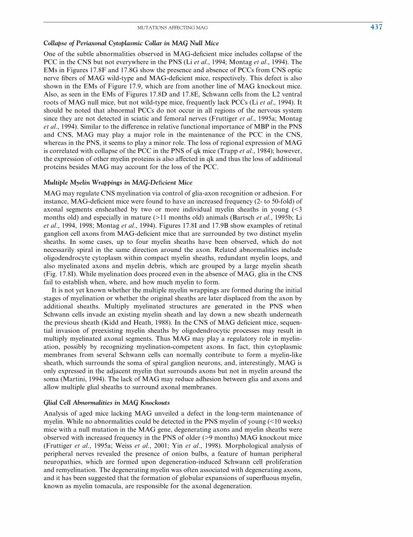
Collapse of Periaxonal Cytoplasmic Collar in MAG Null Mice
One of the subtle abnormalities observed in MAG-deWcient mice includes collapse of the
PCC in the CNS but not everywhere in the PNS (Li et al., 1994; Montag et al., 1994). The
EMs in Figures 17.8F and 17.8G show the presence and absence of PCCs from CNS optic
nerve Wbers of MAG wild-type and MAG-deWcient mice, respectively. This defect is also
shown in the EMs of Figure 17.9, which are from another line of MAG knockout mice.
Also, as seen in the EMs of Figures 17.8D and 17.8E, Schwann cells from the L2 ventral
roots of MAG null mice, but not wild-type mice, frequently lack PCCs (Li et al., 1994). It
should be noted that abnormal PCCs do not occur in all regions of the nervous system
since they are not detected in sciatic and femoral nerves (Fruttiger et al., 1995a; Montag
et al., 1994). Similar to the diVerence in relative functional importance of MBP in the PNS
and CNS, MAG may play a major role in the maintenance of the PCC in the CNS,
whereas in the PNS, it seems to play a minor role. The loss of regional expression of MAG
is correlated with collapse of the PCC in the PNS of qk mice (Trapp et al., 1984); however,
the expression of other myelin proteins is also aVected in qk and thus the loss of additional
proteins besides MAG may account for the loss of the PCC.
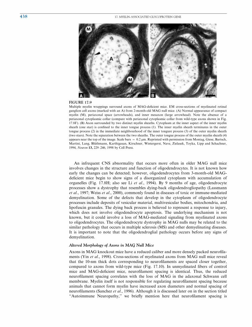
Multiple Myelin Wrappings in MAG-DeWcient Mice
MAGmay regulate CNS myelination via control of glia-axon recognition or adhesion. For
instance, MAG-deWcient mice were found to have an increased frequency (2- to 50-fold) of
axonal segments ensheathed by two or more individual myelin sheaths in young (<3
months old) and especially in mature (>11 months old) animals (Bartsch et al., 1995b; Li
et al., 1994, 1998; Montag et al., 1994). Figures 17.8I and 17.9B show examples of retinal
ganglion cell axons from MAG-deWcient mice that are surrounded by two distinct myelin
sheaths. In some cases, up to four myelin sheaths have been observed, which do not
necessarily spiral in the same direction around the axon. Related abnormalities include
oligodendrocyte cytoplasm within compact myelin sheaths, redundant myelin loops, and
also myelinated axons and myelin debris, which are grouped by a large myelin sheath
(Fig. 17.8I). While myelination does proceed even in the absence of MAG, glia in the CNS
fail to establish when, where, and how much myelin to form.
It is not yet known whether the multiple myelin wrappings are formed during the initial
stages of myelination or whether the original sheaths are later displaced from the axon by
additional sheaths. Multiply myelinated structures are generated in the PNS when
Schwann cells invade an existing myelin sheath and lay down a new sheath underneath
the previous sheath (Kidd and Heath, 1988). In the CNS of MAG deWcient mice, sequen-
tial invasion of preexisting myelin sheaths by oligodendrocytic processes may result in
multiply myelinated axonal segments. Thus MAG may play a regulatory role in myelin-
ation, possibly by recognizing myelination-competent axons. In fact, thin cytoplasmic
membranes from several Schwann cells can normally contribute to form a myelin-like
sheath, which surrounds the soma of spiral ganglion neurons, and, interestingly, MAG is
only expressed in the adjacent myelin that surrounds axons but not in myelin around the
soma (Martini, 1994). The lack of MAG may reduce adhesion between glia and axons and
allow multiple glial sheaths to surround axonal membranes.
Glial Cell Abnormalities in MAG Knockouts
Analysis of aged mice lacking MAG unveiled a defect in the long-term maintenance of
myelin. While no abnormalities could be detected in the PNS myelin of young (<10 weeks)
mice with a null mutation in the MAG gene, degenerating axons and myelin sheaths were
observed with increased frequency in the PNS of older (>9 months) MAG knockout mice
(Fruttiger et al., 1995a; Weiss et al., 2001; Yin et al., 1998). Morphological analysis of
peripheral nerves revealed the presence of onion bulbs, a feature of human peripheral
neuropathies, which are formed upon degeneration-induced Schwann cell proliferation
and remyelination. The degenerating myelin was often associated with degenerating axons,
and it has been suggested that the formation of globular expansions of superXuous myelin,
known as myelin tomacula, are responsible for the axonal degeneration.
MUTATIONS AFFECTING MAG 437
An infrequent CNS abnormality that occurs more often in older MAG null mice
involves changes in the structure and function of oligodendrocytes. It is not known how
early the changes can be detected; however, oligodendrocytes from 3-month-old MAG-
deWcient mice begin to show signs of a disorganized cytoplasm with accumulation of
organelles (Fig. 17.8H; also see Li et al., 1994). By 9 months of age, oligodendrocyte
processes show a dystrophy that resembles dying-back oligodendrogliopathy (Lassmann
et al., 1997; Weiss et al., 2000), commonly found in diseases of toxic or immune-mediated
demyelination. Some of the defects that develop in the cytoplasm of oligodendrocyte
processes include deposits of vesicular material, multivesicular bodies, mitochondria, and
lipofuscin granules. The dying back process is believed to represent a response to injury,
which does not involve oligodendrocyte apoptosis. The underlying mechanism is not
known, but it could involve a loss of MAG-mediated signaling from myelinated axons
to oligodendrocytes. The oligodendrocyte dystrophy in MAG nulls may be related to the
similar pathology that occurs in multiple sclerosis (MS) and other demyelinating diseases.
It is important to note that the oligodendroglial pathology occurs before any signs of
demyelination.
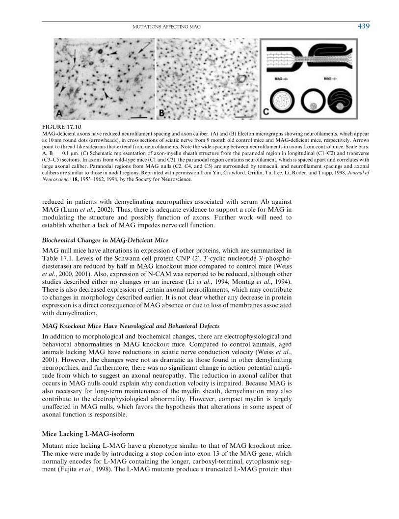
Altered Morphology of Axons in MAG Null Mice
Axons in MAG knockout mice have a reduced caliber and more densely packed neuroWla-
ments (Yin et al., 1998). Cross-sections of myelinated axons from MAG null mice reveal
that the 10-nm thick dots corresponding to neuroWlaments are spaced closer together,
compared to axons from wild-type mice (Fig. 17.10). In unmyelinated Wbers of control
mice and MAG-deWcient mice, neuroWlament spacing is identical. Thus, the reduced
neuroWlament spacing correlates with the loss of MAG in the adaxonal Schwann cell
membrane. Myelin itself is not responsible for regulating neuroWlament spacing because
animals that cannot form myelin have increased axon diameters and normal spacing of
neuroWlaments (Sanchez et al., 1996). Although it is discussed later on in the section titled
‘‘Autoimmune Neuropathy,’’ we brieXy mention here that neuroWlament spacing is
FIGURE 17.9Multiple myelin wrappings surround axons of MAG-deWcient mice. EM cross-sections of myelinated retinal
ganglion cell axons (marked with an A) from 2-month-old MAG null mice. (A) Normal appearance of compact
myelin (M), periaxonal space (arrowheads), and inner mesaxon (large arrowhead). Note the absence of a
periaxonal cytoplasmic collar (compare with periaxonal cytoplasmic collar from wild-type axons shown in Fig.
17.8F). (B) Axon surrounded by two distinct myelin sheaths. Cytoplasm at the inner aspect of the inner myelin
sheath (one star) is conWned to the inner tongue process (1). The inner myelin sheath terminates in the outer
tongue process (2) in the immediate neighbourhood of the inner tongure process (3) of the outer myelin sheath
(two stars). Note the separation between the two sheaths. The outer tongue process of the outer myelin sheath (4)
appears near the top of the image. Scale bars ¼ 0.2 mm. Reprinted with permission fromMontag, Giese, Bartsch,
Martini, Lang, Bluthmann, Karthigasan, Kirschner, Wintergerst, Nave, Zielasek, Toyka, Lipp and Schachner,
1994, Neuron 13, 229–246, 1998 by Cell Press.
au18
438 17. MYELIN-ASSOCIATED GLYCOPROTEIN GENE
reduced in patients with demyelinating neuropathies associated with serum Ab against
MAG (Lunn et al., 2002). Thus, there is adequate evidence to support a role for MAG in
modulating the structure and possibly function of axons. Further work will need to
establish whether a lack of MAG impedes nerve cell function.
Biochemical Changes in MAG-DeWcient Mice
MAG null mice have alterations in expression of other proteins, which are summarized in
Table 17.1. Levels of the Schwann cell protein CNP (2’, 3’-cyclic nucleotide 3’-phospho-diesterase) are reduced by half in MAG knockout mice compared to control mice (Weiss
et al., 2000, 2001). Also, expression of N-CAM was reported to be reduced, although other
studies described either no changes or an increase (Li et al., 1994; Montag et al., 1994).
There is also decreased expression of certain axonal neuroWlaments, which may contribute
to changes in morphology described earlier. It is not clear whether any decrease in protein
expression is a direct consequence of MAG absence or due to loss of membranes associated
with demyelination.
MAG Knockout Mice Have Neurological and Behavioral Defects
In addition to morphological and biochemical changes, there are electrophysiological and
behavioral abnormalities in MAG knockout mice. Compared to control animals, aged
animals lacking MAG have reductions in sciatic nerve conduction velocity (Weiss et al.,
2001). However, the changes were not as dramatic as those found in other demylinating
neuropathies, and furthermore, there was no signiWcant change in action potential ampli-
tude from which to suggest an axonal neuropathy. The reduction in axonal caliber that
occurs in MAG nulls could explain why conduction velocity is impaired. Because MAG is
also necessary for long-term maintenance of the myelin sheath, demyelination may also
contribute to the electrophysiological abnormality. However, compact myelin is largely
unaVected in MAG nulls, which favors the hypothesis that alterations in some aspect of
axonal function is responsible.
Mice Lacking L-MAG-isoform
Mutant mice lacking L-MAG have a phenotype similar to that of MAG knockout mice.
The mice were made by introducing a stop codon into exon 13 of the MAG gene, which
normally encodes for L-MAG containing the longer, carboxyl-terminal, cytoplasmic seg-
ment (Fujita et al., 1998). The L-MAG mutants produce a truncated L-MAG protein that
FIGURE 17.10MAG-deWcient axons have reduced neuroWlament spacing and axon caliber. (A) and (B) Electon micrographs showing neuroWlaments, which appear
as 10 nm round dots (arrowheads), in cross sections of sciatic nerve from 9 month old control mice and MAG-deWcient mice, respectively. Arrows
point to thread-like sidearms that extend from neuroWlaments. Note the wide spacing between neuroWlaments in axons from control mice. Scale bars:
A, B ¼ 0.1 mm. (C) Schematic representation of axon-myelin sheath structure from the paranodal region in longitudinal (C1–C2) and transverse
(C3–C5) sections. In axons from wild-type mice (C1 and C3), the paranodal region contains neuroWlament, which is spaced apart and correlates with
large axonal caliber. Paranodal regions from MAG nulls (C2, C4, and C5) are surrounded by tomaculi, and neuroWlament spacings and axonal
calibers are similar to those in nodal regions. Reprinted with permission from Yin, Crawford, GriYn, Tu, Lee, Li, Roder, and Trapp, 1998, Journal of
Neuroscience 18, 1953–1962, 1998, by the Society for Neuroscience.
au19
MUTATIONS AFFECTING MAG 439
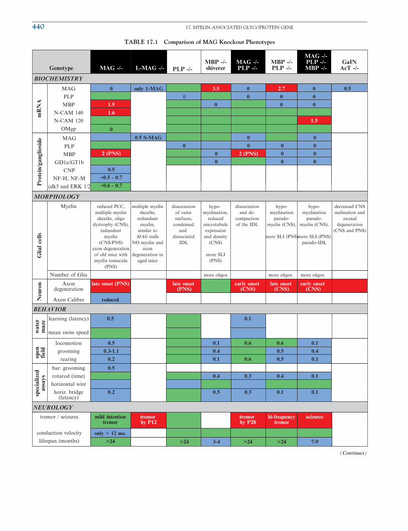
TABLE 17.1 Comparison of MAG Knockout Phenotypes
Genotype
BIOCHEMISTRY
MORPHOLOGY
BEHAVIOR
MAG -/- L-MAG -/- PLP -/-MBP -/-shiverer
MAG -/-PLP -/-
MBP -/-PLP -/-
MAG -/-PLP -/-MBP -/-
GaINAcT -/-
Pro
tein
/gan
glio
side
MAG
PLP
MBP
N-CAM 140
N-CAM 120
OMgp
0
1.5
1.6
0
2 (PNS)
0.5
~0.5 - 0.7
~0.6 - 0.7
0.5 S-MAG
0
0
0
0
0
2 (PNS)
0
0
0
0
0
0
0
only 1-MAG
0
3.5
0
0
0
0.50
0
0
1.5
2.7
0
0
MAG
PLP
MBP
GD1a/GT1b
CNP
NF-H, NF-M
cdk5 and ERK 1/2
Glia
l ce
llsN
euro
n
Myelin
Number of Glia
Axondegeneration
late onset (PNS) late onset (PNS)
early onset (CNS)
early onset (CNS)
late onset (CNS)
Axon Calibre reduced
wat
erm
aze
open
fiel
d
learning (latency)
locomotion
grooming
rearing
0.5
0.3-1.1
0.2
0.1
0.4
0.1
0.6
0.6
0.6
0.5
0.5
0.1
0.4
0.1
spec
ializ
edas
says
bar: grooming
rotarod (time)
horizontal wire
horiz. bridge(latency)
0.5
0.2
0.4
0.5
0.3
0.3
0.4
0.1
0.1
0.1
0.5 0.1
mean swim speed
NEUROLOGY
tremor / seizures
conduction velocity
lifespan (months)
mild intentiontremor
only > 12 mo.
>24
tremor by P12
tremor by P28
hi-frequencytremor
seizures
>243-4>24 >24 7-9
reduced PCC,multiple myelinsheaths, oligo
dystrophy (CNS),redundant
myelin(CNS/PNS)
axon degenerationof old mice withmyelin tomacula
(PNS)
multiple myelinsheaths,
redundantmyelin,
similar toMAG nulls
NO myelin andaxon
degeneration inaged mice
dissociationof outersurfaces,
condensedand
dissociatedIDL
hypo-myelination,
reducedmicrotubuleexpressionand density
(CNS)
more SLI(PNS)
more oligos more oligos more oligos
dissociationand de-
compactionof the IDL
hypo-myelination
pseudo-myelin (CNS),
more SLI (PNS)
hypo-myelination
pseudo-myelin (CNS),
more SLI (PNS)pseudo-IDL
decreased CNSmelination and
axonaldegeneration
(CNS and PNS)
mR
NA
(Continues)
au21
440 17. MYELIN-ASSOCIATED GLYCOPROTEIN GENE

TABLE 17.1 (Continued )
Genotype
BIOCHEMISTRY
MORPHOLOGY
BEHAVIOR
N-CAM -/- MAG -/-N-CAM -/- PO -/-
MAG -/-PO -/- CGT -/-
MAG -/-CGT -/- Fyn -/-
MAG -/-Fyn -/-
Pro
tein
/gan
glio
side
MAG
PLP
MBP
N-CAM 140
N-CAM 120
OMgp
0
0
0
0
0 0 0 0
0 0 00
MAG
PLP
MBP
GD1a/GT1b
CNP
NF-H, NF-M
cdk5 and ERK 1/2
Glia
l ce
llsN
euro
n
Myelin
Number of Glia
Axondegeneration
earlier onset
Axon Calibre
wat
erm
aze
open
fiel
d
learning (latency)
locomotion
grooming
rearing
spec
ializ
edas
says
bar: grooming
rotarod (time)
horizontal wire
horiz. bridge(latency)
mean swim speed
NEUROLOGY
tremor / seizures
conduction velocity
lifespan (months)
tremor by P14
obvioustremor, P12
3 <1
degenerationof axons and
myelin
degenerationof axons andmyelin occursearlier than inN-CAM null
hypo-myelination,abnormalmyelin
compaction
longer delayin myelinformation,abnormalmyelin
compaction
multiple myelinsheaths,
redundant myelin
disorientedparanodal
loops,periaxonal
splitting (CNS)
no PCC, hypo-myelination,
multiple myelinsheaths, severely
disorientedparanodal loops,periaxonal myelin splitting (CNS)
>50% lessmyelin (CNS),myelin sheaths
normal
severe hypo-myelination,80% of opticnerve axonslack myelin,also all of
same defects found inMAG nulls
mR
NA
Regarding quantitative data, an increase greater than 50% appears in red, a reduction to less than 50% appears in blue, and green signifies no change.
These cut-off values are arbitrarily chosen and do not necessarily correspond with differences by statistical significance. Unless noted otherwise,
biochemistry reflects analysis of CNS myelin. Much of the data was obtained from tables found in Uschkureit et al. (2000) and other articles
referenced in the chapter. Acronyms: SLI ¼ Schmidt-Lantermann-incisures, IDL ¼ interperiod dense line
MUTATIONS AFFECTING MAG 441

is four amino acids shorter than S-MAG, whereas the endogenous S-MAG isoform is
unaVected. CNS axons from L-MAG knockout mice were surrounded by multiple myelin
sheaths, there was redundant myelin, and some oligodendrocyte cytoplasm was present in
compact myelin. These abnormalities in the L-MAG mutants are similar to and occur at
the same frequency as in MAG nulls (13 to 15% of CNS axons). In addition, L-MAG
mutants develop a rapid tremor by P12 and tonic seizures in adulthood. Although the total
MAG protein level was reduced by half in the L-MAG null mice, the reduced levels are
likely not responsible for the observed defects because they do not occur in mice that are
heterozygous for the MAG null mutation.
Perhaps more interesting is the observation that older L-MAG mutants do not show
signs of myelin and axon degeneration in the PNS. This is in contrast to the marked
neuropathological abnormalities seen in aged MAG null mice. The simplest interpretation
of the data is that L-MAG is not necessary and that S-MAG suYces for PNS myelin
formation and maintenance. The extracellular domain of MAG appears to mediate pro-
cesses involved in maintaining the integrity of the PNS. In contrast, the longer cytoplasmic
domain found in L-MAG is critical for CNS myelin formation.
Multiple Gene Knockouts That Include Deletion of MAG
There has been a tremendous eVort to Wnd myelin-related molecules that may functionally
compensate for the lack of MAG in MAG knockout mice. Some myelin-related molecules
can have overlapping function, and thus deletion of a single gene do not produce many
defects. Compensation may sometimes be accomplished by up-regulation of related mol-
ecules. For instance, in MAG null mice as well as in MAG/PLP double knockouts, MBP
expression is up-regulated twofold in peripheral nerves (Li et al., 1994; Uschkureit et al.,
2000). There is also a two- or three-fold overexpression of S-MAG in the CNS of MBP
nulls and PLP/MBP double mutants. Even when there are no such changes, the potential
for overlapping function is still there, and it has become necessary to produce multiple gene
knockouts by cross-breeding candidate single mutants to MAG nulls. This search has
excluded a compensatory role for L1 because MAG/L1 double mutants do not have more
deWcits than what occurs in the MAG knockout alone. Compared to control mice, MAG/
L1 double knockout mice have a similar number of myelinated axons (Haney et al., 1999).
However, in several other combinations that are reviewed later in this chapter, there are
more complex phenotypes, and these have conWrmed suspected MAG functions and also
revealed some novel functions of MAG.
Axonal Degeneration in Mice Lacking MAG, PLP, and MBP
Studies on double and triple knockouts of MAG in combination with either PLP or MBP
have corroborated the belief thatMAG has a role in promoting axonal stability (Uschkureit
et al., 2000). As an aid to comparing the phenotypes between the variousMAGknockouts, a
table has been contructed based largely on data from the previously mentioned study (Tab.
17.1). BrieXy, PLP is an integral CNSmyelin sheath protein, which plays a role in stabilizing
the apposition of the adjacent extracellularmembrane surfaces.MBP comprises one-third of
the CNS and one-tenth of PNS myelin and has an important role in the compaction of the
opposing cytosolic surfaces of the plasma membrane processes. In PLP/MAG double
knockouts, severe neurological symptoms develop, which is in contrast to their respective
single mutants. A tremor of the hindlimbs develops by P28 and progresses to dragging and
jerky-like movement with increasing age. Optic nerves from PLP/MAG double mutants
show early onset and rapidly progessing axonal degeneration, which temporally parallels the
neurological symptoms. Behavioral tests also revealed more severe motor and learning
defects in mutant mice lacking both PLP and MAG (see Tab. 17.1). In triple mutant mice
of genotype MAG�/� PLP�/� MBP�/�, a high-frequency tremor develops by 3 months
after birth and the life span is reduced to about 8 months, in contrast to the PLP/MBP
double-mutants that have a normal life span. There is early onset axonal degeneration in the
triple mutant, again suggesting that the deprivation of MAG is detrimental to axons. The
Wndings support a role for MAG in maintaining the integrity of CNS axons.
442 17. MYELIN-ASSOCIATED GLYCOPROTEIN GENE

MAG/N-CAM Double Knockouts
N-CAM partially compensates for MAG functions related to maintenance of axon-myelin
integrity. While there is some uncertainty as to whether N-CAM expression is altered in
MAG knockouts, N-CAM is still present and could take over functions normally carried
out by MAG. To examine the extent of possible compensation by N-CAM in myelin
formation and maintenace, MAG/N-CAM double knockouts were generated by cross-
breeding single mutants (Carenini et al., 1997). Myelin formation was not aVected in the
double mutants; however, degeneration of axons and myelin occurred earlier than in
MAG-deWcient single mutants. More pathological abnormalities were detected in the
MAG/N-CAM double mutants by 4 to 8 weeks of age, although by 26 weeks both mutants
were equally aVected. Therefore, there was no indication that these molecules are necessary
for myelin formation, however, both contribute to maintenance of axon and Schwann cell
function. A subsequent study determined that N-CAM andMAG can contribute to myelin
formation, although these functional roles are only detectable in the absence of P0.
Delayed Myelination in MAG/P0 Null Mice
The most abundant glycoprotein in PNS myelin is P0, which when absent causes severe
dysmelination (Giese et al., 1992). Interestingly, P0 contains an Ig-like extracellular domain,
which confers recognition ability. MAG/P0 null mice were generated to analyze their
combined roles, and the double mutants had abnormal compaction in myelin sheaths that
was similar to that in P0 null mice (Carenini et al., 1999). However, the number of axon-
Schwann cell units devoid of myelin is greater in 10-day-old MAG/P0 double mutants
compared to P0 mutants. This indicates that in mice lacking P0, the formation of turning
loops of Schwann cell processes depends on the presence of MAG. The role of MAG in
promoting the spiraling of myelinating Schwann cell processes cannot be detected in MAG
null mice because P0 seems to compensate. In P0/MAGdouble-deWcientmutants, the lack of
N-CAM also impairs Schwann cell spiraling. However, the eVect is more transient. Thus,
MAG has functions in myelin formation that cannot be duplicated fully by other molecules.
MAG/Galactolipid-DeWcient Mice Show Defects in Adhesion
MAG- and galactolipid-deWcient mice have some common deWciencies, and double
mutants were generated to test for compensatory activity. Mice in which the gene for the
enzyme UDP-galactose:ceramide galactosyltransferase (CGT) is mutated can no longer
convert ceramide to the myelin galactolipids galactocerebroside and its sulfated form
sulfatide (Bosio et al., 1996; Coetzee et al., 1996). CGT mutant mice have a progressive
neuropathological phenotype, which includes tremoring by P14, and they die near P86.
However, similar to the MAG mutant, myelin formation is largely unaVected and in the
CNS of CGT mutant mice, and there are three times as many unmyelinated processes as
well as multiply myelinated axons (Dupree et al., 1998a, 1998b; Marcus et al., 2000).
MAG/CGT double mutants have a more severe phenotype than the single CGT mutant
with a much more obvious tremor and rapid hindlimb paralysis and reduced life expect-
ancy of 22 days (Marcus et al., 2002). The double knockouts have a noticeably absent PCC
whereby eight-fold fewer processes retain a normal periaxonal space and eight times more
spinal cord Wbers display periaxonal splitting. There are additional exacerbations, such as
in paranode structure, which we will not discuss here. The report concludes that MAG and
myelin galactolipids act synergistically at the paranode to promote its structural mainten-
ance. Also, these molecules functionally overlap with one another in maintaining intercel-
lular adhesion along myelinated axons. While there are no recognized axonal ligands for
galactolipids, the authors note that each of sulfatide and MAG have been shown to
interact with members of the tenascin extracellular matrix protein family, which could
modulate adhesion during myelin formation.
Hypomyelination in MAG/Fyn Double Mutants
The multiple knockout approach has been used to assess whether MAG eVects on myelin
involve intracellular signaling via Fyn. Fyn is a nonreceptor-type tyrosine kinase of the Src
au4
MUTATIONS AFFECTING MAG 443
family, which when absent leads to signiWcant impairment of myelination (Umemori et al.,
1994). Fyn is found in myelin-forming cells and is activated by stimulation through cell
receptors including L-MAG, which subsequently leads to transcription of the MBP gene
for myelination (Umemori et al., 1999). Compared to wild-type mice, Fyn-deWcient mice
have only half as much myelin in their brain and even less in spinal cord. To determine
whether MAG and Fyn interact in the myelination process, double knockouts were
generated by cross-breeding the single mutants (BiYger et al., 2000). Double knockout
of MAG and Fyn revealed hypomyelination that is far more severe than that of single
mutants. Half of the axons from optic nerve are unmyelinated in Fyn null mice, but this
ratio increases to 80% in MAG/Fyn double knockouts, which is greater than the 20 to 30%
found in MAG nulls. If MAG and Fyn operate via the same signaling mechanism, then
there should not have been a more severe phenotype in the double mutants. However, it is
still possible that MAG and Fyn actions are not independent because compensatory
mechanisms may exist in the single mutants. For instance, a MAG-associated kinase
activity still persists in Fyn nulls (Umemori et al., 1994), which could partially compensate
for the loss of Fyn.
MAG/Fyn double mutants have the same morphological defects in their myelin sheaths
as in MAG null mice. There is redundant myelin, multiply-myelinated axons, degeneration
of myelin sheaths, impairments in the oligodendrocyte cytoplasmic collar, and noncom-
pacted regions of myelin. It is important to note that Fyn single mutants do not have any
defects in their myelin sheaths, which suggests that Fyn does not participate in the
formation of normal, intact CNS myelin. MAG can therefore activate other downstream
signaling molecules to produce morphologically intact myelin.
MAG Can Act as an Inhibitor of Axon Regeneration
Finally, we will discuss brieXy the inhibitory role of MAG in axonal regeneration, which
was demonstrated in crosses of MAG nulls with C57BL/Wlds mutants. After injury to the
PNS of C57BL/Wlds mutant mice, axon degeneration occurs slowly, as does removal of
myelin due to poor leucocyte invasion (Brown et al., 1991, 1992; Lunn et al., 1989).
Together with the observation that Schwann cells with the greatest amount of lesion-
induced myelin breakdown are more frequently contacted by regrowing axons (Fruttiger
et al., 1995b), this suggested that myelin inhibits axonal regeneration. However, oVspring
generated from crosses of C57BL/Wlds and MAG null mice show much improved axonal
regeneration in regions along Schwann cells with nondegenerated myelin (Schafer et al.,
1996). Still, these mutants have even better axonal growth in regions lacking intact myelin,
which has been interpreted to indicate that either additional myelin-associated inhibitors
exist or that the persistent myelin sheaths can act as a mechanical barrier for sprouting
axons. Nonetheless, the presence of MAG in the PNS of C57BL/Wlds mutant mice
partially contributes to inhibit axon growth. It is not known whether this inhibitory
activity of MAG has any biological signiWcance, because upon injury to nerves from
normal wild-type mice, myelin is removed and Schwann cells down-regulate expression
of myelin-related proteins. Thus, MAG is absent in the injured PNS, and its growth-
inhibitory function may only be important in the normal adult for limiting aberrant
growth. The following section focuses on this inhibitory aspect of MAG, together with
the receptors involved.
CONTROL OF AXONAL GROWTH AND REGENERATION BY MAG
During development of the nervous system, axons send out extensions at a rate and
direction that is regulated by factors in their environment. When axons reach myelin,
their growth is blocked. MAG and several other proteins found in CNS myelin are
inhibitory to axon outgrowth and regeneration. We will discuss the functions that MAG
plays in mediating inhibition, including the role of recently identiWed MAG receptors.
While the focus will be on axon growth and regeneration, it is important to review these
au5
444 17. MYELIN-ASSOCIATED GLYCOPROTEIN GENE

events because the mechanisms will almost certainly have some relevancy to the process of
myelination.
MAG Promotes Axonal Growth during Early Development
Although MAG is a potent axonal growth inhibitor in the adult nervous system, during
early development, MAG is not inhibitory. Indeed, most of the founding experiments that
studied the role of MAG on neurite outgrowth made use of embryonic and neonatal
neurons. MAG increases outgrowth of young cerebellar and spinal cord neurons and
also neonatal DRG neurons (Johnson et al., 1989; Matsuda et al., 1996; Mukhopadhyay
et al., 1994; Turnley and Bartlett, 1998). However, when post-natal neurons are exposed to
MAG, their axonal growth is inhibited. Unfortunately, this developmental switch in the
neuronal response to MAG was not realized for quite some time, causing considerable
confusion to scientists.
It is not clear whether the neurotrophic-like eVect of MAG on young neurons has any
biological signiWcance. For instance, axons have stopped growing by the time MAG
becomes expressed at detectable levels, and thus MAG is likely not normally involved in
the promotion of neurite growth during early development. Nonetheless, a recent investi-
gation has found that the L-MAG isoform is responsible for promoting axonal growth.
SpeciWcally, when cerebellar neurons from newborn mice were cultured on BALB/c 3T3
cells transfected with cDNA for either the S- or L-MAG isoform, only the latter had
growth promoting activity (Shimizu-Okabe et al., 2001). Because L-MAG is the principle
form that is expressed during development, it is speculated to function in enhancing axon
outgrowth. It remains to be seen whether low levels of L-MAG exist during development
or perhaps could assist axonal recovery following injury or disease. Also, by selecting
young neurons that are not inhibited byMAG and do not express receptors that contribute
to inhibiton of axonal outgrowth (discussed later), it may be possible to use young neurons
as an aid for axon regeneration.
In the Adult, MAG Functions as an Inhibitory Molecule
MAG was the Wrst myelin-derived molecule shown to be inhibitory to axonal growth in the
adult nervous system. MAG inhibits growth of neurites (including neuronal-like cells)
regardless of whether it is presented in soluble forms containing the extracellular domain,
is expressed by live cells, or when introduced to axons by way of immobilized membrane
prepared from MAG-expressing cells (DeBellard et al., 1996; Li et al., 1996; McKerracher
et al., 1994; Mukhopadhyay et al., 1994; Shen et al., 1998; Tang et al., 1997b). Further-
more, Schwann cells that are induced to express MAG also inhibit regeneration, demon-
strating that MAG is inhibitory in both the adult CNS and PNS.
Several factors that aVect MAG eYcacy were not recognized until recently, and this also
hindered progress on accepting it is an inhibitory molecule. For instance, neuronal growth
cones were found to collapse upon encountering MAG-coated beads (Li et al., 1996),
whereas another study did not Wnd any such eVect using MAG extracted from myelin
(Bartsch et al., 1995a). However, the inhibitory role of MAG on neurite outgrowth has
also been conWrmed using MAG fused to the Fc portion of human IgG and also dMAG
(Tang et al., 1997b). The authors emphasized several important details, which should be
considered when studying MAG and which likely explain the seeming discrepancies. For
instance, the manner by which MAG is presented can aVect activity, in that MAG is
multimeric when coated onto beads, compared to its monomeric, relatively denatured
format when extracted from myelin and solubilized. Furthermore, the MAG content in
puriWed myelin varies considerably and MAG is also rapidly (within minutes to hours)
converted to dMAG, which would be lost upon any washing.
MAG-mediated inhibition of axonal growth can also be overcome by the presence of
other proteins and also by neurotrophins. Laminin, a component of the Schwann cell basal
lamina, can override the growth inhibitory activity of MAG (David et al., 1995). Hence,
after puriWcation of MAG from myelin, any residual laminin can nullify MAG inhibition
CONTROL OF AXONAL GROWTH AND REGENERATION BY MAG 445

of neuronal outgrowth. MAG also binds the extracellular matrix glycoprotein tenascin-R
which is expressed by oligodendrocytes (Yang et al., 1999). Tenascin-R neutralizes MAG-
mediated inhibition of neurite outgrowth. Finally, neurotrophins such as BDNF or
GDNF have been shown to prevent MAG-mediated inhibition of axonal regeneration
(Cai et al., 1999).
Upon injury to the adult nervous system, myelin-forming glia will down-regulate MAG.
This biological change is of extreme beneWt, given the inhibitory attribute of MAG on
axonal outgrowth. Indeed, axons in the PNS can regenerate successfully, especially when
aided by the additional removal of any remaining MAG by macrophages and also by the
induction of regeneration-associated genes. The inhibitory nature of MAG in the PNS has
been demonstrated by the Wnding that axonal regeneration can be improved in C57BL/
Wlds mutant mice (which do not clear myelin and show hardly any axonal regeneration)
after crossing them to MAG null mice (as already discussed; also see Schafer et al., 1996).
However, axons in the CNS do not grow back, a problem that is of great clinical relevance
for injuries and diseases of the nervous system and that has preoccupied researchers for
many years.
Myelin in the CNS contains two additional proteins that inhibit neuronal growth.
Caroni and Schwab identiWed two membrane proteins, NI35 and NI250, and also raised
IN-1 Abs, which detected these proteins and could counteract the nonpermissive properties
of CNS myelin (Caroni and Schwab, 1988a;1988b). NI250 was next puriWed and shown to
inhibit neurite outgrowth (Spillmann et al., 1998). Several labs then used peptide sequences
of NI250 (now called Nogo-A) to clone the Nogo gene, named for its ability to block
axonal growth (Chen et al., 2000; GrandPre et al., 2000; Prinjha et al., 2000).
Nogo codes for three proteins, Nogo-A, -B, and -C, and there is agreement that Nogo-A
is inhibitory. All three isoforms share a conserved C-terminal region with two transmem-
brane domains connected by a 66-amino acid linker sequence known as Nogo-66. The
Nogo-66 domain is believed to be on the extracellular side of membranes and is most likely
the domain responsible for inhibition of neurite outgrowth. Curiously, the N-terminal
region of Nogo-A is also inhibitory, although the mechanism of action is diVerent from
Nogo-66 (Chen et al., 2000; Fournier et al., 2001; Prinjha et al., 2000). Also, the amino-
Nogo domain is suspected to be cytoplasmic and it may only play a role if released
following disease or injury. Further work is necessary to sort out the details of Nogo
membrane organization and domain diVerences in mediating inhibitory actions.
Finally, the last known myelin-derived inhibitor is the oligodendrocyte-myelin glyco-
protein (OMgp). OMgp is a glycophosphatidylinositol (GPI)-anchored protein, which
remained elusive until very recently when it was identiWed to bind to the same receptor
as Nogo and to inhibit neurite outgrowth in vitro (Wang et al., 2002). A receptor for Nogo-
66, named the Nogo-66 receptor (NgR), was identiWed by expression cloning using a
mouse cDNA library (Fournier et al., 2001). NgR was determined to contain a signal
sequence followed by eight leucine-rich repeat domains and a cysteine-rich region,
followed by a GPI linkage region. The human homologue of the NgR gene has exons
(separated by nearly 30 kb) on chromosome 22q11. The NgR cDNA sequence encodes a
protein 473 amino acids long with a 85 kDa apparent molecular mass. There is widespread
expression in the brain and very little expression in the PNS. In summary, MAG, OMgp,
Nogo, and NgR all contribute to the inhibitory activity of CNS myelin.
Identification of Functional Neuronal MAG Receptors
MAG Binds the Nogo Receptor, NgR
Immediately prior to when this chapter was written, several groups made the important
discovery of identifying the NgR as a common functional receptor for MAG and OMgp
(Domeniconi et al., 2002; Liu et al., 2002; Wang et al., 2002). Figure 17.11 contains
experimental data from the article by Liu and colleagues and elegantly demonstrates that
MAG mediates inhibition of axonal outgrowth via NgRs. First, NgR levels in DRG
neurons are shown to be minimal during embryonic development and to increase into
446 17. MYELIN-ASSOCIATED GLYCOPROTEIN GENE

adulthood (Fig. 17.11A). Second, MAG-mediated neurite outgrowth is eliminated after
blockade of NgR operation (Fig. 17.11B). Several methods have been used in this and
other studies described earlier to interfere with normal NgR activation, including (1)
antibodies against NgRs, (2) soluble NgRs to compete for binding with neuronal recep-
tors, (3) removal of NgRs by cleaving its GPI anchor, and (4) overexpression of truncated
NgRs lacking the extreme C-terminal region (e.g., dominant-negative form of NgR), all of
which prevent MAG/OMgP/Nogo-mediated inhibition. The last panel of Figure 17.11C
shows that embryonic neurons, which are generally unresponsive to growth-inhibition by
MAG and Nogo-66 proteins, become responsive when exogenous NgR is introduced into
them. The assay involves measurement of growth cone collapse in DRG explants infected
with recombinant virus to drive expression of green Xuorescent protein alone or also NgRs.
Similar experiments were used by Wang and coworkers to show that introduction of
FIGURE 17.11The Nogo receptor (NgR) controls inhibitory responses induced by MAG. (A) Confocal microscope images of
mouse DRG neurons showing increase in NgR levels, as detected by anti-NgR Abs and Xuorescent secondary
Abs, from embryonic day 17 (E17) to post-natal day 64 (adult). (B) Comparison of neurite outgrowth under
control conditions containing IgG versus Fc-MAG, the extracellular domain of MAG fused to the Fc portion of
IgG. Some experiments were carried out in the presence (þ) or absence (�) of PI-PLC (to remove GPI-anchored
proteins including NgR) or NgR-Ecto (truncated soluble NgR fragment that antagonizes Nogo-66). The asterisk
denotes a statistically signiWcant diVerence between MAG and control treated adult neurons, and there is no
diVerence after interfering with NgRs. (C) Embryonic (E7) chick DRG explants were infected using recombinant
virus to drive expression of green Xuorescent protein (GFP) or NgR and treated with either GST control
(glutathione S-transferase), GST-Nogo-66, IgG control, or Fc-MAG. (D) Quantitation of growth cone (GC)
collapse shows that embryonic neurons are normally not responsive to MAG or Nogo-66, unless they are forced to
express NgR (asterisks denote statistically signiWcant diVerences). Reprinted with permission from Liu, Fournier,
Grandpre, and Strittmatter, 2002, Science 297, 1190–1193, 2002, American Association for the Advancement of
Science.
CONTROL OF AXONAL GROWTH AND REGENERATION BY MAG 447

exogenous NgRs confers OMgP responsiveness to otherwise insensitive neurons (Wang
et al., 2002). Thus, the developmental switch in neuronal MAG sensitivity very likely
involves an up-regulation of NgRs. Regardless, there is convincing evidence that NgRs
are both necessary and suYcient for MAG-mediated growth inhibition in adult axons.
Is the interaction between NgRs and their multiple binding partners regulated? Al-
though Nogo-66, OMgp, and MAG do not share any sequence similarity, binding studies
show that they all bind to the NgR with very similar aYnities (7, 5, and 8 nM, respectively).
Figure 17.12 contains a model that illustrates the interactions of these three myelin-derived
inhibitors with neuronal receptors including NgRs, as well as gangliosides and neurotro-
phin receptors, which will be discussed later. It is interesting that excess Nogo-66 does not
prevent MAG-NgR binding (see Liu et al., 2002). Whereas MAG and Nogo-66 have
separate binding sites within the same domain of NgR, OMgp and Nogo-66 have an
overlapping binding site. If there is no preferential binding partner for NgRs, the particular
membrane expression level for each NgR ligand may be an important factor that will
determine the extent of axonal inhibition. Also intriguing is the fact that the MAG-NgR
interaction is largely unaVected by the introduction of GT1b, a neuronal ganglioside that
also interacts with MAG and will be covered in the next section. The MAG-NgR inter-
action may represent a more speciWc, biologically relevant interaction. Indeed, Nogo and
MAG appear to be the main inhibitors in myelin, because when myelin is prepared from
mice lacking Nogo-A, there is reduced inhibition of axon growth, and the residual activity
is completely blocked by anti-MAG Abs (unpublished data; see Liu et al., 2002). It will be
crucial to establish whether MAG, Nogo66, and OMgp bound to NgR and gangliosides all
partake to regulate neurite outgrowth in vivo and whether complexes consisting of diVerent
combinations of these players can alter function.
The competition by multiple ligands for binding to the same receptor is a simple
explanation for the redundancy observed when only a single ligand is blocked. The
application of IN-1 Ab to block Nogo has only modest eVects on improving axonal
regeneration (Schnell and Schwab, 1990). Similarly, axons in MAG null mice do not
show any signiWcant improvement in regeneration ( Li et al., 1996; Montag et al., 1994).
Although there are no reports published on whether interfering with OMgp can improve
axon outgrowth, a lack of signiWcant eVect is predicted. In contrast, a peptide antagonist of
NgR (derived from a 40 amino-terminal fragment of Nogo-66, NEP1-40) is an eVective
blocker of myelin-mediated inhibition of axonal outgrowth and also signiWcantly improves
axon growth and functional recovery following spinal cord injury (GrandPre et al., 2002).
Thus, the NgR appears to be a key player in the inhibitory actions of CNS myelin. This
would be consistent with a model by which all three inhibitory ligands only exert their
physiological inhibitory actions via one receptor, NgR. An obvious next step will be to
generate a cocktail containing blockers of Nogo, MAG, OMgp, and also NgR in order
to test whether this allows for superior axonal growth. The blockers will need to be
designed to act in a transient function, because otherwise there may be excessive outgrowth
and possibly an inability to form proper myelin. If this can be overcome, it should yield a
potent theurapeutic agent that promotes functional recovery in many diseases and injuries
aVecting neurons.
Gangliosides Are MAG Receptors
The nerve cell surface gangliosides (sialic acid-containing glycosphingolipids) GD1a and
GT1b are receptors for MAG (see the commentary by McKerracher, 2002). MAG binds
sialic acid containing proteins and glycans, especially the NeuAc a3 Gal b3 GalNAc
structure, which is commonly found on gangliosides (Collins et al., 1997b; Crocker et al.,
1996; Kelm et al., 1994).
Binding studies indicate that GD1a and GT1b are the primary complex gangliosides in
the brain that bind MAG (Collins et al., 1997a; Yang et al., 1996). Mice that lack the
enzyme GM2/GD2 synthase, which is required for biosynthesis of complex gangliosides
including GD1a and GT1b, display pathological features resembling those found in MAG
knockouts (Sheikh et al., 1999). There is decreased CNS myelination and axonal degener-
ation in both CNS and PNS, and demyelination in the PNS. It should be noted that MAG
au6
448 17. MYELIN-ASSOCIATED GLYCOPROTEIN GENE

expression is reduced by half in these GalNAcT �/� mice, although MAG heterozygote
mice have similar reduction inMAGexpression, yet they lack any neuropathology. Neurons
from these GalNAcT knockout mice are less sensitive to MAG-mediated inhibition of
neurite outgrowth (Vyas et al., 2002). Neurite outgrowth mediated by MAG is reduced
upon addition of soluble GD1a and GT1b and also by Abs against GT1b, but not GD1a
(Vinson et al., 2001). A subsequent report conWrms the role of ganglioside GT1b in
blocking neurite outgrowth and shows that anti-GD1a Ab is also eVective (Vyas et al.,
2002). Therefore, gangliosides GT1b and possibly GD1a regulate neurite outgrowth, and
are neuronal receptors for MAG.
While evidence supports that MAG interacts with gangliosides, there is some debate as
to whether gangliosides are necessary for MAG’s inhibition of axonal outgrowth. For
instance, removal of sialic acid residues does not aVect binding of MAG to NgRs, recep-
tors that mediate inhibition (Domeniconi et al., 2002). When MAG is mutated at arginine
118 (R118) to abolish sialic acid-dependent binding, MAG can no longer bind to neurons;
however, when R118-mutated MAG is expressed on the surface of Schwann cells or CHO
FIGURE 17.12Model of MAG-mediated inhibiton of axon regeneration. Oligodendrocytes in the CNS produce three known
growth-inhibitory molecules, MAG (red), OMgp (orange), and Nogo-A (yellow). The exact orientation of Nogo in
the myelin membrane is not yet defined, however, both the N-terminal domain and Nogo-66 domain are inhibi-
tory. MAG, OMgp and Nogo-66 all bind to the same neuronal receptor, NgR (Nogo receptor, pink), which results
in blockade of neurite outgrowth and growth cone collapse. NgR lacks an intracellular domain and requires an
unknown co-receptor to transduce intracellular signals via molecules such as Rho GTPase. MAG also binds to a
complex consisting of gangliosides GD1a and GT1b, the latter of which interacts with the neurotrophin receptor
p75NTR. A signal for MAG-mediated inhibition of neuronal elongation is transduced through GT1b/p75NTR and
activation of Rho. The photomicrographs show DRG neurons grown on a laminin substrate that is permissive for
growth (right, GO) or on a CNS myelin substrate that prevents growth (left, NOGO). Reprinted with permission
from Woolf and Bloechlinger, 2002, Science 297, 1132–1133, 2002, American Association for the Advancement of
Science.
CONTROL OF AXONAL GROWTH AND REGENERATION BY MAG 449

cells, it is still a potent inhibitor of axonal outgrowth (Tang et al., 1997a). These Wndings
were interpreted to indicate sialic acid binding is insuYcient to aVect inhibition of axonal
regeneration and that a second site on MAG must be responsible for this eVect. Subse-
quent work suggested that the residual inhibition in the R118 mutant is dependent on sialic
acid (Vinson et al., 2001). Therefore, the R118-mutant MAG may have enough sialic acid
binding to inhibit neurite outgrowth. However, there is additional evidence that sialic acid-
dependent binding to neurons is insuYcient to induce inhibition. Sialoadhesin (siglec-1)
and a truncated form of MAG, MAG(d1-3)-Fc each recognize the same sialic acid linkage
as MAG and also bind neurons in a sialic-dependent manner; however, none are inhibi-
tory. Thus, there are probably multiple unique recognition sites on MAG, at least one of
which binds sialoglycoproteins and another that mediates inhibition.
MAG may inhibit neurite outgrowth via multivalent clustering of gangliosides on
neurons. Ganglioside-mediated inhibition of neurite outgrowth is independent of MAG
because such inhibition can be induced in the absence of MAG by Ab-mediated clustering
of GT1b (Vinson et al., 2001; Vyas et al., 2002). Compared to bivalent IgG Abs, pre-
complexed Abs, and decavalent IgM Abs are more eYcient in blocking axonal outgrowth
in response to MAG. MAG binding to gangliosides may also function to cluster other
signaling molecules, or even to regulate MAG activity, either of which could help potenti-
ate inhibition of axonal growth (see Fig. 17.6).
Additional neuronal receptors may exist for MAG. For instance, although they are
relatively scarce in brain, the a-series of gangliosides bind MAG with greater potencies
than GD1a and GT1b (Collins et al., 1999). However, the roles of a-gangliosides, if any,remain to be elucidated. In addition, De Bellard and Filbin have used MAG-Fc to
precipitate several neuronal surface proteins (De Bellard and Filbin, 1999). The two
most prominent proteins that bind MAG are of 190 and 250 kDa size, and the former is
a sialoglycoprotein. Further work will need to further identify these potential MAG
receptors as well as their actions.
SIGNALING EVENTS VIA MAG AND INTERACTING PROTEINS
Neuronal Signaling through MAG Receptors
Signaling via Neurotrophin Receptor, Rho GTPase, and cAMP
NgR lacks a transmembrane domain and is attached to the neuronal extracellular mem-
brane by its GPI anchor. Without any cytoplasmic domains, intracellular signal transduc-
tion cannot occur. Thus, MAG (as well as Nogo-66 and OMgp) signaling through NgRs
must be mediated via an associated transmembrane receptor. Similarly, gangliosides
require signaling partners to transduce any signal across the cell membrane. The only
known co-receptor is the low-aYnity nonselective neurotrophin receptor p75 (p75NTR),
which binds GT1b (Yamashita et al., 2002). MAG failed to inhibit outgrowth in neurons
from mice carrying a mutation in the p75NTR gene. Further experiments on neurons from
these mutant mice revealed that activation of the small GTPase Rho was lost. Finally,
MAG fusion protein co-localizes with anti-p75NTR staining, suggesting they spatially
interact. Hence, MAG inhibits neurite outgrowth via p75NTR-mediated activation of
Rho. It is not known whether MAG/NgR-mediated inhibition also operates by way of
Rho signaling.
The p75NTR-mediated signal was suspected to involve Rho because previous experiments
had shown MAG activates Rho. Inactivation of Rho not only blocks growth inhibition by
MAG and myelin, it also permits regeneration in vivo (Lehmann et al., 1999). Neurons that
cannot transduce Rho signals (expression of dominant-negative Rho) do not respond to
MAG. Conversely, blockade of Rho-GTPase activity (using Clostridium botulinum C3
exoenzyme) allows neurons to grow on MAG substrates. An inhibitor of Rho kinase
(Y27632), the downstream eVector of Rho, blocks inhibition of neurite outgrowth induced
either by myelin, MAG or anti-GT1b Ab ( Dergham et al., 2002; Vinson et al., 2001).
Furthermore, the latter study found Y27632 is of theurapeutic value because it promotes
450 17. MYELIN-ASSOCIATED GLYCOPROTEIN GENE
axonal regeneration and functional recovery after spinal cord injury. To summarize, MAG
binds a receptor complex consisting of ganglioside GT1b and p75NTR, which serves as a
signal transducer for neurite outgrowth inhibition via Rho and Rho kinase activation (see
Fig. 17.12). It will be important to decipher the molecular participants in signaling events
downstream of Rho, including activation of the Ser/Thr kinase known as Rho kinase.
Anothe r remai ning issue is wheth er trans duction of an inhibitory signal via p75 NTR can be
modu lated accordi ng to whet her p7 5NTR inter acts with MAG or neurotr ophins. Thus ,
p75NTR may act as a switch that pe rmits outgrowth in environm ents contai ning neu rotro-
phins, only to suppress outgrowth upon binding to MAG.
Neurotrophins and activation of the cAMP pathway can override MAG-mediated
inhibition. Activation of p75NTR by the neurotrophin NGF abolishes Rho activation
and enhances neurite elongation (Yamashita et al., 1999). In addition, the neurotrophins
BDNF and NT-3, which do not directly bind p75NTR but may interact via association with
Trk receptors, can also stimulate neurite outgrowth. Priming of neurons by exposing them
to BDNF or GDNF, or also by providing them with a cAMP analog, suspends the
inhibitory actions of MAG on axonal outgrowth and regeneration (Cai et al., 1999;
Song et al., 1998). It is speculated that MAG binding to neurons activates an inhibitory
G-protein (Gi) that functions to block increases in cAMP and prevents growth. Introduc-
tion of cAMP analogs into spinal cord neurons allows them to regenerate (Neumann et al.,
2002; Qiu et al., 2002). Interestingly, a reduction in endogenous levels of cAMP correlates
with the developmental switch in neuronal response to MAG, which begins with promo-
tion of neurite outgrowth in young neurons, and then changes to inhibiton (Cai et al., 2001).
Do Rho and cAMP signals interact, and what eVectors control neurite outgrowth?
Elevation of cAMP activates protein kinase A (PKA), which subsequently phosphorylates
Rho to induce its dissociation from the cell membrane (Lang et al., 1996). This dual
regulation of Rho activation and inactivation could act as a switch to control neuronal
responses to MAG and neurotrophins. However, there are additional consequences of
neurotrophin/cAMP signaling. Just lately, cAMP was shown to up-regulate Arginase I, an
enzyme involved in the synthesis of polyamines (Cai et al., 2002). After polyamine synthe-
sis is blocked, BDNF and a cAMP analog can no longer suppress MAG- and myelin-
mediated inhibition of neurite outgrowth. Either overexpression of Arginase I or priming
with polyamines blocks inhibition mediated by MAG or myelin. Interestingly, the en-
dogenous levels of Arginase I in DRG neurons drops during the Wrst post-natal week and
appears to coincide with the developmental switch from promotion to inhibition of neurite
outgrowth in response to MAG. The authors discuss potential mechanisms by which
polyamines can modulate neurite outgrowth and regeneration (also see the review by
Skaper et al., 2001). However, as discussed earlier, the developmental increase in NgR
receptor also has been suggested to play a role in the post-natal switch in neuronal MAG
sensitivity. It is possible that NgR and BDNF receptor signaling pathways interact to
control neurite outgrowth. Future work will need to identify the interactions between
downstream eVectors of cAMP and Rho kinase. At the receptor level, it will be important
to determine whether MAG-mediated signaling events through gangliosides and NgRs
have any overlap or interaction. Such research will help in designing strategies that
encourage axons to regrow in the CNS.
MAG Regulates Neuronal Cytoskeletal Proteins
In the section titled ‘‘Altered Morphology of Axons in MAG Null Mice,’’ we noted that
axons in MAG knockout mice have an altered morphology involving more densely packed
neuroWlaments (also see Fig. 17.10). Compared towild-typemice,MAG-deWcientmice older
than 3 months of age have axonal proWles that are smaller with reduced spacing between
neuroWlaments (Yin et al., 1998). The previously stated changes in axon caliber and cyto-
skeleton are likely a result of reduced neuroWlament phosphorylation, which occurs inMAG
knockout mice. This raises the possibility that changes in myelin are secondary to direct
eVects on axonal function. Similar alterations in axons and neuroWlaments also occur in
Trembler mice, which have a mutation in PMP-22 (de Waegh et al., 1992). In the PMP-22
mutants, however, the alterations only occur in regions of sciatic nerve lackingmyelin.MAG
SIGNALING EVENTS VIA MAG AND INTERACTING PROTEINS 451

knockouts have axonal defects even though myelin is present, suggesting that some other
factor is responsible, such as altered signaling between glia and axons.
NeuroWlaments consist of three subunits with low (NF-L), middle (NF-M), and high
(NF-H) molecular weights, respectively. MAG-deWcient mice have less NF-L and NF-H in
nerve, while total NF-M is unchanged (Yin et al., 1998). Phosphorylation of NF-M and
especially NF-H was signiWcantly reduced (30 to 50% reduction depending on assay type).
Phosphorylation of NF-H and NF-M increases the total negative charge and repositioning
(lateral extension) of the neuroWlament sidearms; this in turn normally functions to increase
neuroWlament spacing and axonal caliber. The mechanism of neuroWlament arrangement
has been analyzed using experimental data from wild-type andMAG-deWcient mice and the
most consistentmodel involves electrostatic repulsion between the phosphorylated sidearms
(Kumar et al., 2002). Thus, under conditions of reducedNF-H andNF-M phosphorylation,
the neuroWlament sidearms retract, leading to reduced neuroWlament spacing and smaller
axonal calibers. In MAG-deWcient mice, the diameter of the axons and neuroWlament
spacing are drastically reduced, particularly in paranodal regions. Whereas the paranodal
region in wild-type mice continues to expand between P35 and P90, in MAG null mice the
axon shrinks and this results in the formation of paranodal tomaculi. The schematic shown
in Figure 17.10C illustrates the appearance of these abnormal paranodal myelin structures
and also compares the structure of axons between wild-type andMAG null mice. Finally, it
will be important to establish the signaling cascade that MAG initiates to control the
neuroWlament subunit composition and phosphorylation.
There is evidence that MAG interacts directly with microtubule-associated protein 1B
(MAP1B) found on neurons. Although MAP1B is considered to be an intracelluar cytos-
keletal component, it was proposed to also be a glycoprotein residing on the cell membrane
(Muramoto et al., 1994). Recent analyses by Quarles and colleagues revealed that neurons
express MAP1B, and that its carboxy terminus and glycosylation sites are located in the
extracellular space, whereas the microtuble binding domains are in the neuronal cytoplasm
( Franzen et al., 2001; Tanner et al., 2000). Although MAP1B is expressed by glial cells, a
higher molecular weight isoform is expressed on neurons but not Schwann cells. Co-
immunoprecipitation experiments show that MAG speciWcally interacts with MAP1B,
and immunohistochemistry reveals they colocalize at axon-glia junctions both in vitro
and in vivo. Moreover, when DRG neurons are cocultured with MAG-expressing COS
cells, there is a two-fold increase in expression of phosphorylated MAP1B, as well as
increases in NF-H and NF-M, which are described in the next paragraph. These MAG-
mediated cytoskeletal phosphorylation events are important for control of axon caliber, as
demonstrated by the axonal changes that occur in MAG nulls when such signaling is
missing. Similarly, neuroWlaments in peripheral nerves of Trembler mutants have reduced
phosphorylation and increased density, and interestingly there is less MAP1B phosphoryl-
ation (Kirkpatrick and Brady, 1994). Shiverer mice have a natural mutation, which yields a
truncated form of MBP, and their CNS axon microtubule expression and density is
increased (Kirkpatrick et al., 2001). The signaling mechanisms responsible for altering
the axonal cytoskeleton in these mutants are not known, but potentially involve changes
in MAG expression, which occur in knockouts (see Tab. 17.1). Perhaps most intriguing is
the potential for MAG-MAP1B interactions to provide a structural link between axons
and glia, and directly contribute to the modulation of myelinated axon structure and
stability.
The ability of MAG to regulate the axonal cytoskeleton has been demonstrated in
cultured neurons and has also been extended to expose the underlying mechanisms.
Compared to co-culturing of DRG neurons with control COS cells, co-culturing with
MAG-expressing COS cells results in a Wve- and two-fold increase in NF-H and NF-M
phosphorylation, respectively (Dashiell et al., 2002). MAG also causes the level of NF-M
expression to increase by 50%. Treatment of PC12 neurons with soluble MAG-Fc chimera
also eVectively increases NF-M expression and phosphorylation, thus demonstrating that
MAG is responsible for the axonal changes, rather than other mechanisms such as cell-cell
interactions. MAG also increases MAP1B, phosphorylated MAP1B, MAP2, low-
molecular-weight tau, and growth-associated protein-43 (GAP-43) levels.
452 17. MYELIN-ASSOCIATED GLYCOPROTEIN GENE

Whole extracts of sciatic nerves from MAG null mice were also studied to determine
whether the reduced neuroWlament phosphorylation in MAG null mice is due to altered
kinase activity (Dashiell et al., 2002). Based on an assay that evaluates phosphorylation
of preferred kinase substrates, MAG knockout mice had reduced activity of proline-
directed kinases, including cyclin-dependent kinase-5 (cdk5) and extracellular-signal
regulated kinases 1 and 2 (ERK1/2). The speciWc activities of the cdk5 and ERK1/2
enzymes were subsequently determined to be reduced by 33 and 38%, respectively.
Kinase assays also demonstrate that when these enzymes are immunoprecipitated
from PC12 neurons treated with MAG-Fc, their activity is signiWcantly increased. In
summary, MAG activates cdk5 and ERK1/2 kinases and this signaling process may
be involved in MAG-mediated regulation of the axonal cytoskeleton. Further work
will need to determine whether these or other kinases are obligatory, and whether
MAG modulates phosphatase activities, transcription, and degradation of cytoskeletal
proteins.
Glial Signaling via MAG
The cytoplasmic domains of MAG may be involved in modulating MAG function or
signaling through phosphorylation, association with kinases, and also the cytoskeleton.
Several interesting signaling events that may be of biological relevance to myelin formation
occur within minutes of Ab-induced cross-linking of L-MAG. First, Fyn kinase activity
increases, which leads to phosphorylation of L-MAG on Tyr620 (Jaramillo et al., 1994;
Umemori et al., 1994). Consequently, this phosphorylation event allows MAG to associate
with phospholipase Cg (PLCg). Nonreceptor tyrosine kinases such as Fyn and PLC
associate via their SH2 and SH3 domains with speciWc sequences containing a phosphoryl-
ated Tyr in the cytoplasmic domain of surface receptors and CAMs. Thus, MAG may
serve as a docking protein, which allows interactions between diVerent signaling molecules.
Fyn activation by MAG may be of great importance because myelination is partially
impaired in fyn-deWcient mice. MAG interaction with neuronal receptors could trigger
events such as myelin formation.
The regulation of phosphorylation events may be complex, and it cannot be excluded
that other signaling events through separate receptors and signaling pathways can feed
back onto MAG. We suggested previously that the phosphorylation state of the MAG
cytoplasmic domain modulates adhesion and wrapping of the glial cell membrane around
the axon during the myelination process (see the review by Attia et al., 1989). The strength
of the MAG-neuron interaction could be regulated diVerentially between the leading glial
lip and the previous myelin wrap by phosphorylation. In addition to controlling adhesion,
the regulation of kinase and phosphatase activities may be responsible for initiating and
terminating myelin wrapping.
Besides interactions with kinases, MAG binds other cytoplasmic proteins. There is
evidence that S100b, a 10 kDa calcium- and zinc-binding protein, can bind to the cytoplas-
mic domain of L-MAG (Kursula et al., 1999). A S100b synthetic peptide corresponding to
the L-MAG interacting cytoplasmic domain (putative basic amphipathic alpha-helix)
reproduced this interaction including the Ca2þ-dependency and further revealed a binding
stoichiometry of 1:1 with a dissociation constant (KD) of 7 mM. This represents a fairly
strong interaction that could also take place in vivo and partially contribute to MAG-
mediated signaling. Another interesting Wnding is that in the presence of S100b, phosphor-ylation of the L-MAG cytoplasmic domain by PKA is inhibited (Kursula et al., 2000).
The functional consequences of S100b-mediated inhibition of phosphorylation is being
investigated. However, S100b interacts with a variety of cytoskeleton-associated proteins,
including actin microWlaments, microtubules, and intermediate Wlaments. Thus, MAG-
mediated control of glial cell morphology and function may be modiWed by interactions
with S100b.Finally, there is some progress regarding identiWcation of glial cytoskeletal components
that interact with MAG. A potential cytoskeleton binding site, which is similar in sequence
to a functional site in the b1 subunit of integrin, is found in both isoforms (Salzer et al.,
au7
au8
SIGNALING EVENTS VIA MAG AND INTERACTING PROTEINS 453
1987). Co-localization of MAG and cytoskeletal components such as F-actin and spectrin
is consistent with this prediction (Trapp et al., 1989b).
MAG IN INJURY AND DISEASE
Human Gene Mutations in MAG?
Based on studies of MAG knockout mice, it is probable that human diseases that aVect
only MAG will remain elusive. Analyses of MAG single knockout mice have failed to
show defects in myelin formation, and instead, it is the long-term maintenance of myelin
that is aVected (Fruttiger et al., 1995a; Yin et al., 1998). No electrophysiological defects
in peripheral nerve conduction occur in 4- to 8-week-old MAG-deWcient mice (Montag
et al., 1994), and most behavioral motor and posture patterns are normal in the open
Weld; however, the bar-cross apparatus that tests for Wne motor coordination revealed
several abnormalities (Li et al., 1994). MAG nulls have decreased locomotor activity,
frequency of exploratory sniYng and grooming, and a mild, transient, trunk tremor on
the bars (see Tab. 17-1 for a summary). Similarly, another study found that compared to
wild-type mice, only half of the MAG-deWcient mice were able to cross a horizontal bridge
and there was an increased latency to reach a platform, whereas open Weld behavior was
only mildly aVected (Uschkureit et al., 2000). Thus, there are no gross abnormalities in
MAG mutants; however, their Wner motor coordination abilities are impaired. Aging mice
do reveal neurological dysfunctions in that there is a signiWcant decrease in conduction
velocity, an impairment that also occurs in human demyelinating neuropathies (Weiss
et al., 2001). On the other hand, at least seven other action potential parameters were
unchanged, again demonstrating that the defects are minor. Given that the neuropathol-
ogy in MAG knockout mice is not severe, and together with the ability to only detect it
easily later in life, this suggests that MAG mutations in humans would be diYcult to
diagnose.
As far as we are aware, there is no unequivocal evidence for mutations in MAG as
contributing to human disease. Although there was some suggestion that genes inXuencing
MS reside within or close to human chromosome 19q13, genetic linkage analyses
indicate that MAG is not a leading candidate gene (D’Alfonso et al., 2002). Instead, the
apolipoprotein E gene, which resides in the same chromosomal region, is more favored to
play a role (Schmidt et al., 2002). Still, this locus confers only a part of the genetic
susceptibility in MS. However, MAG mutations may play a role in other neuropathies,
and further linkage studies will be necessary to determine whether the MAG gene is a
candidate. It is also conceivable that alterations in other genes can aVect MAG transcrip-
tion, such as in the abnormal QKI expression found in qk mice. Thus, future studies will
need to consider mutations that are aVecting transcription or post-translational modiWca-
tion of MAG.
Autoimmune Neuropathy
Auto-Abs to MAG and also gangliosides are often associated with immune-mediated
paraproteinemia and peripheral neuropathies (see reviews by Quarles and Weiss, 1999;
Steck et al., 1999). When associated with MAG auto-Ab, the neuropathies involve
neuronal or axonal degeneration or demyelination, the condition is chronic, and it aVects
both sensory and motor nerves. There are many clinical studies describing the presence
of anti-MAG Abs in neuropathies, although they cannot all be referenced here (for
example, see Braun et al., 1982; Melmed et al., 1983). For neuropathies associated with
monoclonal gammopathy, MAG is the most commonly targeted antigen. In a clinical
study of 15 patients with a demyelinating sensory polyneuropathy, most had Abs to MAG
(Van den Berg et al., 1996). Larger studies (40 to 75 patients) involving neuropathies
associated with IgM gammopathy revealed 56 to 65% had anti-MAG monoclonal
Abs (Chassande et al., 1998; Nobile-Orazio et al., 1994;). From other studies, it is
454 17. MYELIN-ASSOCIATED GLYCOPROTEIN GENE

known that the MAG auto-Abs commonly react with the HNK-1 epitope, although there
are diVerences in speciWcities with regard to binding sulfate and carboxlic acid moieties,
and there are also diVerences between patients in the Ab binding strength (Ilyas et al.,
1984, 1990; Weiss et al., 1999; Yeung et al., 1991). In demyelinating neuropathies, the Ab
titers to MAG appear to correlate with the degree of demyelination observed by histology
and electrophysiology (Chassande et al., 1998; Gabriel et al., 1996). The serum anti-MAG
titres correlate inversely with the amount of detectable MAG in nerve biopsies from the
same patients, even though other myelin-related proteins such as P0, MBP, and periaxin
are unaVected.
MAG inXuences the structure and biochemistry of axons in patients that have
demyelinating neuropathies with MAG auto-Abs present. NeuroWlament phosphorylation
and spacing is reduced in MAG null mice (Yin et al., 1998), and similar changes occur
in human patients with MAG auto-Ab-related demyelinating neuropathies (Lunn
et al., 2002). Electron microscopy reveals that neuroWlaments from diseased human
sural nerve axons are more closely spaced in those patients that have anti-MAG auto-
Abs. Axons from normal subjects and also demyelinated axons from patients with
other neuropathies are not aVected, demonstrating that the eVect is speciWc to patients
with MAG auto-Abs. There is a nonsigniWcant trend for diseased axons to be smaller in
diameter. Axonal constriction may account for the reduced conduction velocity
observed in aged MAG null mice. A loss of signaling between Schwann cells and
axons followed by decreased neuroWlament phosphorylation is postulated to be
responsible for the axonal changes in MAG null mice and in patients with MAG auto-
Abs.
Data support the notion that the auto-Abs are indeed targetingMAG on glial cells. First
there is biochemical evidence based on western blotting and also by the ability of auto-Abs
to bind MAG peptides. Furthermore, immunocytochemical data reveal that MAG
auto-Abs from patient serum binds to myelinating Schwann cells (Takatsu et al., 1985).
Prior absorption of serum with MAG abolishes staining. Anti-MAG IgM deposits occur
at sites of uncompacted myelin including Schmidt Lanterman incisures and paranodal
loops (Gabriel et al., 1998; Steck et al., 1999). Furthermore, Abs bind to the Schwann
cell basal lamina of myelinated (but not unmyelinated) Wbers and are co-localized with
collagen IV. In summary, auto-Abs to MAG are associated with demyelination, their
abundance is related to the severity of the neuropathy, and they bind to myelinating
Schwann cells.
There is also an indication that in several diseases of the nervous system, anti-ganglio-
side Abs are involved (see the review by Takamiya et al., 1996). A subset of neuropathies
are associated with auto-Abs to GD1a and GT1b, which bind MAG. Patients with
demyelinating neuropathy have circulating Abs against GD1a, GT1b, and GM3, the latter
of which also binds MAG although with reduced potency ( Mizutani et al., 2001; Vabnick
et al., 1997). In a study of 195 patients having an assortment of motor syndromes, high
anti-GD1a Ab titers were detected in two patients with IgM monoclonal gammopathy and
two patients with Guillain-Barre syndrome (Carpo et al., 1996). The latter disease is an
acute demyelinating inXammatory polyneuropathy often associated with auto-Abs to
gangliosides (including GD1a), and evidence is accumulating to suggest an immune
response to an environmental immunogen is involved (see reviews by Quarles and Weiss,
1999; Nachamkin et al., 2002; Schwerer, 2002). In this case, molecular mimicry occurs
from Campylobacter jejuni bacterial infection, which presents an antigen similar to human
gangliosides.
Gangliosides and MAG auto-Abs may also play a role in a variety of other diseases,
including MS, optic neuritis, and myasthenia gravis (for example, see Link et al., 1992;
Moller et al., 1987; Sato et al., 1986; Wajgt and Gorny, 1983). Among 25 MS patients, 15
had anti-MAG Abs (12 IgG, 3 IgM) in their CNS Xuid, compared to only 1 out 27
control patients (Baig et al., 1991). A study of 32 MS and 20 optic neuritis patients
found 23% and 18%, respectively, had anti-GD1a Abs (Mata et al., 1999). Thus, there is
clinical evidence that gangliosides which bind to MAG are involved in neurological
disease.
MAG IN INJURY AND DISEASE 455

While auto-Abs can induce demyelination, the reasons for their production in neu-
ropathies is unclear. There is suYcient evidence that anti-MAG and anti-GD1a Abs can
induce axonal demyelination and degeneration. Injection of cats, rabbits, or chickens
with anti-MAG Abs causes demyelination (Hays et al., 1987; Tatum, 1993; Trojaborg
et al., 1989; Willison et al., 1988). The ultrastructural pathology in these animal experi-
ments is very similar to that found in human neuropathy, including the widening of
myelin lamellae. Mice exposed to anti-GD1a Abs develop axonal degeneration (see the
report by Willison et al., 2002). Regarding neuropathologies, the trigger for auto-Ab
production by T cells could be antigens that enter the circulation from damaged myelin.
Damage to the myelin sheath releases the soluble proteolytic fragment of MAG,
dMAG, which inhibits axonal regeneration (Tang et al., 1997b, 2001) and has been
suggested to contribute to neurological disease (Johnson et al., 1986). A study of Wve
MS patients found that the dMAG form was more abundant than in control subjects
and seemed to account for the preferential loss of MAG at MS plaques (Moller et al.,
1987). Another study found that lymphocytes in patients with MS and polyneuropathy
responded to MAG and to Wve synthetic MAG peptides to a greater extent relative to
healthy subjects (Andersson et al., 2002). However, since none of the peptides appear to
be immunodominant, the authors conclude that MAG auto-Abs arise through second-
ary responses associated with myelin breakdown. However, they did not exclude a
possibility that these Abs play a role in the initiation or progression of the diseases.
Since there is no evidence for molecular mimicry with a MAG-like epitope, an immune
response to endogenous myelin does appear to be a simple reason why MAG is targeted
so frequently in a variety of neurological disorders, particularly those involving demye-
lination. Finally, it is not known whether the pathogenic eVects of MAG Abs are
moderated by disruption of normal MAG function or also by cell death through
complement-mediated lysis.
SUMMARY AND CLOSING REMARKS
MAG is a glycoprotein localized in the periaxonal membranes of glia that form the
specialized wrappings known as myelin. Evidence supports multiple functional roles
for MAG, including the formation and maintenance of myelin. MAG also aVects
axonal viability and cytoskeletal organization, and recent studies show that MAG
inhibits axonal sprouting and regeneration. Regarding this latter role, a wealth of infor-
mation recently became available including the remarkable discovery that MAG,
Nogo, and OMgp all bind to NgR. This Wnding explains why removal of only some
of the myelin-derived inhibitory molecules is not suYcient to block inhibition mediated
by myelin from the adult CNS. Furthermore, the absence of NgR during early develop-
ment appears to explain why these myelin-derived molecules do not inhibit young
neurons. The identiWcation of NgR, together with additional ganglioside receptors
for MAG, now makes it possible to focus on and further characterize MAG-mediated
molecular interactions and signals. Inhibition of neurite outgrowth by way of MAG/NgR
signaling may either overlap or be distinct from ganglioside/p75NTR-mediated intra-
cellular signaling events involving Rho kinase. It is now possible to determine whether
these molecular events also apply to processes involving the genesis and stability of
myelin.
Studies of transgenic mice have revealed important details regarding MAG function.
For instance, the L- and S-MAG isoforms have diVerent functions, the former being
crucial for formation of CNS myelin and the latter contributing to maintenance of PNS
myelin. Mice lacking MAG entirely have mild defects in the formation and maintenance of
myelin. Myelination is delayed, superXuous myelin is formed, there is increased degener-
ation of myelin and axons, and both the ultrastructure and biochemistry of axons are
altered. The defects are not prominent, and superWcially it may seem that MAG does not
have an essential role. However, there is signiWcant molecular redundancy in the nervous
system, and it is diYcult to appreciate the full contribution of endogenous MAG function.
456 17. MYELIN-ASSOCIATED GLYCOPROTEIN GENE

For instance, the role of MAG in the spiraling process of myelination only becomes
apparent in knockout mice lacking both MAG and P0.
Behavioral and electrophysiological changes in MAG-deWcient mice present a pheno-
type similar to that found in human neuropathies and demyelinating diseases. MAG and
its ganglioside receptors are a frequent target of auto-Abs in immune-mediated neuropa-
thies. Mice exposed to anti-MAG Abs develop axonal degeneration and demyelination,
and the pathology is similar in human patients with MAG auto-Abs. However, there is no
human disease that has been mapped to the MAG gene, possibly because the phenotype is
not severe in younger individuals and thus is diYcult to detect.
References
Afar, D. E., Salzer, J. L., Roder, J., Braun, P. E., and Bell, J. C. (1990). DiVerential phosphorylation of myelin-
associated glycoprotein isoforms in cell culture. J Neurochem 55, 1418–1426.
Afar, D. E., Marius, R. M., Salzer, J. L., Stanners, C. P., Braun, P. E., and Bell, J. C. (1991). Cell adhesion
properties of myelin-associated glycoprotein in L cell Wbroblasts. J Neurosci Res 29, 429–436.Agrawal, H. C., Noronha, A. B., Agrawal, D., and Quarles, R. H. (1990). The myelin-associated glycoprotein is
phosphorylated in the peripheral nervous system. Biochem Biophys Res Commun 169, 953–958.
Almazan, G., Tropak, M., and Roder, J. (1992). Myelin associated glycoproteins confer heterophilic adhesion
properties to an immortalized optic nerve derived cell line. Cell Struct Funct 17, 407–415.Amzel, L. M., and Poljak, R. J. (1979). Three-dimensional structure of immunoglobulins. Annu Rev Biochem 48,
961–997.
Andersson, M., Yu, M., Soderstrom, M., Weerth, S., Baig, S., Solders, G., and Link, H. (2002). Multiple MAG
peptides are recognized by circulating T and B lymphocytes in polyneuropathy and multiple sclerosis. Eur JNeurol 9, 243–251.
Angata, T., Kerr, S. C., Greaves, D. R., Varki, N. M., Crocker, P. R., and Varki, A. (2002). Cloning and
characterization of human Siglec-11. A recently evolved signaling molecule that can interact with SHP-1 and
SHP-2 and is expressed by tissue macrophages, including brain microglia. J Biol Chem 277, 24466–24474.
Arquint, M., Roder, J., Chia, L. S., Down, J., Wilkinson, D., Bayley, H., Braun, P., and Dunn, R. (1987).
Molecular cloning and primary structure of myelin-associated glycoprotein. Proc Natl Acad Sci USA 84,
600–604.
Attia, J. (1992). Myelin-associated glycoprotein: Production and characterization of a recombinant, soluble form
and search for a neuronal ligand. Ph. D. thesis, University of Toronto. 1–179.
Attia, J., Hicks, L., Oikawa, K., Kay, C. M., and Dunn, R. J. (1993). Structural properties of the myelin-
associated glycoprotein ectodomain. J Neurochem 61, 718–726.
Attia, J., Tropak, M., Johnson, P. W., Newerly-Abranow, W., Pawson, T., Roder, J. C., and Dunn, R. J. (1989).
Modulated adhesion: A proposal for the role of myelin-associated glycoprotein in myelin wrapping. ClinChem 35, 717–720.
Bachmann, M., Conscience, J. F., Probstmeier, R., Carbonetto, S., and Schachner, M. (1995). Recognition
molecules myelin-associated glycoprotein and tenascin-C inhibit integrin-mediated adhesion of neural cells
to collagen. J Neurosci Res 40, 458–470.
Baig, S., Olsson, T., Yu-Ping, J., Hojeberg, B., Cruz, M., and Link, H. (1991). Multiple sclerosis: Cells secreting
antibodies against myelin-associated glycoprotein are present in cerebrospinal Xuid. Scand J Immunol 33,
73–79.
Bajt, M. L., Schmitz, B., Schachner, M., and Zipser, B. (1990). Carbohydrate epitopes involved in neural cell
recognition are conserved between vertebrates and leech. J Neurosci Res 27, 276–285.Bambrick, L. L., and Braun, P. E. (1991). Phosphorylation of myelin-associated glycoprotein in cultured
oligodendrocytes. Dev Neurosci 13, 412–416.Barnes, Y. C., Skelton, T. P., Stamenkovic, I., and Sgroi, D. C. (1999). Sialylation of the sialic acid binding lectin
sialoadhesin regulates its ability to mediate cell adhesion. Blood 93, 1245–1252.
Barton, D. E., Arquint, M., Roder, J., Dunn, R., and Francke, U. (1987). The myelin-associated glycoprotein
gene: Mapping to human chromosome 19 and mouse chromosome 7 and expression in quivering mice.
Genomics 1, 107–112.Bartoszewicz, Z. P., Noronha, A. B., Fujita, N., Sato, S., Bo, L., Trapp, B. D., and Quarles, R. H. (1995).
Abnormal expression and glycosylation of the large and small isoforms of myelin-associated glycoprotein in
dysmyelinating quaking mutants. J Neurosci Res 41, 27–38.
Bartsch, S., Montag, D., Schachner, M., and Bartsch, U. (1997). Increased number of unmyelinated axons in optic
nerves of adult mice deWcient in the myelin-associated glycoprotein (MAG). Brain Res 762, 231–234.
Bartsch, U., Bandtlow, C. E., Schnell, L., Bartsch, S., Spillmann, A. A., Rubin, B. P., Hillenbrand, R., Montag,
D., Schwab, M. E., and Schachner, M. (1995a). Lack of evidence that myelin-associated glycoprotein is a
major inhibitor of axonal regeneration in the CNS. Neuron 15, 1375–1381.
Bartsch, U., KirchhoV, F., and Schachner, M. (1989). Immunohistological localization of the adhesion
molecules L1, N-CAM, and MAG in the developing and adult optic nerve of mice. J Comp Neurol 284,
451–462.
SUMMARY AND CLOSING REMARKS 457

Bartsch, U., Montag, D., Bartsch, S., and Schachner, M. (1995b). Multiply myelinated axons in the optic nerve of
mice deWcient for the myelin-associated glycoprotein. Glia 14, 115–122.
BiYger, K., Bartsch, S., Montag, D., Aguzzi, A., Schachner, M., and Bartsch, U. (2000). Severe hypomyelination
of the murine CNS in the absence of myelin-associated glycoprotein and Fyn tyrosine kinase. J Neurosci 20,7430–7437.
Bo, L., Quarles, R. H., Fujita, N., Bartoszewicz, Z., Sato, S., and Trapp, B. D. (1995). Endocytic depletion of
L-MAG from CNS myelin in quaking mice. J Cell Biol 131, 1811–1820.Bollensen, E., Steck, A. J., and Schachner, M. (1988). Reactivity with the peripheral myelin glycoprotein P0 in
serum from patients with monoclonal IgM gammopathy and polyneuropathy. Neurology 38, 1266–1270.
Bosio, A., Binczek, E., and StoVel, W. (1996). Functional breakdown of the lipid bilayer of the myelin membrane
in central and peripheral nervous system by disrupted galactocerebroside synthesis. Proc Natl Acad Sci USA93, 13280–13285.
Bradel, E. J., and Prince, F. P. (1983). Cultured neonatal rat oligodendrocytes elaborate myelin membrane in the
absence of neurons. J Neurosci Res 9, 381–392.Braun, P. E., Frail, D. E., and Latov, N. (1982). Myelin-associated glycoprotein is the antigen for a monoclonal
IgM in polyneuropathy. J Neurochem 39, 1261–1265.
Brown, M. C., Lunn, E. R., and Perry, V. H. (1992). Consequences of slow Wallerian degeneration for regener-
ating motor and sensory axons. J Neurobiol 23, 521–536.Brown, M. C., Perry, V. H., Lunn, E. R., Gordon, S., and Heumann, R. (1991). Macrophage dependence of
peripheral sensory nerve regeneration: Possible involvement of nerve growth factor. Neuron 6, 359–370.
Burger, D., Perruisseau, G., Simon, M., and Steck, A. J. (1992). Comparison of the N-linked oligosaccharide
structures of the two major human myelin glycoproteins MAG and P0: Assessment of the structures bearing
the epitope for HNK-1 and human monoclonal immunoglobulin M found in demyelinating neuropathy.
J Neurochem 58, 854–861.
Burger, D., Perruisseau, G., and Steck, A. J. (1991). Anti-myelin-associated glycoprotein antibodies in patients
with a monoclonal IgM gammopathy and polyneuropathy, and a simpliWed method for the preparation of
glycolipid antigens. J Immunol Methods 140, 31–36.Butt, A. M., Ibrahim, M., Gregson, N., and Berry, M. (1998). DiVerential expression of the L- and S-isoforms of
myelin associated glycoprotein (MAG) in oligodendrocyte unit phenotypes in the adult rat anterior medullary
velum. J Neurocytol 27, 271–280.Cai, D., Deng, K., Mellado, W., Lee, J., Ratan, R. R., and Filbin, M. T. (2002). Arginase I and Polyamines Act
Downstream from Cyclic AMP in Overcoming Inhibition of Axonal Growth MAG and Myelin in Vitro.
Neuron 35, 711–719.
Cai, D., Qiu, J., Cao, Z., McAtee, M., Bregman, B. S., and Filbin, M. T. (2001). Neuronal cyclic AMP controls
the developmental loss in ability of axons to regenerate. J Neurosci 21, 4731–4739.Cai, D., Shen, Y., De Bellard, M., Tang, S., and Filbin, M. T. (1999). Prior exposure to neurotrophins blocks
inhibition of axonal regeneration by MAG and myelin via a cAMP-dependent mechanism. Neuron 22,
89–101.
Cameron-Curry, P., and Le Douarin, N. M. (1995). Oligodendrocyte precursors originate from both the dorsal
and the ventral parts of the spinal cord. Neuron 15, 1299–1310.
Carenini, S., Montag, D., Cremer, H., Schachner, M., and Martini, R. (1997). Absence of the myelin-associated
glycoprotein (MAG) and the neural cell adhesion molecule (N-CAM) interferes with the maintenance, but not
with the formation of peripheral myelin. Cell Tissue Res 287, 3–9.Carenini, S., Montag, D., Schachner, M., and Martini, R. (1999). Subtle roles of neural cell adhesion mole-
cule and myelin-associated glycoprotein during Schwann cell spiralling in P0-deWcient mice. Glia 27,
203–212.
Caroni, P., and Schwab, M. E. (1988a). Antibody against myelin-associated inhibitor of neurite growth neutral-
izes nonpermissive substrate properties of CNS white matter. Neuron 1, 85–96.
Caroni, P., and Schwab, M. E. (1988b). Two membrane protein fractions from rat central myelin with inhibitory
properties for neurite growth and Wbroblast spreading. J Cell Biol 106, 1281–1288.Carpo, M., Nobile-Orazio, E., Meucci, N., Gamba, M., Barbieri, S., Allaria, S., and Scarlato, G. (1996). Anti-
GD1a ganglioside antibodies in peripheral motor syndromes. Ann Neurol 39, 539–543.Chassande, B., Leger, J.M., Younes-ChennouW, A. B., Bengoufa,D.,Maisonobe, T., Bouche, P., andBaumann,N.
(1998). Peripheral neuropathy associated with IgM monoclonal gammopathy: Correlations between M-pro-
tein antibody activity and clinical/electrophysiological features in 40 cases. Muscle Nerve 21, 55–62.
Chen, M. S., Huber, A. B., van der Haar, M. E., Frank, M., Schnell, L., Spillmann, A. A., Christ, F., and Schwab,
M. E. (2000). Nogo-A is a myelin-associated neurite outgrowth inhibitor and an antigen for monoclonal
antibody IN-1. Nature 403, 434–439.
Chou, D. K., Ilyas, A. A., Evans, J. E., Costello, C., Quarles, R. H., and Jungalwala, F. B. (1986). Structure of
sulfated glucuronyl glycolipids in the nervous system reacting with HNK-1 antibody and some IgM para-
proteins in neuropathy. J Biol Chem 261, 11717–11725.
Coetzee, T., Fujita, N., Dupree, J., Shi, R., Blight, A., Suzuki, K., and Popko, B. (1996). Myelination in the
absence of galactocerebroside and sulfatide: Normal structure with abnormal function and regional instability.
Cell 86, 209–219.Collins, B. E., Ito, H., Sawada, N., Ishida, H., Kiso, M., and Schnaar, R. L. (1999). Enhanced binding of the
neural siglecs, myelin-associated glycoprotein and Schwann cell myelin protein, to Chol-1 (-series) gangliosides
and novel sulfated Chol-1 analogs. J Biol Chem 274, 37637–37643.
458 17. MYELIN-ASSOCIATED GLYCOPROTEIN GENE

Collins, B. E., Kiso, M., Hasegawa, A., Tropak, M. B., Roder, J. C., Crocker, P. R., and Schnaar, R. L. (1997a).
Binding speciWcities of the sialoadhesin family of I-type lectins. Sialic acid linkage and substructure require-
ments for binding of myelin-associated glycoprotein, Schwann cell myelin protein, and sialoadhesin. J BiolChem 272, 16889–16895.
Collins, B. E., Yang, L. J., Mukhopadhyay, G., Filbin, M. T., Kiso, M., Hasegawa, A., and Schnaar, R. L.
(1997b). Sialic acid speciWcity of myelin-associated glycoprotein binding. J Biol Chem 272, 1248–1255.
CorWeld, A. P., Wember, M., Schauer, R., and Rott, R. (1982). The speciWcity of viral sialidases. The use of
oligosaccharide substrates to probe enzymic characteristics and strain-speciWc diVerences. Eur J Biochem 124,
521–525.
Crocker, P. R., Clark, E. A., Filbin, M., Gordon, S., Jones, Y., Kehrl, J. H., Kelm, S., Le Douarin, N., Powell, L.,
Roder, J., Schnaar, R. L., Sgroi, D. C., Stamenkovic, K., Schauer, R., Schachner, M., van den Berg, T. K., van
der Merwe, P. A., Watt, S. M., and Varki, A. (1998). Siglecs: A family of sialic-acid binding lectins.
Glycobiology 8, v–vi.
Crocker, P. R., Freeman, S., Gordon, S., and Kelm, S. (1995). Sialoadhesin binds preferentially to cells of the
granulocytic lineage. J Clin Invest 95, 635–643.Crocker, P. R., Kelm, S., Dubois, C., Martin, B., McWilliam, A. S., Shotton, D. M., Paulson, J. C., and Gordon,
S. (1991). PuriWcation and properties of sialoadhesin, a sialic acid-binding receptor of murine tissue macro-
phages. Embo J 10, 1661–1669.
Crocker, P. R., Kelm, S., Hartnell, A., Freeman, S., Nath, D., Vinson, M., and Mucklow, S. (1996). Sialoadhesin
and related cellular recognition molecules of the immunoglobulin superfamily. Biochem Soc Trans 24,
150–156.
Crocker, P. R., Mucklow, S., Bouckson, V., McWilliam, A., Willis, A. C., Gordon, S., Milon, G., Kelm, S., and
BradWeld, P. (1994). Sialoadhesin, a macrophage sialic acid binding receptor for haemopoietic cells with 17
immunoglobulin-like domains. Embo J 13, 4490–4503.
Cyster, J. G., and Goodnow, C. C. (1997). Tuning antigen receptor signaling by CD22: Integrating cues from
antigens and the microenvironment. Immunity 6, 509–517.
D’Alfonso, S., Mellai, M., Giordano, M., Pastore, A., Malferrari, G., Naldi, P., Repice, A., Liguori, M., Cannoni,
S., Milanese, C., Caputo, D., Savettieri, G., and Momigliano-Richiardi, P. (2002). IdentiWcation of single
nucleotide variations in the coding and regulatory regions of the myelin-associated glycoprotein gene and
study of their association with multiple sclerosis. J Neuroimmunol 126, 196–204.Dashiell, S. M., Tanner, S. L., Pant, H. C., and Quarles, R. H. (2002). Myelin-associated glycoprotein modulates
expression and phosphorylation of neuronal cytoskeletal elements and their associated kinases. J Neurochem81, 1263–1272.
Datta, A. K., and Paulson, J. C. (1995). The sialyltransferase ‘‘sialylmotif’’ participates in binding the donor
substrate CMP-NeuAc. J Biol Chem 270, 1497–1500.
David, S., Braun, P. E., Jackson, D. L., Kottis, V., and McKerracher, L. (1995). Laminin overrides the inhibitory
eVects of peripheral nervous system and central nervous system myelin-derived inhibitors of neurite growth.
J Neurosci Res 42, 594–602.
De Bellard, M. E., and Filbin, M. T. (1999). Myelin-associated glycoprotein, MAG, selectively binds several
neuronal proteins. J Neurosci Res 56, 213–218.
DeBellard, M. E., Tang, S., Mukhopadhyay, G., Shen, Y. J., and Filbin, M. T. (1996). Myelin-associated
glycoprotein inhibits axonal regeneration from a variety of neurons via interaction with a sialoglycoprotein.
Mol Cell Neurosci 7, 89–101.de Waegh, S. M., Lee, V. M., and Brady, S. T. (1992). Local modulation of neuroWlament phosphorylation,
axonal caliber, and slow axonal transport by myelinating Schwann cells. Cell 68, 451–463.Dergham, P., Ellezam, B., Essagian, C., Avedissian, H., Lubell, W. D., and McKerracher, L. (2002). Rho
signaling pathway targeted to promote spinal cord repair. J Neurosci 22, 6570–6577.D’Eustachio, P., Colman, D. R., and Salzer, J. L. (1988). Chromosomal location of the mouse gene that encodes
the myelin-associated glycoproteins. J Neurochem 50, 589–593.
Domeniconi, M., Cao, Z., Spencer, T., Sivasankaran, R., Wang, K., Nikulina, E., Kimura, N., Cai, H., Deng, K.,
Gao, Y., He, Z., and Filbin, M. T. (2002). Myelin-associated glycoprotein interacts with the nogo66 receptor
to inhibit neurite outgrowth. Neuron 35, 283–290.
Dorken, B., Moldenhauer, G., Pezzutto, A., Schwartz, R., Feller, A., Kiesel, S., and Nadler, L. M. (1986). HD39
(B3), a B lineage-restricted antigen whose cell surface expression is limited to resting and activated human B
lymphocytes. J Immunol 136, 4470–4479.Dubois-Dalcq, M., Behar, T., Hudson, L., and Lazzarini, R. A. (1986). Emergence of three myelin proteins in
oligodendrocytes cultured without neurons. J Cell Biol 102, 384–392.Dulac, C., Cameron-Curry, P., Ziller, C., and Le Douarin, N. M. (1988). A surface protein expressed by
avian myelinating and nonmyelinating Schwann cells but not by satellite or enteric glial cells. Neuron 1,
211–220.
Dulac, C., Tropak, M. B., Cameron-Curry, P., Rossier, J., Marshak, D. R., Roder, J., and Le Douarin, N. M.
(1992). Molecular characterization of the Schwann cell myelin protein, SMP: Structural similarities within the
immunoglobulin superfamily. Neuron 8, 323–334.
Dupree, J. L., Coetzee, T., Blight, A., Suzuki, K., and Popko, B. (1998a). Myelin galactolipids are essential for
proper node of Ranvier formation in the CNS. J Neurosci 18, 1642–1649.Dupree, J. L., Coetzee, T., Suzuki, K., and Popko, B. (1998b). Myelin abnormalities in mice deWcient in
galactocerebroside and sulfatide. J Neurocytol 27, 649–659.
SUMMARY AND CLOSING REMARKS 459
Ebersole, T. A., Chen, Q., Justice, M. J., and Artzt, K. (1996). The quaking gene product necessary in
embryogenesis and myelination combines features of RNA binding and signal transduction proteins. NatGenet 12, 260–265.
Edwards, A. M., Arquint, M., Braun, P. E., Roder, J. C., Dunn, R. J., Pawson, T., and Bell, J. C. (1988). Myelin-
associated glycoprotein, a cell adhesion molecule of oligodendrocytes, is phosphorylated in brain. Mol CellBiol 8, 2655–2658.
Edwards, A. M., Braun, P. E., and Bell, J. C. (1989). Phosphorylation of myelin-associated glycoprotein
in vivo and in vitro occurs only in the cytoplasmic domain of the large isoform. J Neurochem 52,
317–320.
Engel, P., Nojima, Y., Rothstein, D., Zhou, L. J., Wilson, G. L., Kehrl, J. H., and Tedder, T. F. (1993). The same
epitope on CD22 of B lymphocytes mediates the adhesion of erythrocytes, T and B lymphocytes, neutrophils,
and monocytes. J Immunol 150, 4719–4732.Fahrig, T., Probstmeir, R., Spiess, E., Meyer-Franke, A., Kirchhoff, F., Drescher, B., Schachner, M. (1993).
Functional topography of the myelin-associated glycoprotein. I. mapping of domains by electron microscopy. Eur J Neurosci 5, 1118-1126. Fischer, C., Schroth-Diez, B., Herrmann, A., Garten, W., and Klenk, H. D. (1998). Acylation of the in Xuenza
hemagglutinin modulates fusion activity. Virology 248, 284–294.Fournier, A. E., GrandPre, T., and Strittmatter, S. M. (2001). IdentiW cation of a receptor mediating Nogo-66
inhibition of axonal regeneration. Nature 409, 341–346.Frail, D. E., and Braun, P. E. (1984). Two developmentally regulated messenger RNAs diVering in their coding
region may exist for the myelin-associated glycoprotein. J Biol Chem 259, 14857–14862.
Frail, D. E., and Braun, P. E. (1985). Abnormal expression of the myelin-associated glycoprotein in the central
nervous system of dysmyelinating mutant mice. J Neurochem 45, 1071–1075.
Frail, D. E., Webster, H. D., and Braun, P. E. (1985). Developmental expression of the myelin-associated
glycoprotein in the peripheral nervous system is diVerent from that in the central nervous system. J Neuro-chem 45, 1308–1310.
Franzen, R., Tanner, S. L., Dashiell, S. M., Rottkamp, C. A., Hammer, J. A., and Quarles, R. H. (2001).
Microtubule-associated protein 1B: A neuronal binding partner for myelin-associated glycoprotein. J CellBiol 155, 893–898.
Freeman, S. D., Kelm, S., Barber, E. K., and Crocker, P. R. (1995). Characterization of CD33 as a new member of
the sialoadhesin family of cellular interaction molecules. Blood 85, 2005–2012.Freigang, J., Proba, K., Leder, L., Diederichs, K., Sonderegger, P., and Welte, W. (2000). The crystal structure of
the ligand binding module of axonin-1/TAG-1 suggests a zipper mechanism for neural cell adhesion. Cell 101,425–433.
Fruttiger, M., Montag, D., Schachner, M., and Martini, R. (1995a). Crucial role for the myelin-associated
glycoprotein in the maintenance of axon-myelin integrity. Eur J Neurosci 7, 511–515.Fruttiger, M., Schachner, M., and Martini, R. (1995b). Tenascin-C expression during wallerian degeneration in
C57BL/Wlds mice: Possible implications for axonal regeneration. J Neurocytol 24, 1–14.Fujita, N., Kemper, A., Dupree, J., Nakayasu, H., Bartsch, U., Schachner, M., Maeda, N., Suzuki, K., and
Popko, B. (1998). The cytoplasmic domain of the large myelin-associated glycoprotein isoform is needed for
proper CNS but not peripheral nervous system myelination. J Neurosci 18, 1970–1978.Fujita, N., Sato, S., Ishiguro, H., Inuzuka, T., Baba, H., Kurihara, T., Takahashi, Y., and Miyatake, T. (1990).
The large isoform of myelin-associated glycoprotein is scarcely expressed in the quaking mouse brain.
J Neurochem 55, 1056–1059.
Fujita, N., Sato, S., Kurihara, T., Inuzuka, T., Takahashi, Y., and Miyatake, T. (1988). Developmentally
regulated alternative splicing of brain myelin-associated glycoprotein mRNA is lacking in the quaking
mouse. FEBS Lett 232, 323–327.Fujita, N., Sato, S., Kurihara, T., Kuwano, R., Sakimura, K., Inuzuka, T., Takahashi, Y., and Miyatake, T.
(1989). cDNA cloning of mouse myelin-associated glycoprotein: A novel alternative splicing pattern. Biochem
Biophys Res Commun 165, 1162–1169.Gabriel, J. M., Erne, B., Miescher, G. C., Miller, S. L., Vital, A., Vital, C., and Steck, A. J. (1996). Selective loss of
myelin-associated glycoprotein from myelin correlates with anti-MAG antibody titre in demyelinating para-
proteinaemic polyneuropathy. Brain 119, 775–787.Gabriel, J. M., Erne, B., Bernasconi, L., Tosi, C., Probst, A., Landmann, L., and Steck, A. J. (1998). Confocal
microscopic localization of anti-myelin-associated glycoprotein autoantibodies in a patient with
peripheral neuropathy initially lacking a detectable IgM gammopathy. Acta Neuropathol (Berl) 95,540–546.
Georgiou, J., and Charlton, M. P. (1999). Non-myelin-forming perisynaptic schwann cells express protein zero
and myelin-associated glycoprotein. Glia 27, 101–109.Giese, K. P., Martini, R., Lemke, G., Soriano, P., and Schachner, M. (1992). Mouse P0 gene disruption leads to
hypomyelination, abnormal expression of recognition molecules, and degeneration of myelin and axons. Cell71, 565–576.
GrandPre, T., Li, S., and Strittmatter, S. M. (2002). Nogo-66 receptor antagonist peptide promotes axonal
regeneration. Nature 417, 547–551.GrandPre, T., Nakamura, F., Vartanian, T., and Strittmatter, S. M. (2000). IdentiW cation of the Nogo inhibitor of
axon regeneration as a Reticulon protein. Nature 403, 439–444.GriYth, L. S., Schmitz, B., and Schachner, M. (1992). L2/HNK-1 carbohydrate and protein-protein interactions
mediate the homophilic binding of the neural adhesion molecule P0. J Neurosci Res 33, 639–648.
460 17. MYELIN-ASSOCIATED GLYCOPROTEIN GENE

Grubinska, B., Laszkiewicz, I., Royland, J., Wiggins, R. C., and Konat, G. W. (1994). DiVerentiation-speciWc
demethylation of myelin associated glycoprotein gene in cultured oligodendrocytes. J Neurosci Res 39,
233–242.
Hall, H., Liu, L., Schachner, M., and Schmitz, B. (1993). The L2/HNK-1 carbohydrate mediates adhesion of
neural cells to laminin. Eur J Neurosci 5, 34–42.Haney, C. A., Sahenk, Z., Li, C., Lemmon, V. P., Roder, J., and Trapp, B. D. (1999). Heterophilic binding of L1
on unmyelinated sensory axons mediates Schwann cell adhesion and is required for axonal survival. J Cell Biol146, 1173–1184.
Hardy, R. J. (1998). Molecular defects in the dysmyelinating mutant quaking. J Neurosci Res 51, 417–422.Hardy, R. J., Loushin, C. L., Friedrich, V. L., Jr., Chen, Q., Ebersole, T. A., Lazzarini, R. A., and Artzt, K.
(1996). Neural cell type-speciWc expression of QKI proteins is altered in quakingviable mutant mice. J Neurosci16, 7941–7949.
Harpaz, Y., and Chothia, C. (1994). Many of the immunoglobulin superfamily domains in cell adhesion molecules
and surface receptors belong to a new structural set which is close to that containing variable domains. J MolBiol 238, 528–539.
Hays, A. P., Latov, N., Takatsu, M., and Sherman, W. H. (1987). Experimental demyelination of nerve induced
by serum of patients with neuropathy and an anti-MAG IgM M-protein. Neurology 37, 242–256.
Ilyas, A. A., Chou, D. K., Jungalwala, F. B., Costello, C., and Quarles, R. H. (1990). Variability in the structural
requirements for binding of human monoclonal anti-myelin-associated glycoprotein immunoglobulin M
antibodies and HNK-1 to sphingoglycolipid antigens. J Neurochem 55, 594–601.
Ilyas, A. A., Quarles, R. H., MacIntosh, T. D., Dobersen, M. J., Trapp, B. D., Dalakas, M. C., and Brady, R. O.
(1984). IgM in a human neuropathy related to paraproteinemia binds to a carbohydrate determinant in the
myelin-associated glycoprotein and to a ganglioside. Proc Natl Acad Sci USA 81, 1225–1229.
Inuzuka, T., Fujita, N., Sato, S., Baba, H., Nakano, R., Ishiguro, H., and Miyatake, T. (1991). Expression of the
large myelin-associated glycoprotein isoform during the development in the mouse peripheral nervous system.
Brain Res 562, 173–175.
Jaramillo, M. L., Afar, D. E., Almazan, G., and Bell, J. C. (1994). IdentiWcation of tyrosine 620 as the major
phosphorylation site of myelin-associated glycoprotein and its implication in interacting with signaling
molecules. J Biol Chem 269, 27240–27245.
Jensen, N. A., West, M. J., and Celis, J. E. (1999). Oligodendrocyte programmed cell death and central
myelination deWciency induced in transgenic mice by synergism between c-Myc and Oct-6. J Biol Chem 274,
29921–29926.
Jessen, K. R., and Mirsky, R. (2002). Signals that determine Schwann cell identity. J Anat 200, 367–376.Johnson, D., Sato, S., Quarles, R. H., Inuzuka, T., Brady, R. O., and Tourtellotte, W. W. (1986). Quantitation of
the myelin-associated glycoprotein in human nervous tissue from controls and multiple sclerosis patients.
J Neurochem 46, 1086–1093.
Johnson, P. W., Abramow-Newerly, W., Seilheimer, B., Sadoul, R., Tropak, M. B., Arquint, M., Dunn, R. J.,
Schachner, M., and Roder, J. C. (1989). Recombinant myelin-associated glycoprotein confers neural adhesion
and neurite outgrowth function. Neuron 3, 377–385.
Jones, E. Y., Davis, S. J., Williams, A. F., Harlos, K., and Stuart, D. I. (1992). Crystal structure at 2.8 A
resolution of a soluble form of the cell adhesion molecule CD2. Nature 360, 232–239.
Kelm, S. (2001). Ligands for siglecs. Results Probl Cell DifferV 33, 153–176.
Kelm, S., Pelz, A., Schauer, R., Filbin, M. T., Tang, S., de Bellard, M. E., Schnaar, R. L., Mahoney, J. A.,
Hartnell, A., BradWeld, P., and Crocker, P. R. (1994). Sialoadhesin, myelin-associated glycoprotein and CD22
deWne a new family of sialic acid-dependent adhesion molecules of the immunoglobulin superfamily. Curr Biol4, 965–972.
Kidd, G. J., and Heath, J. W. (1988). Double myelination of axons in the sympathetic nervous system of the
mouse. II. Mechanisms of formation. J Neurocytol 17, 263–276.KirchhoV, F., Hofer, H. W., and Schachner, M. (1993). Myelin-associated glycoprotein is phosphorylated by
protein kinase C. J Neurosci Res 36, 368–381.
Kirkpatrick, L. L., and Brady, S. T. (1994). Modulation of the axonal microtubule cytoskeleton by myelinating
Schwann cells. J Neurosci 14, 7440–7450.Kirkpatrick, L. L., Witt, A. S., Payne, H. R., Shine, H. D., and Brady, S. T. (2001). Changes in microtubule
stability and density in myelin-deWcient shiverer mouse CNS axons. J Neurosci 21, 2288–2297.Kruse, J., Mailhammer, R., Wernecke, H., Faissner, A., Sommer, I., Goridis, C., and Schachner, M. (1984).
Neural cell adhesion molecules and myelin-associated glycoprotein share a common carbohydrate moiety
recognized by monoclonal antibodies L2 and HNK-1. Nature 311, 153–155.
Kumar, S., Yin, X., Trapp, B. D., Paulaitis, M. E., and Hoh, J. H. (2002). Role of long-range repulsive forces in
organizing axonal neuroWlament distributions: Evidence from mice deWcient in myelin-associated glycopro-
tein. J Neurosci Res 68, 681–690.
Kunemund, V., Jungalwala, F. B., Fischer, G., Chou, D. K., Keilhauer, G., and Schachner, M. (1988). The L2/
HNK-1 carbohydrate of neural cell adhesion molecules is involved in cell interactions. J Cell Biol 106,
213–223.
Kursula, P. (2001). The current status of structural studies on proteins of the myelin sheath (Review). Int J MolMed 8, 475–479.
Kursula, P., Lehto, V. P., and Heape, A. M. (2000). S100b inhibits the phosphorylation of the L-MAG
cytoplasmic domain by PKA. Brain Res Mol Brain Res 76, 407–410.
SUMMARY AND CLOSING REMARKS 461
Kursula, P., Tikkanen, G., Lehto, V. P., Nishikimi, M., and Heape, A. M. (1999). Calcium-dependent interaction
between the large myelin-associated glycoprotein and S100b. J Neurochem 73, 1724–1732.
Lai, C., Brow, M. A., Nave, K. A., Noronha, A. B., Quarles, R. H., Bloom, F. E., Milner, R. J., and SutcliVe, J.
G. (1987a). Two forms of 1B236/myelin-associated glycoprotein, a cell adhesion molecule for postnatal neural
development, are produced by alternative splicing. Proc Natl Acad Sci USA 84, 4337–4341.
Lai, C., Watson, J. B., Bloom, F. E., SutcliVe, J. G., and Milner, R. J. (1987b). Neural protein 1B236/myelin-
associated glycoprotein (MAG) deWnes a subgroup of the immunoglobulin superfamily. Immunol Rev 100,
129–151.
Lang, P., Gesbert, F., Delespine-Carmagnat, M., Stancou, R., Pouchelet, M., and Bertoglio, J. (1996). Protein
kinase A phosphorylation of RhoA mediates the morphological and functional eVects of cyclic AMP in
cytotoxic lymphocytes. Embo J 15, 510–519.
Lassmann, H., Bartsch, U., Montag, D., and Schachner, M. (1997). Dying-back oligodendrogliopathy: A late
sequel of myelin-associated glycoprotein deWciency. Glia 19, 104–110.
Laszkiewicz, I., Grubinska, B., Wiggins, R. C., and Konat, G. W. (1997). Structural characterization of myelin-
associated glycoprotein gene core promoter. J Neurosci Res 50, 928–936.
Lehmann, M., Fournier, A., Selles-Navarro, I., Dergham, P., Sebok, A., Leclerc, N., Tigyi, G., andMcKerracher,
L. (1999). Inactivation of Rho signaling pathway promotes CNS axon regeneration. J Neurosci 19, 7537–7547.Li, C., Trapp, B., Ludwin, S., Peterson, A., and Roder, J. (1998). Myelin associated glycoprotein modulates glia-
axon contact in vivo. J Neurosci Res 51, 210–217.Li, C., Tropak, M. B., Gerlai, R., ClapoV, S., Abramow-Newerly, W., Trapp, B., Peterson, A., and Roder, J.
(1994). Myelination in the absence of myelin-associated glycoprotein. Nature 369, 747–750.
Li, M., Shibata, A., Li, C., Braun, P. E., McKerracher, L., Roder, J., Kater, S. B., and David, S. (1996). Myelin-
associated glycoprotein inhibits neurite/axon growth and causes growth cone collapse. J Neurosci Res 46,
404–414.
Li, Z., Zhang, Y., Li, D., and Feng, Y. (2000). Destabilization and mislocalization of myelin basic protein
mRNAs in quaking dysmyelination lacking the QKI RNA-binding proteins. J Neurosci 20, 4944–4953.Link, H., Sun, J. B., Wang, Z., Xu, Z., Love, A., Fredrikson, S., and Olsson, T. (1992). Virus-reactive and
autoreactive T cells are accumulated in cerebrospinal Xuid in multiple sclerosis. J Neuroimmunol 38, 63–73.Liu, B. P., Fournier, A., GrandPre, T., and Strittmatter, S. M. (2002). Myelin-Associated Glycoprotein as a
Functional Ligand for the Nogo-66 Receptor. Science 27, 27.
Lunn, E. R., Perry, V. H., Brown, M. C., Rosen, H., and Gordon, S. (1989). Absence of Wallerian Degeneration
does not Hinder Regeneration in Peripheral Nerve. Eur J Neurosci 1, 27–33.Lunn, M. P., Crawford, T. O., Hughes, R. A., GriYn, J. W., and Sheikh, K. A. (2002). Anti-myelin-associated
glycoprotein antibodies alter neuroWlament spacing. Brain 125, 904–911.
Marcus, J., Dupree, J. L., and Popko, B. (2000). EVects of galactolipid elimination on oligodendrocyte develop-
ment and myelination. Glia 30, 319–328.
Marcus, J., Dupree, J. L., and Popko, B. (2002). Myelin-associated glycoprotein and myelin galactolipids stabilize
developing axo-glial interactions. J Cell Biol 156, 567–577.Martini, R. (1994). Myelin-associated glycoprotein is not detectable in perikaryal myelin of spiral ganglion
neurons of adult mice. Glia 10, 311–314.
Martini, R., and Schachner, M. (1986). Immunoelectron microscopic localization of neural cell adhesion mol-
ecules (L1,N-CAM, and MAG) and their shared carbohydrate epitope and myelin basic protein in developing
sciatic nerve. J Cell Biol 103, 2439–2448.Mata, S., Lolli, F., Soderstrom, M., Pinto, F., and Link, H. (1999). Multiple sclerosis is associated with enhanced
B cell responses to the ganglioside GD1a. Mult Scler 5, 379–388.Matsuda, Y., Okitsu, A., Sato, S., Koito, H., and Yamamoto, H. (1996). Soluble myelin-associated glycoprotein-
immunoglobulin G1 chimera protein promotes neurite outgrowth from mouse cerebellar neurons. NeurosciLett 205, 87–90.
Matthieu, J. M., Brady, R. O., and Quarles, R. H. (1974). Anomalies of myelin-associated glycoproteins in
‘Quaking’ mice. J Neurochem 22, 291–296.
Matthieu, J. M., Quarles, R. H., Poduslo, J. F., and Brady, R. O. (1975). [35S]sulfate incorporation into myelin
glycoproteins. I. Central nervous system. Biochim Biophys Acta 392, 159–166.
May, A. P., and Jones, E. Y. (2001). Sialoadhesin structure. Results Probl Cell DiVer 33, 139–151.
May, A. P., Robinson, R. C., Vinson, M., Crocker, P. R., and Jones, E. Y. (1998). Crystal structure of
the N-terminal domain of sialoadhesin in complex with 3’ sialyllactose at 1.85 A resolution. Mol Cell 1,
719–728.
McGarry, R. C., Helfand, S. L., Quarles, R. H., and Roder, J. C. (1983). Recognition of myelin-associated
glycoprotein by the monoclonal antibody HNK-1. Nature 306, 376–378.
McKerracher, L. (2002). Ganglioside rafts as MAG receptors that mediate blockade of axon growth. Proc NatlAcad Sci USA 99, 7811–7813.
McKerracher, L., David, S., Jackson, D. L., Kottis, V., Dunn, R. J., and Braun, P. E. (1994). IdentiWcation of
myelin-associated glycoprotein as a major myelin-derived inhibitor of neurite growth. Neuron 13, 805–811.
Melmed, C., Frail, D., Duncan, I., Braun, P., DanoV, D., Finlayson, M., and Stewart, J. (1983). Peripheral
neuropathy with IgM kappa monoclonal immunoglobulin directed against myelin-associated glycoprotein.
Neurology 33, 1397–1405.
Meyer-Franke, A., and Barres, B. (1994). Axon myelination. Myelination without myelin-associated glycoprotein.
Curr Biol 4, 847–850.
462 17. MYELIN-ASSOCIATED GLYCOPROTEIN GENE
Meyer-Franke, A., Tropak, M. B., Roder, J. C., Fischer, P., Beyreuther, K., Probstmeier, R., and Schachner, M.
(1995). Functional topography of myelin-associated glycoprotein. II. Mapping of domains on molecular
fragments. J Neurosci Res 41, 311–323.
Miescher, G. C., Lutzelschwab, R., Erne, B., Ferracin, F., Huber, S., and Steck, A. J. (1997). Reciprocal
expression of myelin-associated glycoprotein splice variants in the adult human peripheral and central nervous
systems. Brain Res Mol Brain Res 52, 299–306.Mizutani, K., Oka, N., Kusunoki, S., Kaji, R., Mezaki, T., Akiguchi, I., and Shibasaki, H. (2001). Sensorimotor
demyelinating neuropathy with IgM antibody against gangliosides GD1a, GT1b and GM3. J Neurol Sci 188,9–11.
Moller, J. R., Yanagisawa, K., Brady, R. O., Tourtellotte, W. W., and Quarles, R. H. (1987). Myelin-associated
glycoprotein in multiple sclerosis lesions: A quantitative and qualitative analysis. Ann Neurol 22, 469–474.Montag, D., Giese, K. P., Bartsch, U., Martini, R., Lang, Y., Bluthmann, H., Karthigasan, J., Kirschner, D. A.,
Wintergerst, E. S., Nave, K. A., Zielasek, J., Toyka, K. V., Lipp, H., and Schachner, M. (1994). Mice deWcient
for the myelin-associated glycoprotein show subtle abnormalities in myelin. Neuron 13, 229–246.
Mucklow, S., Hartnell, A., Mattei, M. G., Gordon, S., and Crocker, P. R. (1995). Sialoadhesin (Sn) maps to
mouse chromosome 2 and human chromosome 20 and is not linked to the other members of the sialoadhesin
family, CD22, MAG, and CD33. Genomics 28, 344–346.Mukhopadhyay, G., Doherty, P., Walsh, F. S., Crocker, P. R., and Filbin, M. T. (1994). A novel role for myelin-
associated glycoprotein as an inhibitor of axonal regeneration. Neuron 13, 757–767.
Muramoto, K., Taniguchi, H., Kawahara, M., Kobayashi, K., Nonomura, Y., and Kuroda, Y. (1994). A
substrate of ecto-protein kinase is microtubule-associated protein 1B in cortical cell cultures undergoing
synaptogenesis. Biochem Biophys Res Commun 205, 1467–1473.
Nachamkin, I., Liu, J., Li, M., Ung, H., Moran, A. P., Prendergast, M. M., and Sheikh, K. (2002). Campylobacter
jejuni from Patients with Guillain-Barre Syndrome Preferentially Expresses a GD(1a)-Like Epitope. InfectImmun 70, 5299–5303.
Nakano, R., Fujita, N., Sato, S., Inuzuka, T., Sakimura, K., Ishiguro, H., Mishina, M., and Miyatake, T. (1991).
Structure of mouse myelin-associated glycoprotein gene. Biochem Biophys Res Commun 178, 282–290.
Nath, D., van der Merwe, P. A., Kelm, S., BradWeld, P., and Crocker, P. R. (1995). The amino-terminal
immunoglobulin-like domain of sialoadhesin contains the sialic acid binding site. Comparison with CD22.
J Biol Chem 270, 26184–26191.
Neumann, S., Bradke, F., Tessier-Lavigne, M., and Basbaum, A. I. (2002). Regeneration of sensory axons within
the injured spinal cord induced by intraganglionic cAMP elevation. Neuron 34, 885–893.
Nobile-Orazio, E., Manfredini, E., Carpo, M., Meucci, N., Monaco, S., Ferrari, S., Bonetti, B., Cavaletti, G.,
Gemignani, F., Durelli, L., Barbieri, S., Allaria, S., Sgarzi, M., and Scarlato, G. (1994). Frequency and
Clinical Correlates of Anti-Neural IgM Antibodies in Neuropathy Associated with IgMMonoclonal Gammo-
pathy. Ann Neurol 36, 416–424.Noronha, A. B., Hammer, J. A., Lai, C., Kiel, M., Milner, R. J., SutcliVe, J. G., and Quarles, R. H. (1989).
Myelin-associated glycoprotein (MAG) and rat brain-speciWc 1B236 protein: Mapping of epitopes and
demonstration of immunological identity. J Mol Neurosci 1, 159–170.Noronha, A. B., Ilyas, A., Antonicek, H., Schachner, M., and Quarles, R. H. (1986). Molecular speciWcity of L2
monoclonal antibodies that bind to carbohydrate determinants of neural cell adhesion molecules and their
resemblance to other monoclonal antibodies recognizing the myelin-associated glycoprotein. Brain Res 385,
237–244.
Ong, E., Suzuki, M., Belot, F., Yeh, J. C., Franceschini, I., Angata, K., Hindsgaul, O., and Fukuda, M. (2002).
Biosynthesis of HNK-1 glycans on O-linked oligosaccharides attached to the neural cell adhesion molecule
(NCAM): The requirement for core 2b1, 6-N-acetylglucosaminyltransferase and the muscle-speciWc domain
in NCAM. J Biol Chem 277, 18182–18190.
O’Shannessy, D. J., Willison, H. J., Inuzuka, T., Dobersen, M. J., and Quarles, R. H. (1985). The species
distribution of nervous system antigens that react with anti-myelin-associated glycoprotein antibodies.
J Neuroimmunol 9, 255–268.Owens, G. C., Boyd, C. J., Bunge, R. P., and Salzer, J. L. (1990). Expression of recombinant myelin-associated
glycoprotein in primary Schwann cells promotes the initial investment of axons by myelinating Schwann cells.
J Cell Biol 111, 1171–1182.Owens, G. C., and Bunge, R. P. (1989). Evidence for an early role for myelin-associated glycoprotein in the
process of myelination. Glia 2, 119–128.
Owens, G. C., and Bunge, R. P. (1991). Schwann cells infected with a recombinant retrovirus expressing myelin-
associated glycoprotein antisense RNA do not form myelin. Neuron 7, 565–575.
Paulson, J. C., and Colley, K. J. (1989). Glycosyltransferases. Structure, localization, and control of cell type-
speciWc glycosylation. J Biol Chem 264, 17615–17618.
Pedraza, L., Frey, A. B., Hempstead, B. L., Colman, D. R., and Salzer, J. L. (1991). DiVerential expression of
MAG isoforms during development. J Neurosci Res 29, 141–148.
Pedraza, L., Owens, G. C., Green, L. A., and Salzer, J. L. (1990). The myelin-associated glycoproteins: Membrane
disposition, evidence of a novel disulWde linkage between immunoglobulin-like domains, and posttranslational
palmitylation. J Cell Biol 111, 2651–2661.Pedraza, L., Spagnol, G., Latov, N., and Salzer, J. L. (1995). Biosynthesis and regulation of expression of the
HNK-1 epitope on myelin-associated glycoprotein in a transfected cell model system. J Neurosci Res 40,
716–727.
SUMMARY AND CLOSING REMARKS 463

Peiper, S. C., Ashmun, R. A., and Look, A. T. (1988). Molecular cloning, expression, and chromosomal
localization of a human gene encoding the CD33 myeloid diVerentiation antigen. Blood 72, 314–321.
Peirano, R. I., Goerich, D. E., Riethmacher, D., and Wegner, M. (2000). Protein zero gene expression is regulated
by the glial transcription factor Sox10. Mol Cell Biol 20, 3198–3209.Pierelli, L., TeoWli, L., Menichella, G., Rumi, C., Paoloni, A., Iovino, S., Puggioni, P. L., Leone, G., and Bizzi, B.
(1993). Further investigations on the expression of HLA-DR, CD33 and CD13 surface antigens in puriWed
bone marrow and peripheral blood CD34þ haematopoietic progenitor cells. Br J Haematol 84, 24–30.Poltorak, M., Sadoul, R., Keilhauer, G., Landa, C., Fahrig, T., and Schachner, M. (1987). Myelin-associated
glycoprotein, a member of the L2/HNK-1 family of neural cell adhesion molecules, is involved in neuron-
oligodendrocyte and oligodendrocyte-oligodendrocyte interaction. J Cell Biol 105, 1893–1899.Powell, L. D., and Varki, A. (1995). I-type lectins. J Biol Chem 270, 14243–14246.
Prinjha, R., Moore, S. E., Vinson, M., Blake, S., Morrow, R., Christie, G., Michalovich, D., Simmons, D. L., and
Walsh, F. S. (2000). Inhibitor of neurite outgrowth in humans. Nature 403, 383–384.
Probstmeier, R., Fahrig, T., Spiess, E., and Schachner, M. (1992). Interactions of the neural cell adhesion
molecule and the myelin-associated glycoprotein with collagen type I: Involvement in Wbrillogenesis. J CellBiol 116, 1063–1070.
Qiu, J., Cai, D., Dai, H., McAtee, M., HoVman, P. N., Bregman, B. S., and Filbin, M. T. (2002). Spinal axon
regeneration induced by elevation of cyclic AMP. Neuron 34, 895–903.
Quarles, R. H. (1983). Myelin-associated glycoprotein in development and disease. Dev Neurosci 6, 285–303.Quarles, R. H., Barbarash, G. R., Figlewicz, D. A., and McIntyre, L. J. (1983). PuriWcation and partial
characterization of the myelin-associated glycoprotein from adult rat brain. Biochim Biophys Acta 757,
140–143.
Quarles, R. H., Everly, J. L., and Brady, R. O. (1973). Myelin-associated glycoprotein: A developmental change.
Brain Res 58, 506–509.Quarles, R. H., and Weiss, M. D. (1999). Autoantibodies associated with peripheral neuropathy. Muscle Nerve
22, 800–822.
Resh, M. D. (1994). Myristylation and palmitylation of Src family members: The fats of the matter. Cell 76,411–413.
Riopelle, R. J., McGarry, R. C., and Roder, J. C. (1986). Adhesion properties of a neuronal epitope recognized by
the monoclonal antibody HNK-1. Brain Res 367, 20–25.Rome, L. H., Bullock, P. N., Chiappelli, F., Cardwell, M., AdinolW, A. M., and Swanson, D. (1986). Synthesis of a
myelin-like membrane by oligodendrocytes in culture. J Neurosci Res 15, 49–65.
Sadoul, R., Fahrig, T., Bartsch, U., and Schachner, M. (1990). Binding properties of liposomes containing the
myelin-associated glycoprotein MAG to neural cell cultures. J Neurosci Res 25, 1–13.Salzer, J. L., Holmes, W. P., and Colman, D. R. (1987). The amino acid sequences of the myelin-associated
glycoproteins: Homology to the immunoglobulin gene superfamily. J Cell Biol 104, 957–965.Sanchez, I., Hassinger, L., Paskevich, P. A., Shine, H. D., and Nixon, R. A. (1996). Oligodendroglia regulate the
regional expansion of axon caliber and local accumulation of neuroWlaments during development independ-
ently of myelin formation. J Neurosci 16, 5095–5105.Sato, S., Baba, H., Inuzuka, T., and Miyatake, T. (1986). Anti-myelin-associated glycoprotein antibody in sera
from patients with demyelinating diseases. Acta Neurol Scand 74, 115–120.
Sato, S., Fujita, N., Kurihara, T., Kuwano, R., Sakimura, K., Takahashi, Y., and Miyatake, T. (1989). cDNA
cloning and amino acid sequence for human myelin-associated glycoprotein. Biochem Biophys Res Commun163, 1473–1480.
Sato, S., Yanagisawa, K., and Miyatake, T. (1984). Conversion of myelin-associated glycoprotein (MAG) to a
smaller derivative by calcium activated neutral protease (CANP)-like enzyme in myelin and inhibition by E-64
analogue. Neurochem Res 9, 629–635.Schachner, M., and Bartsch, U. (2000). Multiple functions of the myelin-associated glycoprotein MAG (siglec-4a)
in formation and maintenance of myelin. Glia 29, 154–165.
Schafer, M., Fruttiger, M., Montag, D., Schachner, M., and Martini, R. (1996). Disruption of the gene for the
myelin-associated glycoprotein improves axonal regrowth along myelin in C57BL/Wlds mice. Neuron 16,
1107–1113.
Schauer, R. (1982). Chemistry, metabolism, and biological functions of sialic acids. Adv Carbohydr ChemBiochem 40, 131–234.
Schmidt, S., Barcellos, L. F., DeSombre, K., Rimmler, J. B., Lincoln, R. R., Bucher, P., Saunders, A. M., Lai, E.,
Martin, E. R., Vance, J. M., Oksenberg, J. R., Hauser, S. L., Pericak-Vance, M. A., and Haines, J. L. (2002).
Association of polymorphisms in the apolipoprotein E region with susceptibility to and progression of
multiple sclerosis. Am J Hum Genet 70, 708–717.Schnell, L., and Schwab, M. E. (1990). Axonal regeneration in the rat spinal cord produced by an antibody against
myelin-associated neurite growth inhibitors. Nature 343, 269–272.
Schurmann, G., Haspel, J., Grumet, M., and Erickson, H. P. (2001). Cell adhesion molecule L1 in folded
(horseshoe) and extended conformations. Mol Biol Cell 12, 1765–1773.Schwerer, B. (2002). Antibodies against gangliosides: A link between preceding infection and immunopathogen-
esis of Guillain-Barre syndrome. Microbes Infect 4, 373–384.Sgroi, D., Nocks, A., and Stamenkovic, I. (1996). A single N-linked glycosylation site is implicated in the
regulation of ligand recognition by the I-type lectins CD22 and CD33. J Biol Chem 271, 18803–18809.
464 17. MYELIN-ASSOCIATED GLYCOPROTEIN GENE
Sheikh, K. A., Sun, J., Liu, Y., Kawai, H., Crawford, T. O., Proia, R. L., GriYn, J. W., and Schnaar, R. L. (1999).
Mice lacking complex gangliosides develop Wallerian degeneration and myelination defects. Proc Natl AcadSci USA 96, 7532–7537.
Shen, Y. J., DeBellard, M. E., Salzer, J. L., Roder, J., and Filbin, M. T. (1998). Myelin-associated glycoprotein in
myelin and expressed by Schwann cells inhibits axonal regeneration and branching. Mol Cell Neurosci 12,79–91.
Shimizu-Okabe, C., Matsuda, Y., Koito, H., and Yoshida, S. (2001). L-isoform but not S-isoform of myelin
associated glycoprotein promotes neurite outgrowth of mouse cerebellar neurons. Neurosci Lett 311, 203–205.Sidman, R. L., Dickie, M. M., and Appel, S. H. (1964). Mutant mice (quaking and jimpy) with deWcient
myelination in the central nervous system. Science 144, 309–311.
Simmons, D., and Seed, B. (1988). Isolation of a cDNA encoding CD33, a diVerentiation antigen of myeloid
progenitor cells. J Immunol 141, 2797–2800.Skaper, S. D., Moore, S. E., and Walsh, F. S. (2001). Cell signalling cascades regulating neuronal growth-
promoting and inhibitory cues. Prog Neurobiol 65, 593–608.Snipes, G. J., Suter, U., and Shooter, E. M. (1993). Human peripheral myelin protein-22 carries the L2/HNK-
1 carbohydrate adhesion epitope. J Neurochem 61, 1961–1964.
Song, H., Ming, G., He, Z., Lehmann, M., McKerracher, L., Tessier-Lavigne, M., and Poo, M. (1998). Conver-
sion of neuronal growth cone responses from repulsion to attraction by cyclic nucleotides. Science 281,
1515–1518.
Spagnol, G., Williams, M., Srinivasan, J., Golier, J., Bauer, D., Lebo, R. V., and Latov, N. (1989). Molecular
cloning of human myelin-associated glycoprotein. J Neurosci Res 24, 137–142.Spillmann, A. A., Bandtlow, C. E., Lottspeich, F., Keller, F., and Schwab, M. E. (1998). IdentiWcation and
characterization of a bovine neurite growth inhibitor (bNI-220). J Biol Chem 273, 19283–19293.
Stamenkovic, I., and Seed, B. (1990). The B-cell antigen CD22 mediates monocyte and erythrocyte adhesion.
Nature 345, 74–77.
Stamenkovic, I., Sgroi, D., AruVo, A., Sy, M. S., and Anderson, T. (1991). The B lymphocyte adhesion molecule
CD22 interacts with leukocyte common antigen CD45RO on T cells and α 2–6 sialyltransferase, CD75, on B
cells. Cell 66, 1133–1144.Steck, A. J., Erne, B., Gabriel, J. M., and Schaeren-Wiemers, N. (1999). Paraproteinaemic Neuropathies. Brain
Pathol 9, 361–368.Sternberger, N. H., Quarles, R. H., Itoyama, Y., and Webster, H. D. (1979). Myelin-associated glycoprotein
demonstrated immunocytochemically in myelin and myelin-forming cells of developing rat. Proc Natl AcadSci USA 76, 1510–1514.
Stolt, C. C., Rehberg, S., Ader, M., Lommes, P., Riethmacher, D., Schachner, M., Bartsch, U., and Wegner, M.
(2002). Terminal diVerentiation of myelin-forming oligodendrocytes depends on the transcription factor
Sox10. Genes Dev 16, 165–170.
Su, X. D., Gastinel, L. N., Vaughn, D. E., Faye, I., Poon, P., and Bjorkman, P. J. (1998). Crystal structure of
hemolin: A horseshoe shape with implications for homophilic adhesion. Science 281, 991–995.
SutcliVe, J. G., Milner, R. J., Shinnick, T. M., and Bloom, F. E. (1983). Identifying the protein products of brain-
speciWc genes with antibodies to chemically synthesized peptides. Cell 33, 671–682.Takamiya, K., Yamamoto, A., Furukawa, K., Yamashiro, S., Shin, M., Okada, M., Fukumoto, S., Haraguchi,
M., Takeda, N., Fujimura, K., Sakae, M., Kishikawa, M., Shiku, H., and Aizawa, S. (1996). Mice with
disrupted GM2/GD2 synthase gene lack complex gangliosides but exhibit only subtle defects in their nervous
system. Proc Natl Acad Sci USA 93, 10662–10667.
Takatsu, M., Hays, A. P., Latov, N., Abrams, G. M., Nemni, R., Sherman, W. H., Nobile-Orazio, E., Saito, T.,
and Freddo, L. (1985). ImmunoXuorescence study of patients with neuropathy and IgM M proteins. AnnNeurol 18, 173–181.
Tang, S., Qiu, J., Nikulina, E., and Filbin, M. T. (2001). Soluble myelin-associated glycoprotein released from
damaged white matter inhibits axonal regeneration. Mol Cell Neurosci 18, 259–269.Tang, S., Shen, Y. J., DeBellard, M. E., Mukhopadhyay, G., Salzer, J. L., Crocker, P. R., and Filbin, M. T.
(1997a). Myelin-associated glycoprotein interacts with neurons via a sialic acid binding site at ARG118 and a
distinct neurite inhibition site. J Cell Biol 138, 1355–1366.Tang, S., Woodhall, R. W., Shen, Y. J., deBellard, M. E., SaVell, J. L., Doherty, P., Walsh, F. S., and Filbin, M.
T. (1997b). Soluble Myelin-Associated Glycoprotein (MAG) Found in Vivo Inhibits Axonal Regeneration.
Mol Cell Neurosci 9, 333–346.Tanner, S. L., Franzen, R., JaVe, H., and Quarles, R. H. (2000). Evidence for expression of some microtubule-
associated protein 1B in neurons as a plasma membrane glycoprotein. J Neurochem 75, 553–562.
Tatum, A. H. (1993). Experimental paraprotein neuropathy, demyelination by passive transfer of human IgM
anti-myelin-associated glycoprotein. Ann Neurol 33, 502–506.Tchilian, E. Z., Beverley, P. C., Young, B. D., and Watt, S. M. (1994). Molecular cloning of two isoforms of the
murine homolog of the myeloid CD33 antigen. Blood 83, 3188–3198.
Topilko, P., Schneider-Maunoury, S., Levi, G., Baron-Van Evercooren, A., ChennouW, A. B., Seitanidou, T.,
Babinet, C., and Charnay, P. (1994). Krox-20 controls myelination in the peripheral nervous system. Nature371, 796–799.
Trapp, B. D. (1988). Distribution of the myelin-associated glycoprotein and P0 protein during myelin compaction
in quaking mouse peripheral nerve. J Cell Biol 107, 675–685.
SUMMARY AND CLOSING REMARKS 465

Trapp, B. D., Andrews, S. B., Cootauco, C., and Quarles, R. (1989a). The myelin-associated glycoprotein is
enriched in multivesicular bodies and periaxonal membranes of actively myelinating oligodendrocytes. J CellBiol 109, 2417–2426.
Trapp, B. D., Andrews, S. B., Wong, A., O’Connell, M., and GriYn, J. W. (1989b). Co-localization of the myelin-
associated glycoprotein and the microWlament components, F-actin and spectrin, in Schwann cells of myeli-
nated nerve Wbres. J Neurocytol 18, 47–60.Trapp, B. D., and Quarles, R. H. (1982). Presence of the myelin-associated glycoprotein correlates with alterations
in the periodicity of peripheral myelin. J Cell Biol 92, 877–882.Trapp, B. D., Quarles, R. H., and Suzuki, K. (1984). Immunocytochemical studies of quaking mice support a role
for the myelin-associated glycoprotein in forming and maintaining the periaxonal space and periaxonal
cytoplasmic collar of myelinating Schwann cells. J Cell Biol 99, 594–606.Trask, B., Fertitta, A., Christensen, M., Youngblom, J., Bergmann, A., Copeland, A., de Jong, P., Mohrenweiser,
H., Olsen, A., Carrano, A., and Tynan, K. (1993). Fluorescence in situ hybridization mapping of human
chromosome 19: Cytogenetic band location of 540 cosmids and 70 genes or DNA markers. Genomics 15,
133–145.
Trojaborg, W., Galassi, G., Hays, A. P., Lovelace, R. E., Alkaitis, M., and Latov, N. (1989). Electrophysiologic
study of experimental demyelination induced by serum of patients with IgM M proteins and neuropathy.
Neurology 39, 1581–1586.
Tropak, M. B., Johnson, P. W., Dunn, R. J., and Roder, J. C. (1988). DiVerential splicing of MAG transcripts
during CNS and PNS development. Brain Res 464, 143–155.
Tropak, M. B., and Roder, J. C. (1997). Regulation of myelin-associated glycoprotein binding by sialylated cis-
ligands. J Neurochem 68, 1753–1763.
Turnley, A. M., and Bartlett, P. F. (1998). MAG and MOG enhance neurite outgrowth of embryonic mouse
spinal cord neurons. Neuroreport 9, 1987–1990.Umemori, H., Kadowaki, Y., Hirosawa, K., Yoshida, Y., Hironaka, K., Okano, H., and Yamamoto, T. (1999).
Stimulation of myelin basic protein gene transcription by Fyn tyrosine kinase for myelination. J Neurosci 19,1393–1397.
Umemori, H., Sato, S., Yagi, T., Aizawa, S., and Yamamoto, T. (1994). Initial events of myelination involve Fyn
tyrosine kinase signalling. Nature 367, 572–576.
Uschkureit, T., Sporkel, O., Stracke, J., Bussow, H., and StoVel, W. (2000). Early onset of axonal degeneration in
double (plp�/�mag�/�) and hypomyelinosis in triple (plp�/�mbp�/ �mag�/�) mutant mice. J Neurosci 20,5225–5233.
Vabnick, I., Messing, A., Chiu, S. Y., Levinson, S. R., Schachner, M., Roder, J., Li, C., Novakovic, S., and
Shrager, P. (1997). Sodium channel distribution in axons of hypomyelinated and MAG null mutant mice.
J Neurosci Res 50, 321–336.
Van den Berg, L., Hays, A. P., Nobile-Orazio, E., Kinsella, L. J., Manfredini, E., Corbo, M., Rosoklija, G.,
Younger, D. S., Lovelace, R. E., Trojaborg, W., Lange, D. E., Goldstein, S., DelWner, J. S., Sadiq, S. A.,
Sherman, W. H., and Latov, N. (1996). Anti-MAG and anti-SGPG antibodies in neuropathy. Muscle Nerve19, 637–643.
van den Berg, L. H., Sadiq, S. A., Lederman, S., and Latov, N. (1992a). The gp120 glycoprotein of HIV-1 binds to
sulfatide and to the myelin associated glycoprotein. J Neurosci Res 33, 513–518.van den Berg, T. K., Breve, J. J., Damoiseaux, J. G., Dopp, E. A., Kelm, S., Crocker, P. R., Dijkstra, C. D., and
Kraal, G. (1992b). Sialoadhesin on macrophages: Its identiWcation as a lymphocyte adhesion molecule. J ExpMed 176, 647–655.
van der Merwe, P. A., and Barclay, A. N. (1994). Transient intercellular adhesion: The importance of weak
protein-protein interactions. Trends Biochem Sci 19, 354–358.Varki, A. (1992). Selectins and other mammalian sialic acid-binding lectins. Curr Opin Cell Biol 4, 257–266.Vernet, C., and Artzt, K. (1997). STAR, a gene family involved in signal transduction and activation of RNA.
Trends Genet 13, 479–484.Vinson, M., Strijbos, P. J., Rowles, A., Facci, L., Moore, S. E., Simmons, D. L., and Walsh, F. S. (2001). Myelin-
associated glycoprotein interacts with ganglioside GT1b. A mechanism for neurite outgrowth inhibition. J BiolChem 276, 20280–20285.
Voshol, H., van Zuylen, C. W., Orberger, G., Vliegenthart, J. F., and Schachner, M. (1996). Structure of the
HNK-1 carbohydrate epitope on bovine peripheral myelin glycoprotein P0. J Biol Chem 271, 22957–22960.
Vyas, A. A., Patel, H. V., Fromholt, S. E., HeVer-Lauc, M., Vyas, K. A., Dang, J., Schachner, M., and Schnaar,
R. L. (2002). Gangliosides are functional nerve cell ligands for myelin-associated glycoprotein (MAG), an
inhibitor of nerve regeneration. Proc Natl Acad Sci USA 99, 8412–8417.
Wajgt, A., and Gorny, M. (1983). CSF antibodies to myelin basic protein and to myelin-associated glycoprotein in
multiple sclerosis. Evidence of the intrathecal production of antibodies. Acta Neurol Scand 68, 337–343.
Wang, K. C., Koprivica, V., Kim, J. A., Sivasankaran, R., Guo, Y., Neve, R. L., and He, Z. (2002). Oligodendro-
cyte-myelin glycoprotein is a Nogo receptor ligand that inhibits neurite outgrowth. Nature 417, 941–944.
Wegner, M. (2000). Transcriptional control in myelinating glia: Flavors and spices. Glia 31, 1–14.
Weiss, M. D., Dalakas, M. C., Lauter, C. J., Willison, H. J., and Quarles, R. H. (1999). Variability in the binding
of anti-MAG and anti-SGPG antibodies to target antigens in demyelinating neuropathy and IgM parapro-
teinemia. J Neuroimmunol 95, 174–184.Weiss, M. D., Hammer, J., and Quarles, R. H. (2000). Oligodendrocytes in aging mice lacking myelin-associated
glycoprotein are dystrophic but not apoptotic. J Neurosci Res 62, 772–780.
466 17. MYELIN-ASSOCIATED GLYCOPROTEIN GENE

Weiss, M. D., Luciano, C. A., and Quarles, R. H. (2001). Nerve conduction abnormalities in aging mice deWcient
for myelin-associated glycoprotein. Muscle Nerve 24, 1380–1387.
Williams, A. F., and Barclay, A. N. (1988). The immunoglobulin superfamily--domains for cell surface recogni-
tion. Annu Rev Immunol 6, 381–405.Willison, H., Stoll, G., Toyka, K. V., Berger, T., and Hartung, H. P. (2002). Autoimmunity and inXammation in
the peripheral nervous system. Trends Neurosci 25, 127–129.Willison, H. J., Trapp, B. D., Bacher, J. D., Dalakas, M. C., GriYn, J. W., and Quarles, R. H. (1988).
Demyelination induced by intraneural injection of human antimyelin-associated glycoprotein antibodies.
Muscle Nerve 11, 1169–1176.
Wilson, G. L., Fox, C. H., Fauci, A. S., and Kehrl, J. H. (1991). cDNA cloning of the B cell membrane protein
CD22: A mediator of B-B cell interactions. J Exp Med 173, 137–146.
Wilson, G. L., Najfeld, V., Kozlow, E., Menniger, J., Ward, D., and Kehrl, J. H. (1993). Genomic structure and
chromosomal mapping of the human CD22 gene. J Immunol 150, 5013–5024.Wu, J. I., Reed, R. B., Grabowski, P. J., and Artzt, K. (2002). Function of quaking in myelination: Regulation of
alternative splicing. Proc Natl Acad Sci USA 99, 4233–4238.
Yamashita, T., Higuchi, H., and Tohyama, M. (2002). The p75 receptor transduces the signal from myelin-
associated glycoprotein to Rho. J Cell Biol 157, 565–570.Yamashita, T., Tucker, K. L., and Barde, Y. A. (1999). Neurotrophin binding to the p75 receptor modulates Rho
activity and axonal outgrowth. Neuron 24, 585–593.
Yang, H., Xiao, Z. C., Becker, B., Hillenbrand, R., Rougon, G., and Schachner, M. (1999). Role for myelin-
associated glycoprotein as a functional tenascin-R receptor. J Neurosci Res 55, 687–701.
Yang, L. J., Zeller, C. B., Shaper, N. L., Kiso, M., Hasegawa, A., Shapiro, R. E., and Schnaar, R. L. (1996).
Gangliosides are neuronal ligands for myelin-associated glycoprotein. Proc Natl Acad Sci USA 93, 814–818.
Ye, P., Laszkiewicz, I., Wiggins, R. C., and Konat, G. W. (1994). Transcriptional regulation of myelin associated
glycoprotein gene expression by cyclic AMP. J Neurosci Res 37, 683–690.Yeung, K. B., Thomas, P. K., King, R. H., Waddy, H., Will, R. G., Hughes, R. A., Gregson, N. A., and
Leibowitz, S. (1991). The clinical spectrum of peripheral neuropathies associated with benign monoclonal
IgM, IgG and IgA paraproteinaemia. Comparative clinical, immunological and nerve biopsy Wndings.
J Neurol 238, 383–391.Yim, S. H., Toda, K., Goda, S., and Quarles, R. H. (1995). Comparison of the phosphorylation of myelin-
associated glycoprotein in cultured oligodendrocytes and Schwann cells. J Mol Neurosci 6, 63–74.Yin, X., Crawford, T. O., GriY n, J. W., Tu, P., Lee, V. M., Li, C., Roder, J., and Trapp, B. D. (1998). Myelin-
associated glycoprotein is a myelin signal that modulates the caliber of myelinated axons. J Neurosci 18,
SUMMARY AND CLOSING REMARKS 467
1953–1962.


