
2019
Nerve and Nerve Injuries” Sunderland : 50 years laterFaye Chiou Tan, MDProfessor, Dir. EDX, H. Ben Taub PMR, Baylor College of MedicineChief PMR, Dir. EDX, Harris Health System
2019
Financial Disclosure
• Elsevier Book Royalties for “EMG Secrets” textbook• Revance, consultation panel
2019
WarningVideotaping or taking pictures of the slides associated with this presentation is prohibited. The information on the slides is copyrighted and cannot be used without permission and author attribution.

Introduction
– Sydney Sunderland was Professor of Experimental Neurology at the University of Melbourne.
– His textbook “Nerve and Nerve lnjuries” published in 1968 is no longer in print (copies $1000 on the internet)
– Here is a review as relates to new technology: Ultrahigh frequency musculoskeletal ultrasound

Part I
– I. Anatomic and physiologic features of
A. Peripheral nerve fibers
B. Peripheral nerve trunks

I.A. Peripheral nerve fibers
– Axoplasm– Increased flow of cytoplasm from cell body into axons during
electrical stimulation (Grande and Richter 1950)
– Although overall proximal to distal axoplasmic flow, the pattern of streaming in the axon is bidirectional and faster (up to 3-7 cm/day) (Lubinska 1964).

I. A. Peripheral nerve fibers
– Sheath
– Myelinated
– Length of internode elongates with growth (Vizoso and Young 1948, Siminoff 1965)
– In contrast, remyelination in adults produce short internodes of same length (Leegarrd 1880, Young 1945,…)
– Incisures of Schmidt-Lantermann are clefts conical clefts that open when a nerve trunk is stretched thereby preventing distortion of myelin. (Glees, 1943)

Schmidt-Lantermann Clefts
Sunderland S. Nerve and Nerve Injuries, Sunderland, Livingstone,LTD, Edinburgh/London, 1968, p. 8

I. A. Peripheral nerve fibers
– Branching – Neuron cell bodies can branch to more than one main nerve (eg. Median
and ulnar) and possibly more than one tissue (skin and blood vessel) (Sunderland, p. 16-17. 1968)
– Nerve fiber/Axon– Constriction of the axon 25-50% at nodes of Ranvier (De Renyi 1929, …
and Young, 1949)
– Peripheral axon modified by (Sunderland, p.9, 1968)
– Tapering of the nerve proximally to distally
– Indentation by nuclei of Schwann cell
– Constriction near nodes of Ranvier
– Reduction in size at at clefts of Schmidt-Lantermann

Cross section nerve fibres
Sunderland S. Nerve and Nerve Injuries, Sunderland, Livingstone,LTD, Edinburgh/London, 1968, pp. 8-9

Questions for Dr. Strakowski:
– Can you see with ultrahigh frequency MSKUS:
– Nodes of Ranvier?
– Schmitt- Lantermann Clefts?
– Measure internodal distance?
I. B. Peripheral Nerve Trunks
– Funiculus = “bundle of nerve fibres invested by a thin strong sheath of connective tissue, the perineurium” (Sunderland, p. 26, 1968)
– Funicular plexus patterns change rapidly within mm
– This is the origin of sensory fibers superficial and motor fibers deep (Sunderland, p.27, 1968) and that bundles more superficially placed are more at greater risk of injury (Sunderland, p. 29, 1968)

Figure of Funicular Plexus
Sunderland S. Nerve and Nerve Injuries, Sunderland, Livingstone,LTD, Edinburgh/London, 1968, pp. 26-27.
I. B. Peripheral Nerve Trunks
– Funiculi tend to be multiple and small as they cross joints and reverse between joints EXCEPT single funiculus at:
– Ulnar nerve – medial epicondyle
– Radial nerve – spiral groove
– Fibular (Lateral popliteal) – distal thigh
– Axillary – shoulder joint

Questions for Dr. Strakowski:
– Can you see with ultrahigh frequency MSKUS:
– Funicular changes?
– As they cross joints?

I. B. Peripheral Nerve Trunks
– Epineurial tissue
– Composed of collagen and elastin
– Eg. 88% sciatic at gluteal region, 22% ulnar medial epicondyle (Sunderland, p. 37, 1968)
– Adipose interfunicular tissue
– Eg. Increased at sciatic nerve, little at fibular nerve (Sunderland, p. 36, 1968)
– More when crossing joints
– Undulations permit stretch without harm to funiculi

I. B. Peripheral Nerve Trunks
– Perineurium
– Cell body exerts intracellular pressure that drives the proximo-distal axoplasmic flow.
– During Wallerian degeneration the intrafunicular pressure falls and funiculus shrinks (Sunderland, p. 39, 1968) and nerve fiber drops 80-90% diameter (Sunderland and Bradley, 1950)
– Endoneurium
– Also has undulations to protect the axons during stretch (Sunderland and Bradley, 1961)

Questions for Dr. Strakowski:
– Nerve structure
– Can you point out?
– Epineurium
– Perineurium/ Funiculi
– Funicular Plexus – longitudinal and cross section
– Endoneurium/Axon

I. B. Peripheral Nerve Trunks
– Blood supply can keep a nerve functioning 6-8 hours after nerve sectioning (Causey and Schoepfle, 1951)
– Conversely, ligation of small nutrient arteries does not affect nerve function (Adams 1943, Denny-Brown and Brenner, 1944)
– Damage to major artery can develop ischaemic lesions (Adams, 1943)
– There is a lymphatic capillary network in the epineurium that drains to regional lymph nodes (Alford and Schwab, 1918)
Questions for Dr. Strakowski:
– Can you see
– Blood supply/flow to nerves? On Doppler?
– Lymphatics

I. B. Peripheral Nerve Trunks- Take home points
– Mechanical properties (Sunderland, p. 65-66, 1968)
– Epineurium provides cushioning
– Perineurium provides tensile strength
– Increased number and small funiculi contribute to nerve strength and resistance to stretch
– Small nerve bundles with large epineurial cushioning are less at risk to compression
– Stretching over greater lengths of time (months to years) does not disturb nerve function.
Part II
II. Degeneration/Regeneration: Nerve Injury
A. Conduction block
B. Axonal degeneration
C. Axonal regeneration
II. A. Conduction Block
– Blocking conduction is a functional term that may not be relevant to imaging (Chiou-Tan)
– Cuff/ tourniquet studies both cause ischaemia and compression
– Sensory impairment withing 15-45 min (Waller, 1862 – his own arm!, Reid 1928,…)
– Proprioception, touch, temperature, pain are lost in sequence. Not proven to be related to size of nerve necessarily.

II. A. Conduction Block
– Anatomic changes include edema, infiltration of lymphocytes, macrophages near nodes and Schwann cells. (Denny-Brown, 1944)
– However– Conductivity can exist despite histological changes
– Conduction block can exist without histological changes (Sunderland, p. 75, 1968)
– “Molecular structure of the axon is temporarily deranged by mechanical deformation and/or ischemia” (Sunderland, p. 77, 1968)

II. B. Axonal degeneration
– Retrograde changes– Transynaptic: Contralateral spread of failure of transmission (Homen, 1888
and many other authors)
– Neuron:
– Swelling week 1 (Gersh and Bodian, 1943) followed by 40% cell atrophy day 10 (Cavanaugh, 1948), depletion of cytoplasm through axon (Bodian and Mellors, 1945)
– More damage if closer to cell body, less if more distal (Marinesco, 1909)
– 6% (Barr and Hamilton, 1948) to 83% cell death (Turner, 1943)
– Fiber:
– Few mm to several cm depending on severity (Becker, 1952)
– Nucleated Schwann cell region survives only – may compromise node of Ranvier if not nucleated portion damaged.

II. B. Axonal degeneration
– At and below the site of injury
– 2-3 days: Axon “varicose appearance”, “twisted fragments” (Sunderland, p. 82, 1968)
– 3-5 days: Conduction failure (Erlanger and Schopfle, 1946)
– 1-8 days: Myelin irregular, folds, split, fracture, droplets, myelin globules (Ohmi, 1961), Schwann cell hyperactivity, proliferation, Wallerian degeneration and phagocytic debris removal (multiple authors) tapers 2-4 weeks, fibroblasts proliferate leading to scar tissue (Denny-Brown, 1946)
– 5-8 weeks: central core of Schwann tissue (Band of Bungner) (Sunderland, p.85, 1968)
– Final state: endoneurial tube shrinkage 80-90% funicular area decreased 60-70% > 140 days (Sunderland and Bradley, 1950)

II. C. Axonal Regeneration
– Delays: Neuron recovery, scar delay, blood supply, rate of axon growth, end organ contact, functional recovery
– If endoneurial wall intact
– Axon diameter can increase to original dimensions (Ramon y Cajal 1928, Gutmann and Sanders 1943)
– Conduction velocity recovered by day 200 (Cragg and Thomas 1961)
– If endoneurial wall severed
– Human nerves retain capacity to sprout for several years (Sunderland, p. 102, 1968)
– Nerve grafting of digital nerve worked 13 months post injury (Seddon, 1954)
– Overloading muscle causes peripheral nerve fiber hypertrophy to the muscle they innervate (Edds, 1949, 1950)

II. C. Axonal Regeneration
– “Generally held to be true for Man, for whom a rate of 1 mm per day was commonly quoted in the literature” (Sunderland, p. 109, 1968)
– Rate varies from 8.5 mm/day upper arm to 1 mm/ day ankle. Proximal limb faster than distal, arm faster than leg (Sunderland, p. 113, 1968)
– Rate declines further away from cell body (Sunderland, p. 119, 1968)
– Different nerve trunks : fibular 1.38 mm/day vs tibial 0.95 mm/day (Seddon, 1943)

Questions for Dr. Strakowski:
– Can you see
– Evidence of Denervation?
– Axon, myelin, scar tissue, neuroma
– Evidence of Reinnervation?
– Fixed internodal distances, sprouting

Part III
– Clinico-pathologic considerations
A. Causative agents
B. Neuroma, Fiber interaction, Artificial synapse
C. Applied anatomy of nerve trunks
– Internal features

III. A. Causative agents
– Radiation – UV, ionizing- enhanced activity (eg. CV) followed by decline (eg. Conduction block) sensory fibers more susceptible followed by motor fibers (1-3 weeks later), Wallerian degeneration, little regeneration, fibrous tissue (Sunderland, p. 171, 1968)
– Ischemia – embolic, interference with blood supply incl. main artery, severe pain is prominent feature (Richards, 1954)
– Freezing – necrosis of funiculi, inflammatory reaction, extravasation of RBC
– Friction – eg. CTS, tardy ulnar
– Compression – rate, surface area, magnitude, at risk areas
– Types: blunt, crush, constricting band
– Stretch – duration, rate
– Resulting in rupture of blood vessels, nerve fibers, funiculi

III. B. Neuromas
– Spindle – lesion confined within the funiculi by perineurium
– Lateral bulb – funiculi and perineurium breaks
– Distal bulb – eg. Amputation stump neuromas
– Onion bulb formation due to concentric layers of Schwann cell.
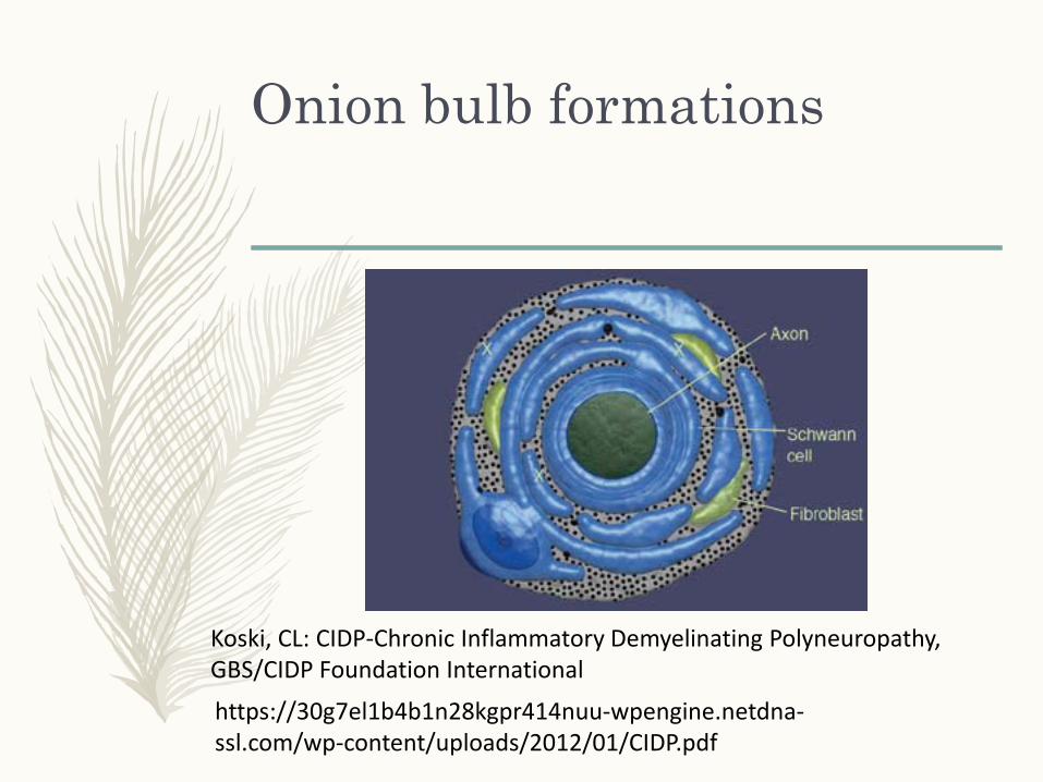
Onion bulb formations
Koski, CL: CIDP-Chronic Inflammatory Demyelinating Polyneuropathy, GBS/CIDP Foundation International
https://30g7el1b4b1n28kgpr414nuu-wpengine.netdna-ssl.com/wp-content/uploads/2012/01/CIDP.pdf

III. B. Fiber interaction/Artificial Synapses
– Axons /funiculi are separated by myelin, endoneurialsheath preventing spread of current to adjacent fibers
– After damage the currents may spread to adjacent fibers causing excitation (Otani, 1937, … many others)
– Especially true of sensory fibers (less myelin)

III. C. Applied anatomy to Nerve injuries – Internal features– Regeneration
– Scar tissue can redirect funicular pathways, blocking access to branches.
– The higher the lesion, the greater chance growing fibers will enter foreign tubes
– Ideally, it is important to have correct orientation of the nerve ends before resuturing, suture bundle groups separately, nerve grafts should be selected for large tightly packed funiculi.
(Sunderland, p. 204 – 211, 1968)

References
– Sunderland S, Nerve and Nerve Injuries, Livingstone LTD, Ediburgh and London, 1968
– Please email request for list of other references
2019
Share Your Feedback
• Please use the 2019 AANEM Annual Meeting app to rate this presentation and the speaker(s).
• Your feedback helps us enhance our annual meeting to ensure we are continuing to meet your needs.

2019
• Claiming CME• Course and Plenary Presentations
Visit: www.aanem.org/resources
Record your attendance hours after each session or do it all at once after the meeting is complete! Credit not recorded by December 15, 2019 will not be reported to ABPN and ABPMR. The AANEM will report ALL Annual Meeting attendees’ credit to ABPN and ABPMR by December, 31, 2019.

“


