HAL Id: hal-00297294https://hal.archives-ouvertes.fr/hal-00297294
Submitted on 16 Jul 2008
HAL is a multi-disciplinary open accessarchive for the deposit and dissemination of sci-entific research documents, whether they are pub-lished or not. The documents may come fromteaching and research institutions in France orabroad, or from public or private research centers.
L’archive ouverte pluridisciplinaire HAL, estdestinée au dépôt et à la diffusion de documentsscientifiques de niveau recherche, publiés ou non,émanant des établissements d’enseignement et derecherche français ou étrangers, des laboratoirespublics ou privés.
Neurofeedback improves executive functioning inchildren with autism spectrum disorders
Mirjam Kouijzer, Jan De Moor, Berrie Gerrits, Marco Congedo, Hein VanSchie
To cite this version:Mirjam Kouijzer, Jan De Moor, Berrie Gerrits, Marco Congedo, Hein Van Schie. Neurofeedback im-proves executive functioning in children with autism spectrum disorders. Research in Autism SpectrumDisorders, Elsevier, 2009, 3 (1), pp.145-162. <10.1016/j.rasd.2008.05.001>. <hal-00297294>
Running head: NEUROFEEDBACK IMPROVES EXECUTIVE FUNCTIONING IN AUTISM
Neurofeedback improves Executive Functioning in Children with Autism Spectrum Disorders
Mirjam E. J. Kouijzer1,2, Jan M. H. de Moor1, Berrie J. L. Gerrits2, Marco Congedo3, and Hein T.
van Schie1
1 Behavioral Science Institute, Radboud University Nijmegen, Nijmegen, The Netherlands
2 Neurofeedback Nijmegen, Nijmegen, The Netherlands
3 Centre National de la Recherche Scientifique, France
Corresponding author: Hein T. van Schie; [email protected]

Neurofeedback in ASD 2
Abstract
Seven autistic children diagnosed with autism spectrum disorders (ASD) received a
neurofeedback treatment that aimed to improve their level of executive control. Neurofeedback
successfully reduced children’s heightened theta/beta ration by inhibiting theta activation and
enhancing beta activation over sessions. Following treatment children’s executive capacities were
found to have improved greatly relative to pre-treatment assessment on a range of executive
function tasks. Additional improvements were found in children’s social, communicative and
typical behavior, relative to a waiting list control group. These findings suggest a basic executive
function impairment in ASD that can be alleviated through specific neurofeedback treatment.
Possible neural mechanisms that may underlie neurofeedback mediated improvement in
executive functioning in autistic children are discussed.
Keywords: neurofeedback, autism spectrum disorder, executive function, theta/beta ratio,
anterior cingulate cortex
Neurofeedback in ASD 3
Neurofeedback refers to a form of operant conditioning of electrical brain activity, in
which desirable brain activity is rewarded and undesirable brain activity is inhibited.
Neurofeedback is believed to elicit growth and changes at cellular levels of the brain, which in
turn support brain functioning and behavioral cognitive performance (Demos, 2005). In the
domain of intervention, neurofeedback training is useful in treatment of different disorders in
adults and children. Positive effects of neurofeedback in adults have been found for Attention
Deficit Hyperactivity Disorder (ADHD) (Kropotov et al., 2005), traumatic brain injury
(Thornton, 2000), epilepsy (Sterman, 2000), depression (Hammond, 2003), migraine (Kropp,
Siniatchkin & Gerber, 2002), addiction (Trudeau, 2005), anxiety disorders (Moore, 2000), and
general cognitive performance (Vernon et al., 2003).
Less is known about the effects of neurofeedback in children. In children, research on the
effects of neurofeedback is mainly carried out in the area of ADHD (Fuchs et al., 2003; Monastra
et al., 2005; Vernon, Frick, & Gruzelier, 2004), but positive effects of neurofeedback have also
been found for children with migraine (Kropp, Siniatchkin, & Gerber, 2002) and learning
disorders (Fernandez et al., 2003; Thornton, & Carmody, 2005). ADHD is typically characterized
by a heightened ratio between theta (4-8 Hz) and beta (12-21 Hz) activity in the ongoing EEG
during rest. Neurofeedback protocols that have aimed at inhibiting theta activity while rewarding
beta activity have led to successful alleviation of symptoms associated with ADHD such as
deficits in sustained attention, impulsivity, and control over hyperactive behaviors (reviews in
Butnik, 2005; Fox, Tharp, & Fox, 2005).
Several studies suggest that neurofeedback protocols that have been successful for
treatment of ADHD may also be efficacious for treating children with autistic related deficits.
Sichel, Fehmi, and Goldstein (1995) report about Frankie, a 8,5 year old boy with a mild form of
autism and attention impairments suggesting ADHD. Frankie’s 19-channel QEEG demonstrated

Neurofeedback in ASD 4
theta (4-8 Hz) to beta (13-21 Hz) ratios of 3.59 (Cz), 3.40 (C3), 3.03 (C4), 3.98 (Pz), 4.07 (P3),
3.63 (P4), and 3.02 (Fz). After 31 neurofeedback sessions aimed at inhibiting theta (4-8 Hz) and
rewarding low beta (12-15 Hz), his mother reported positive changes in all the diagnostic criteria
defining autism in DSM-III-R (e.g. attending and reacting to others, imaginative play, seeking
comfort, more talking and eye contact). QEEG furthermore revealed that theta/beta power ratios
had dropped below 3.0 at C3, C4, Fz, Pz, and P4.
Further support for a relation between theta/beta power and autism was provided by
Jarusiewicz (2002) who conducted a group study investigating effects of neurofeedback in 12
autistic children, compared with matched controls. The main protocol aimed at inhibiting theta
(2-7 Hz) and increasing sensory motor rhythm (SMR) activity (10-13 Hz) over the right motor
area. Results indicated a substantial decline in autistic behavior (26% as compared to 3% for the
controls) as reflected by the Autism Treatment Evaluation Checklist (ATEC). Parent reports
furthermore indicated considerable improvements on socialization, vocalization, school work,
anxiety, tantrums, and sleep, whereas no or minimal changes were found for the control group.
More recently Scolnik (2005) conducted a neurofeedback study with five children
diagnosed with Asperger disorder (a subclass of Autism Spectrum Disorder; ASD), each with
unique behavioral problems , i.e. poor social skills, lack of empathy, and inflexibility, coupled
with abnormal high theta/beta ratios varying from 2.19 to 6.89. Each child’s protocol was
determined on the basis of their individual QEEG and consisted of variations on the theme of
rewarding 12-15 Hz in the lower beta range while inhibiting slower 4-10 Hz activity in the theta
band. After 24 sessions of neurofeedback, parents and teachers reported improvements in
behavior, i.e. less anxiety, more flexibility, higher self-esteem, more empathy, improvement in
frustration toleration, increased social interaction, and fewer severe mood changes. Furthermore,
in two of the five children, theta/beta ratios changed into a positive direction.

Neurofeedback in ASD 5
The above studies suggest that neurofeedback protocols that inhibit theta and reward beta
and SMR may hold particular value for the treatment of autistic children, similar to the treatment
of ADHD. Surprisingly, however, no functional explanations currently exist for these
improvements and little is known about the neural mechanisms involved. In light of the
increasing popularity and clinical use of neurofeedback, however, fundamental explanations
become increasingly more relevant. Vice versa, the efficacy of evolved protocols and practices
may help to advance more fundamental insight into impairments underlying neuropsychological
deficits such as autism and ADHD. The aim of the current study therefore is twofold. On the one
hand we wish to contribute to the clinical practice by evaluating the efficacy of neurofeedback for
treatment of autism, whereas on the other hand we intend to further our understanding of the
possible (neural) mechanisms supporting treatment effects.
In order to optimize the neurofeedback treatment protocol for children with ASD and its
rationale, further methodological improvement is necessary in the form of controlled studies,
larger sample sizes, a more accurate description of sample characteristics and collection of
follow-up data. Another guiding principle should be the assessment of the clients’ satisfaction
with the treatment and procedure to enhance the social validity of the approach. Social validity
refers to the use of evaluative feedback from clients to guide program planning and evaluation
(Schwartz & Baer, 1991). Social validity may be evaluated at three levels of treatment: goals,
procedures, and outcomes (Wolf, 1978). In the current study we included the above guidelines
and evaluative measures (cf. Heinrich, Gevensleben, & Strehl, 2007) to further validate the use of
neurofeedback treatment of ASD.
In addition to the practical evaluation of neurofeedback treatment of ASD, the current
study aims to contribute to understanding the cognitive and neural mechanisms that underlie
neurofeedback improvements in ASD. We hypothesize that the reason for the efficacy of
Neurofeedback in ASD 6
neurofeedback protocols that reduce theta and reward beta lies primarily in the enhancement of
activation in the anterior cingulated cortex (ACC). The ACC is one of the main generators of
theta (Onton, Delorme, & Makeig, 2005; Tsujimoto, Shimazu, & Isomura, 2006; Meltzer,
Negishi, Mayes, & Constable, 2007), and is well known for its role in regulating cognitive and
emotional processes in the brain contributing to cognitive control and executive function (review
in Bush, Luu, & Posner, 2000). Neuroimaging studies investigating the neural basis of ADHD
and ASD have reported hypo-activation and functional under-connectivity of the ACC (Bush,
Valera, & Seidman, 2005; Cherkassky, Kana, Keller, & Just, 2006) which could explain why
cognitive deficits associated with ADHD and ASD often seem to fall within the domain of self-
regulation and executive function (Barkley, 1997). Furthermore, combined EEG-fMRI studies
have indicated a negative relationship between theta power and BOLD signal in the ACC
(Meltzer, Negishi, Mayes, & Constable, 2007), in line with the hypothesis that theta activation in
autistic children is associated with under-activation of the ACC (Murias, Webb, Greenson, &
Dawson, 2007).
Following the above reasoning we predict that down-regulation of theta activity should
enhance activation of the ACC and executive control mechanisms of the brain, which should lead
to more efficient behavior of ASD children on tasks requiring executive function. To investigate
the hypothesized relationship between theta and executive function, a group of children from the
autism spectrum diagnosed with ASD were selected for neurofeedback training that reduced theta
activity while rewarding low beta activity, in accordance with the standard ADHD treatment
protocol. A waiting-list control group, also diagnosed with ASD, received neurofeedback training
at a later time and served as a baseline to determine treatment effects of neurofeedback on
children’s’ executive, social and neurophysiological levels of functioning.

Neurofeedback in ASD 7
Method
Participants
Fourteen children with ASD (12 males; 2 females) with a mean age of 10.1 years (range 8
to 12 years) were recruited by an advertisement in a magazine for parents of ASD children.
Inclusion criteria were an IQ-score of 70 and above and the presence of ASD as diagnosed by a
child psychiatrist or health care psychologist. All participants had the diagnosis pervasive
developmental disorder – not otherwise specified (PDD-NOS). Each diagnosis was confirmed by
a clinical psychologist and by results on the CCC questionnaire. Excluded were children using
medication, children with a history of severe brain injury, and children with co-morbidity such as
ADHD and epilepsy. The seven children who applied first, were assigned to the intervention
group. The control group included seven children who were recruited out of a larger group of
children who applied later and were selected to match children of the intervention group on age,
sex, and intelligence scores. Table 1 represents the demographic characteristics of the
intervention and the control group. There were no significant differences between the both groups
with respect to the variables sex, mean age, total IQ, verbal IQ, and performal IQ. Children in the
control group were invited for neurofeedback training after finishing the present study.
----------------------------
Insert Table 1 about here
-----------------------
Procedure
A non-randomized pretest-posttest control group design with individual matching was
used with follow-up measurements after three months. During a baseline period, all participants
were pre-tested on QEEG and a range of executive functions tasks, and parents completed a
communication checklist (CCC-2). After 40 sessions of neurofeedback, or comparable time

Neurofeedback in ASD 8
interval for the waiting-list control group, QEEG, executive functions skills, and communicative
abilities were re-collected. During follow-up, three months after ending neurofeedback sessions,
again, QEEG, executive functions skills and communicative abilities were measured together
with a questionnaire (AUTI-R) to estimate behavioral improvements in children. For the
intervention group, the follow-up measurement furthermore included a social validity
questionnaire. The research design was authorized by an ethics committee for behavioral
sciences.
An interview was conducted with the parents prior to the neurofeedback treatment to
survey the anamneses of the child, family history, and current problems of the participant.
Procedures and possible side effects were explained to all participants. All participants signed an
informed consent. Pre and post-treatment measures took two hours for each participant to
complete. Tasks for executive functioning were given to all participants in a fixed order, with the
first five tasks before QEEG assessment and the rest after QEEG assessment. The CCC-2, the
adapted AUTI-R, and the social validity questionnaire were filled out by the parents at home.
QEEG measurement
Children’s QEEG (quantitative electroencephalogram) was recorded and digitized with a
TruScan 32 Acquisition EEG System (Deymed Diagnostic, USA). Data were acquired using a
stretchable electrode cap embedded with 19 sensors at scalp locations Fp1, Fp2, F7, F3, Fz, F4,
F8, T3, C3, Cz, C4, T4, T5, P3, Pz, P4, T6, O1, and O2, according to the International 10/20
System (Jasper, 1958). A ground electrode was placed between Fp2 and F8 and two ear clips
were used as reference electrodes (A1 and A2). Impedance was kept below 5 kΩ, with a
maximum difference of 1 kΩ between electrodes. Data was collected for three minutes in an eyes
open and an eyes closed condition.

Neurofeedback in ASD 9
Neurofeedback training
A portable NeXus-4 amplifier and recording system (Mindmedia, The Netherlands) was
used for neurofeedback training and concurrent data collection. Ag/AgCl disposable snap-on
sensors (MedCaT, The Netherlands) were applied to the patients’ scalp at the locations C3 and
C4.
Each participant in the intervention group visited a private practice twice a week until 40
sessions were completed. Training was carried out by a state licensed psychotherapist with
extensive training in neurofeedback. During each session a protocol was carried out, which
consisted of a baseline of three minutes (i.e., no feedback), followed by seven three-minute
intervals of neurofeedback. Neurofeedback intervals were separated by one-minute rest intervals,
in which the participant was instructed to sit still and relax, without receiving feedback.
Neurofeedback training followed a standard ADHD training protocol (Heinrich, Gevensleben, &
Strehl, 2007 for review) aimed at reducing theta activity (4-7 Hz) while increasing activity in the
low beta band (12-15 Hz)1 at C4 (reference at A1). The signal at location C4 was fed back to the
patient in visual form. Theta and beta activity were visualized in separate bar graphs on the
computer screen and participants were instructed to “try to move down the theta activity below
the criterion line on the computer screen and to move up the beta activity above the criterion line,
using the feedback to guide you”. During intervals when specified amplitude conditions were
met, subjects were rewarded by the continuation of a short movie that was selected to fit each
child's individual interest and age. All movies were presented with audio. When subjects failed to
maintain power within the required range, the movie and music would stop playing. Individual
criteria were set to allow each participant to reach the reward.

Neurofeedback in ASD 10
Executive function tasks
According to Smidts (2003), executive functions are typically divided into separate
subdomains, each including one or more executive functiontests.
Attentional control
Attentional control encompasses selective attention, visual as well as auditory, and
response inhibition. Visual selective attention was measured by the Continuous Performance Test
(CPT), a subtest of the neurocognitive test battery CNS Vital Signs (CNSVS). In the CPT, the
participant has to respond to one particular character on the computer screen while ignoring other
characters during five minutes. The score for visual selective attention is based on the amount of
errors of the CPT (range 0 – 200). Selective attention for auditory stimuli was measured by the
Test of Sustained Selected Attention (TOSSA; Kovács, 2005b). In the TOSSA, participants have
to respond to sets of 3 beeps while ignoring sets of 2 or 4 beeps. Beeps are presented during eight
minutes with variable speed. The test score reflects the percentage of good answers, calculated by
dividing the number of hits by the total amount of items, times 100. Response inhibition is
divided in a verbal and a motor variant. Verbal response inhibition was assessed by the Stroop
test (Stroop, 1935). In this test, participants have to read aloud as soon as possible A) 100 words
(green, red, yellow, and blue), B) the color of 100 colored rectangles, and C) the color of the ink
of 100 written incongruent color names. The goal in part C is to pronounce the name of the color
of the ink, while ignoring reading the word. The score on this test is represented by the
interferential time (time C minus time B). Motor response inhibition was assessed with the
response inhibition score (RIS; range 0 – 100) of the TOSSA, based on the number of
commission errors.
Cognitive flexibility
Cognitive flexibility covers verbal memory and visual memory, set-shifting, concept

Neurofeedback in ASD 11
generation, and feedback utilization. Verbal memory and visual memory were assessed by the
Verbal Memory Test (VBM) and the Visual Memory Test (VIM) of the CNSVS, respectively. In
the VBM and the VIM, participants have to memorize words (n=15, VBM) and geometric figures
(n=15, VIM) and later recognize them in a series of distracters (n=15 for both tests). The sum of
correct responses (maximum = 60) was calculated to get a final score for verbal memory
(maximum = 60) and a final score for visual memory (maximum = 60). Set-shifting was
examined by the Trail Making Test (TMT; Reitan, 1956). In the TMT, participants have to switch
between the numerical mode and the alphabetic mode by connecting 26 numbers and characters
in the 1-A-2-B-3-C – order. A score on the TMT is comprised of the total time needed to finish
the test, translated into an age related t - score (range 20 – 75). Concept generation and feedback
utilization were examined by the Milwaukee Card Sorting Test (MCST; Kovács, 2005a), a
computerized version of the Wisconsin Card Sorting Test. The participant has to generate and
apply a non-spoken rule for sorting cards (n=60), based on feedback (e.g. ‘good’ or ‘fault’).
These card sorting principles can be either color, shape, or number and change after every 10
correct answers. An indicator for cognitive flexibility is the number of categories (range 0-6) a
child creates with 60 cards.
Goal setting
Goal setting was assessed by the Tower of London (TOL; Kovács, 2005c). Participants
have to copy a construction of blocks and bars by moving three prearranged different colored
blocks along three bars of different lengths. The score on the TOL is a percentage calculated by
dividing the participants’ score by the maximum score, times 100.
Speed and efficiency
Speed and efficiency is measured by the Symbol Digit Coding (SDC) of the CNSVS.
Participants have to code as many symbols as possible within two minutes, according to a set of

Neurofeedback in ASD 12
eight symbol–digit pairings that are displayed continuously for reference on screen. A score for
speed and efficiency is calculated by the number of correct responses minus the number of errors
on the SDC.
Questionnaires
Children’s Communication Checklist (CCC-2-NL)
The CCC-2-NL (Geurts, 2007) was used to assess improvement in children’s language
structure, pragmatics, and social interaction. Language structure includes the subscales speech
production, syntax, semantics, and coherence. The domain of pragmatics consists of the subscales
inappropriate initiation, stereotyped conversation, use of context, and non-verbal communication.
The domain of social interaction includes the subscales social relations and interests. Response
categories for each question are ‘never or less than once a week’, ‘at least once a week, but not
every day’, ‘once or twice a day’ or ‘more than twice a day or always’. An age-related standard
score was calculated for each subscale and for the composed scales general communication
(language structure and social interaction) and pragmatics.
AUTI-R
An adapted version of the AUTI-R (Berckelaer-Onnes & Hoekman, 1991) was used to
study improvement of children in the intervention group on social interaction, communication,
and restricted, repetitive, and stereotyped patterns of behavior, interests and activities. Eleven
items of the AUTI-R that were considered not relevant for the present study and five items that
did not fall into the categories social interaction, communication or restricted, repetitive, and
stereotyped patterns of behavior, interests and activities, were excluded from the list. The adapted
questionnaire contained 33 items, subdivided into the scales Social interaction (n=10),
Communication (n=8), and Behavior (n=15). Items on the questionnaire were rated on a 5-point

Neurofeedback in ASD 13
scale, with 1 point indicating low progression and 5 points indicating strong progression. Mean
scores were calculated for the subscales Social interaction, Communication, and Behavior, and
for the complete questionnaire.
Social validity
Social validity was assessed by a self-constructed anonymous 5-point scale questionnaire
with 15 items about the Goals of treatment (n=4), Treatment procedures (n=8), and Outcomes
(n=3) (Wolf, 1978). All items were scored, with 1 point indicating low satisfaction and 5 points
indicating high satisfaction. Sum scores were calculated for each subscale to evaluate the
acceptability of the neurofeedback treatment. Three open response questions were added to assess
whether parents had any remarks, whether they had suggestions to improve neurofeedback
treatment, and whether they would recommend neurofeedback treatment to others.
Data analysis
QEEG
Eye blinks and other artifacts were manually removed from the raw EEG data by an
independent EEG specialist and statistician, who was blind to the subject’s classification (i.e.,
intervention group vs. control group) and the type of EEG (i.e., pre vs. post training). The raw
data were processed with fast Fourier transformation to determine the magnitude of each
frequency band in microvolt. Separate power measures were calculated for delta (1-4 Hz), theta
(4-8 Hz), alpha (8-12 Hz), low beta (12-15 Hz), beta 2 (15-18 Hz), beta 3 (18-25 Hz), and high
beta (25-30 Hz). EEG data of all individuals were compared with the Neuroguide (Thatcher et al.,
2003) database, which provides reliable descriptors of normative brain electrical activity (John et
al., 1988). Linked ears montages were used. Data from all 19 electrode sites were used for
analysis. The split-half reliability and test-retest reliability of the artifact free data of all subjects

Neurofeedback in ASD 14
were above .95 (p<.05). Absolute power (the amount of energy in µV²), relative power (the
percentage of power in a frequency band relative to the total power contained by all other
frequency bands), and coherence were calculated for each participant, frequency band, and
individual electrode lead. All power and coherence values were subsequently transformed to Z-
scores, reflecting deviancy from the normative database (Hughes & John, 1999). A 2 (Time:
time1 vs. time2) x 2 (Group: intervention vs. control) mixed MANOVA was performed to look
for treatment effects in the intervention group relative to the control group.
Session data
Eye blinks and other artifacts were manually removed from the raw EEG data of 40
sessions, collected at C3 and C4 during training intervals. The raw data were Fast Fourier
Transformed (FFT) to determine the power of each frequency. Separate power measures were
calculated for delta (1.5-3.5 Hz), theta (4-8 Hz), alpha (8-12 Hz), low beta (12-15 Hz), beta 2 (13-
21 Hz), and high beta (22-30 Hz). Power values of each frequency band were log-transformed. A
2 (Time: first sessions vs. last sessions) x 2 (Location: C3 vs. C4) mixed MANOVA was
conducted to compare power during the first 20 sessions with the final 20 sessions. Furthermore,
the efficacy of neurofeedback over sessions per frequency band was estimated for each individual
subject by calculating a linear regression line and Spearman regression coefficient fitting the
progression of power values over sessions.
Executive function tasks
Results of a one-sample Kolmogorov -Smirnov test showed that data of each variable did
not deviate significantly from normality. A MANOVA was conducted to test differences in
executive functions for the intervention group and the control group at time1. Neurofeedback
related changes in executive functions were verified by performing a 2 (Time: time1 vs. time2) x
2 (Group: intervention vs. control) mixed MANOVA.

Neurofeedback in ASD 15
Questionnaires
Results of a one-sample Kolmogorov -Smirnov test showed that data of each variable of
the CCC-2 did not deviate significantly from normality. MANOVA was conducted to test for
differences on the CCC-2 between the intervention group and the control group at time1.
Neurofeedback related changes on the CCC-2 were verified by performing a 2 (Time: time1 vs.
time2) x 2 (Group: intervention vs. control) mixed MANOVA.
In order to assess whether the intervention group decreased in ASD-symptoms more than
the control group, a comparison between scores on the adapted AUTI-R of the intervention group
and the control group was made using a MANOVA with between-subjects factor Group.
The social validity of the neurofeedback treatment was evaluated via the sum scores of
the subscales Goals, Procedures, Outcomes, and open response questions.
Results
Session data
At the individual level, Spearman’s correlation coefficients showed a significant reduction
of theta power (4-7 Hz) over 40 sessions of neurofeedback in five participants at C4 (p’s <.05, r =
-.596 ~ -.718) and in the same five participants at C3 (p’s <.05, r = -.496 ~ -.771). Two
participants did not show significant reduction of theta power at C4 (p =.411, r =.035; p =.359, r
=.056) and C3 (p =.018, r =.453; p =.170, r =.135 ). Results of theta reduction at C3 and C4 for
all participants can be found in Figure 1.
----------------------------
Insert Figure 1 about here
-----------------------
Low beta power (12-15 Hz) increased significantly over time for five participants at C4 (p’s <

Neurofeedback in ASD 16
.05, r = .218 ~ .410) and for six participants at C3 (p’s <.05, r = .253 ~ .529). Two participants
did not show significant increase of low beta power at C4 (p =.311, r =.079; p =.173, r =-.145)
and one participant did not show significant increase at C3 (p =.372, r =.051) (see Figure 2).
----------------------------
Insert Figure 2 about here
-----------------------
Besides changes in theta and low beta power, changes in delta power (1.5-3.5 Hz) were found as
well. Delta power decreased significantly in five participants at C4 (p’s <.05, r = -.449 ~ -.555)
and in five participants at C3 (p <.05, r = -.291 ~ -.562). No increase in delta power was found in
two participants at C4 (p =.125, r =.177; p =.356, r =.125 ) and at C3 (p =.263, r =.098; p =.054,
r =-.243). Results can be found in Figure 3. In alpha power (8-12 Hz), beta 2 power (13-21 Hz),
and high beta power (22-30 Hz), no unanimous patterns of change were found.
----------------------------
Insert Figure 3 about here
-----------------------
Analysis at group level further supported the correlation results. A 2 (Time: first sessions
vs. last sessions) x 2 (Location: C3 vs. C4) mixed MANOVA showed significant reduction of
theta power (4-7 Hz) (F (1,6) =11.419, p <.05, η =.656) and significant increase of low beta (12-
15 Hz) (F (1,6) =21.922, p <.01, η =.785) at C3 and C4 over 40 sessions of neurofeedback.
Besides power changes in theta and low beta, a significant decrease of delta power (1.5-3.5 Hz)
over time was found as well (F (1,6) =6.982, p <.05, η =.538). For alpha power (8-12 Hz), beta2
power (13-21 Hz), and high beta power (22-30 Hz), no significant effects of time were found.
Decrease of delta power was significantly correlated with decrease in theta power (r =
0.667, p <.01) and with increase in low beta power (r =-0.695, p <.01). The correlation between
Neurofeedback in ASD 17
decrease in theta power and increase in low beta power was highly significant (r =-0.811, p
<0.001).
QEEG
The absolute and relative power of each frequency band for all 19 channels for the
intervention group and the control group were compared using MANOVA. In order to claim a
treatment effect, we need the interaction between Time (time1 vs. time2) and Group (intervention
vs. control) to be significant. The mixed MANOVA suggested no significant multivariate
interaction between Time and Group in the target frequency bands, i.e. absolute (F (1,12) =2.382,
p =.149, η =.166) or relative theta power (F (1,12) =.986, p =.340, η =.076) and absolute (F
(1,12) =.018, p =.897, η =.001) or relative low beta power (F (1,12) =.614, p =.449, η =.049).
Univariate results of absolute and relative theta and low beta power in 19 separate electrodes
revealed no significant interaction effects either (range of F-values = .000-3.977, p’s >.05). A
similar 2 (Time: time1 vs. time2) x 2 (Group: intervention vs. control) MANOVA for the other
frequency bands, i.e. delta, alpha, beta2, beta3 and high beta revealed no significant multivariate
effects, neither for absolute nor relative power (range of F-values = .000-1.820, p’s >.05).
For the analysis of coherence, a 2 (Time: time1 vs. time2) x 2 (Group: intervention vs.
control) mixed MANOVA was performed. Univariate results revealed a significant reduction of
hypo connectivity in theta power at time2 (F-values up to 17.572, p’s < .05), especially between
frontal and central/temporal electrodes. However, since this reduction was found in both the
intervention and the control group, no significant interaction effects were found (range of F-
values = .000 – 2.914, p’s > .05).
Executive function tasks
A MANOVA was conducted to test the hypothesis that participants in the intervention
group would display the same scores as participants in the control group at time1. No statistical

Neurofeedback in ASD 18
significant differences between intervention and control group were found on tests for executive
functioning at time1 (F (1,12) =1.066, p =.577, η =.842).
To analyze whether children in the intervention group scored significantly higher on tests
for executive functioning at time2 compared to the matched control group, a 2 (Time: time1 vs.
time2) X 2 (Group: intervention vs. control) mixed MANOVA was performed. In order to claim
a treatment effect and to control for practice effects, we need the interaction to be significant.
Attentional control
Subjects’ capacity for attentional control was tested using separate measures targeting
children’s attentional capacity in the visual and auditory domains and their ability to inhibit
verbal and manual response tendencies. Table 2 reports the behavioral results of all executive
function tests gathered for both groups at time1 and time2. No significant interaction between
Time and Group was found for measures of visual selective attention (F (1,11) =.047, p=.832, η
=.004). Both groups made very little errors in detecting a target letter in a continuous stream of
distractors, leaving little or no room for improvement at time2 (values for visual selective
attention in Table 2 represent the amount of errors found with 200 items). However, a significant
Time x Group interaction effect was found for measures of auditory selective attention (F (1,11)
=8.437, p =.014, η =.434). Children in the intervention group showed a considerable
improvement in their ability to correctly detect auditory targets in the TOSSA, from 48% to 62%
correct responses after neurofeedback training, as compared to the control group who showed
minimal improvement from 68% to 69% correctly detected targets. In addition, a significant
interaction between Time and Group was found for children’s capacity to inhibit verbal
responses (F (1,11) =4.890, p =.049, η =.308). Interference effects of written names were
strongly reduced from 68 seconds before to 30 seconds after neurofeedback training for the
intervention group. The control group also showed a difference between interference effects at

Neurofeedback in ASD 19
time1 and time2 (66 seconds and 50 seconds respectively) but this reduction was about half the
size of the effect found with the intervention group. Consistent with the increased ability to
inhibit verbal responses, children of the intervention group were also better able to inhibit
impulsive tendencies in responding on the TOSSA, suggesting improved inhibition capacity after
neurofeedback training (78% correctly inhibited before training vs. 90% after neurofeedback
training). Only minimal improvements in impulse control were found for the control group (89%
correct inhibitions at time1 followed by 91% correct inhibitions at time2), resulting in a
significant Time x Group interaction, F (1,11) =5.064, p =.046, η =.315.
Cognitive flexibility
Children’s cognitive flexibility was investigated using measures of visual and verbal
memory, set-shifting and concept generation. Neurofeedback training did not influence children’s
capacity to memorize and recognize words (F (1,11) = .021, p =.889, η =.002) and geometric
shapes (F (1,11) =.004, p =.952, η =.000). Both groups showed a minimal non-significant
reduction of performance from time1 to time2 (see Table 2), on verbal memory, F (1,11) =0.355,
p =.563, η =.031, and visual memory, F (1,11) =0.138, p =.717, η =.012. However, children’s set-
shifting ability as indexed by the TMT did show a significant Time x Group interaction (F (1,11)
= 5.602, p =.037, η =.337), reflecting improved cognitive flexibility and sequencing after
neurofeedback treatment. For the intervention group t-scores improved from 30 (time1) to 47
(time2), whereas only a small improvement was found for the control group with t-scores
improving from 30 (time1) to 34 (time2). Also concept generation and use of feedback, as
measured by the MCST, were found to improve significantly for the intervention group as
compared to the control group, F (1,11) =5.081, p =.046, η =.316. After neurofeedback, ASD
children discovered an average of 5 (out of 6) card sorting rules, whereas before training they
only reached an average of 2.5. In contrast, the performance of the control group was comparable

Neurofeedback in ASD 20
at time1 (3.5 rules) and time2 (3.8 rules).
Goal Setting
Analysis of children’s goal setting capacity as assessed by the TOL showed a significant
interaction between Time and Group, F (1,11) = 7.198, p =.021, η =.396, reflecting a clear
improvement in complex sequential problems after neurofeedback training, as compared to the
control children. At time1 children from both groups reached an average performance of 55
(range 0-138). However, whereas children of the control group showed little improvement (57 at
time2), children of the intervention group drastically improved their capacity score to 76 at time2.
Speed and efficiency
Children’s combined score for speed and efficiency on the SDC indicated a stronger
improvement for the intervention group than for the control group (see Table 2), but the required
interaction between Group and Time was not found significant (F (1,11) =.397, p =.542, η
=.035).
----------------------------
Insert Table 2 about here
-----------------------
A 2 (Time: time2 vs. follow-up) x 2 (Group: intervention vs. control) mixed MANOVA
indicated no significant differences between post treatment and 3-month follow-up measurements
of children’s executive functioning at time3, F (1,11) =.987, p =.602, η =.832.
Questionnaires
CCC-2
The CCC-2 measured parents’ appreciation of their children’s communication skills for
different aspects (subscales) of communication. A MANOVA was conducted in order to test the

Neurofeedback in ASD 21
hypothesis that participants in the intervention group would display the same scores on the CCC-
2 questionnaire as participants in the control group at time1. No statistically significant
differences between intervention and control group were found on the CCC-2 questionnaire
collected at time1, F (1,12) =54.149, p =.106, η =.998.
To analyze whether children in the intervention group scored significantly higher on the
CCC-2 at time2 compared to the matched control group, a 2 (Time: time1 vs. time2) x 2 (Group:
intervention vs. control) mixed MANOVA was performed. Separate analysis of the
communication subscales of the CCC-2 showed a significant Time x Group interaction effect for
non-verbal communication, F (1,12) =5.505, p =.037, η =.314, reflecting an improvement in
non-verbal communication for the intervention group, relative to the control group. For none of
the other subscales the interaction between Time and Group was found significant, all p’s > .05.
In Table 3 the average ratings of children’s communication skills are reported for sub- and
compound-scales of the CCC-2 for the control group and the intervention group at time1 and
time2. Lower values in Table 3 reflect better communication skills. Analysis of the two
compound scales, general communication and pragmatics, revealed a significant interaction
effect between Time and Group for general communication, F (1,12) =5.379, p =.039, η =.310,
but not for pragmatics, F (1,12) =.036, p =.852, η =.003. Parents of children in the intervention
regarded their children’s communication skills as more advanced after neurofeedback training
than before, whereas no such difference was found for the control group.
----------------------------
Insert Table 3 about here
-----------------------

Neurofeedback in ASD 22
A 2 (Time: time2 vs. follow-up) x 2 (Group: intervention vs. control) mixed MANOVA
indicated no significant changes in scores on the CCC-2 three months after neurofeedback
training was ended (F (1,12) =.253, p =.930, η =.752).
AUTI-R
The AUTI-R measured parents’ evaluation of children’s improvements on social
interaction, communication, and typical behavior. Table 4 shows the average improvement for
the intervention and control group for each subscale of the AUTI-R. Following treatment,
parents’ ratings suggested improvements for children in the intervention group on social
interaction, communication, and typical behavior as compared to children in the control group. A
MANOVA with between subjects factor Group was used to analyze the results of the three
subscales of the adapted AUTI-R. Results indicated a significant increase in desired behavior
after neurofeedback training for the intervention group in comparison with the control group.
Children’s social interaction ability was valued to be improved following treatment, as compared
to the control group, F (1,12) =17.775, p =.001, η=.618. Children’s communication ability was
assessed to be enhanced in comparison to the assessment of children in the control group, F
(1,12) =29.054, p =.000, η=.725. Furthermore, typical autistic behavior was found to be
attenuated as compared to the assessment of children in the control group, F (1,12) =7.782, p
=.018, η=.414.
----------------------------
Insert Table 4 about here
-----------------------
Social validity
Social validity of the intervention was assessed using 5-point rating scales (5= high
satisfaction, 1= low satisfaction). Results indicated neurofeedback treatment to be a socially

Neurofeedback in ASD 23
acceptable treatment method with respect to its goals, procedures, and outcomes. Parents of
children in the intervention group indicated to be well informed about the goals of treatment
before treatment started (M=4.67). Parents rated the treatment as being neither aggravating for
their child (M=4.47), nor for themselves (M=3.34). Viewing video’s during training was rated
not to be aggravating at all for the children (M=5), as was placement of electrodes on the scalp
(M=4.83). The requirement of visiting the private practice twice a week for training (M=3.17)
and for pre and post assessment (M=3.17) was considered the most aggravating part of the
procedure for the parents, although the mean scores on these items are still relatively positive, i.e.
in the direction of ‘not aggravating’. Parents indicated to be satisfied with the outcomes of the
treatment with respect to children’s social behavior (M=3.83), communication skills (M=3.83),
and typical behavior (M=3.83). All parents would recommend neurofeedback treatment to other
parents of children with ASD. Only two parents had suggestions for improvement of
neurofeedback treatment, which were a real life experience of neurofeedback treatment for the
parents themselves and more time for evaluation during treatment. No parents had any other
further remarks in addition to their personal explanation of their answers on the 5 point scales.
Discussion
The present study evaluated the effects of a specific ADHD neurofeedback training
protocol for treatment of autistic children diagnosed with ASD. Reduction of theta power was
hypothesized to improve children’s executive capacities by enhancing activation of the ACC,
which is one of the main generators of theta activation over central areas. Consistent with our
prediction, children of the intervention group made large improvements in performance on a
range of executive function tasks after neurofeedback training, whereas no such effects were
found for a matched control group. These findings provide further support for the impairment of

Neurofeedback in ASD 24
executive functions in autism, and reinforce existing neurobiological views on autism that
suggested abnormal functioning of the ACC. Furthermore our findings provide further evidence
in support of the view that neurofeedback may hold particular value for treatment of children
with ASD which might be comparable with the effects found with ADHD.
At a neurophysiological level, neurofeedback training successfully reduced theta power
(4-7 Hz) and significantly increased low beta power (12-15 Hz) in all but two of seven
participants in the intervention group. Interestingly, and consistent with our hypothesis that
neurofeedback protocols that target children’s theta/beta ratio mainly work because they reduce
theta power, attenuation of theta power was found more reliable than enhancement of beta power
over sessions. Children’s individual Spearman correlation coefficients reflected significant
reductions of theta in five participants showing consistent effects over both hemispheres at C4
(average r = .68) and C3 (average r = .64), and enhancement of beta in five participants at C4
(average r = .30) and C3 (average r = .38). Furthermore, consistent decreases in delta power (1.5-
3.5 Hz) were found for 5 participants at C4 (average r = .55) and at C3 (average r = .45). The
gradual reduction in delta power probably co-occurred in conjunction with the reduction in theta
power, which is further supported by the strong correlation between power reductions of both
frequencies over time (r = .67).
Considering the consistent suppression of theta and delta frequencies and enhancement of
low beta activation over time across sessions, one could imagine structural changes in QEEG to
develop between pre- and post-test recordings. However, no significant changes were found in
the QEEG of the intervention group as compared with QEEG data of the control group. Our
findings are in line with results of Kropotov and colleagues (2007) who found no notable changes
in QEEG power spectra of children with ADHD after neurofeedback training, although
neurofeedback was found to affect the amplitude of event-related potential (ERP) components.

Neurofeedback in ASD 25
Coben and Padolsky (2007) found changes in children’s QEEG coherence after
neurofeedback training reflecting a decrease in cerebral hyper-connectivity in 76% of all children
of the intervention group. QEEG coherence values were only available for the intervention group,
not for the control group. In the present study, changes in connectivity were found for both the
intervention and the control group. These findings suggest a test-retest effect between pre- and
post-test EEG assessment which could e.g. reflect differences in vigilance or arousal between the
two assessments. That is, young children may be more alert and attentive during their first EEG
assessment as compared to the second time. This different mental state may be responsible for the
observed differences in QEEG between the pre- and post-test in both groups. Another
explanation for the absence of differences in QEEG is the small sample size that was used in the
present study.
At a cognitive level, neurofeedback training was hypothesized to improve the executive
functions of children with ASD, comparable with the success of the protocol in the treatment of
ADHD (Butnik, 2005). Results indicated significant improvement in attentional control,
cognitive flexibility and goal setting for children in the intervention group when compared to
children in the control group. These results are important because they reflect a serious cognitive
improvement in the intervention group that cannot be reduced to differences in perceived well-
being e.g. by parents. Instead, these findings indicate that neurofeedback training was associated
with a clear improvement in cognitive functioning on tasks requiring executive control.
Improvements were found for the majority of tasks taxing executive control, with strong
improvements on sustained auditory selective attention (30% more correct responses), inhibition
of verbal responses (55% reduction in response interference time), inhibition of motor responses
(15% reduction of commission errors), set shifting (57% reduction of time needed to switch
between the numerical and alphabetical mode), concept generation (50% increase in the number

Neurofeedback in ASD 26
of card sorting categories created), and planning ability (37% increase in performance on the
Tower of London task). Symbol digit coding was found improved (20% more accurate) for the
treatment group, but the difference with the improvement of the control group (7%) was not
significant. No noteworthy improvements were found on tasks taxing verbal and visual memory,
and sustained visual attention. Most children showed to be already highly efficient on these tasks
before the start of the neurofeedback treatment at Time 1, leaving little room for further
improvement. Coben and Padolsky (2007) evaluated executive functioning of children with ASD
after neurofeedback training using a questionnaire completed by parents and teachers. In
agreement with the present results a significant improvement on measures of executive functions
was reported. The present experimental findings further extend these previous results by showing
enhanced performance on a range of cognitive tasks requiring executive control. Whereas the
appraisal of a child’s level of executive functioning might be influenced by wishful thinking or
social expectation, such factors can not explain a 40% average increase in cognitive performance.
The fact that similar improvements were found over a range of different executive tasks further
strengthens the conclusion that neurofeedback substantially enhanced the executive capacity of
children with ASD. These results are furthermore in line with recent models that suggest a single
genetic to underlie most executive functions (Friedman, Miyake, Young, DeFries, Corley, &
Hewitt, in press).
We hypothesized that the elevated theta power that characterizes autistic children is
functionally related to their executive impairment. Electroencephalographic and
magnetoencephalographic studies have localized frontal theta activation to the rostral ACC
(Gevins, Smith, & McEvoy, 1997; Ishii et al., 1999) and studies combining EEG and fMRI have
consistently found correlations between theta power and BOLD signal in rostral ACC (Meltzer,
Negishi, Mayes, & Constable, 2007; Pizzagalli, Oakes, & Davidson, 2003; Sammer et al., 2007).

Neurofeedback in ASD 27
Interestingly, ACC activation and theta power appear to be inversely related. High-functioning
autistic individuals show hypoactivation and reduced connectivity of the ACC (Kana, Keller,
Minshew, & Just, 2007; Cherkassky, Kana, Keller, & Just, 2006) whereas EEG measures
consistently indicate elevated levels of theta power over medial frontal areas in ASD (e.g.
Murias, Webb, Greenson, & Dawson, 2007). Meltzer et al. (2007) found increasing working
memory load to be associated with enhancements of EEG theta power which correlated
negatively with BOLD signal in a network of areas including the rostral ACC (Meltzer et al.,
2007). Similar findings were reported by Sammer et al. (2007) using mental arithmetic-induced
workload and Kana et al. (2007) using a response inhibition paradigm. Interestingly, deactivation
of the ACC during cognitive demanding tasks is often found in association with deactivations of
other (medial) areas, such as the precuneus, which together have been labeled the default mode
network (DMN) reflecting its high default metabolism during rest (Gusnard & Raichle, 2001).
Much interest has developed in understanding the function of the DMN and several interesting
views have been formulated which appear to converge on the idea that the DMN is involved in
self-referential processing (Northoff et al., 2006) and understanding others’ intentions through
mental simulation (Uddin et al., 2007). These findings may have implications for understanding
social impairments in ASD. However, for the present discussion it is first important to note that
the rostral ACC is not directly involved in executing cognitive control (Rushworth et al., 2004),
but that its activation is inversely related to other areas that are activated during cognitive tasks,
such as the lateral prefrontal cortex (Greicius, Krasnow, Reiss, & Menon, 2003). Following this
suggestion, Fox and colleagues (2005b) discovered strong spontaneous anticorrelations between a
"task-negative" DMN and an opposing "task-positive" attentional network, in a resting state.
Kelly et al. (2008) furthermore found differences in individual attentional capacity to depend on
the strength of the negative correlation between the two opposing networks, with a reduced
Neurofeedback in ASD 28
antiphase relation resulting in more variable behavioral performance. In addition, a recent fMRI
study by Weissman et al. (2006) indicated that a failure to suppress the DMN may result in lapses
of attention. Uddin et al. (in press) yield further support for this view by indicating that the
balance between the two networks is primarily controlled by the DMN.
Importantly, these findings provide a possible mechanism through which we can
understand the relation between theta power, ACC activation, and executive function. The
enhancement of theta that is consistently found during cognitive effortful tasks, such as use of
working memory (Jensen & Tesche, 2002), mental arithmetic (Mizuhara, Wang, Kobayashi, &
Yamaguchi, 2004), error monitoring (Luu, Tucker, & Makeig, 2004), and sentence
comprehension (Bastiaansen, van Berkum, & Hagoort, 2002), probably reflects deactivation of
the rostral ACC / DMN, to allow activation in (task-positive) areas supporting the processing of
external goals (cf. Fransson, 2005). Consistent with the hypothesis that the executive problems of
autistic children may originate from a defective DMN, Kennedy et al. (2006) recently found that
autistic subjects, as compared with controls, did not deactivate their DMN during a range of
cognitive and emotional Stroop tasks. Inability to deactivate or modulate activation of the DMN
might thus impair the engagement of task-positive areas exerting cognitive control.
So far we mainly focused on theta and its possible contribution to improvements in
executive control. However, in addition to theta reduction the neurofeedback protocol also
operated to enhance beta activation, which might also have contributed to the success of the
treatment. Interestingly, whereas theta activation is negatively related to activation in medial
frontal areas, beta power appears to be positively related to activation in those same areas, as is
indicated by recent EEG-fMRI studies (Laufs et al., 2003; Mantini, Perrucci, Del Gratta, Romani,
& Corbetta, 2007) and intracerebral recordings studying the neural origins of the beta rhythm
(Bočková, Chládek, Jurák, Halámek, & Rektor, 2007). That is, comparable with the effect of

Neurofeedback in ASD 29
theta, enhancing beta should also increase activation in the DMN. In other words, the effects of
reducing theta and at the same time enhancing beta power may actually work together in parallel
to increase activation of hypoactive areas of the DMN in ASD patients.2
Interestingly, the hypothesis that ASD is primarily characterized by underactivation of the
DMN may explain both executive dysfunctioning and social deficits that are typical of ASD. As
was indicated earlier, parts of the DMN are known to be involved in self-referential processes
and internal models of the self (reviews in Northoff, 2004, 2006). Importantly, the capacity to
mentalize about others’ intentions and their internal states is thought to rely for a large part on our
ability to simulate others' thoughts and feelings via the self. That is, we can understand what
others might be feeling, thinking, or aiming for, by putting ourselves into their shoes, i.e. by
imagining what we would feel, think or do in their situation (Keysers & Gazzola, 2007). In other
words, impairments of the DMN supporting self-referential thought could well be held
responsible for a reduced ability to represent intentions and mental states of others, which in turn
would result in various social impairments. Consistent with this perspective, several studies have
indicated similar activations of DMN areas in conditions that required subjects to either think
about themselves or think about close others (see review in Uddin et al., 2007; Mitchell, Macrae,
& Banaji, 2006; Moriguchi et al., 2006; Seger, Stone, & Keenan, 2004; Ochsner et al., 2005).
Furthermore, studies investigating structural abnormalities in autistic brains have been identified
to overlap areas that are known to support theory of mind tasks and social cognition (Barnea-
Goraly, Kwon, & Menon, 2004; Haznedar et al., 2000; Abell et al., 1999).
In line with the above suggestion that neurofeedback enhancement of DMN activation
may both reduce ASD executive dysfunctions and at the same time improve children’s social and
communicative abilities, a significant improvement in general communication was found for
children in the treatment group (14%), but not for children in the control group (-7%) on the
Neurofeedback in ASD 30
CCC-2. This result was furthermore supported by the estimated improvement of children in the
treatment group on levels of social interaction (16%), communication (17%), and typical
behavior (9%) as measured by the AUTI-R. These findings are in line with previous studies that
reported significant reductions in ASD symptoms (Coben & Padolsky, 2007; Jarusiewicz, 2002)
and improvements in behavior on several social and cognitive factors (Scolnick, 2004; Sichel et
al., 1995) following neurofeedback training inhibiting theta activation.
Although the present findings are encouraging, studies with improved methodology
regarding the effectiveness of neurofeedback training for children with ASD and other types of
ASD are needed. This study used the same training protocol for each participant, but evidence is
now growing for the use of an individualized protocol based on the individual EEG. We intend to
incorporate protocols based on individualized EEGs in future research. The most important
methodological improvement would be to control for direct, unintentional effects of
neurofeedback training, such as providing extra time and attention to participants in the
intervention group twice a week and learning them to handle an attention-demanding task like
neurofeedback (Heinrich et al., 2007). We also expect indirect influence of neurofeedback
training on children in the intervention group via their parents. Parents have brief talks or
conversations with the neurofeedback trainer the minutes before and after neurofeedback sessions
and during evaluations, and they get advice, encouragement, support, and compliments. These
occasions raise expectations of improvement in parents, act upon parents’ answers on behavior
questionnaires, and change the parents’ approach to their children. A solution for this problem
would be randomized double blind studies with random feedback for the control group. However,
the use of such a placebo condition raises ethical questions and therefore does not seem feasible.
Instead of placebo feedback, neurofeedback training could be compared with established
interventions like medication and behavior therapy (Heinrich et al., 2007), like Fuchs and

Neurofeedback in ASD 31
colleagues (2003) did in ADHD. However, in the case of ASD it does not seem easy to create
such a design. Comparison with medication is not attainable, since no appropriate medication is
available for ASD (Buitelaar & Willemsen-Swinkels, 2000). Comparison with an intervention
like behavior therapy seems almost impossible, since time and intensity of both the
neurofeedback training and the time-consuming and more intensive behavior therapy should be
kept constant (Matson & Smith, 2007).
In conclusion, application of a typical ADHD neurofeedback protocol to a group of ASD
children diagnosed with ASD was found to be highly affective. Neurofeedback treatment resulted
in clear improvements in children’s executive functioning as reflected in a wide range of
executive function tasks. These findings provide further evidence for a basic executive function
impairment in ASD and suggest a relationship between enhanced theta / beta ratio’s in these
children and hypoactivation of the ACC as a possible neural origin of this impairment.
Neurofeedback in ASD 32
References
Abell, F., Krams, M., Ashburner, J., Passingham, R., Friston, K., Frackowiak, R., et al. (1999).
The neuroanatomy of autism: a voxel-based whole brain analysis of structural scans.
Neuroreport, 10(8), 1647-1651.
Barkley, R. A. (1997). Behavioral inhibition, sustained attention, and executive functions:
constructing a unifying theory of ADHD. Psychological Bulletin, 121(1), 65-94.
Barnea-Goraly, N., Kwon, H., Menon, V., Eliez, S., Lotspeich, L., & Reiss, A. L. (2004). White
matter structure in autism: preliminary evidence from diffusion tensor imaging. Biological
Psychiatry, 55(3), 323-326.
Bastiaansen M. C. M., van Berkum, J. J. A, & Hagoort, P. (2002). Event-related theta responses
in the human EEG during online sentence processing. Neuroscience letters, 324, 121-124.
Berckelaer- Onnes, I.A. & Hoekman, J. (1991). AUTI-R Schaal. Lisse: Swets & Zeitlinger.
Bocková, M., Chládek, J., Jurák, P., Halámek, J., & Rektor, I. (2007). Executive functions
processed in the frontal and lateral temporal cortices: intracerebral study. Clinical
Neurophysiology, 118(12), 2625-2636.
Buitelaar, J.K. & Willemsen-Swinkels, S.H.N. (2000). Medication treatment in subjects with
autism spectrum disorder. European Child & Adolescent Psychiatry, 9, I/85 – I/97.
Bush, G., Luu, P., & Posner, M. I. (2000). Cognitive and emotional influences in anterior
cingulate cortex. Trends in Cognitive Science, 4(6), 215-222.
Bush, G., Valera, E. M., & Seidman, L. J. (2005). Functional neuroimaging of attention-
deficit/hyperactivity disorder: a review and suggested future directions. Biological
Psychiatry, 57(11), 1273-1284.
Butnik, S. M. (2005). Neurofeedback in adolescents and adults with attention deficit
hyperactivity disorder. Journal of Clinical Psychology, 61(5), 621-625.

Neurofeedback in ASD 33
Cherkassky, V. L., Kana, R. K., Keller, T. A., & Just, M. A. (2006). Functional connectivity in a
baseline resting-state network in autism. Neuroreport, 17(16), 1687-1690.
Coben, R. & Padolsky, I. (2007). Assessment-guided neurofeedback for autistic spectrum
disorders. Journal of Neurotherapy, 11 (1), 5-23.
Demos, J. N. (2005). Getting Started with Neurofeedback. New York: W.W. Norton & Company,
Inc.
Fernandez, T., Herrera, W., Harmony, T., Diaz-Comas, L., Santiago, E., Sanchez, L., Bosch, J.,
Fernandez-Bouzas, A., Otero, G., Ricardo-Garcell, J., Barraza, C., Aubert, El., Galan, L.,
& Valdes, R. (2003). EEG and behavioral changes following neurofeedback treatment in
learning disabled children. Clinical Electroencephalography, 34 (3), 145-152.
Fox, D. J., Tharp, D. F., Fox, & L. C. (2005). Neurofeedback: An Alternative and Efficacious
Treatment for Attention Deficit Hyperactivity Disorder. Applied Psychophysiology and
Biofeedback, 30 (4), 365-373.
Fox, M. D., Snyder, A. Z., Vincent, J. L., Corbetta, M., Van Essen, D. C., & Raichle, M. E.
(2005b). The human brain is intrinsically organized into dynamic, anticorrelated
functional networks. Proceedings of the National Academy of Sciences USA, 102, 9673-
9678
Fransson, P. (2005). Spontaneous low-frequency BOLD signal fluctuations: an fMRI
investigation of the resting-state default mode of brain function hypothesis. Human Brain
Mapping, 26(1), 15-29.
Friedman, N. P., Miyake, A., Young, S. E., DeFries, J. C., Corley, R. P., & Hewitt, J. K. (in
press). Individual differences in executive functions are almost entirely genetic in origin.
Journal of Experimental Psychology: General.
Fuchs, T., Birbaumer, N., Lutzenberger, W., Gruzelier, J. H., & Kaiser, J. (2003) Neurofeedback

Neurofeedback in ASD 34
treatment for attention-deficit/hyperactivity disorder in children: a comparison with
methylphenidate. Applied Psychophysiology and Biofeedback, 28 (1), 1-12.
Geurts, H. M. (2007). Children’s communication checklist. Amsterdam: Harcourt Assessment.
Gevins, A., Smith, M. E., McEvoy, L., & Yu, D. (1997). High-resolution EEG mapping of
cortical activation related to working memory: effects of task difficulty, type of
processing, and practice. Cerebral Cortex, 7(4), 374-385.
Greicius, M. D., Krasnow, B., Reiss, A. L., & Menon, V. (2003). Functional connectivity in the
resting brain: a network analysis of the default mode hypothesis. Proceedings of the
National Academy of Sciences USA, 100(1), 253-258.
Gusnard, D. A., Raichle, M. E., & Raichle, M. E. (2001). Searching for a baseline: functional
imaging and the resting human brain. Nature Reviews Neuroscience, 2(10), 685-694.
Hammond, D. C. (2003). QEEG-guided neurofeedback in the treatment of obsessive-compulsive
disorder. Journal of Neurotherapy, 7(2), 25-52.
Haznedar, M. M., Buchsbaum, M. S., Wei, T. C., Hof, P. R., Cartwright, C., Bienstock, C. A., et
al. (2000). Limbic circuitry in patients with autism spectrum disorders studied with
positron emission tomography and magnetic resonance imaging. American Journal of
Psychiatry, 157(12), 1994-2001.
Heinrich, H., Gevensleben, H., & Strehl, U. (2007). Annotation: Neurofeedback – train your
brain to train behavior. Journal of Child Psychology and Psychiatry, 48, 1, 3-16.
Hughes, J. R. & John, E. R. (1999). Conventional and quantitative electroencephalography in
psychiatry. Journal of Neuropsychiatry and Clinical Neurosciences, 11(2), 190-208.
Ishii, R., Shinosaki, K., Ukai, S., Inouye, T., Ishihara, T., Yoshimine, T., et al. (1999). Medial
prefrontal cortex generates frontal midline theta rhythm. Neuroreport, 10(4), 675-679.
Jarusiewicz, B. (2002). Efficacy of neurofeedback for children in the autistic spectrum: A pilot
Neurofeedback in ASD 35
study. Journal of Neurotherapy, 6 (4), 39-49.
Jasper, H. H. (1958). The 10-20 electrode system of the International Federation.
Electroencephalography and Clinical Neurophysiology, 10, 371-375.
Jensen, O. & Tesche, C. D. (2002). Frontal theta activity in humans increases with memory load
in a working memory task. European Journal of Neuroscience, 15(8), 1395-1399.
John, E. R., Prichep, L. S., Fridman, J., & Easton, P. (1988). Neurometrics: computer-assisted
differential diagnosis of brain dysfunctions. Science, 239 (4836), 162-9.
Kana, R. K., Keller, T. A., Minshew, N. J., & Just, M. A. (2007). Inhibitory control in high-
functioning autism: decreased activation and underconnectivity in inhibition networks.
Biological Psychiatry, 62(3), 198-206.
Kelly, A. M. C., Uddin, L. Q., Biswal, B. B., Castellanos, F. X., & Milhama, M. P. (2008)
Competition between functional brain networks mediates behavioral variability,
Neuroimage 39, 527-537.
Kennedy, D. P., Redcay, E., & Courchesne, E. (2006). Failing to deactivate: resting functional
abnormalities in autism. Proceedings of the National Academy of Sciences USA, 103(21),
8275-8280.
Keysers, C. & Gazzola, V. (2007). Integrating simulation and theory of mind: from self to social
cognition. Trends in Cognitive Science, 11(5), 194-196.
Kovács, F. (2005a). Milwaukee card sorting test (3rd ed.): Handleiding. Voorhout: Pyramid
Productions.
Kovács, F. (2005b). Test of Sustained Selected Attention (3rd ed.): Handleiding. Voorhout:
Pyramid Productions.
Kovács, F. (2005c). Tower of London Test (3rd ed.): Handleiding. Voorhout: Pyramid
Productions.
Neurofeedback in ASD 36
Kropotov, J. D., Grin-Yatsenko, V. A., Ponomarev, V. A., Chutko, L. S., Yakovenko, E. A., &
Nikishena, I. S. (2005). ERP correlates of EEG relative beta training in ADHD Children.
International Journal of Psychophysiology, 55, 23-34.
Kropotov, J. D., Grin-Yatsenko, V. A., Ponomarev, V. A., Chutko, L. S., Yakovenko, E. A., &
Nikishena, I. S. (2007). Changes in EEG spectrograms, event-related potentials and event-
related desynchronization induced by relative beta training in ADHD children. Journal of
Neurotherapy, 11 (2), 3-11.
Kropp, P., Siniatchkin, M., & Gerber, W. D. (2002). On the pathophysiology of migraine – links
for “empirically based treatment” with neurofeedback. Applied Psychophysiology and
Biofeedback, 27, 3, 203-213.
Laufs, H., Krakow, K., Sterzer, P., Eger, E., Beyerle, A., Salek-Haddadi, A., & Kleinschmidt, A.,
(2003). Electroencephalographic signatures of attentional and cognitive default modes in
spontaneous brain activity fluctuations at rest. Proceedings of the National Academy of
Sciences USA, 100 (19), 11053-11058.
Luu, P., Tucker, D. M., & Makeig, S. (2004). Frontal midline theta and the error-related
negativity: neurophysiological mechanisms of action regulation. Clinical
Neurophysiology, 115(8), 1821-1835.
Mantini, D., Perrucci, M. G., Del Gratta, C., Romani, G. L., & Corbetta, M. (2007).
Electrophysiological signatures of resting state networks in the human brain. Proceedings
of the National Academy of Sciences USA, 104(32), 13170-13175.
Matson, J. L. & Smith, K. R. (2008). Current status of intensive behavioral interventions for
young children with autism and ASD. Research in Autism Spectrum Disorders, 2, 60-74.
Meltzer, J. A., Negishi, M., Mayes, L. C., & Constable, R. T. (2007). Individual differences in
EEG theta and alpha dynamics during working memory correlate with fMRI responses

Neurofeedback in ASD 37
across subjects. Clinical Neurophysiology, 118(11), 2419-2436.
Mitchell, J. P., Macrae, C. N., & Banaji, M. R. (2006). Dissociable medial prefrontal
contributions to judgments of similar and dissimilar others. Neuron, 50(4), 655-663.
Mizuhara, H., Wang, L. Q., Kobayashi, K., & Yamaguchi, Y. (2004). A long-range cortical
network emerging with theta oscillation in a mental task. Neuroreport, 15(8), 1233-1238.
Monastra, V. J., Lynn, S., Linden, M., Lubar, J. F., Gruzelier, J., & LaVaque, T. J. (2005).
Electroencephalographic biofeedback in the treatment of attention-deficit/hyperactivity
disorder. Applied Psychophysiology and Biofeedback, 30 (2), 95-114.
Moore, N. C. (2000). A review of EEG biofeedback treatment of anxiety disorders. Clinical
Electroencephalography, 31 (1), 1-6.
Moriguchi, Y., Ohnishi, T., Lane, R. D., Maeda, M., Mori, T., Nemoto, K., et al. (2006).
Impaired self-awareness and theory of mind: an fMRI study of mentalizing in
alexithymia. Neuroimage, 32(3), 1472-1482.
Murias, M., Webb, S. J., Greenson, J., & Dawson, G. (2007). Resting state cortical connectivity
reflected in EEG coherence in individuals with autism. Biological Psychiatry, 62(3), 270-
273.
Northoff, G. & Bermpohl, F. (2004). Cortical midline structures and the self. Trends in Cognitive
Science, 8(3), 102-107.
Northoff, G., Heinzel, A., de Greck, M., Bermpohl, F., Dobrowolny, H., & Panksepp, J. (2006).
Self-referential processing in our brain - a meta-analysis of imaging studies on the self.
Neuroimage, 31, 440-457
Ochsner, K. N., Beer, J. S., Robertson, E. R., Cooper, J. C., Gabrieli, J. D., Kihsltrom, J. F., et al.
(2005). The neural correlates of direct and reflected self-knowledge. Neuroimage, 28(4),
797-814.

Neurofeedback in ASD 38
Onton, J., Delorme, A., & Makeig, S. (2005). Frontal midline EEG dynamics during working
memory. NeuroImage, 27(2), 341-356.
Pizzagalli, D. A., Oakes, T. R., & Davidson, R. J. (2003). Coupling of theta activity and glucose
metabolism in the human rostral anterior cingulate cortex: an EEG/PET study of normal
and depressed subjects. Psychophysiology, 40(6), 939-949.
Reitan, R. (1956). Trail Making Test: Manual for administration, scoring and interpretation.
Indiana University, Bloomington.
Rushworth, M. F., Walton, M. E., Kennerley, S. W., & Bannerman, D. M. (2004). Action sets
and decisions in the medial frontal cortex. Trends in Cognitive Science, 8(9), 410-417.
Sammer, G., Blecker, C., Gebhardt, H., Bischoff, M., Stark, R., Morgen, K., et al. (2007).
Relationship between regional hemodynamic activity and simultaneously recorded EEG-
theta associated with mental arithmetic-induced workload. Human Brain Mapping, 28(8),
793-803.
Schwartz, I. S. & Baer, D.M. (1991). Social validity assessments: is current practice state of the
art? Journal of Applied Behavior Analysis, 24, 189-204.
Scolnick, B. (2005). Effects of electroencephalogram biofeedback with Asperger’s syndrome.
International Journal of Rehabilitation Research, 28, 2, 159-163.
Seger, C. A., Stone, M., & Keenan, J. P. (2004). Cortical activations during judgments about the
self and an other person, Neuropsychologia, 42, 1168-1177.
Sichel, A. G., Fehmi, L. G., & Goldstein, D. M. (1995). Positive outcome with neurofeedback
treatment in a case of mild autism. Journal of Neurotherapy, 1, 60-64.
Smidts, D. P. (2003). Development of executive processes in early childhood. Unpublished
doctoral dissertation, University of Melbourne, Australia.
Sterman, M. B. (2000). Basic concepts and clinical findings in the treatment of seizure disorders

Neurofeedback in ASD 39
with EEG operant conditioning. Clinical Electroencephalography, 31, 1, 45-55.
Stroop, J. R. (1935). Studies of interference in serial verbal reactions. Journal of Experimental
Psychology, 18, 643-662.
Thatcher, R. W., Walker, R. A., Biver, C. J., North, D. N., & Curtin, R. (2003). Quantitative EEG
normative databases: Validation and clinical correlation. In Lubar, J. F. (Ed.) Quantitative
electroencephalographic analysis (QEEG) databases for neurotherapy: Description,
validation, and application (pp. 87-121). New York: Haworth Press.
Thornton, K. (2000). Improvement/rehabilitation of memory functioning with
neurotherapy/QEEG biofeedback. Journal of Head Trauma Rehabilitation, 15(6), 1285-
1296.
Thornton, K. E. & Carmody, D. P. (2005). Electroencephalogram biofeedback for reading
disability and traumatic brain injury. Child and Adolescent Psychiatric Clinics of North
America, 14, 137-162.
Trudeau, D.L. (2005). Applicability of brain wave biofeedback to substance use disorder in
adolescence. Child and Adolescent Psychiatric Clinics of North America, 14, 125-136.
Tsujimoto, T., Shimazu, H., & Isomura, Y. (2006). Direct recording of theta oscillations in
primate prefrontal and anterior cingulate cortices. Journal of Neurophysiology, 95(5),
2987-3000.
Uddin, L. Q., Clare Kelly, A. M., Biswal, B. B., Xavier Castellanos, F., & Milham, M. P. (in
press). Functional connectivity of default mode network components: Correlation,
anticorrelation, and causality. Human Brain Mapping.
Uddin, L. Q., Iacoboni, M., Lange, C., & Keenan, J. P. (2007). The self and social cognition: the
role of cortical midline structures and mirror neurons. Trends in Cognitive Science, 11(4),
153-157.

Neurofeedback in ASD 40
Vernon, D., Egner, T., Cooper, N., Compton, T., Neilands, C., Sheri, A, & Gruzelier, J. (2003).
The effect of training distinct neurofeedback protocols on aspects of cognitive
performance. International Journal of Psychophysiology, 47, 75-85.
Vernon, D., Frick, A., & Gruzelier, J. (2004). Neurofeedback as a treatment for ADHD: a
methodological review with implications for future research. Journal of Neurotherapy, 8
(2), 53-82.
Weissman, D. H., Roberts, K. C., Visscher, K. M., & Woldorff, M. G. (2006). The neural bases
of momentary lapses in attention. Nature Neuroscience, 9(7), 971-978.
Wolf, M. M. (1978). Social Validity: The case for subjective measurement or how applied
behavior analysis is finding its heart. Journal of Applied Behavior Analysis, 11, 203-214.
Neurofeedback in ASD 41
Author Note
We thank all the families who participated in the study. We also thank Erwin Hartsuiker
(MindMedia, The Netherlands) for his share in the availability of neurofeedback equipment.
Correspondence concerning this article should be addressed to Hein T. van Schie, Ph.D.,
Department of Social and Cultural Psychology, Behavioral Science Institute (BSI), Radboud
University Nijmegen, P.O. Box 9104, 6500 HE Nijmegen, The Netherlands. E-mail:

Neurofeedback in ASD 42
Footnotes
1. Sometimes low beta activity is also referred to as SMR (sensorimotor rhythm) to indicate
its assumed rolandic origin.
2. As a side-note, however, bear in mind that both theta and beta rhythms are not the sole
property of the DMN areas or its associated function. Both theta and beta rhythms have
been found in association with other areas and in support of different cognitive functions
(e.g. Mantini et al., 2007).

Neurofeedback in ASD 43
Tables
Table 1. Demographic characteristics of the Intervention Group (IG) and the Control Group (CG)
Variable Intervention group
(n=7)
Control group
(n=7)
p
Gender (male/female) 6/1 6/1 -
Mean age (years) 1 9.63 (1.53) 10.64 (1.41) .220
Mean total IQ 2 92.50 (16.05) 93.83 (13.67) .891
Mean verbal IQ 3 97.80 (18.38) 95.40 (18.15) .841
Mean performal IQ 4 99.60 (25.77) 93.40 (9.71) .628
Note. Standard deviations are in parentheses. 1 Age range IG: 8-12, CG: 9-12,
2 Total IQ range
IG: 73-111, CG: 82-119, 3 Verbal IQ range IG: 77-119, CG: 78-125,
4 Performal IQ range IG: 73-
134, CG: 81-108.
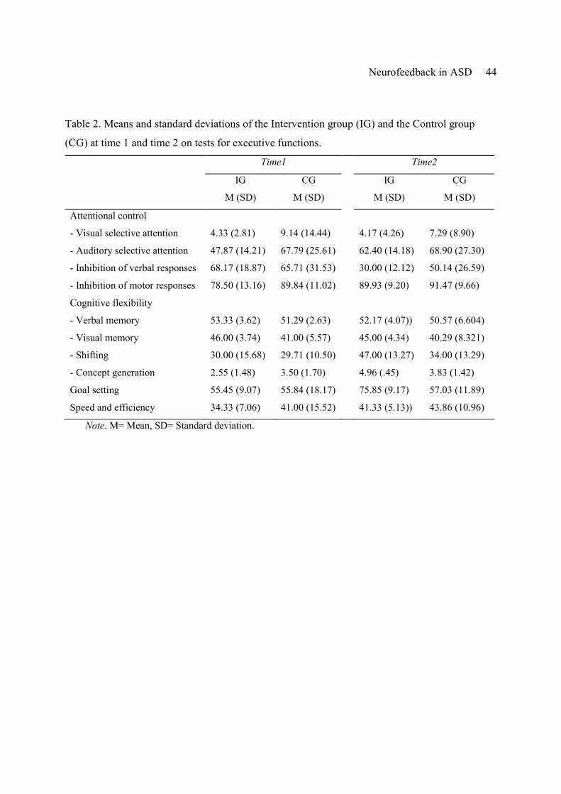
Neurofeedback in ASD 44
Table 2. Means and standard deviations of the Intervention group (IG) and the Control group
(CG) at time 1 and time 2 on tests for executive functions.
Time1 Time2
IG
M (SD)
CG
M (SD)
IG
M (SD)
CG
M (SD)
Attentional control
- Visual selective attention 4.33 (2.81) 9.14 (14.44) 4.17 (4.26) 7.29 (8.90)
- Auditory selective attention 47.87 (14.21) 67.79 (25.61) 62.40 (14.18) 68.90 (27.30)
- Inhibition of verbal responses 68.17 (18.87) 65.71 (31.53) 30.00 (12.12) 50.14 (26.59)
- Inhibition of motor responses 78.50 (13.16) 89.84 (11.02) 89.93 (9.20) 91.47 (9.66)
Cognitive flexibility
- Verbal memory 53.33 (3.62) 51.29 (2.63) 52.17 (4.07)) 50.57 (6.604)
- Visual memory 46.00 (3.74) 41.00 (5.57) 45.00 (4.34) 40.29 (8.321)
- Shifting 30.00 (15.68) 29.71 (10.50) 47.00 (13.27) 34.00 (13.29)
- Concept generation 2.55 (1.48) 3.50 (1.70) 4.96 (.45) 3.83 (1.42)
Goal setting 55.45 (9.07) 55.84 (18.17) 75.85 (9.17) 57.03 (11.89)
Speed and efficiency 34.33 (7.06) 41.00 (15.52) 41.33 (5.13)) 43.86 (10.96)
Note. M= Mean, SD= Standard deviation.

Neurofeedback in ASD 45
Table 3. Test results of the CCC-2 for the intervention group (IG) and the control group (CG) at time1 and
time2.
Time1 Time2
IG
M (SD)
CG
M (SD)
IG
M (SD)
CG
M (SD)
General communication 115.14 (10.45) 115.86 (9.42) 101.29 (12.09) 114.29 (16.45)
Pragmatics 60.57 (7.00) 60.71 (7.25) 54.14 (5.579) 65.86 (20.84)
- Speech production 12.86 (2.54) 12.14 (3.63) 10.86 (2.96) 11.43 (4.08)
- Syntax 12.71 (1.89) 14.43 (1.40) 11.29 (2.69) 14.71 (1.89)
- Semantics 12.29 (2.29) 13.14 (1.57) 12.00 (2.08) 13.43 (1.40)
- Coherence 15.43 (1.81) 15.43 (1.51) 14.28 (1.50) 14.14 (3.58)
- Inappropriate initialization 14.29 (1.89) 13.57 (2.76) 13.86 (1.57) 14.57 (2.57)
- Stereotyped conversation 15.14 (2.27) 15.57 (1.40) 13.57 (1.81) 14.43 (3.64)
- Context use 15.14 (1.77) 16.71 (1.89) 13.71 (1.80) 16.14 (2.54)
- Non-verbal communication 15.86 (2.34) 14.86 (2.85) 13.71 (2.50) 15.57 (2.76)
- Social relations 15.57 (1.90) 14.42 (2.63) 14.57 (2.07) 14.57 (2.44)
- Interests 13.57 (1.90) 14.00 (2.16) 12.14 (3.67) 14.14 (2.04)
Note. M= Mean, SD= Standard deviation.

Neurofeedback in ASD 46
Table 4. Means and standard deviations of the subscales of the adapted AUTI-R for the
intervention group (IG) and the control group (CG)
IG
M (SD)
CG
M (SD)
Social interaction 36.50 (3.51) 30.71 (.92)
Communication 29.00 (1.79) 24.14 (.64)
Typical behavior 48.33 (3.44) 44.14 (1.06)
Total 113.83 (7.17) 99.00 (1.95)
Note. M= Mean, SD= Standard deviation.

Neurofeedback in ASD 47
Figure captions
Figure 1. Average theta (4-7 Hz) power during neurofeedback sessions recorded over C3 (left
graph) and C4 (right graph) indicating the reduction in theta power over consecutive sessions.
Regression lines reflect the slope of theta reduction over time for each individual patient, with
* <.05 and **<.01.
Figure 2. Average low beta (12-15 Hz) power during neurofeedback sessions recorded over C3
(left graph) and C4 (right graph) indicating the increase in low beta power over consecutive
sessions. Regression lines reflect the slope of beta enhancement over time for each individual
patient, with * <.05 and **<.01.
Figure 3. Average delta (1.3-3.5 Hz) power during neurofeedback sessions recorded over C3 (left
graph) and C4 (right graph) indicating the reduction in delta power over consecutive sessions.
Regression lines reflect the slope of delta reduction over time for each individual patient, with
* <.05 and **<.01.
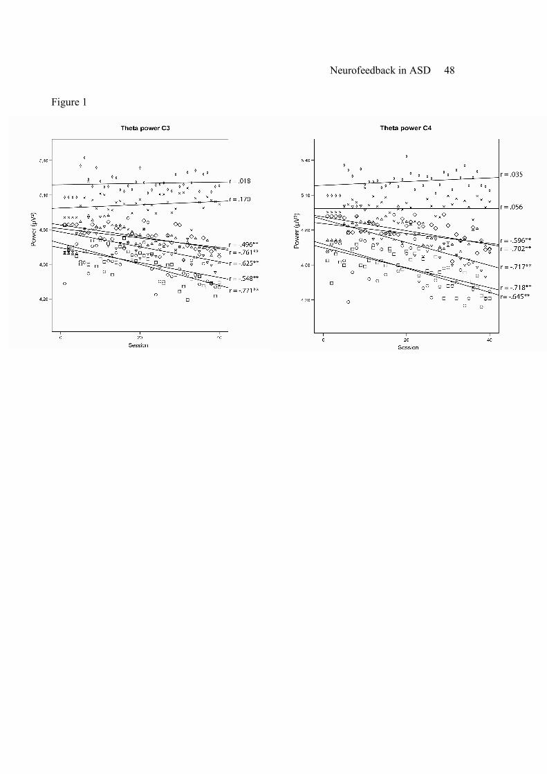
Neurofeedback in ASD 48
Figure 1
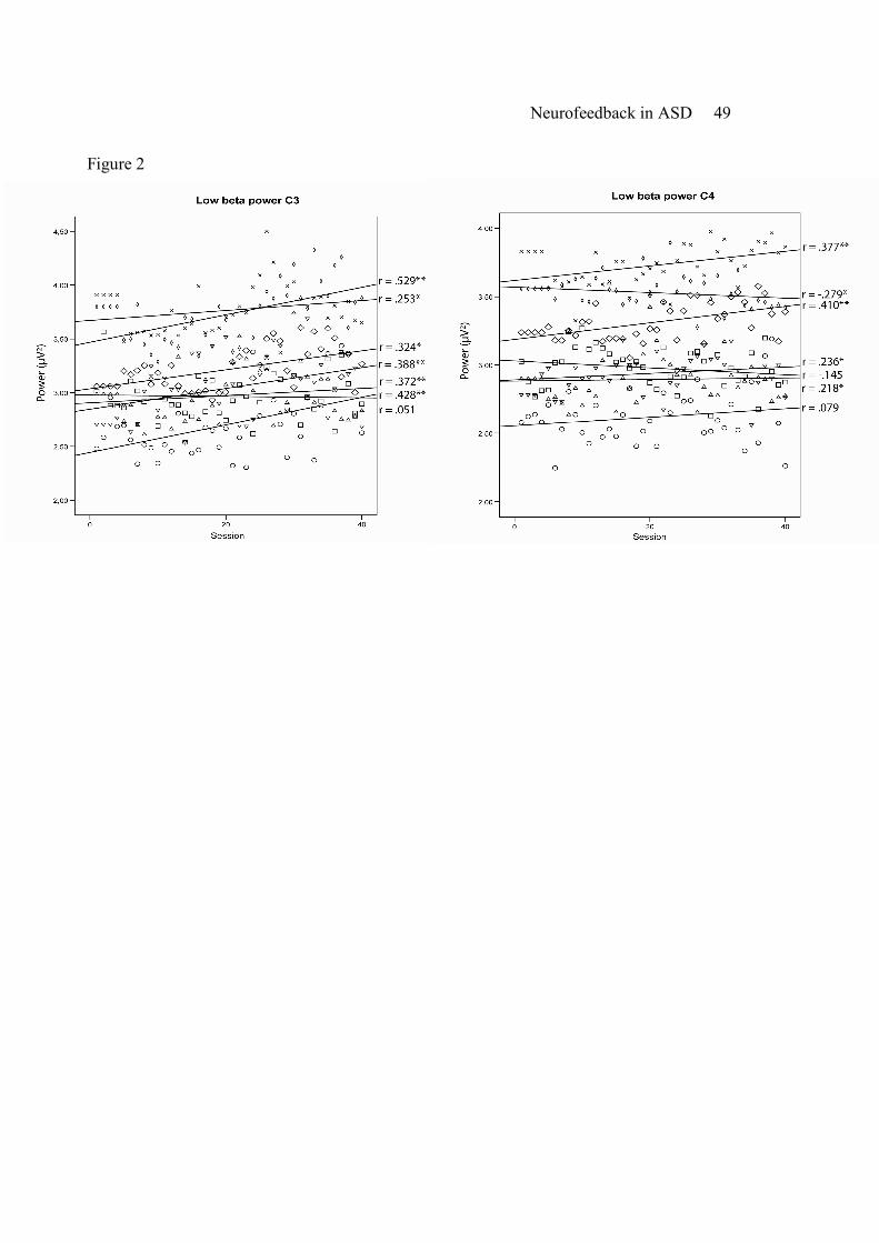
Neurofeedback in ASD 49
Figure 2

Neurofeedback in ASD 50
Figure 3


