Download - Nitrate Reductase Biochemistry Comes of Age
Plant Physiol. (1 996) 11 1 : 355-361
Nitrate Reductase Biochemistry Comes of Age’
Wilbur H. Campbell*
Phytotechnology Research Center and Department of Biological Sciences, Michigan Technological University, Houghton, Michigan 49931 -1 295
NR (EC 1.6.6.1-3) was first isolated and characterized more than 40 years ago, and each decade of study of this key enzyme of nitrate assimilation has been associated with a new understanding of its structure and function. Briefly, NR is a homodimeric enzyme (native form = A,) with each subunit containing a 100-kD polypeptide and one each of molybdate, Mo-pterin, Fe, heme, and FAD (Redinbaugh and Campbell, 1985). NR has two active sites, one where NADH donates electrons to FAD to begin the transport of electrons via the heme-Fe to the Mo / Mo-pterin in the second active site, where nitrate is reduced to nitrite. NR has two-site, ping-pong, steady-state kinetics, where the enzyme “pings and pongs” between oxidized and re- duced forms, as NADH/NADC bind at the electron donor active site and nitrate/nitrite bind at the electron acceptor site. The most recent advances have resulted from the cloning of the NR gene (Campbell and Kinghorn, 1990; Solomonson and Barber, 1990; Rouze and Caboche, 1992). The deduced amino acid sequence of higher-plant NR showed that it contained about 900 residues with a pre- dicted molecular size of approximately 100 kD. Although this size is a bit smaller than the NR polypeptide appears on SDS-PAGE gels (110-115 kD; Redinbaugh and Camp- bell, 1985), this may be explained by the runs of acidic residues near the N terminus of well-characterized and sequenced NR forms such as squash (Hyde et al., 1991).
In this general review, I will focus on recent advances in NR biochemistry. I last reviewed this topic in a general way in 1988 (Campbell, 1988). Reviews by Solomonson and Barber (1990), Rouze and Caboche (1992), and Crawford (1995) provide more detailed accounts of various aspects of this topic than will be presented here. Recently, Kaiser and Huber (1994) reviewed posttranslational regulation of NR in an Update.
N R STRUCTURE
The important discovery from the NR amino acid se- quence was that the apparent cofactor-binding regions of NR were laid out linearly in the backbone (Campbell and Kinghorn, 1990; Solomonson and Barber, 1990; Rouze and Caboche, 1992). This was demonstrated by similarity of the
The research in the author’s laboratory on nitrate reductase was supported by grants from the National Science Foundation and the U.S. Department of Agriculture.
* E-mail [email protected]; fax 1-906-487-3355.
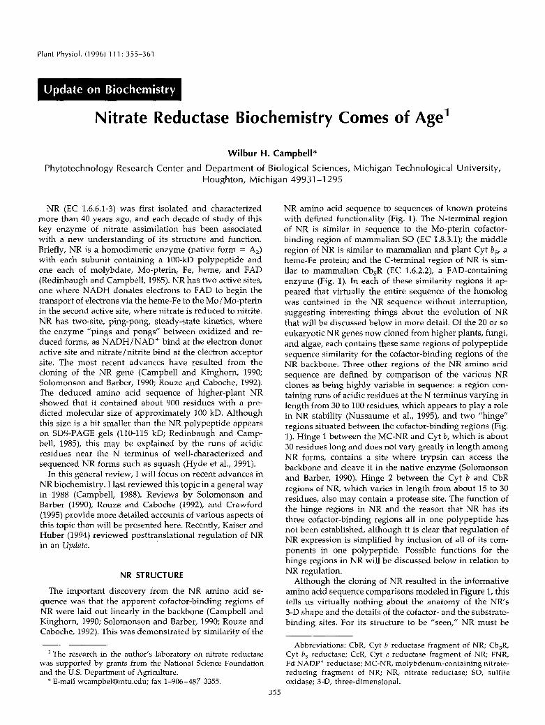
NR amino acid sequence to sequences of known proteins with defined functionality (Fig. 1). The N-terminal region of NR is similar in sequence to the Mo-pterin cofactor- binding region of mammalian SO (EC 1.8.3.1); the middle region of NR is similar to mammalian and plant Cyt b,, a heme-Fe protein; and the C-terminal region of NR is sim- ilar to mammalian Cb,R (EC 1.6.2.2), a FAD-containing enzyme (Fig. 1). In each of these similarity regions it ap- peared that virtually the entire sequence of the homolog was contained in the NR sequence without interruption, suggesting interesting things about the evolution of NR that will be discussed below in more detail. Of the 20 or so eukaryotic NR genes now cloned from higher plants, fungi, and algae, each contains these same regions of polypeptide sequence similarity for the cofactor-binding regions of the NR backbone. Three other regions of the NR amino acid sequence are defined by comparison of the various NR clones as being highly variable in sequence: a region con- taining runs of acidic residues at the N terminus varying in length from 30 to 100 residues, which appears to play a role in NR stability (Nussaume et al., 1995), and two “hinge” regions situated between the cofactor-binding regions (Fig. 1). Hinge 1 between the MC-NR and Cyt b, which is about 30 residues long and does not vary greatly in length among NR forms, contains a site where trypsin can access the backbone and cleave it in the native enzyme (Solomonson and Barber, 1990). Hinge 2 between the Cyt b and CbR regions of NR, which varies in length from about 15 to 30 residues, also may contain a protease site. The function of the hinge regions in NR and the reason that NR has its three cofactor-binding regions a11 in one polypeptide has not been established, although it is clear that regulation of NR expression is simplified by inclusion of a11 of its com- ponents in one polypeptide. Possible functions for the hinge regions in NR will be discussed below in relation to NR regulation.
Although the cloning of NR resulted in the informative amino acid sequence comparisons modeled in Figure 1, this tells us virtually nothing about the anatomy of the NRs 3-D shape and the details of the cofactor- and the substrate- binding sites. For its structure to be ”seen,” NR must be
Abbreviations: CbR, Cyt b reductase fragment of NR; Cb,R, Cyt b, reductase; CcR, Cyt c reductase fragment of NR; FNR, Fd NADP+ reductase; MC-NR, molybdenum-containing nitrate- reducing fragment of NR; NR, nitrate reductase; SO, sulfite oxidase; 3-D, three-dimensionai.
355
356 Campbell Plant Physiol. Vol. 111, 1996
CcR
MC-NR ' Cb CbR '
N—I(FHD/NBDH)-Figure T . Model of the NR amino acid sequence showing its rela-tionship to other eukaryotic enzymes and proteins with similar se-quences. This model is based on amino acid sequence comparisonsand alignments (Campbell and Kinghorn, 1990). The N and C terminiof the sequence models are designated N- and -C, respectively. Themajor fragments of NR with functionality as catalysts and stability asstructural units are designated at the top: MC-NR, Cb (Cyt b fragmentor domain), CbR, and CcR, which comprises the Cb domain, hinge 2,and the CbR fragment. The designations for the enzymes/proteins areshown along the side: NR, SO, Cbs (Cyt b5), and Cb5R. In the modelfor NR, the circled A, 1, and 2 designate the acidic N-terminal regionand the two hinge regions of variable sequence that join the majorfunctional/structural fragments of the enzyme. Within the fragmentsthemselves are shown the cofactors and substrates of the enzymes/proteins: NO,", nitrate-binding and reduction site; Mo, molybdate/molybdenum-pterin binding site; Fe, heme-Fe binding site; FAD,FAD-binding site; NADH, NADH-binding and reaction site; andSO3
2~, sulfite-binding and oxidation site. NR and SO are solubleenzymes, NR in the cytosol and SO in the inner membrane fluid ofmitochondria, whereas Cb5 and Cb5R are membrane bound on theER, thecytosolic face of the outer membrane of mitochondria and thecytosolic face of the plasma membrane; the positions of the mem-brane anchor sequences of the latter two proteins are shown. Solubleforms of Cb,; and Cb^R also exist in red blood cells that are identicalin sequence to the soluble portions of the membrane-bound forms.
crystallized and the crystals analyzed by x-ray diffraction.The limited quantities of pure NR available and, perhaps tosome extent, NR's complex structure and large size haveprevented this goal from being achieved. Another ap-proach is to use recombinant protein expression technol-ogy to provide the large quantities of protein needed forcrystallization and x-ray analysis. Unfortunately, to date noone has succeeded in producing by recombinant expres-sion holo-NR in an active form in sufficient quantities forstructural analysis. On the other hand, recombinant expres-sion of fragments of NR has been achieved, which turns outto be possible because of the linear arrangement of the NRfragments in both the polypeptide and the gene or cDNA(Fig. 1). The proof that the sequence comparisons werevalid came when redox active and functional fragments ofNR were produced by expression in Escherichia coll of genefragments for corn NR's CbR (originally called NR's flavinor FAD domain) and Chlorella NR's Cyt /) (Hyde and Camp-bell, 1990; Cannons et al., 1991). The recombinant CbR of
corn NR, a 30-kD protein, is an active NADHrferricyanidereductase that is also a partial activity of holo-NR, depend-ing only on the enzyme's FAD. Subsequently, the Cyt b andCbR fragments of corn NR and the CcR fragment of NRwere recombinantly produced as a 58-kD protein, havingNADH:Cyt c reductase activity, which is also a property ofholo-NR, depending on both the FAD and the heme-Fe(Campbell, 1992). Recently, a fusion of mammalian Cyt b5and spinach NR's CbR fragment was recombinantly pro-duced, which also catalyzes NADH-dependent Cyt c re-duction (Quinn et al., 1994). More recently, my laboratoryhas produced CcR fragments of spinach NR with the 42-kDminimum size predicted for the region of the enzyme in E.col: and Pichia pastoris, a methylotrophic yeast that pro-duces very high levels of recombinant proteins (N. Shi-raishi and W.H. Campbell, unpublished observations).
Since the recombinant corn CbR was a soluble proteinproduced in large quantities and easily purified (Hyde andCampbell, 1990), it was crystallized and its 3-D structurewas determined (Lu et al., 1994). It had been predicted byamino acid sequence comparisons that NR's CbR fragmentwas a member of a family of flavin-containing oxidoreduc-tases that reduce cytochromes (Hyde et al., 1991; Karplus etal., 1991). This family is known as the FNR family offlavoproteins because FNR was the first member to have its3-D structure determined (Karplus et al., 1991). The familyconsists of FNR (EC 1.18.1.2), NR, nitric oxide synthase (EC1.14.13.39), Cb5R, Cyt P450 reductase (EC 1.6.2.3), andsome other enzymes containing FAD or FMN, and differsfrom the other well-known flavoproteins whose structureshave been determined, namely glutathione reductase (EC1.6.4.2) and glycolate oxidase (EC 1.1.3.15) (Karplus et al.,1991; Lu et al., 1994). This is one of the first times that plantproteins have taken a leading role in establishing a newstructural family of enzymes, which exists in many differ-ent eukaryotic and prokaryotic species (Correll et al., 1992).Subsequently, crystals of NR's CbR were produced withthe inhibitor ADP bound to the apparent NADH-bindingsite, and an active site mutant called C242S was also crys-tallized (Lu et al., 1995). In addition, it has been possible tomodel the CcR fragment of NR, since NR's Cyt b domain ishighly similar in sequence to mammalian Cyt frs, which isof known 3-D structure (Fig. 2; Mathews et al., 1971; Lu etal., 1995).
NR's CbR fragment has two domains: one for bindingFAD at its N terminus and one presumed to be for bindingNADH at its C terminus, with the two domains joined bya short linker sequence (Fig. 2). There is a central cleftbetween the two domains, which is the active site whereNADH transfers electrons to FAD. Reduced FAD in turnreduces ferricyanide in the recombinant fragment or theinternal Cyt b of NR in the holoenzyme and recombinantCcR fragment. The FAD domain of CbR is a six-stranded,/3-barrel structure with a single a helix important in FADbinding. NR's FAD domain has virtually the same shape asFNR's FAD domain, confirming the amino acid sequenceprediction that these two key plant enzymes are closelyrelated in structure while differing significantly in se-quence and function. The apparent NADH-binding do-
Nitrate Reductase Biochemistry 357
NflDH Domain
Cut b DomainFflD Domain
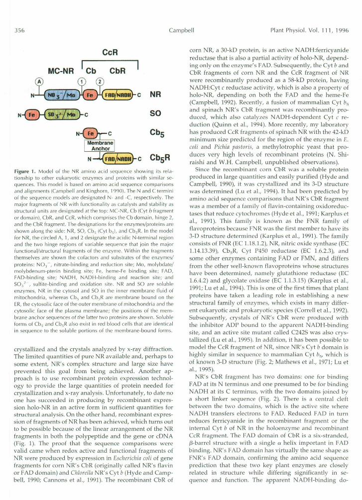
Figure 2. Model of the Cyt c reductase fragment of NR. This model was derived by docking the 3-D structure of mammalianCyt fa, to the 3-D structure of corn NR's CbR using electrostatic calculations as a guide in positioning the two protein modelsrelative to one another (Lu et al., 1 995). In the ribbon model, the NADH domain of the CbR fragment of NR with bound ADP(shown as space filling model) is blue, the FAD domain of CbR with bound FAD (space filling model) is green, andmammalian Cyt fa5 with bound heme-Fe (space filling model—central Fe atom) is yellow. The 3-/3 strand linker between theFAD and NADH domains of NR's CbR is also blue in the ribbon model; it is to the left of the main portion of the NADHdomain, with its fusion to the green ribbon of the FAD domain hidden behind Cyt b$. Hinge 2 is not modeled here; however,it would most probably extend along the surface of Cyt fa5 from its C terminus to the N terminus of the FAD domain of CbR,which is visible just below one of the a helices of Cyt for>. In the cameo inset, the same color scheme is used and space-fillingrepresentations for the major domains of the Cyt c reductase model are shown with the cofactors and ADP inhibitor bound,to provide a better sense of the size and shape of the CcR fragment of NR.
main of CbR is a six-stranded /3 sheet that resembles the"Rossman" fold of dehydrogenases (Lu et al., 1994). NR'sNADH domain differs in some minor aspects when com-pared to the NADP+ domain of FNR, which is not surpris-ing, since different pyridine nucleotides are bound to thesestructures; but it is clear that they are closely related. Adifference between NR's CbR and FNR is that the FADdomain is a bit more separated from the NADH domain inNR than the corresponding domains are in FNR (Karplus etal., 1991; Lu et al., 1994). Although the biochemical signif-icance of this difference is not clear, it also has been foundin the 3-D model recently reported for Cb5R, which is muchmore closely related in sequence and functionality to NR'sCbR than it is to FNR (Nishida et al., 1995).
The model of the CcR fragment of NR illustrates how theheme-Fe of the Cyt b domain may approach the FAD of theCbR fragment to facilitate electron transfer (Fig. 2; Lu et al.,1995). It is generally predicted that the isoalloxazine ring of
FAD should be in the same plane as the heme-Fe foroptimum electron transfer, and this optimum alignment isonly partially achieved in the model. The model of NR'sCcR also predicts amino acid side chains that may beimportant at the interface of the enzyme's Cyt b and FADdomains, which are charged side chains of amino acids,and one interaction between a carboxylic acid on the hemeand a His of the FAD domain (Lu et al., 1995). Thesepredictions can now be tested by site-directed mutagenesisof NR's recombinantly expressed CcR fragment. Of course,this model of CcR only provides an idea of how NR reallylooks, and current experiments working toward crystalli-zation of the recombinant CcR fragment of spinach NR willpossibly provide a more definitive look at this part of NR'sstructure. The elusive prize of "seeing" NR's MC-NR frag-ment and the overall shape of the native holo-NR dimerremain for future investigations. Recently, Su et al. (1996)succeeded in expressing low levels of active holo-NR in P.
358 Campbel I Plant Physiol. Vol. 11 1 , 1996
pastoris using an Arabidopsis NR cDNA, which may be the breakthrough needed to propel structural studies of holo-NR.
NR FUNCTIONALITY
When a11 the available NR sequences are compared, only two Cys residues remain as invariant and absolutely con- served residues in the sequence: one in the MC-NR frag- ment and one in the CbR fragment. Tomsett and co-work- ers (Garde et al., 1995) carried out site-directed mutagenesis on the invariant Cys residue in the MC-NR fragment of Aspergillus NADPH:NR to change it to an Ala residue and found that this mutant had no NR activity. This strongly supports the concept that this Cys is essential for NR functionality, especially since no other mutation generated by this group resulted in complete loss of a11 nitrate-reducing activities, including activities driven by reduced dyes (Garde et al., 1995). It has been suggested that this Cys residue of NR is involved in binding the Mo-pterin cofactor to the MC-NR fragment and holding it in the nitrate-reducing active site of the enzyme (Solomonson and Barber, 1990). NADPH:NR activity was lost in an Aspergil- Lus NR mutant where one of the reputed His ligands of the heme-Fe in the Cyt b domain was changed to an Ala, and reduced bromphenol b1ue:NR activity was retained (Garde et al., 1995). A similar mutant of a His ligand of the heme-Fe was found among tobacco plants lacking NADH:NR activity when the mutant NR gene was se- quenced (Meyer et al., 1991). This NR mutant also retained reduced bromphenol b1ue:NR activity, but lacked reduced methyl vio1ogen:NR activity, suggesting that these reduced dyes donate electrons to different sites in NR. This is an important result, since it was long thought that methyl viologen donated electrons directly to the Mo / Mo-pterin of NR, which is a functionality that must now be assigned to reduced bromphenol blue. Reduced methyl vio1ogen:NR activity clearly depends on the Cyt b domain in some way; most probably this dye donates an electron to the heme-Fe, which transfers it to the Mo/Mo-pterin in the usual man- ner for nitrate reduction.
The other invariant Cys of all NR forms has been studied by site-directed mutagenesis of the recombinant CbR frag- ment of corn NADH:NR (Dwivedi et al., 1994). There are five Cys residues in recombinant corn CbR, each of which was converted to a Ser residue and the mutant enzyme forms purified from the E. coli extracts by affinity chroma- tography. Only when the invariant Cys, which is residue 242 in the recombinant protein, thus is called C242S, was mutated did CbR largely lose its ferricyanide reductase activity while retaining its native shape, as indicated by visible and circular dichroism spectra being identical to those of the wild type (Dwivedi et al., 1994; Ratnam et al., 1995). Inhibitor studies using p-hydroxymercuribenzoate, which reacts with thiol groups, showed that only the C242S mutant of the corn CbR was resistant to inhibition, whereas a11 other Cys to Ser mutants, as well as the wild type, lost their activity in the presence of the inhibitor. Kinetic stud- ies indicated that CbRs V,,, for ferricyanide reduction by NADH and the rate of reduction of CbRs FAD by NADH,
assayed by pre-steady-state methods, were decreased by more than 5-fold in the C242.5 mutant compared to wild type. The NADH K , and NADH binding constant were almost unchanged (Dwivedi et al., 1994; Ratnam et al., 1995). Thus, the longstanding suggestion that this Cys thiol in the NADH active site of NR has a role in binding NADH was largely disproved, and it now can be convincingly stated that the role of this invariant Cys thiol is in assisting electron transfer from NADH to FAD in the active site of the CbR.
Clearly, since CbR retains some NADH:ferricyanide re- ductase activity in its C242S mutant, the invariant Cys is not absolutely required for catalytic activity. So, this Cys residue is not redox active in the electron transfer process and it is not involved in the chemistry of FAD reduction by NADH. The much greater efficiency of electron transfer in the wild type versus the C242S mutant of CbR indicates that the Cys thiol is "essential" for making a highly effi- cient NR catalyst, suggesting that this is sufficient evolu- tionary pressure for retaining this residue in a11 NR forms so far cloned as well as in a11 other members of the FNR structural family of flavoenzymes (Dwivedi et al., 1994; Ratnam et al., 1995). The crystal structure of the (2242s mutant of CbR helps to explain the role of the invariant Cys thiol in efficient electron transfer (Lu et al., 1995). In wild- type CbRs 3-D structure, the C242 thiol projects into the active-site cleft, where it may interact with the carboxam- ide side chain on the pyridine ring of NADH, whereas in the C242S mutant of CbR, the Ser hydroxyl group is hy- drogen bonded to the backbone of the protein at residue 147, which leaves a large void in the active site (Lu et al., 1995). Thus, the invariant Cys's thiol may be for position- ing NADH in the active site for efficient electron transfer to FAD.
Another aspect of NRs functionality brought into better focus by the availability of the 3-D structure of its CbR fragment is how pyridine nucleotide specificity is deter- mined. NR exists in three forms: NADH specific (EC 1.6.6.1), NAD(P)H bispecific (EC 1.6.6.2), and NADPH spe- cific (EC 1.6.6.3). The 3-D structure of the recombinant CbR of corn NR with the inhibitor ADP bound in the apparent NADH binding site shows that the negatively charged side chain of the Asp residue at position 205 (D205) is within bonding distance of the 2' hydroxyl group of the ADP's Rib (Lu et al., 1995). No other polar amino acid side chains of CbR are in the vicinity of the 2' hydroxyl group of the ADP Rib. Since most NADH-specific NR forms have either an aspartic acid or Glu residue at their position corresponding to D205 of corn N R s CbR, we have suggested that the negative charge of this side chain probably plays a role in excluding the negatively charged 2' phosphate of NADPH from the active site of these NR forms (Lu et al., 1994). Higher-plant NAD(P)H:NR and funga1 NADPH:NR forms have either a Ser or a Thr in the position corresponding to that of corn CbRs D205 when the amino acid sequences are aligned. Thus, the lack of a negatively charged side chain here may allow NADPH into the active site. However, this simple theory cannot completely explain why some NR forms are highly specific for NADPH and others are bispe-
Nitrate Reductase Biochemistry 359
cific and accept electrons from both NADH and NADPH. Recent results for birch NAD(P)H:NR suggest that residues more remote from the actual ligands to the 2' hydroxyl/2' phosphate of NADH/NADPH (as judged by the 3-D struc- ture of ADP bound to corn's CbR) also play a role in determining pyridine nucleotide specificity (Schondorf and Hatchtel, 1995). Pyridine nucleotide specificity of NR is currently under more complete investigation.
NR RECULATION
Control of NR activity can be achieved in two ways: altering the activity level of existing enzyme or controlling the amount of enzyme by synthesizing new enzyme and degrading old enzyme. De novo synthesis of new NR, stimulated by nitrate, was established as a mechanism for controlling enzyme level when combined with NR protein degradation (Remmler and Campbell, 1986; Campbell, 1988; Solomonson and Barber, 1990). After NR clones be- came available, it was shown that the NR gene is tran- scribed in response to nitrate application to plants, leading to increased levels of NR mRNA, which is the underlying mechanism for nitrate stimulation of de novo NR synthesis (Solomonson and Barber, 1990; Rouze and Caboche, 1992). The signals triggering NR protein degradation in the pres- ente of nitrate, even in the light, have not been identified. However, it is likely that NR degradation begins by attack either in the readily available hinge regions or the N- terminal region and eventually leads to total degradation of NR. NR activity level can be controlled by a posttrans- lational mechanism involving phosphorylation of the NR protein and binding of Mg2+ or another divalent cation and an inhibitor protein, which was recently reviewed here (Kaiser and Huber, 1994). These observations describe the end result of a signal transduction pathway where the triggering signals are light/ dark transitions, as well as other environmental factors impacting dominant plant pro- cesses such as carbon assimilation. The mediators of this signal transduction pathway from the triggering signal(s) have just begun to be identified by the recent isolation of a calcium-dependent protein kinase catalyzing the phos- phorylation of spinach NR in vitro (Bachmann et al., 1995).
Most recently, the NR protein kinase was used to iden- tify the apparent key Ser residue in spinach NR, which is involved in activity regulation (Bachmann et al., 1996). Two approaches were used in this study. One involved the recombinant fragments of spinach NR, where the CcR frag- ment was produced with and without the hinge-1 region. Since the CcR-hinge-1 fragment was phosphorylated by the NR protein kinase and the CcR without hinge 1 was not, the only conserved Ser (residue 543 in spinach NR) in the hinge-1 region was the likely site of phosphorylation. Sub- sequently, synthetic peptides containing hinge-1 sequences were prepared and used to establish that Ser543 was indeed the phosphorylation site (Bachmann et al., 1996). Based on the tentative location of the target Ser for the NR protein kinase in hinge 1, a site-directed mutant of Arabidopsis NR (Ser534-Asp534; Arabidopsis NR is 9 residues shorter in length than spinach NR at the N terminus) was prepared and expressed in Pichia (Su et al., 1996). This mutant was
not inactivated in vitro by treatment with ATP and extracts of Arabidopsis leaf (source of the NR protein kinase and inhibitor protein), but the wild type was, which is consis- tent with Ser534 being the site of regulatory phosphoryla- tion in Arabidopsis Nia2. Since Arg540 in hinge 1 of spinach NR has been identified as the target site for trypsin cleav- age of the native enzyme (Campbell and Kinghorn, 1990), it can be concluded that this region in hinge 1 of NR is at the surface of the 3-D structure. This surface location allows the NR protein kinase to recognize and bind the sequence LKRTAS (residues 538-543 of spinach NR hinge l), leading to the catalytic transfer of phosphate from ATP to form p h ~ s p h o - S e r ~ ~ ~ (Bachmann et al., 1996). After this Ser res- idue is phosphorylated, presumably Mg2+ (or another di- valent cation) will bind to NR and the complex with the NR inhibitor protein will form, resulting in loss of NR activity. It seems líkely that other parts of NR beside the phosphor- ylation site may be involved with the interaction of NR and the inhibitor protein, but these are yet to be identified. In this regard, an NR mutant found in an NR-deficient Ara- bidopsis plant, where an invariant Gly residue in the MC-NR fragment of NR is converted to an acidic residue, renders the NR both inactive and without phosphorylation (LaBrie and Crawford, 1994). Thus, the overall3-D shape of NR may be important in controlling phosphorylation, and further understartding of how phosphorylation of NR at the regulatory Ser leads to binding of the inhibitors and activity loss may not be gained until the 3-D shape of NR is known.
NR GENES AND EVOLUTION
NR genes, like many other eukaryotic genes, contain introns. There are basically two theories to explain the common existence of introns in eukaryotic genes encoding proteins and their absence from similar prokaryotic genes: (a) introns were present in ancestral genes of a11 species and selectively lost during the evolution of prokaryotes, probably for reasons of "economics of survival," but were retained during evolution of eukaryotes, which have more energy to spare for maintaining less useful DNA and a greater need for the complex regulation and manipulation of the genome made possible by intervening DNA se- quences in genes; and (b) introns were introduced into eukaryotic genes after the split in evolution of prokaryote and eukaryote lineages (Alberts et al., 1994). Although I do not wish to enter into the dispute over these two possibil- ities, it is interesting to look at intron locations in NR genes, because the regions encoding the functionalities of the enzyme are so clearly laid out in a linear array in the gene, which along with the sequence similarities to independent enzymes suggests that the NR gene was formed by fusion of existing exons (Fig. 1). Introns occur at one or a11 of three locations in higher-plant NR genes, although a fourth in- tron has been found in a bean NR gene (Campbell and Kinghorn, 1990; Jensen et al., 1994). Funga1 NR genes have from 1 to 6 introns and for the most part the introns are found at different locations in the sequence than those in higher plants (Campbell and Kinghorn, 1990). Perhaps the most interesting NR gene is from Volvox carteri, a green
3 60 Campbell Plant Physiol. Vol. 1 1 1, 1996
alga, which has 10 introns dividing the coding sequence into 11 exons (Gruber e t al., 1992). The Volvox NR gene has 6 introns i n the MC-NR fragment, with the 7th exon con- taining the hinge-1 region; exon 8 contains the core of the Cyt b domain, exon 9 contains hinge 2 and the first par t of the FAD domain, exon 10 contains the end of the FAD domain and the beginning of the NAD(P)H domain, a n d exon 11 contains the remainder of the sequence beginning just before the key pyridine nucleotide-specificity-deter- mining amino acid residues described above. Thus, the Volvox NR gene illustrates a way the ancestral NR gene may have formed from groups of exons representing prim- itive forms of a Mo-containing protein, a Cyt b-type heme-Fe protein, a n d a flavin-containing reductase enzyme.
Codon usage is another aspect of genome evolut ion well illustrated i n N R genes (Campbell and Gowri, 1990). Since N R is a very large gene, a11 61 codons of the genetic code a re used in these genes. However , NR genes reflect the codon usage i n t h e species f rom which they w e r e isolated rather t h a n following a pa t te rn representative of the gene. Thus, monocot NR genes a r e highly biased toward codons ending in the nucleotides G and C, like m a n y o ther monocot genes, whereas dicot NR genes have the more evenly dis t r ibuted codon usage typical of dicot genes (Campbell and Gowri, 1990). This f ine tun ing of genes d u r i n g the evolut ion of species is a fascinating aspect of the evolut ion of individual species and families of genera, which t o my knowledge has no known mech- anism. To make t h e n a t u r e of t h e puzz le clear, it can b e restated i n this way: t h e a m i n o acid sequences of NR proteins h a v e been m o r e conserved i n evolut ion of the NR gene t h a n the codons used t o encode these a m i n o acid sequences. One supposes tha t codon usage i n a species' t ranslated genes was fine t u n e d t o t h e abun- dance of the var ious tRNA molecules available for pro- tein synthesis and perhaps also to opt imize m R N A sta- bility. But why plant families differ so greatly i n codon usage o r tRNA abundance, if that is t h e underlying dr iv ing force, remains mysterious. Furthermore, how i n evolut ion were individual codons i n a gene fine t u n e d t o arr ive a t the distinct codon usage pat terns found today in plant species?
Received February 9, 1996; accepted February 20, 1996. Copyright Clearance Center: 0032-0889/96/ 111 /0355/07.
LITERATURE CITED
Alberts B, Bray D, Lewis J, Raff M, Roberts K, Watson JD (1994) Molecular Biology of the Cell, Ed 3. Garland Publishing, New York, pp 389-39T
v
Bachmann M. McMichael RW. Huber 1L. Kaiser WM. Huber SC . . (1995) Partia1 purification and characterization of a calcium- dependent protein kinase and an inhibitor protein required for inactivation of spinach leaf nitrate reductase. Plant Physiol 108:
Bachmann M, Shiraishi N, Campbell WH, Yoo B-C, Harmon AC, Huber SC (1996) Identification of Ser-543 as the major regula- tory phosphorylation site in spinach leaf nitrate reductase. Plant Cell 8: 505-517
Campbell WH (1988) Higher plant nitrate reductase and its role in regulation of nitrate assimilation. Physiol Plant 7 4 214-219
1083-1091
Campbell WH (1992) Expression in Escherichia coli of cytochrome c reductase activity from a maize NADH:nitrate reductase cDNA. Plant Physiol 99: 693-699
Campbell WH, Gowri G (1990) Codon usage in higher plants, green algae, and cyanobacteria. Plant Physiol 92: 1-11
Campbell WH, Kinghorn JR (1990) Functional domains of assimi- latory nitrate reductases and nitrite reductases. Trends Biochem Sci 15: 315-319
Cannons AC, Iida N, Solomonson LP (1991) Expression of a cDNA clone encoding the heme binding domain of Chlorella nitrate reductase. Biochem J 278: 203-209
Corre11 CC, Batie JC, Ballou DP, Ludwig ML (1992) Phthalate dioxygenase reductase: a modular structure for electron transfer from pyridine nucleotides to 2Fe-2s. Science 258: 1604-1610
Crawford NM (1995) Nitrate: nutrient and signal for plant growth. Plant Cell 7: 859-868
Dwivedi UN, Shiraishi N, Campbell WH (1994) Identification of an "essential" cysteine of nitrate reductase via mutagenesis of its recombinant cytochrome b reductase domain. J Biol Chem 269:
Garde J, Kinghorn JR, Tomsett AB (1995) Site-directed mutagen- esis of nitrate reductase from Aspergillus nidulans. Identification of some essential and nonessential amino acids among con- served residues. J Biol Chem 270: 6644-6650
Gruber H, Goetinck SD, Kirk DL, Schmitt (1992) The nitrate reductase-encoding gene of Vo[uox curteri: map location, se- quence and induction kinetics. Gene 120: 75-83
Hyde GE, Campbell WH (1990) High-leve1 expression in Esche- richia coli of the catalytically active flavin domain of corn leaf NADH:nitrate reductase and its comparison to human NADH: cytochrome b, reductase. Biochem Biophys Res Commun 168:
Hyde GE, Crawford NM, Campbell WH (1991) The sequence of squash NADH:nitrate reductase and its relationship to the se- quences of other flavoprotein oxidoreductases. A family of fla- voprotein pyridine nucleotide cytochrome reductases. J Biol Chem 266 23542-23547
Jensen PE, Hoff T, Moller MG, Stummann BM, Henningsen KW (1994) Identification and characterization of a nitrate reductase gene from bean (Phust.olus uulguris) containing four introns. Physiol Plant 92: 613-623
Kaiser WM, Huber SC (1994) Posttranslational regulation of ni- trate reductase in higher plants. Plant Physiol 106: 817-821
Karplus PA, Daniels MJ, Herriott JR (1991) Atomic structure of ferredoxin: NADP+ reductase: prototype for a structurally nove1 flavoenzyme family. Science 251: 60-66
LaBrie ST, Crawford NM (1994) A glycine to aspartic acid change in the MoCo domain of nitrate reductase reduces both activity and phosphorylation levels in Arubidopsis. J Biol Chem 269:
Lu G, Campbell WH, Schneíder G, Lindqvist Y (1994) Crystal structure of the FAD-containing fragment of corn nitrate reduc- tase at 2.5 A resolution: relationship to other flavoprotein reduc- tases. Structure 2 809-821
Lu G, Lindqvist Y, Schneider G, Dwivedi UN, Campbell WH (1995) Structural studies on corn nitrate reductase: refined struc- ture of the cytochrome b reductase fragment at 2.5 A, its ADP complex and an active site mutant and modeling of the cyto- chrome b domain. J Mo1 Biol 248: 931-948
Mathews FS, Levine M, Argos P (1971) The structure of calf liver cytochrome b, at 2.8 A resolution. Nature New Biol 233: 15-16
Meyer C, Levin JM, Roussel JM, Rouze P (1991) Mutational and structural analysis of nitrate reductase heme domain of Nicotiana plumbaginifolia. J Biol Chem 266: 20561-20566
Nishida H, Inaka K, Yamanaka M, Kaida S, Kobayashi K, Miki K (1995) Crystal structure of NADH-cytochrome b, reductase from pig liver at 2.4 A resolution. Biochemistry 34: 2763-2767
Nussaume L, Vincentz M, Meyer C, Boutin J-P, Caboche M (1995) Post-transcriptional regulation of nitrate reductase by light is abol- ished by an N-terminal deletion. Plant Cell 7: 611-621
Quinn GB, Trimboli AJ, Barber MJ (1994) Construction and ex- pression of a flavocytochrome b, chimera. J Biol Chem 269:
13785-13791
1285-1291
14497-14501
13375-13381
Nitrate Reductase Biochemistry 361
Ratnam K, Shiraishi N, Campbell WH, Hille R (1995) Spectro- scopic and kinetic characterization of the recombinant wild-type and C242S mutant of the cytochrome b reductase fragment of nitrate reductase. J Biol Chem 270: 24067-24072
Redinbaugh MG, Campbell WH (1985) Quaternary structure and composition of squash NADH:nitrate reductase. J Biol Chem
Remmler JL, Campbell WH (1986) Regulation of corn leaf nitrate reductase. 11. Synthesis and turnover of the enzyme’s activity and protein. Plant Physiol 80: 442-447
Rouze P, Caboche M (1992) Inducible plant proteins-nitrate re- duction. In JL Wray, ed, Higher Plants: Approaches to Function
260: 3380-3385
and Regulation. Cambridge University Press, Cambridge, UK,
Schondorf T, Hatchtel W (1995) The choice of reducing substrate is altered by replacement of an alanine by a proline in the FAD domain of a bispecific NAD(P)H-nitrate reductase from birch. Plant Physiol 108: 203-210
Solomonson LP, Barber MJ (1990) Assimilatory nitrate reductase: functional properties and regulation. Annu Rev Plant Physiol Plant Mo1 Biol 41: 225-253
Su W, Huber SC, Crawford NM (1996) Identification in vitro of a posttranslational regulatory site in the hinge 1 region of Arabi- dopsis nitrate reductase. Plant Cell 8: 519-527
pp 45-77


