Palisades and tumors
Lung metastatic tumor in brain
QuickTime™ and aTIFF (Uncompressed) decompressor
are needed to see this picture.
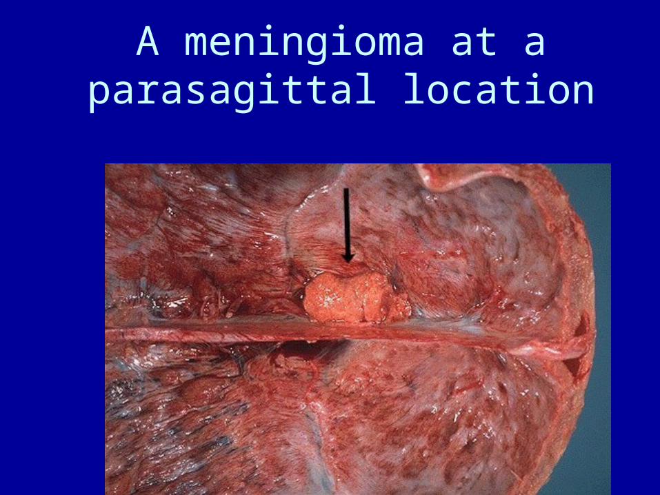
A meningioma at a parasagittal location
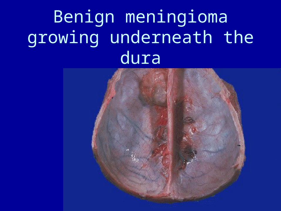
Benign meningioma growing underneath the dura
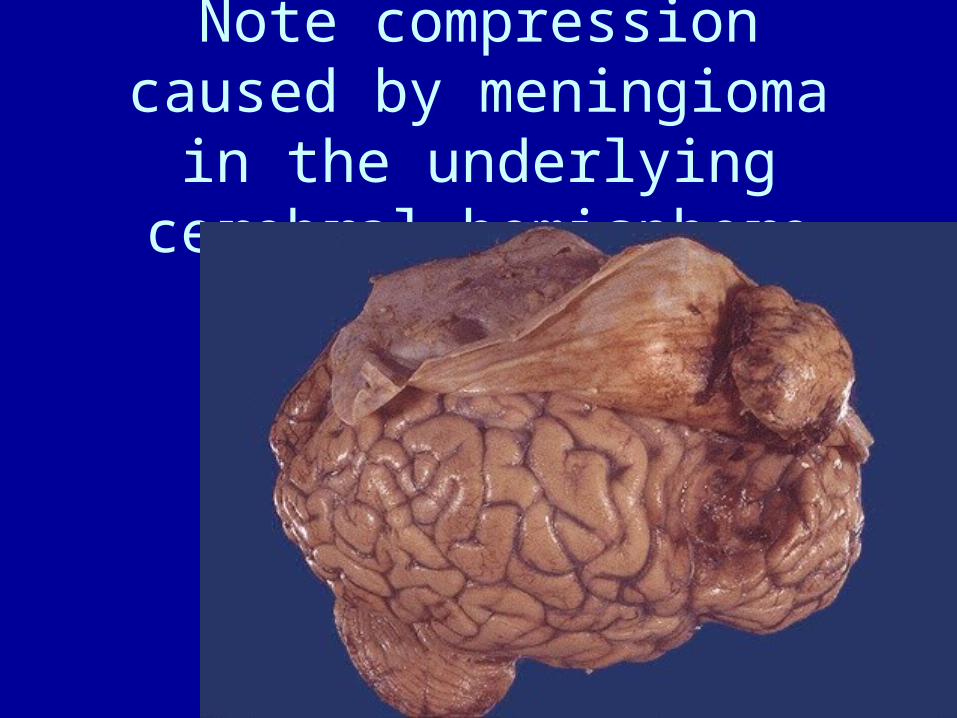
Note compression caused by meningioma in the underlying
cerebral hemisphere
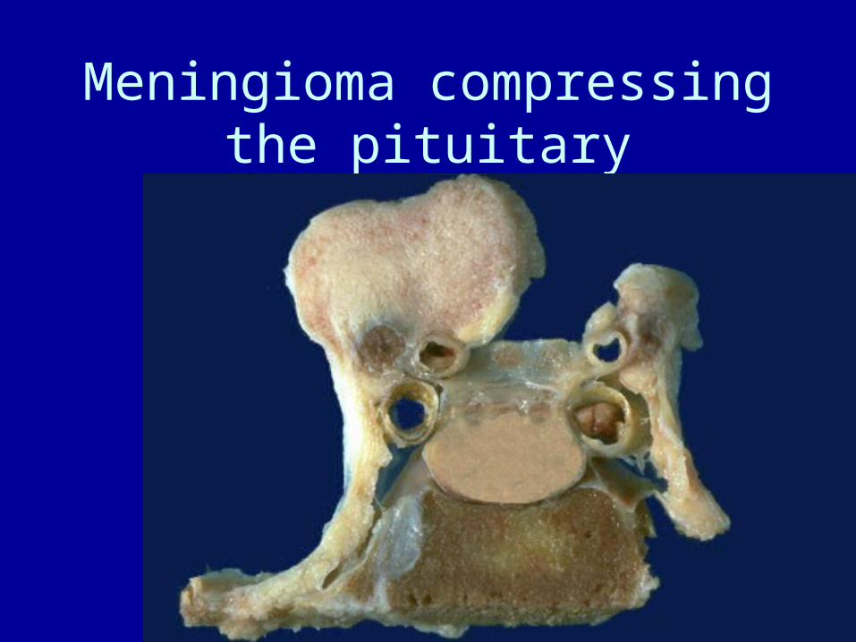
Meningioma compressing the pituitary
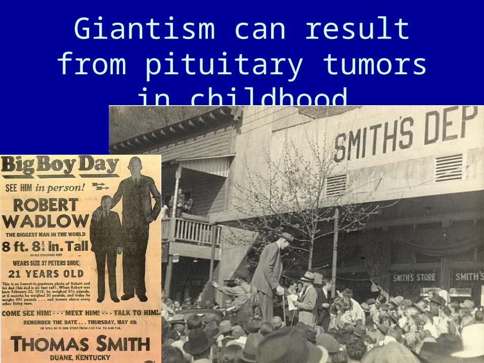
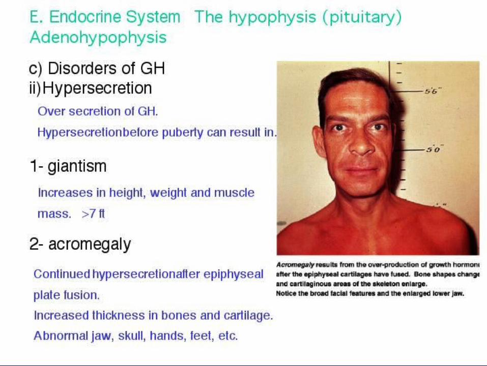
Giantism can result from pituitary tumors in childhood
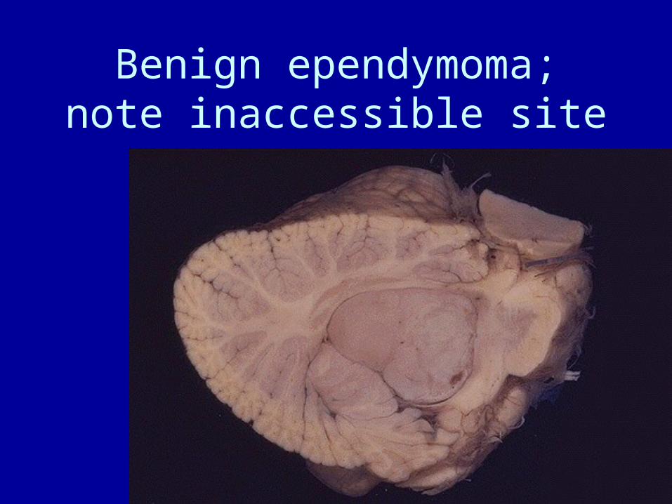
Benign ependymoma; note inaccessible site
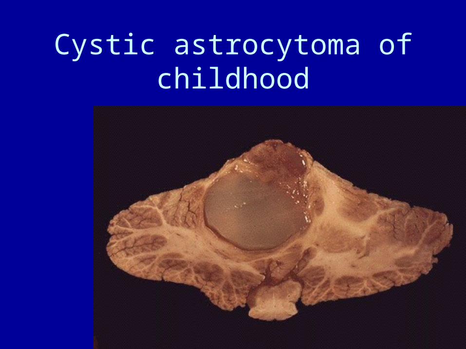
Cystic astrocytoma of childhood
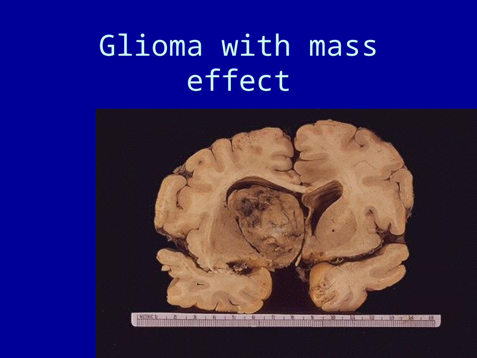
Glioma with mass effect
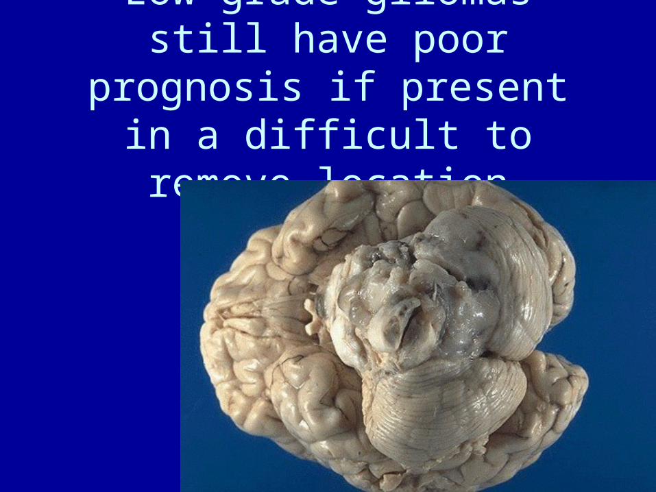
Low grade gliomas still have poor prognosis if present in a difficult to remove location
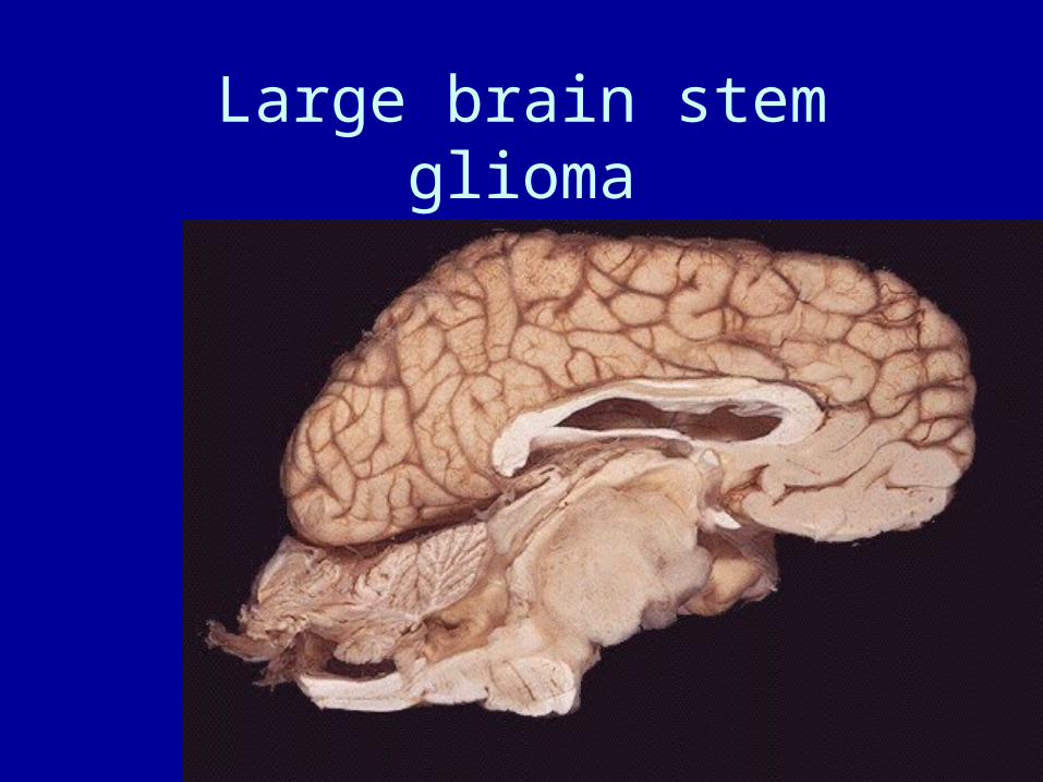
Large brain stem glioma
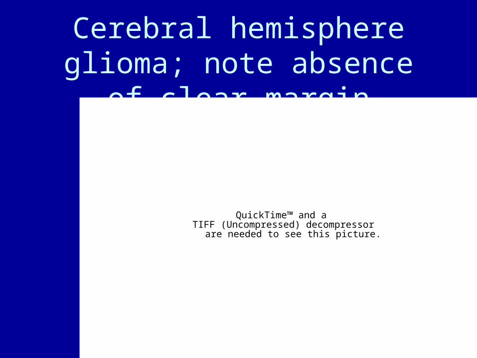
Cerebral hemisphere glioma; note absence of clear margin
QuickTime™ and aTIFF (Uncompressed) decompressor
are needed to see this picture.
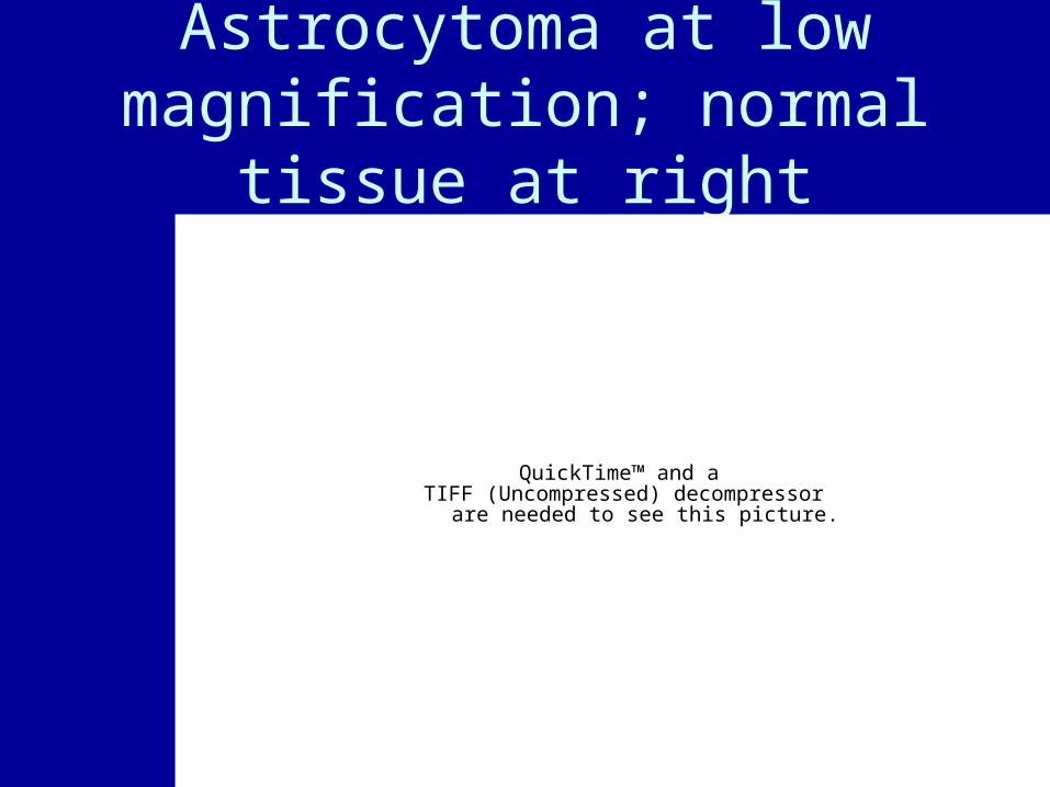
Astrocytoma at low magnification; normal tissue at
right
QuickTime™ and aTIFF (Uncompressed) decompressor
are needed to see this picture.
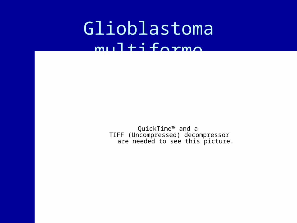
Glioblastoma multiforme
QuickTime™ and aTIFF (Uncompressed) decompressor
are needed to see this picture.

GBM: note necrosis, hemorrhage
QuickTime™ and aTIFF (Uncompressed) decompressor
are needed to see this picture.
Note palisading effect around necrotic region
QuickTime™ and aTIFF (Uncompressed) decompressor
are needed to see this picture.
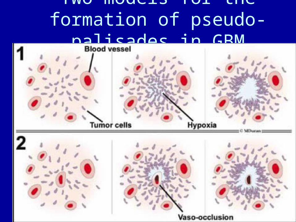
Two models for the formation of pseudo-palisades in GBM
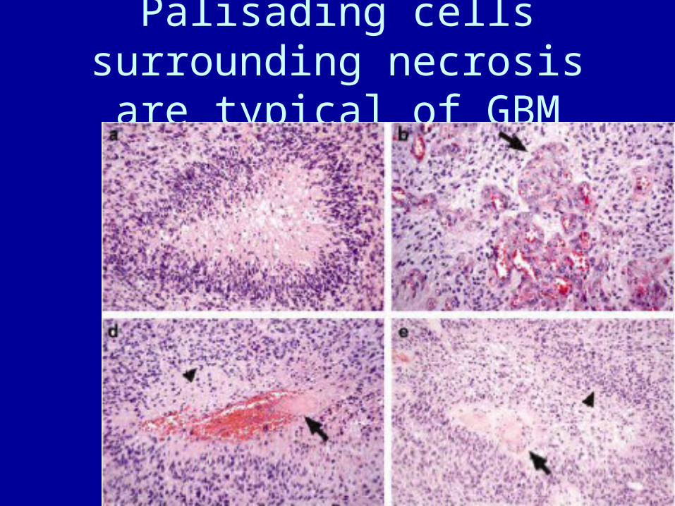
Palisading cells surrounding necrosis are typical of GBM
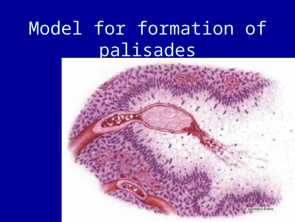
Model for formation of palisades


