
The University of Manchester Research
Parallelized Biocatalytic Scanning Probe Lithography forthe Additive Fabrication of Conjugated Polymer StructuresDOI:10.1039/C8NR01283K
Document VersionAccepted author manuscript
Link to publication record in Manchester Research Explorer
Citation for published version (APA):Hosford, J., Valles, M., Krainer, F. W., Glieder, A., & Wong, L. S. (2018). Parallelized Biocatalytic Scanning ProbeLithography for the Additive Fabrication of Conjugated Polymer Structures. Nanoscale, 10, 7185-7193.https://doi.org/10.1039/C8NR01283K
Published in:Nanoscale
Citing this paperPlease note that where the full-text provided on Manchester Research Explorer is the Author Accepted Manuscriptor Proof version this may differ from the final Published version. If citing, it is advised that you check and use thepublisher's definitive version.
General rightsCopyright and moral rights for the publications made accessible in the Research Explorer are retained by theauthors and/or other copyright owners and it is a condition of accessing publications that users recognise andabide by the legal requirements associated with these rights.
Takedown policyIf you believe that this document breaches copyright please refer to the University of Manchester’s TakedownProcedures [http://man.ac.uk/04Y6Bo] or contact [email protected] providingrelevant details, so we can investigate your claim.
Download date:01. Jul. 2020

Parallelized Biocatalytic Scanning Probe Lithography for the Additive Fabrication of
Conjugated Polymer Structures
Joseph Hosford,† Morgane Valles,† Florian W. Krainer,‡ Anton Glieder,‡ Lu Shin Wong†*
† Manchester Institute of Biotechnology and School of Chemistry, University of Manchester,
131 Princess Street, Manchester M1 7DN, United Kingdom.
‡ Institute of Molecular Biotechnology, Graz University of Technology, NAWI Graz,
Petersgasse 14, 8010 Graz, Austria.
* email: [email protected]
Abstract
Scanning probe lithography (SPL) offers a more accessible alternative to conventional
photolithography as a route to surface nanofabrication. In principle, the synthetic scope of
SPL could be greatly enhanced by combining the precision of scanning probe systems with
the chemoselectivity offered by biocatalysis. This report describes the development of
multiplexed SPL employing probes functionalized with horseradish peroxidase, and its
subsequent use for the constructive fabrication of polyaniline features on both silicon oxide
and gold substrates. This polymer is of particular interest due to its potential applications in
organic electronics, but its use is hindered by its poor processability, which could be
circumvented by the direct in situ synthesis at the desired locations. Using parallelized arrays
of probes, the lithography of polymer features over 1 cm2 areas was achieved with individual
feature widths as small as 162 ± 24 nm. The nature of the deposited materials was confirmed
by Raman spectroscopy, and it was further shown that the features could be chemically

derivatized postlithographically by Huisgen [2+3] “click” chemistry, when
propargyloxyaniline was used as the monomer in the initial lithography step.
Introduction
Scanning probe lithography (SPL) represents a family of surface nanolithography methods
based on scanning probe microscopy platforms, where the probe tip is used to localize a
physical or chemical manipulation of the surface. Such SPL-based methods have attracted
much interest in nanoscience since they can achieve nanoscale resolution and registry, and
are able to “write” any user-defined pattern, using instrumentation that is readily accessible.
Indeed, the major impetus for the development of new SPL methods has been the need to
address the limitations of classical “hard” lithography techniques derived from the
microelectronics sector, such as the lack of flexibility for rapid design prototyping and the
patterning of “soft” materials such as small organic molecules, polymers or biomolecules. 1, 2
In terms of physical manipulation, scanning probes have been used for the mechanical
removal of material (c.f. nanoshaving and nanografting),3-6 the application of an electrical
bias to induce oxidation (local anodic oxidation or local oxidation nanolithography),7, 8 and
heating of the probes to perform thermally induced effects (thermochemical
nanolithography).9
The use of scanning probes for the local delivery of materials has also been
demonstrated. In dip-pen nanolithography (DPN), an atomic force microscopy (AFM) probe
tip directs the local deposition of “ink” molecules onto a surface by diffusion of the
molecules from the probe to the surface.1, 2, 10 Scanning probes with microfluidic channels
(“nanopipettes” and “nano-fountain pens”) for the delivery of inks to surfaces have also been
reported.11 This group of methods is particularly interesting as they are examples of “additive”

or “constructive” fabrication, since the surface features are formed by the deposition of
material, rather than the removal or ablation of materials (“subtractive” or “destructive”) that
typify conventional hard lithography.
In principle, an attractive approach to additive nanofabrication is to use scanning
probes bearing a catalyst to directly synthesize materials in situ. Such an approach would
combine the precision of SPL with the chemical selectivity of catalysis, thereby greatly
increasing the chemical scope and complexity that can be achieved by SPL.1, 12
This type of direct synthesis is of particular relevance to the lithography of materials
that are otherwise difficult to process by other approaches, such as conducting polymers. For
example, polyaniline is a conjugated polymer that exhibits high electrical conductivity and
electrochemical stability, and is therefore of interest in the development of organic electronic
devices.13-15 However, polyaniline has very low solubility in organic solvents and its
decomposition temperature is below its glass transition temperature, making the practical
manipulation of this material challenging.14, 16 Methods for the generation of polyaniline
features, to any user-defined design template, would therefore be highly desirable.15
As an exemplification of this strategy, the use of a biocatalytically functionalized
scanning probes to perform the additive this report demonstrates lithography of polyanilines,
by oxidative cross-coupling of anilines. Here, a recombinant horseradish peroxidase (HRP) is
employed, which has been engineered to enable its use in an SPL context. This biocatalyst is
particularly suitable for this application as it has a high redox potential (i.e. it is able to
catalyze oxidations of otherwise stable aromatic substrates), uses H2O2 as a terminal oxidant
that is safe and readily available, operates under mild ambient conditions, and has previously
been shown to catalyze oxidations when placed at the tip of an AFM probe.17 Furthermore,
parallelization of this biocatalytic SPL over large (cm2) areas using an array of elastomeric

probes (c.f. polymer pen lithography),18 and the postlithographic derivatization of the
polyaniline features, are also demonstrated.
Results and Discussion
Biocatalytic probe array preparation.
In order to realize the proposed biocatalytic nanolithography, a number of considerations
needed to be addressed. Firstly, in order for the nanolithography to be precise (i.e. only at the
point of contact between the probes and the surface) it was necessary to confine the enzymes
to the apex of each probe.17 Since each probe tip would therefore only carry a relatively small
number of enzyme molecules, it was also necessary to employ a site-specific method of
protein immobilization that gives a uniform protein orientation relative to the probes, and
thus achieve maximum activity from each protein. For this purpose, the HRP was
recombinantly engineered so that it was fused to a ybbR tag, which can be immobilized onto
coenzyme A-functionalized materials by the phosphopantetheinyl transferase enzyme Sfp.1, 19
Taking these issues into consideration, the elastomeric probe arrays with HRP confined to the
tip apex were constructed and employed following the workflow shown in Scheme 1.
Scheme 1. Workflow for the functionalization of the scanning probes and their use in the
nanolithography of polyaniline features. Steps: (i) functionalization of PDMS tips with
coenzyme A (see also scheme 2a); (ii) site-selective immobilization of ybbR-HRP mediated
by Sfp (see scheme 2b); (iii) biocatalytic nanolithography of polyaniline features.

Polydimethylsiloxane (PDMS) elastomeric probe arrays of the type typically used for
PPL20 were used as the basis of the biocatalytic probe array. In all cases arrays of 1 × 1 cm2
were used. The array was first amino-functionalized by vapor deposition of 3-
aminopropyltriethoxysilane 1, followed by the attachment of an oligoethylene glycol linker
with a terminal maleimide group 2 (Scheme 2a). Any unreacted amines were blocked by
treatment with acetic anhydride. The array was mounted on an AFM, which was used to
precisely bring the probe into contact with a silicon substrate drop-coated with coenzyme A,
thus ensuring that only the tips of the probes would be ligated with the coenzyme required for
subsequent protein immobilization. The remaining unreacted maleimide groups, which
coated the entire surface of the array, were blocked by immersion in a solution of 2-
mercaptoethanol. Following washing to remove any unreacted reagents, the arrays were
finally submerged in a solution containing Sfp and ybbR-HRP to allow enzyme conjugation
(scheme 2b). Alignment of the probe array relative to the coenzyme A-bearing surface, as
well as controlling its contact to the surface was vital to ensure the uniformity of protein
immobilization and subsequent lithography performance. It was found that excessive probe-
surface contact resulted in the lithography of overly large features, while lack of contact
would result in no features being produced.

Scheme 2. (a) Coenzyme A functionalization onto the PDMS probes. Steps: (i) surface
oxidation by oxygen plasma; (ii) vapor deposition of 1 followed by thermal curing; (iii)
ligation of 2, then blocking of unreacted amines with acetic anhydride; (iv) ligation of 3 then
blocking of unreacted maleimides with 2-mercapotethanol. (b) Site-specific ybbR-HRP
immobilization catalyzed by the transferase enzyme Sfp.
The peroxidase activity of HRP immobilized on the probes was confirmed with a
colorimetric assay using 2,2'-azino-bis(3-ethylbenzothiazoline-6-sulphonic acid) (ABTS) and
H2O2.21 Thus, upon immersing the array into the assay solution, the immobilized HRP
catalyzes the generation of a colored product, the intensity of which is monitored by UV-vis
spectrometry at 405 nm (Figure S1 in SI). For comparison, the assay was also performed on
arrays entirely coated with HRP, rather than only at the apices, as a positive control. Negative
control experiments were conducted using unfunctionalized arrays, arrays where all
(a)
(b)

maleimide groups were blocked with 2-mercaptoethanol prior to the protein immobilization,
and arrays with full coenzyme A coverage where either Sfp or ybbR-HRP were omitted from
the protein immobilization step (step ii in Scheme 1). Arrays with a full coverage of HRP
show a rapid increase in absorbance, indicating the immobilization approach was effective.
Those that were subject to the enzyme confinement only to the probe tips also showed an
increase in activity, though it was lower compared to the fully coated arrays, as expected. In
contrast, the negative controls gave essentially no activity.
Lithography of poly(2-methoxyaniline) by enzyme-probe arrays.
The HRP-functionalized probe arrays were then used to demonstrate the lithography of
polymer features by biocatalytic polymerization of 2-methoxyaniline onto a silicon dioxide
surface. Here, the surface was functionalized with 4-aminophenol that served as the site of
attachment and subsequent growth of the polymer chains (Scheme 3). 2-methoxyaniline was
chosen as a model enzyme substrate because the methoxy group provided a readily detectable
signal by Raman spectroscopic imaging (see below). The lithography setup consisted of the
HRP-bearing probes and silicon surface being placed in a fluid cell, with a solution of 2-
methoxyaniline and H2O2 being added upon the initiation of the lithography; followed by
rapid retraction of the catalytic probes upon completion of the lithography.

Scheme 3. Illustration of the HRP-catalyzed polymerization of 2-methoxyaniline reaction on
aniline-functionalized silicon oxide or gold surfaces to form lithographic patterns.
As an example, lithography was performed such that each probe in the array patterned
a series of lines composed of either 80 or 160 individual dot features, with a dwell time of 2
or 1 s per dot feature. AFM analysis post-lithography clearly showed topographic features
that were consistent with the desired pattern (Figures 1a and b). Measurements taken across
the line features in 20 representative locations across the printed area gave an average feature
size of 489 ± 52 nm (full width at half maximum height, fwhm) and an average height of 1.9
± 1.1 nm (Figure S2 in SI). Further optimization enabled these features to be reduced to an
fwhm of 162 ± 24 nm, with a similar height (2.1 ± 0.9 nm, Figures 1c and d, Figure S3 in SI).

Figure 1. Illustrative AFM topographic images of poly(2-methoxyaniline) features and
graphs of height against distance for representative cross-sections. (a and b) A series of
parallel lines produced with 2 s dwell time per dot feature, 80 dot features per line, 2.6 mM 2-
methoxyaniline and 1 mM H2O2. (c and d) A series of diagonal lines produced with 1 s dwell
time per dot feature, 160 features per line, 2.6 mM 2-methoxyaniline and 1 mM H2O2. The
scale bars in the images represent 10 μm.
These results compare favorably with the additive lithography of conducting polymers
by DPN. For example, the deposition of proprietary formulations of pre-synthesized
sulfonated polyaniline or polypyrrole colloids on to a polyamine surface gave lines that were
310 or 290 nm, respectively.22 In another report, an approach whereby 4-aminothiophenol
was first deposited by DPN on a gold surface, followed by exposure of the patterned surface
to a solution of HRP and H2O2, gave features with a width of 210 nm.23 It also compares

favorably to a nanopipette method that used local electropolymerisation of 3,4-
ethylenedioxylthiophene or aniline, which gave features of ~600 nm.24 However, the reported
features are larger than those achieved by “field assisted nanopatterning”, whereby an AFM
probe coated with pre-synthesized polyaniline can be made to deposit the polymer by
applying an electrical bias between the probe and the conducting surface.25 This approach
gave polyaniline features as small as 20 nm, but can only be used with probes and surfaces
that are electrically conductive. In all these cases, only lithography with a single probe was
reported.
The typical apex width of the elastomeric probes20 are approximately 60 nm and the
oligoethylene linker is approximately 10 nm in length when fully extended, which implies
that the theoretical maximum distance that can be reached by the tethered HRP molecule
would encompass a diameter of ~80 nm. However, since the radical monomers that are
generated must leave the enzyme active site prior to polymerization, it is postulated that the
observed feature sizes are thus related to the distance these radicals can diffuse prior to
deposition.
Raman mapping on gold 4-aminothiophenol functionalized surfaces.
In order to demonstrate the applicability of this lithographic approach to another substrate,
the biocatalytic lithography was conducted in an analogous manner onto a self-assembled
monolayer of 4-aminothiophenol on a gold surface. Since such metal substrates exhibit
surface-enhanced Raman scattering (SERS),26-29 this experiment also enabled direct chemical
detection and identification of the features by Raman spectroscopy.
Raman microscopy imaging was performed over an area of 100 × 100 µm with a
spectra collected in a 200 × 200 grid (i.e. every 0.5 µm apart). A plot of the signal intensity at
1398 cm–1, which corresponds to the quinoid C-N+ Raman band, shows strong signals

corresponding to the pattern programmed into the instrumentation (Figure 2a and b). In
contrast, the unpatterned areas show no corresponding signal. The representative spectrum
for the patterned area (Figure 2c, Table S1 in SI) was essentially identical to that of poly(2-
methoxyaniline) synthesized in solution by the conventional route.30 Indeed, the spectrum
from the features displayed a peak at 1140 cm–1 corresponding to a C-O bond vibration,
which was attributed to the methoxy groups of the polymer. Its presence therefore indicated
that the features arose from the polymerization of the methoxyaniline monomers, and not
simply the oxidative crosslinking of the underlying aminothiophenol monolayer. In
comparison, the spectrum for the unpatterned area is identical to that of a control surface
consisting of only the 4-aminothiophenol monolayer (Figure 2d).

Figure 2. Raman chemical imaging of poly-2-methoxyaniline patterns obtained by HRP-
catalyzed nanolithography on 4-aminothiophenol functionalized gold surfaces. (a) Raman
microscopy map of signal intensity at 1398 cm–1 and (b) the template image used for
lithography. Representative Raman spectra of: (c) the patterned area and (d) the unpatterned
surface. The scale bar in the microscopy image represents 20 μm.
Postlithography derivatization of polymer features.
In order to demonstrate that the deposited polyaniline can be further chemically derivatized,
and as a means to assess the wide-area nature of the parallelized lithography, an experiment
to fluorescently tag the polymer patterns after the lithography was performed. Here, the
Huisgen 1,3-dipolar “click” cycloaddition between an azide and alkyne was used as a

convenient means for ligating the tags.31, 32 For this purpose, 3-(prop-2-yn-1-yloxy)aniline
was synthesized for use as the monomer in the biocatalytic lithography, which was then
carried out in the same way as before. The patterned substrate was then submerged in a
solution of azide-functionalized Alexafluor 488 in the presence of a Cu(I) catalyst to affect
the click ligation. Subsequently, the surfaces were washed and imaged under a fluorescence
microscope, where fluorescent features were observed that conformed to the patterns used
(Figure 3); confirming successful parallelized lithography and post-lithographic
derivatization.
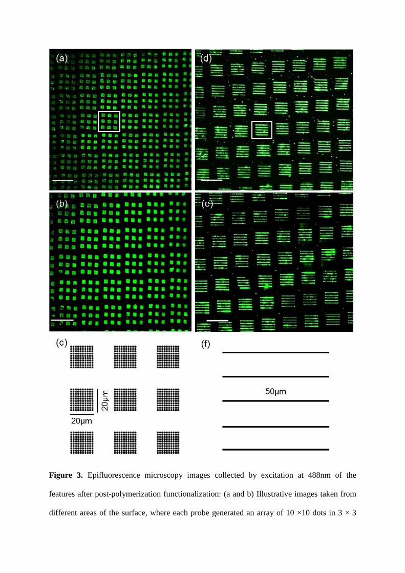
Figure 3. Epifluorescence microscopy images collected by excitation at 488nm of the
features after post-polymerization functionalization: (a and b) Illustrative images taken from
different areas of the surface, where each probe generated an array of 10 ×10 dots in 3 × 3
20µm
(a) (b)
(c)

grids (the white box indicating the pattern written by a single probe); (c) the template pattern
written by a single probe; (d and e) Illustrative images taken from different areas of the
surface where each probe generated a series of 5 lines, each line is 50 μm in length and
generated from 50 dot features each (the white box indicating the pattern written by a single
probe); (c) the template pattern written by a single probe. Scale bars on the microscopy
images represent 100 µm.
Conclusions
Overall, this report demonstrates the feasibility of parallelized constructive biocatalytic
nanolithography of conjugated polyaniline on both silicon dioxide and gold surfaces, over
wide areas (cm2) and under ambient conditions. The polyanilines were catalytically formed
and deposited at high resolution, with feature widths of 162 ± 24 nm. These patterns were
chemically analyzed by Raman microscopy, which confirmed that aniline polymers were
being formed from the corresponding monomer feedstocks. Wide area patterning was further
confirmed by modifying the deposited aniline polymers with a fluorescent moiety, which
could then be imaged by wide-field fluorescence microscopy.
From a practical perspective, since a single round of enzyme production yielded
sufficient ybbR-HRP for ~ 50 arrays, and PDMS is very inexpensive, these probe arrays can
be treated as single-use “disposable” items. This approach thus ensures no cross-
contamination of monomers between different rounds of lithography and maximally active
enzymes are used on each occasion. One possible route to improve the resolution of the
reported method would be to use arrays of probes made from hard materials which would
give sharper probe tips, such as those reported in hard-tip, soft-spring lithography.18

In comparison with previous attempts at enzymatically-catalyzed nanolithography that
only utilized serial (single probe) lithography,1 these patterns were of higher chemical
complexity while giving superior resolution over larger areas. The generation of features
consisting of conjugated polymers is of particular interest since these materials can be made
to be electrically conducting, with potential applications in flexible electronic devices.13
While there are several SPL examples involving conjugated polymers, none show large-area
parallelization, despite its potential usefulness in the fabrication of devices containing such
materials.
Key to the success of this approach was the implementation of covalent and site
specific protein immobilization, using a homogeneous and highly active enzyme preparation.
Indeed, the approach described here is generally applicable to other enzymatic reactions,
which would further widen the scope of biocatalytic nanolithography. Indeed, with the
increasing availability of convenient tools for the genetic engineering of proteins,33, 34 it is
now possible to rationally design proteins specifically for use in conjunction with scanning
probes. Such developments would be a possible route to incorporating biological catalysis in
any future universal “desk-top fab” system.
Experimental
Materials and Equipment.
The maleimide linker 1 (MAL-dPEG24-NHS ester) was purchased from Quanta Biodesign
(Plain City, OH) and Azide-fluor 488 from Sigma-Aldrich (Poole, UK). The 2-
naphthylhydroxamic acid-functionalized agarose used for the protein purification was
prepared according to procedures adapted from previous reports.35 The phosphopantetheinyl
transferase Sfp was produced heterologously in E. coli and purified by metal affinity

chromatography according to previously reported procedures.19 Gold substrates and silicon
wafers were purchased from Sigma-Aldrich, and atomically flat glass microarray slides
(Arrayit Superclean 2) from Arrayit (Sunnyvale, CA). All other reagents were purchased
from the usual laboratory suppliers.
The lithography was performed on a custom-built AFM (Nanosurf AG, Liestal,
Switzerland) equipped with the automated alignment algorithm reported elsewhere.36 Raman
microscopy was performed on a Renishaw inVia confocal Raman microscope using a 532nm
laser source. Epifluorescence microscopy was carried out with a Fluorescent Stereo
Microscope, (Leica Microsystems GmbH, Germany).
Production of ybbR-HRP.
The C1A isoform of HRP was used for this work,37 which was cloned into a pET28a plasmid
vector using standard procedures to give a protein fused to an N-terminal hexahistidine tag,
followed by the ybbR tag. This protein was heterologously produced using standard methods
in E. coli. Briefly, the plasmid was transformed into E. coli BL21(DE3). Cells were grown to
an optical density of 0.6 OD600, then gene expression was induced by the addition of β-D-1-
thiogalactopyranoside (0.1 mM) at 37 °C for 3 h, after which the cells were collected by
centrifugation (4000 g for 15 min) and the supernatant removed.
These cells were resuspended in 50 mM Tris buffer pH 8 containing 1 mM EDTA and
10 mM DTT (buffer A); and left to stand for 1 h at 4 C prior to lysis by sonication (5 × 30 s).
The lysate was separated by centrifugation (8000 g for 40 min) and the supernatant discarded.
The pellet was thoroughly resuspended in 50 mM Tris buffer pH 8 containing 2 M urea
(buffer B), subjected to centrifugation (8000 g for 40 min) and supernatant discarded. The
pellet containing washed inclusion bodies were finally solubilized in 50 mM Tris buffer pH 8
containing 1 mM DTT and 6 M urea (buffer C), and subjected to centrifugation (8000 g for

40 min). The supernatant, now containing the solubilized inclusion bodies, was then purified
by Ni-affinity chromatography under denaturing conditions in Buffer C containing increasing
concentrations of imidazole. The purified denatured protein (final concentration
approximately 0.18 mg/mL) was then added drop-wise to the refolding buffer (1.7 M urea, 2
mM CaCl2, 7% glycerol, 0.35 mM oxidized glutathione, 0.044 mM dithiothreitol) and stirred
at 4°C for 16 h.38 The solution was then filtered (0.2 m pore size) and purified by Ni-affinity
chromatography to remove any aggregated protein. Refolded apoprotein was then
reconstituted with heme by drop wise addition of hemin (from a 1 mM solution of hemin in
20 mM NaOH).
The reconstituted holoprotein was then buffer exchanged by centrifugal filtration into
50 mM pH 5.5 succinate buffer. This material was then further purified by chromatography
through 2-naphthylhydroxamic acid-functionalized agarose column, at a flow rate of 1 mL
min–1, by elution with 50 mM succinate and 200 mM sodium borate buffer at pH 5.5. The
pure protein was then buffer exchanged into 25 mM Tris 50 mM NaCl pH 7.5 and
concentrated approximately 5 mg mL–1. This final HRP preparation typically gave an RZ
(A404/A280) ratio of 2.93.
Synthesis of 3-(Prop-2-yn-1-yloxy)aniline.39-41
3-nitrophenol (2.75 g, 20 mmol) was dissolved in diethyl ether (20 mL) and potassium
carbonate (3.17 g, 23 mmol) was added. The mixture was stirred for 10 min, after which
propargyl bromide (2.5 mL, 22 mmol) was added and the mixture refluxed for 5 h.
Subsequently, the reaction was cooled to room temperature and the organic phase was
washed with water (2 × 20 mL), brine (2 × 10 mL) and saturated potassium carbonate
solution (20 mL). The organic phase was then dried over anhydrous magnesium sulfate,

filtered and the solvent removed under reduced pressure to yield 1-nitro-3-(prop-2-yn-1-
yloxy)benzene (2.409 g, 13.6 mmol, 69 %) as an orange solid.
The 1-nitro-3-(prop-2-yn-1-yloxy)benzene (147 mg, 0.83 mmol) and iron powder
(279 mg, 5.00 mmol) were suspended in a solution of glacial acetic acid (2 mL), ethanol (2
mL) and water (1 mL). The resulting suspension was ultrasonically agitated at 30 C until the
reaction was observed to be complete (by TLC, typically 2 h). The solution was filtered and
the residue washed with ethyl acetate (30 mL). The combined filtrate and washings were
extracted with potassium hydroxide (2 M, 20 mL), and this aqueous phase extracted with
diethyl ether (2 × 25 mL). The combined organic phases were washed with brine (2 × 25 mL),
followed by water (3 × 50 mL), dried over anhydrous magnesium sulfate, filtered and the
solvent removed under reduced pressure. The residue was subjected to flash column
chromatography (SiO2, hexane: ethyl acetate; 2:1) to yield the desired 3-(prop-2-yn-1-
yloxy)aniline (57 mg, 0.39 mmol, 47 %) as a yellow oil. Rf: 0.41 (hexane: ethyl acetate; 2:1),
1H NMR (400 MHz, CDCl3) δ 7.07 (t, J = 8.0 Hz, 1H), 6.39 (ddd, J = 8.2, 2.4, 0.7 Hz, 1H),
6.33 (ddd, J = 7.9, 2.1, 0.8 Hz, 1H), 6.30 (t, J = 2.2 Hz, 1H), 4.64 (d, J = 2.4 Hz, 2H), 3.65 (s,
2H), 2.53 (t, J = 2.4 Hz, 1H); 13C NMR (101 MHz, CDCl3) δ 158.77, 147.89, 130.16, 108.81,
104.70, 102.04, 78.89, 75.44, 55.70; m/z (ESI+) 148.1 (100 %, [M+H]+), HRMS found
148.0754; C9H10NO+ requires 148.0760.
Synthesis of Tert-butyl(4-mercaptophenyl)carbamate.42
To a stirred slurry of di-tert-butyl dicarbonate (554 mg, 2.5 mmol) and indium chloride (90
mg, 0.025 mmol), 4-aminobenzenethiol (313 mg, 2.5 mmol) was added and heated at 35 C.
The reaction was monitored by TLC and was observed to be complete after 1 h. The resulting
reaction mixture was suspended in ethyl acetate (25 mL) and washed with water (3 × 25 mL).
The organic phase was dried over anhydrous magnesium sulfate, filtered and the solvent

removed under reduced pressure. Flash column chromatography (SiO2, hexane: ethyl acetate;
2:1) subsequently yielded tert-butyl (4-mercaptophenyl)carbamate (0.545 g, 2.4 mmol, 97%)
as a white solid. Rf: 0.36 (hexane: ethyl acetate; 2:1), Mp: 179-180°C. 1H NMR (400 MHz,
CDCl3) δ 7.39 (d, J = 8.7 Hz, 2H), 7.29 (d, J = 8.7 Hz, 2H), 6.48 (s, 1H), 1.57 (s, 1H), 1.51 (s,
9H); 13C NMR (101 MHz, CDCl3) δ 152.60, 138.53, 131.14 (2C), 130.96, 119.05 (2C), 81.02,
28.46 (3C); m/z (ESI+) 226.1 (100%, [M+H]+).
Preparation of 4-Aminobenzenethiol-Functionalized Silicon Oxide Surfaces.
The following procedure was performed in a glove bag. Microarray glass slides were treated
with O2 plasma (200 mTorr) for 1 min immediately prior to use, and were then immersed in a
freshly prepared 1% v/v solution of (3-iodopropyl)trimethoxysilane in anhydrous toluene
under an argon atmosphere at 80 °C for 24 h. The slides were then removed, washed by
rinsing with anhydrous toluene (3 × 5 mL), by sonication in anhydrous acetone for 1 minute,
then further washed with a stream of acetone (1 min) and dried with a stream of nitrogen gas.
Finally the surfaces were annealed at 130 C for 10 min.
These freshly prepared (3-iodopropyl)trimethoxysilane functionalized surfaces were
immersed in a freshly prepared solution of tert-butyl (4-mercaptophenyl)carbamate (50 mM
in anhydrous methanol) under an argon atmosphere at 60 °C for 96 h. Subsequently, the
surfaces were washed by submerging them in 2-propanol (3 × 15 min), acetone (3 × 15 min)
and dried under a stream of nitrogen gas.
Tert-butyl (4-mercaptophenyl)carbamate terminated surfaces were then immersed in a
solution hydrochloric acid (4 M in 1,4-dioxane) under an argon atmosphere at room
temperature for 12 h. The surfaces were then washed by immersion in 1,4-dioxane (3 × 15
min), followed by acetone (3 × 15 min) and dried with a stream of nitrogen gas. The prepared
surfaces were kept under vacuum and used within 2 weeks of preparation.

Preparation of 4-Aminobenzenethiol-Functionalized Gold Surfaces.
Gold substrates (10 nm gold with 2 nm titanium adhesion layer on aluminasilicate glass
microscope slides) were immersed in a freshly prepared 5 mM solution of 4-aminothiophenol
in ethanol under an argon atmosphere for 96 h at room temperature. The surfaces were then
washed by immersing them in ethanol (3 × 5 min), sonication in ethanol for 1 min, further
rinsed with a stream of ethanol (1 min) and dried with a stream of nitrogen. These monolayer
coated surfaces were kept under vacuum and used within 2 weeks of preparation.
Ligation of Maleimido-Linker 1 onto PDMS Probe Arrays.
PDMS probe arrays (50 µm or 100 µm pitch), prepared according to previously reported
procedures, were freshly peeled off the master and treated with O2 plasma for 30 s at 200
mTorr.36 The plasma-treated arrays and an empty glass vial were immediately placed inside
a desiccator under an argon atmosphere and 100 μL of (3-aminopropyl)triethoxysilane added
to the vial. The desiccator was evacuated to 2-3 Torr and left to stand overnight at room
temperature. The surfaces were then removed and cured at 110 C for 10 min, before washing
by submerging them in DMSO (3 × 5 min), rinsing with an ethanol stream and drying under a
stream of nitrogen.
The amino-functionalized surfaces were then submerged in a 5 mM solution of
maleimide-linker 1 in DMSO for 2 h at room temperature with gentle agitation, then washed,
rinsed and dried as above.
Ligation of Coenzyme A to Probe Apices.
An 8 mM coenzyme A trilithium salt aqueous solution was drop-coated on to half a silicon
wafer (approximately 4 × 2 cm) and allowed to air dry. The maleimide-functionalized array
was then aligned (using auto alignment software as previously described)36 to the uncoated
half of the wafer surface. Upon completion of the alignment the stage was retracted on its z-
axis and moved in its y/x-axis until the array was over the drop-coated area, and the humidity
was set at 40 %. The stage was then moved in its z-axis until contact was observed (using
minimum force or visual inspection). Contact was held for 5 min then the stage retracted in
its z-axis. The stage was moved diagonally on the x/y-axis by10 µm and this contacting
process was repeated twice, following which the array was incubated at 60 % humidity for 2
h. The array was then washed by submerging in water (3 × 5 min) followed by an aqueous
solution of 80 mM 2-mecaptoethanol to block the remaining unreacted maleimide groups.
ybbR-HRP Immobilization onto Probe Arrays.
A reaction mixture consisting of 50 mM sodium phosphate pH 7.4 buffer, 10 mM MgCl2, 5
mM Sfp, 1 μM TCEP, and 20 μM ybbR-C1A was pipetted on to CoA-functionalized surfaces,
incubated at 37 C for 2 h at 100% relative humidity. The surfaces were then washed by
submerging them in 50 mM sodium phosphate buffer pH 7.4, with 0.5% tween (3 × 15 min),
50 mM sodium phosphate buffer pH 7.4 (3 × 5 min), and finally rinsing with 50 mM sodium
phosphate buffer pH 7.4. The arrays were stored in 50 mM HEPES buffer at pH 7.4 and used
within one week. Surfaces were rinsed with water and dried with a stream of nitrogen prior to
use in the lithography experiments.
ABTS Peroxidase Activity Assays on Immobilized HRP.
The probe arrays were attached inside individual wells in a 24-well microtiter plate with
double-sided tape. 500 µL of assay solution (50 mM sodium acetate pH 4.5, 0.77 mM
hydrogen peroxide, 1.00 mM ABTS) added to each well, and the UV-vis absorbance at 405

nm was read every 15 s with the plate swirled between each reading to ensure homogenous
mixing of the solution.
Lithography on 4-Aminothiophenol Surfaces.
The array was then mounted onto the AFM scan head using double-sided tape and the
substrate (functionalized silicon oxide or gold surfaces) was mounted at the bottom of a 1.5 ×
1.5 × 0.5 cm fluid well on to the sample stage using double-sided tape. The fluid well then
filled with a 2.6 mM solution of enzyme substrate (2-methoxyanilne or 3-(prop-2-yn-1-
yloxy)aniline) in 50 mM acetate buffer pH 5 to submerge the surface (approximately 3 mL).
Alignment of the probe array relative to the surface was performed36 and the array was then
retracted to above the level of fluid. Hydrogen peroxide added to a final concentration of 1
mM (final volume of solution typically 4 mL). The array was then very rapidly brought
within 10 μm of the surface and patterning commenced with the predefined template to
produce the desired pattern using the standard lithography software provided with the
instrument, with each dot feature representing at a 2 s dwell time at that location. Patterning
was undertaken at 60% relative humidity to prevent evaporation of the solution in the open
fluid well.
Upon completion the array was immediately lifted from the solution and the enzyme-
functionalised probe arrays discarded (i.e. not reused). The substrate was then removed and
washed by immersion in 50 mM acetate buffer pH 5 (3 × 5 min) followed by 1 M aq. HCl (3
× 5 min) and finally water (3 × 5 min); dried under a stream of nitrogen and immediately
imaged by AFM or Raman microscopy.
Postlithographic Functionalization of Poly(3-(prop-2-yn-1-yloxy)aniline.

The substrates bearing the polymer were submerged in an aqueous solution of 1 mM Azide-
fluor 488 and 0.1 mM copper(I) iodide in the dark for 24 h at room temperature. The surfaces
were then washed by submersion in water (6 × 15 min), dried under a stream of nitrogen and
immediately visualized by epifluorescence microscopy with fluorescent excitation at 488 nm.
Conflicts of Interest.
The authors declare no conflicts of interest.
Acknowledgements
Support is acknowledged from the Engineering and Physical Sciences Research Council (UK)
under grants EP/K024485/1 and EP/K011685/1, as well as a graduate studentship to JH under
grant EP/J50032X/1. Support is also acknowledged from the Biotechnology and Biological
Sciences Research Council (UK) for support of the Raman microscopy equipment under
grant BB/L014823/1 and a Doctoral Training Partnership studentship to MV under grant
BB/J014478/1.
Footnotes.
Time course plots of ABTS peroxidase activity assays with probe arrays, AFM topographic
cross-sections of the polymer features, and a table of assignments for the Raman spectra of
the polymers are provided in the supplementary information.

References
1. S. A. Carnally and L. S. Wong, Nanoscale, 2014, 6, 4998.
2. R. Garcia, A. W. Knoll and E. Riedo, Nat. Nanotechnol., 2014, 9, 577.
3. S. Xu and G.-Y. Liu, Langmuir, 1997, 13, 127.
4. H. Sugihara, A. Takahara and T. Kajiyama, J. Vac. Sci. Technol. B., 2001, 19, 593.
5. J. N. Ngunjiri, D. J. Stark, T. Tian, K. A. Briggman and J. C. Garno, Anal. Bioanal.
Chem., 2013, 405, 1985.
6. Y. Yan, Y. Geng and Z. Hu, Int. J. Mach. Tools Manuf., 2015, 99, 1.
7. H. C. Day and D. R. Allee, Appl. Phys. Lett., 1993, 62, 2691.
8. D. H. Lee, C. K. Kim, J.-H. Lee, H.-J. Chung and B. H. Park, Carbon, 2016, 96, 223.
9. W.-K. Lee, M. Haydell, J. T. Robinson, A. R. Laracuente, E. Cimpoiasu, W. P. King
and P. E. Sheehan, ACS Nano., 2013, 7, 6219.
10. R. Garcia, R. V. Martinez and J. Martinez, Chem. Soc. Rev., 2006, 35, 29.
11. M. Ghatkesar, H. Garza, F. Heuck and U. Staufer, Micromachines, 2014, 5, 954.
12. V. Mesquita, J. Botton, D. A. Valyaev, C. François, L. Patrone, T. S. Balaban, M.
Abel, J.-L. Parrain, O. Chuzel and S. Clair, Langmuir, 2016, 32, 4034.
13. T.-H. Le, Y. Kim and H. Yoon, Polymers, 2017, 9, 150.
14. S. Bhadra, D. Khastgir, N. K. Singha and J. H. Lee, Prog. Polym. Sci., 2009, 34, 783-
810.
15. L. Jiang, X. Wang and L. Chi, Small, 2011, 7, 1309-1321.
16. C. D. Liu, S. Y. Wu, J. L. Han and K. H. Hsieh, J. Appl. Polym. Sci., 2010, 115, 2271-
2276.
17. X. Luo, V. A. Pedrosa and J. Wang, Chem. Eur. J., 2009, 15, 5191.

18. L. R. Giam, A. J. Senesi, X. Liao, L. S. Wong, J. Chai, D. Eichelsdorfer, W. Shim, B.
Rasin, S. He and C. A. Mirkin, Proc. SPIE, 2011, 8031, 803103.
19. L. S. Wong, J. Thirlway and J. Micklefield, J. Am. Chem. Soc., 2008, 130, 12456.
20. F. Huo, Z. Zheng, G. Zheng, L. R. Giam, H. Zhang and C. A. Mirkin, Science, 2008,
321, 1658.
21. H. Gallati, Clin. Chem. Lab. Med., 1979, 17, 1.
22. J. H. Lim and C. A. Mirkin, Adv. Mater., 2002, 14, 1474-1477.
23. P. Xu and D. L. Kaplan, Adv. Mater., 2004, 16, 628-633.
24. C. Laslau, D. E. Williams, B. Kannan and J. Travas‐Sejdic, Adv. Funct. Mater., 2011,
21, 4607-4616.
25. J.-F. Liu and G. P. Miller, J. Phys. Chem. C, 2007, 111, 10758-10760.
26. P. A. Kilmartin and G. A. Wright, Synth. Met., 1999, 104, 145.
27. F. Flory, L. Escoubas and G. Berginc, J. Nanophoton., 2011, 5, 052502.
28. N. Tiwari, M. Y. Liu, S. Kulkarni and Y. Fang, J. Nanophoton., 2011, 5, 053513.
29. S. Mondal, U. Rana and S. Malik, ACS Appl. Mater. Interfaces, 2015, 7, 10457.
30. W. Liu, J. Kumar, S. Tripathy, K. J. Senecal and L. Samuelson, J. Am. Chem. Soc.,
1999, 121, 71.
31. V. V. Rostovtsev, L. G. Green, V. V. Fokin and K. B. Sharpless, Angew. Chem., 2002,
114, 2708.
32. J.-F. Lutz, Angew. Chem. Int. Ed., 2007, 46, 1018.
33. U. T. Bornscheuer, G. W. Huisman, R. J. Kazlauskas, S. Lutz, J. C. Moore and K.
Robins, Nature, 2012, 485, 185.
34. N. J. Turner, Nat. Chem. Biol., 2009, 5, 567-573.
35. L. Reimann and G. R. Schonbaum, Methods Enzymol., 1978, 52, 514.
36. S. Wang, J. Hosford, W. P. Heath and L. S. Wong, RSC Adv., 2015, 5, 61402.

37. F. W. Krainer, R. Pletzenauer, L. Rossetti, C. Herwig, A. Glieder and O. Spadiut,
Protein Expr. Purif., 2014, 95, 104.
38. S. Asad, B. Dabirmanesh, N. Ghaemi, S. M. Etezad and K. Khajeh, Mol. Biotechnol.,
2013, 54, 484.
39. F. Liu, J. Liu and T. Zhao, J. Appl. Polym. Sci., 2010, 115, 3103.
40. B. K. Patel, H. Ghosh, A. Baneerjee and S. K. Rout, ARKIVOC, 2011, 2011, 209.
41. K.-C. Tiew, D. Dou, T. Teramoto, H. Lai, K. R. Alliston, G. H. Lushington, R.
Padmanabhan and W. C. Groutas, Bioorg. Med. Chem., 2012, 20, 1213.
42. D. Niculescu-Duvaz, C. Gaulon, H. P. Dijkstra, I. Niculescu-Duvaz, A. Zambon, D.
Ménard, B. M. J. M. Suijkerbuijk, A. Nourry, L. Davies, H. Manne, F. Friedlos, L.
Ogilvie, D. Hedley, S. Whittaker, R. Kirk, A. Gill, R. D. Taylor, F. I. Raynaud, J.
Moreno-Farre, R. Marais and C. J. Springer, J. Med. Chem., 2009, 52, 2255.

Table of Contents Graphic
Enzyme-functionalized scanning probe
Peroxidase


