Open a new chapterin radiographyPhilips DigitalDiagnost Digital Radiography Solutions

2
A whole new worldThere’s no turning back the clock, especially when it comes to medical systems.
Be assured that with DigitalDiagnost you’ll get solutions that are way ahead in
technical developments. The scalability concept of Philips digital radiography
supports technologists and practitioners in the best possible way.

3
Table of contents
Chapter 1: Digital Rad Rooms
6-7 Dedicated chest room
8-9 General Rad Room – Compact
10-11 General Rad Room – Standard
12-13 General Rad Room – High Performance
Chapter 2: Features
15 Image quality
16-17 Automatic image stitching, tracking and
move-to-position, VM horizontal movement
18-19 CR integration, digital tomography,
vertical stand display, DICOM
20-21 CAD Chest
22 Security
Chapter 3: Rad Room components
24 Scalability concept
25 Detector technology
26 Generators, X-ray tubes
27 Tube carriers, tables
28 Vertical stands
29 Acquisition console, digital detector
Chapter 4: Reliability
31 Service, CustomerCare, EasyUpgrade

4
Orthopedic imaging with
automatic image stitching
UNIQUE image
processing software
Integrated
workflow with CR
Digital Imaging and
COmmunication in
Medicine – DICOM
DigitalDiagnost
acquisition console
Why should you choose Philips?
When developing medical solutions
Philips strictly focuses on customers.
Our DigitalDiagnost solutions are designed
for you. We invite you to access our holistic
concept with consulting, finance, service,
IT maintenance and networking all from one
source. And when it comes to the digital
radiography system itself, another concept
unfolds: the scalability concept.
Because the choice is yours!
What is the scalability concept? It means that
you can determine the sum of the individual
parts. Guided by us you can choose from
special hardware and software ranges to
configure a digital system that perfectly fits
your needs. This way you will benefit from a
truly filmless workflow. The advantages gained
may have a positive impact not only in your
Rad room but throughout the entire depart-
ment as well. Experience your new tailor-made
digital Rad Room!
Discover
5
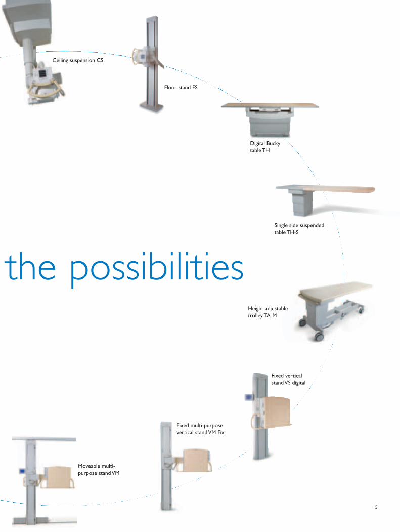
Ceiling suspension CS
Floor stand FS
Digital Bucky
table TH
Single side suspended
table TH-S
Height adjustable
trolley TA-M
Moveable multi-
purpose stand VM
Fixed vertical
stand VS digital
the possibilities
Fixed multi-purpose
vertical stand VM Fix

6
Automated workflow
With Philips’ DigitalDiagnost chest room you’ll get
workstation controlled collimation, asymmetric beam
alignment, tracking, remote control and near real-time
image display to create a highly automated workflow.
UNIQUE, Philips’ advanced image processing system,
delivers superb chest images within seconds.
Extending the range
Two features may drastically improve efficiency in the
chest room. On one hand, the tracking function maintains
a constant source-detector distance. On the other hand,
the tube assembly automatically tracks the vertical
movement of the detector. Using a tiltable digital vertical
stand and a ceiling suspended tube can extend the
application range to skeletal examinations. And combined
with a trolley, even under-table examinations are easily
possible.
Standard configuration:
DigitalDiagnost VR
• Generator
• Fixed floor stand with X-ray tube assembly,
control grip and collimator (FS Fix)
• Digital vertical stand with integrated flat
detector (VS)
• Acquisition console with monitor, keyboard
and mouse
• Tracking
• UNIQUE image processing
Why everything in your rooms runslike clockwork

7
Supporting clinical excellence
The addition of Philips’ CAD solution, xLNA, aids
physicians in visualizing, identifying, evaluating and repor t -
ing pulmonary lesions/nodules in digital radio graphic chest
images. For more information, see pages 20/21.
It’s your choice
Add efficiency with the following options (selection):
• Vertical stand display
• Automatic image stitching
All features and options in chapter 2 starting on page 15.
Single detector /Dedicated chest room
When performing
thorax examinations,
the tube assembly
automatically tracks
the vertical movement
of the detector.
For more flexibility,
horizontal examinations
are also possible.
Some X-ray departments specialize in thorax examinations. An output of
more than 200 thorax images per day sets the benchmark in these
environments. Philips’ response to this situation is the dedicated DigitalDiagnost
chest room.
Choose the chest champion
Room
s

8
Have you set your sights on going digital in your medical facility? Then let
DigitalDiagnost Compact be your entry to filmless workflow. Benefit from an
affordable X-ray system which integrates seamlessly into your hospital network
environment.
Your easy move to digital
Multi-purpose use, medium workflow
The DigitalDiagnost Compact is a cost-effective X-ray
system for multi-purpose use and medium workflow.
It offers the full range of standard radio graphy applications
and all the advantages of digital work flow. Medical facilities
often use this room as a chest room, which can also
serve as a back-up general DR room.
It’s never been easier
With DigitalDiagnost Compact, patient data and work
lists are received directly from your RIS. Anatomical
program parameters are ready as soon as you select the
type of examination. That automatically sets pre-filtration
and collimation for each exposure. The images are
Standard configuration:
DigitalDiagnost Compact
• Generator
• Fixed multi-purpose stand with swiveling
C-arm and integrated digital detector
(VM Fix)
• Ceiling suspension with X-ray tube
assembly, control grip and collimator (CS 2)
• Optional height adjustable trolley
(TA-M)
• Acquisition console with monitor, keyboard
and mouse
• UNIQUE image processing
Single detector /General Rad Room – Compact

9 Room
sprocessed with UNIQUE image processing software
and sent to PACS or printer. In addition, the system can
simultaneously transfer exa mination information back to
the RIS.
It’s your choice
Add efficiency with the following options (selection):
• Automatic collimation
• Tracking and move-to-position
All features and options in chapter 2 starting on page 15.

10
Philips has redefined the benchmark for standard rooms with a highly flexible
configuration. Renowned DigitalDiagnost image quality together with easy
handling, comfort and excellent ergonomics set this single-detector solution
apart.
All-in-one for all applications
Very versatile
The DigitalDiagnost standard room solution is a very
versatile system for environments with a medium to high
patient load. It features a moveable multi-purpose stand
including an integrated detector combined with a single
side suspended table. By moving the detector to the end
of the table, the system becomes a digital chest unit.
Positioning it vertically alongside the table enables easy
lateral projections.
Standard configuration:
DigitalDiagnost – one-detector
standard room
• Generator
• Moveable multi-purpose stand with swiveling
C-arm and integrated digital detector (VM)
• Ceiling suspension with X-ray tube
assembly, control grip and collimator (CS 4)
• Single side suspended height adjustable
table (TH-S) or moveable trolley (TA-M)
• Acquisition console with monitor,
keyboard and mouse
• UNIQUE image processing
• Tracking
Single detector /General Rad Room – Standard

11
More convenient
Working with DigitalDiagnost is even more convenient
than before since the system’s motorized detector moves
both vertically and horizontally. By simply pressing a
button you switch from under-table to chest positions,
thus facilitating smooth working procedures.
It’s your choice
Add efficiency with the following options (selection):
• Horizontal motorized movements, plus extended move-
to-position and alignment functionality
• Vertical stand display
• Automatic image stitching
• Clinical QC (quality control)
All features and options in chapter 2 starting on page 15.
Benefit from cross table projections: lateral projections even for difficult angulated views.
Room
s

12
If your department cares for a high number of patients and carries out a large
variety of applications and projections, a high performance room is absolutely
essential. The Philips DigitalDiagnost dual-detector system is the ideal solution
to these demands.
The easy switch
The two detectors make it easy to switch from table
exams to chest. In addition, automated functions such as
auto collimation and move-to-position help increase
workflow. For higher demands, you have the option of
adding a second tube.
Faster procedures
In a high performance room from Philips, DICOM
Worklist Management (WLM) for easy scheduling and
fast distribution maximizes efficiency via the DICOM
standard. Instant availability and optimized image display
also greatly contribute to faster working procedures.
Standard configuration:
DigitalDiagnost dual-detector high
performance room
• Generator
• Height adjustable table with integrated
digital detector (TH)
• Vertical stand with integrated detector
(VS) with motorized tilting
• Ceiling suspension with X-ray tube
assembly, control grip and collimator (CS 4)
• Acquisition console with monitor, keyboard
and mouse
• UNIQUE image processing
• Tracking
Dual detector /General Rad Room – High Performance
Excellence and efficiency

13
It’s your choice
Add efficiency with the following options (selection):
• Vertical stand display
• Second table control
• Automatic stitching
• Clinical QC
• PCR integration
All features and options in chapter 2 starting on page 15.
Extend your applications:
By using the moveable
multi-purpose stand VM
instead of the VS, all
vertical Bucky exams as
well as lateral examina -
tions such as axial hip
are fast and convenient.
This combination signifi -
cantly increases the
range of applications.
Room
s

14
Which benefitsimprove your work -flow and results

15
Creating brilliance in diagnostic viewing
DigitalDiagnost supports diagnostic viewing by:
• Image processing especially suited to flat detector
characteristics
• Detecting the appropriate region of interest
• Adapting to the output medium
• Application-driven image processing with UNIQUE
UNIQUE image processing
With UNIQUE you can expect consistently high image
quality whether working with Computed Radiography,
Direct Radiography or CR/DR combinations. UNIQUE
enhances the detail contrast and harmonizes the image
quality for all digital radiography modalities. UNIQUE
image processing is especially suited to those applications
where high-definition detail is absolutely essential.
UNIQUE at a glance
• Harmonizes contrast
• Enhances weak details and achieves detail accuracy in
all areas
• Eliminates processing artifacts
• Permits a visually uniform impression for DR and CR
images
• Achieves consistently high image quality
UNIQUE is ideal for both viewing on the monitor and for
printing. Image quality is enhanced while simultaneously
preserving the images’ natural appearance. Plus, the
parameters can be adapted to suit each individual.
Image verification
The image is available within a matter of seconds after
the exposure, which reduces waiting time for each
individual patient. In addition, the user can use a range
of parameters to further enhance the image:
• Contrast/brightness
• Rotation/mirror
• Annotations
• Shutters
Image processing is of major importance in achieving consistent, excellent image
quality for all anatomical areas in order to support quality of care. In all its
radiography systems, Philips has always placed special emphasis on enabling
excellent image processing.
The visibledifference
Feat
ure
s
16
DigitalDiagnost’s ergonomic features, such as motorized movements, reduce the
physical demands on the technologist substantially. Combined with automated
procedures such as automatic stitching, you will benefit from digital efficiency in
the Rad room and throughout the entire medical facility.
Designed to improve your
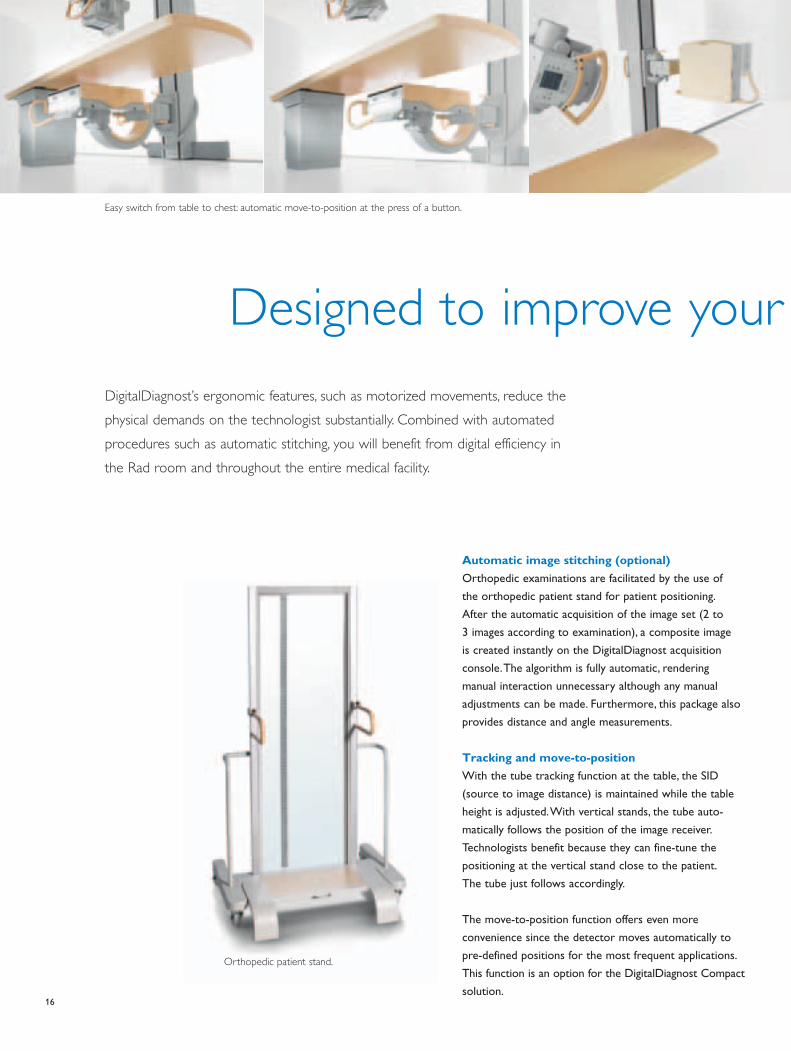
Automatic image stitching (optional)
Orthopedic examinations are facilitated by the use of
the orthopedic patient stand for patient positioning.
After the automatic acquisition of the image set (2 to
3 images according to examination), a composite image
is created instantly on the DigitalDiagnost acquisition
con sole. The algorithm is fully automatic, rendering
manual interaction unnecessary although any manual
adjustments can be made. Furthermore, this package also
provides distance and angle measurements.
Tracking and move-to-position
With the tube tracking function at the table, the SID
(source to image distance) is maintained while the table
height is adjusted. With vertical stands, the tube auto -
matically follows the position of the image receiver.
Technologists benefit because they can fine-tune the
positioning at the vertical stand close to the patient.
The tube just follows accordingly.
The move-to-position function offers even more
con venience since the detector moves automatically to
pre-defined positions for the most frequent applications.
This function is an option for the DigitalDiagnost Compact
solution.
Orthopedic patient stand.
Easy switch from table to chest: automatic move-to-position at the press of a button.
17
workflow
VM horizontal movement (optional)
This option contains three functions:
Motorized horizontal movements
In addition to the standard motorized movements of
the DigitalDiagnost vertical stands, the VM stand can be
moto rized horizontally as well to enhance comfort and
workflow.
Move-to-position extended
With the motorized VM stand, the move-to-position func -
tion is expanded to include more pre-defined posi tions,
which can switch automatically between table and chest
positions by simply pressing a button.
Alignment
Furthermore, the tube and the detector can be aligned
automatically. Just press the light button for 2 seconds and
the detector automatically moves to the field of radia tion.
This is especially convenient for positioning patients for
cross-lateral applications with the light field
of the tube. The detector follows automatically.
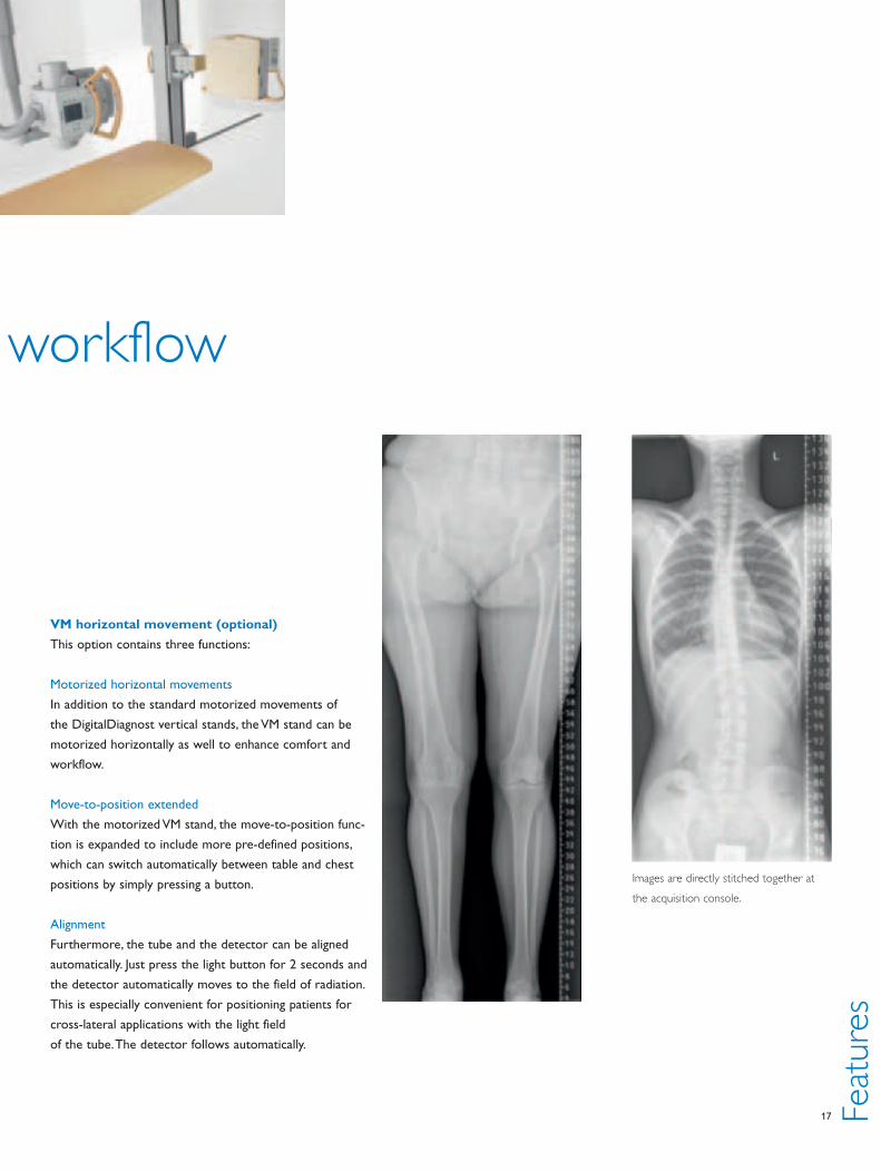
Images are directly stitched together at
the acquisition console.
Feat
ure
s

18
Digital tomography (optional)
Digital tomography combines ergonomics, image quality
and economic aspects in a unique manner. All movements
are motorized and electronically controlled. Superb image
quality is achieved by using a digital flat detector in com -
bi nation with UNIQUE image processing.
Vertical stand display (optional)
The display on the vertical stand simplifies workflow and
permits closer contact to the patient, which is particularly
important for a high patient throughput. All vital parame-
ters are directly displayed while positioning the patient.
This allows the patient to be addressed personally and
enables the technologist to
check parameters without
frequently walking over to the
acquisition con sole.
The display shows the
following parameters:
• Patient name, patient ID and
date of birth
• Selected examination
• Grid status
Patient data organization
Depending on hospital infrastructure, patient data can
be entered via the keyboard, barcode reader (optional)
or directly via the DICOM RIS interface (optional, see
DICOM functions). The system automatically creates the
worklist from the data. Connection to non-DICOM
compatible interfaces (NFS/FTP) is included.
DICOM functions
Optional DICOM WLM (Work List Management)
DICOM WLM connects DigitalDiagnost to the RIS.
DigitalDiagnost automatically retrieves the work list from
the RIS, thus supporting efficient and seamless workflow
in the digital X-ray room.
Exposure on
CR cassette.
Cassette is read out via
the Philips CR reader.
The image appears
on the DigitalDiagnost
acquisition console and
is stored in the same
patient folder as the
images made with the
DigitalDiagnost.
Making it even easier
Portable examinations with integrated CR
(optional)
Some applications cannot be performed with a flat panel
detector system, e.g. special projections taken in bed.
Fully integrated Computed Radiography (CR) is the cost
efficient alternative to using a heavy, cable limited and
fragile portable detector. DigitalDiagnost’s streamlined
workflow also covers the seamless acquisition and
processing of CR images. Schedu ling, examination, data
handling and image pro cessing are identical for both
DR and CR acquisitions. For patients receiving DR and
portable examinations, the images will have the same
impression and will be stored in one folder on PACS –
because it is fully inte grated!
19
DigitalDiagnost supports DICOM GSDF (Grayscale
Standard Display). This provides optimum consistency
between quality control and reading situations by
ensuring consistent high-quality image display on both
printouts and PACS viewing monitors when exporting
to DICOM imagers and PACS systems with the same
func tion.
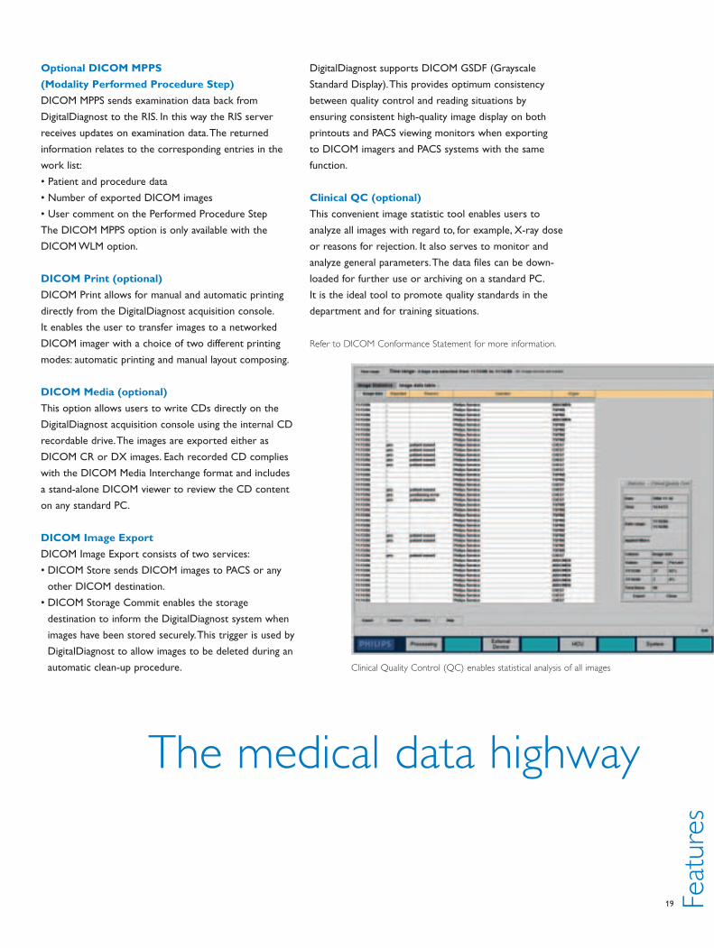
Clinical QC (optional)
This convenient image statistic tool enables users to
analyze all images with regard to, for example, X-ray dose
or reasons for rejection. It also serves to monitor and
analyze general parameters. The data files can be down -
loaded for further use or archiving on a standard PC.
It is the ideal tool to promote quality standards in the
depart ment and for training situations.
Refer to DICOM Conformance Statement for more information.
Optional DICOM MPPS
(Modality Performed Procedure Step)
DICOM MPPS sends examination data back from
DigitalDiagnost to the RIS. In this way the RIS server
receives updates on examination data. The returned
information relates to the corresponding entries in the
work list:
• Patient and procedure data
• Number of exported DICOM images
• User comment on the Performed Procedure Step
The DICOM MPPS option is only available with the
DICOM WLM option.
DICOM Print (optional)
DICOM Print allows for manual and automatic printing
directly from the DigitalDiagnost acquisition console.
It enables the user to transfer images to a networked
DICOM imager with a choice of two different printing
modes: automatic printing and manual layout composing.
DICOM Media (optional)
This option allows users to write CDs directly on the
DigitalDiagnost acquisition console using the internal CD
recordable drive. The images are exported either as
DICOM CR or DX images. Each recorded CD complies
with the DICOM Media Interchange format and includes
a stand-alone DICOM viewer to review the CD content
on any standard PC.
DICOM Image Export
DICOM Image Export consists of two services:
• DICOM Store sends DICOM images to PACS or any
other DICOM destination.
• DICOM Storage Commit enables the storage
destination to inform the DigitalDiagnost system when
images have been stored securely. This trigger is used by
DigitalDiagnost to allow images to be deleted during an
automatic clean-up procedure.
The medical data highway
Feat
ure
sClinical Quality Control (QC) enables statistical analysis of all images

20
Philips xLNA Enterprise lung nodule assessment software
Computer assisted detection (CAD) for digital chest X-ray images
See for yourself
Clinical report
• Automatic generation of clinical report on physician
confirmed diagnostic information
• Allows physicians to input notes and digital signature
• Secures report with time stamp
• Report stored in DICOM format ready for PACS
archiving
Integration into your PACS workflow
xLNA integrates into your PACS based solely on
DICOM connectivity. Neither code-level integration
nor the installation of any software on your PACS
will be required.
It’s like having a second pair of eyes on every chest exam.
Philips xLNA lung nodule assessment CAD software for
digital chest X-rays supports you in visualizing, identifying,
evaluating, and reporting pulmonary lesions and nodules.
A first-class second look
Unlike conventional CAD software, xLNA includes
exclusive real-time interactive image-reading features,
region of interest (ROI) analysis, easy reporting, and
direct integration with PACS. It provides CAD capabilities
(Computer Assisted Detection) and interactive toolkits to
assist in the identification of lung nodules, including small
ones.
Image reading and ROI (Region of Interest)
analysis
• Image visualization toolkits with multiple viewing modes
• Nodule-specific contrast-enhanced and nodule-enhanced
view
• Tools for physicians to perform lesion marking and
selection
• Lesion/nodule segmentation in automated or manual
mode
• Instantaneous automatic computation of quantitative
measurements from segmentation results
• Tools for physicians to add additional diagnostic
assessment comments
For more information please visit also our internet at:
www.medical.philips.com

21 Feat
ure
s
Instantaneous automatic computation of quantitative
measurements from segmentation results.
22
Information in safe hands
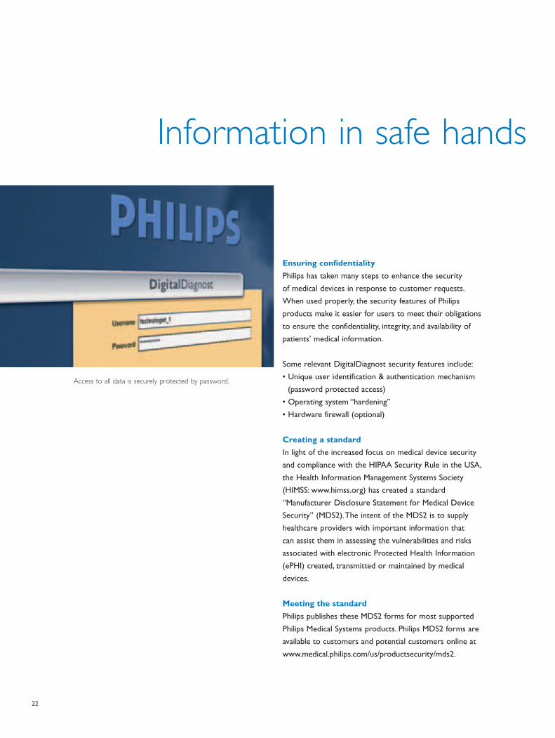
Ensuring confidentiality
Philips has taken many steps to enhance the security
of medical devices in response to customer requests.
When used properly, the security features of Philips
products make it easier for users to meet their obligations
to ensure the confidentiality, integrity, and availability of
patients’ medical information.
Some relevant DigitalDiagnost security features include:
• Unique user identification & authentication mechanism
(password protected access)
• Operating system “hardening”
• Hardware firewall (optional)
Creating a standard
In light of the increased focus on medical device security
and compliance with the HIPAA Security Rule in the USA,
the Health Information Management Systems Society
(HIMSS: www.himss.org) has created a standard
“Manufacturer Disclosure Statement for Medical Device
Security” (MDS2). The intent of the MDS2 is to supply
healthcare providers with important information that
can assist them in assessing the vulnerabilities and risks
associated with electronic Protected Health Information
(ePHI) created, transmitted or maintained by medical
devices.
Meeting the standard
Philips publishes these MDS2 forms for most supported
Philips Medical Systems products. Philips MDS2 forms are
available to customers and potential customers online at
www.medical.philips.com/us/productsecurity/mds2.
Access to all data is securely protected by password.

23
What componentsmake up your tailor-made system

24
Scalability – more than the sum of the parts
The advantages of scalability
Following this rigid evaluation, the advantages of scalability
truly pay off. That is when our sales representatives plan
with you the exact composition of your digital Rad room.
Together we select the components and features to place
your radiography department precisely where you, your
colleagues, your patients and your administration want
it to be. Rest assured that your DigitalDiagnost system
will integrate perfectly into your department and beyond.
Plus, it will run with peak performance for years to come
thanks to the individual excellence of each component
combining to form an absolutely reliable whole. The
united components will be more than just the sum of
their parts.
Quality in every detail
This is made possible because every single component of
our DigitalDiagnost solutions is a technological master -
piece. Take a look inside and see the precision with which
every item is manufactured.
The figure to the right shows our flat detector in every
detail. With in-depth knowledge of material and physical
phenomena, we have created a precision tool that easily
masters the constant demands placed on everyday
radio graphy operation. For example, our best-in-class
elec tro nics ensure excellent image quality while allowing
a low radiation dose. Or look at the scintillation layer.
It in cor porates a columnar structure (only 2–3 μm thick)
to accurately guide the light to the silicon with minimized
spatial spread.
Inside your head
Philips has more than 100 years of experience in medical
technology. In the 1980s we introduced CR and DR
radiography systems. From our experience and
interaction with customers, we know what is on the
minds of those responsible for various medical areas:
What are my resources?
How many experienced technologists and practitioners
are in the department? How do the patient profiles
change? How do I make the most out of a limited budget?
We have specific answers to your changing requirements.
The close look
We guide you through the decision making process.
We pave your way to filmless efficiency. We boil all those
questions down to a few basic essentials by minutely
evaluating your application mix, your throughput and your
financial resources.

25
Refresh Light
Philips eliminates ghosting
and blur. Refresh Light
“wipes” the silicon layer
clean immediately before
every new image.
Scintillator
Converts X-rays to light.
The 550μm thick, highly
efficient cesium iodide
scintillator layer offers
the best combination of
sensitivity and image
sharpness.
Com
ponen
ts
Electronic control
lines to trigger the
switching diodes.
Sensor matrix made
of amorphous silicon
Converts light to electrons.
Philips uses a 9 Mpixel array
with 143 μm pixels which
gives high resolution to all
medical applications.
Switching diodes
Connects each pixel to
read-out line (blue lines) for
image data read-out.
26
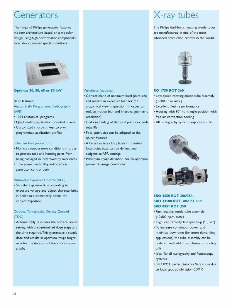
GeneratorsThe range of Philips generators features
modern architecture based on a modular
design using high performance components
to enable customer specific solutions.
Optimus 30, 50, 65 or 80 kW
Basic features:
Anatomically Programmed Radiography
(APR)
• 1024 anatomical programs
• Quick-to-find application oriented menus
• Customized short-cut keys to pre-
programmed application profiles
Tube overload protection
• Monitors temperature conditions in order
to protect tube and housing parts from
being damaged or destroyed by overstress
• Tube power availability indicated on
generator control desk
Automatic Exposure Control (AEC)
• Sets the exposure time according to
exposure voltage and object characteristics
in order to automatically obtain the
correct exposure
Optional Tomography Density Control
(TDC)
• Automatically calculates the correct power
setting with predetermined dose steps and
the time required. This guarantees a steady
dose and results in optimum image bright-
ness for the duration of the entire tomo-
graphy
Variofocus (optional)
• Correct blend of minimum focal point size
and maximum exposure load for the
anatomical view in question (in order to
reduce motion blur and improve geometric
resolution)
• Uniform loading of the focal points extends
tube life
• Focal point size can be adapted to the
object features
• A broad variety of application oriented
focal point sizes can be defined and
assigned to APR settings
• Maximum image definition due to optimum
geometric image conditions
X-ray tubesThe Philips dual-focus rotating anode tubes
are manufactured in one of the most
advanced production centers in the world.
RO 1750 ROT 360
• Low-speed rotating anode tube assembly
(3,600 r.p.m. max.)
• Excellent lifetime performance
• Housing with 90° horn angle position with
free air convection cooling
• All radiography systems, esp. chest units
SRO 2550 ROT 306/351,
SRO 33100 ROT 306/351 and
SRO 0951 ROT 350
• Fast rotating anode tube assembly
(10,800 r.p.m. max.)
• High load capacity, fast speed-up (1.0 sec)
• To increase continuous power and
minimize downtime (for more demanding
applications) the tube assembly can be
ordered with additional blower or cooling
unit
• Ideal for all radiography and fluoroscopy
systems
• SRO 0951: perfect tube for Variofocus due
to focal spot combination 0.3/1.0

27
Tube carriersAll DigitalDiagnost systems come with a
ceiling suspension (CS). The dedicated chest
system DigitalDiagnost VR comes with an
optional fixed floor stand.
Ceiling suspension CS
• Longitudinal and transverse movements
• Four-part telescopic column
• Award-winning Bucky control grip for easy,
one-handed operation and positioning
close to the patient
• Various optional functions include sensing,
tracking, alignment
• High projection flexibility, plus quick and
easy handling saves time
• Available in two versions, CS 2 and CS 4
for a full range of transverse movement
Floor stand FS Fix
• Optimized to the needs of modern, high
performance, fully automated chest rooms
• Column is mounted on the floor with a
fixed distance to the vertical stand with its
integrated detector
• Standard functions include automatic colli-
mation and tracking
TablesPhilips offers a variety of different tables and
trolleys to fit every requirement. All tables
feature a high patient load and convenient
access to the patient from all sides.
Digital Bucky table TH
• X-ray from head to toe – all radiographic
applications (skeletal, tomography etc.)
• X-ray transparent and floating table top
(two widths available)
• Integrated digital detector
• Motorized height adjustment
• Easy horizontal and vertical patient
positioning due to large movement range
• Electromagnetic brakes for a high level of
patient security
• Hands-free operation via a foot switch
• Additional optional hand switch for all table
movements, which can be flexibly
positioned even on the rear of the table
• Maximum patient load 375 kg / 820 lbs
Single side suspended table TH-S
• Single side suspended X-ray transparent
table especially designed for combining with
the moveable vertical stand, DigitalDiagnost
VM (see also page 10/11)
• X-rays from head to toe due to its large
X-ray transparent area
• Wide floating table top
• Motorized height adjustment
• Easy horizontal and vertical patient
positioning due to large movement range
• Easy patient transfer at any working height
• Hands-free operation via foot switch
• Electromagnetic brakes for a high level of
patient security
• Maximum patient load: 225 kg /496 lbs
(dynamic load)
Height adjustable trolley TA-M
• Single side suspended trolley with floating
table top (two widths) and central pedal
control
• Hydraulic height adjustment
• To be used in combination with the vertical
stands DigitalDiagnost VS and
DigitalDiagnost VM and VM Compact
(see also pages 8/9)
• Full application flexibility
• Excellent access to the patient from all
sides
• Floating table top, fully X-ray transparent
• Easy and precise to maneuver due to
central pedal control
• Maximum patient load: 225 kg /496 lbs
Com
ponen
ts

28
Vertical standsOur range of vertical stands caters to all
your individual application needs. Motorized
height adjustment, customizable pre-defined
detector positions and numerous other well
thought-out features significantly reduce the
physical demands placed on the technologist.
Fixed vertical stand VS digital
• Vertical stand mounted on the floor with
integrated digital detector
• Motorized height adjustment with two
different speeds plus manual operation for
precise positioning
• Customizable pre-defined detector
positions (move-to-position)
• Large detector format (43cm x 43cm /
17" x 17")
• Removable grid and storage unit for two
grids within the detector unit for immed-
iate availability and safe storage
• Two user interfaces
• Wireless remote control unit
• Additional optional display for patient data
in the examination room
• Five AEC measuring chambers to ensure
correct dosage
• Projection with angulated beam
• Tilting (-20° to +90°) to support exami-
nations of patients on a stretcher, plus
straightforward exams of extremities
for seated or standing patients (optional)
Moveable multi-purpose vertical
stand VM
• Motorized stand with multi-purpose
swiveling C-arm and integrated digital
detector
• Full application flexibility with just one
detector
• Motorized height adjustment with two
different speeds plus manual operation for
precise positioning
• Customized, pre-defined detector positions
(move-to-position)
• Optional customized, pre-defined system
positions switch from chest exam to table
position at the push of a button (extended
move-to-position)
• Motorized horizontal stand includes
detector alignment movement for more
convenience (optional)
• Horizontal alignment at the push of a
button
• Vertical tracking
• Cross table lateral exams
• Large detector format (43cm x 43cm /
17" x 17")
• Removable grid and storage unit for two
grids within the detector for instant access
and secure storage
• Two user interfaces
• Wireless remote control unit
• Additional optional display for patient data
in the examination room
• Five AEC measuring chambers, to ensure
correct dosage
• Angulated beam projection
Fixed multi-purpose vertical stand
VM Fix
• Fixed floor-mounted stand with multi-
purpose swiveling C-arm and integrated
digital detector for combining with a single
side suspended trolley
• Full application flexibility with just one
detector
• Motorized height adjustment with two
different speeds plus manual operation for
precise positioning
• Optional customized, pre-defined detector
positions (switch from chest exam to
extremity position at the push of a button)
• Vertical tracking (optional)
• Large detector format (43cm x 43cm /
17" x 17")
• Removable grid and grid storage for two
grids within the detector for instant access
and secure storage
• One user interface either on the left or
the right side of the detector unit
• Additional optional display for patient data
in the examination room
• Three AEC measuring chambers
• Angulated beam projection

29
AcquisitionconsoleOn the DigitalDiagnost acquisition console,
the clinical image is available within seconds
after the exposure.
Basic features
• Consists of a powerful SUN computer,
19" TFT color monitor, keyboard and
mouse
• A central operating console for the whole
X-ray examination, with emphasis on:
- Monitoring the entire DigitalDiagnost
system
- Reading patient data arriving automatically
via the RIS, or manual patient entry
- Selecting patient and exam
- Generator control
- Data transfer from the digital detector
- Image processing with UNIQUE, Philips’
advanced image processing software
- Image quality check
- Link to the hospital’s digital infrastructure
(PACS, etc.)
- Optional image archiving on CD
(DICOM Media)
Digital detectorThe DigitalDiagnost flat detector is made
of amorphous silicon and cesium iodide
scintillator for excellent image quality even
with low dose.
Basic features
• Completely integrated into the radio-
graphic table TH and all vertical stands
• Large size (43cm x 43cm / 17" x 17") for
high projection flexibility even with large
patients
• Resolution up to 3.5 lp/mm, 143 μm pixel
size, pixel matrix of approx. 9 Mpixels
More details, see page 25.
Contact your nearest Philips Medical
Systems representative to find out
how the scalability concept supports
you to shape your ideal digital
radiography room.
Com
ponen
ts

30
Why you can alwaysrely on Philips
31
Prized quality
Place your trust in our integrated concepts. From
financing to system maintenance, we’re at your disposal.
Our philosophy is to offer you fast support and excellent
quality. Benefit from our global service network, our
highly qualified service engineers, our service technicians’
individual attention and our international availability of
spare parts. Maintaining this high level of competence is
one of our greatest priorities.
CustomerCare portfolio
Our CustomerCare service programs ensure excellent
support, flexible solutions and effective relationships –
providing the service you need to guarantee that your
DigitalDiagnost system always operates at its peak.
Our range can be tailored to any individual customer
situation. We offer customized Service Agreement
solutions to help enhance the quality of patient care,
increase your productivity and improve your profitability.
Our Service Agreements come in silver, gold or platinum
levels. Regardless of the level, a Philips expert is always
just around the corner, whether via proactive remote
support or in person.
EasyUpgrade program
Another feature of our digital service concept is
its excellent upgrade opportunities. We call it the
EasyUpgrade program. With EasyUpgrade you can
conveniently switch to digital state-of-the-art technology
in no time. Whether you work with the conventional
BuckyDiagnost* solutions or are equipped with Thora -
vision, you can easily upgrade to DigitalDiagnost. So make
the switch when your medical facility feels ready for it.
*Not applicable to for BuckyDiagnost TS systems
Let Philips be your partner before, during and after the purchase of a system.
It pays off. While your system is in your medical facility, our service professionals,
numbering more than 6,000, offer you predictable life cycle costs and peak
perfor mance now and in the future.
We’re at your service
Serv
ice

© 2008 Koninklijke Philips Electronics N.V.
All rights are reserved.
Philips Healthcare reserves the right to make changes in specifications and/or to discontinue any product at any time without notice or obligation and will
not be liable for any consequences resulting from the use of this publication.
Printed in The Netherlands.
4522 962 15201/712 * APR 2008
Philips Healthcare is part of
Royal Philips Electronics
Interested?
Would you like to know more about our imaginative
products? Please do not hesitate to contact us.
We would be glad to hear from you.
On the web
www.philips.com/healthcare
Via email
By fax
+31 40 27 64 887
By mail
Philips Healthcare
Global Information Center
P.O. Box 1286
5602 BG Eindhoven
The Netherlands
By phone
Asia
Tel: +852 2821 5888
Europe, Middle East, Africa
Tel: +49 7031 463 2254
Latin America
Tel: +55 11 2125 0764
North America
Tel: +425 487 7000
1 800 285 5585 (toll free, US only)


