Download - Port position
WORLD LAPAROSCOPY HOSPITALCyberciti, DLF Phase II, NCR Delhi, Gurgaon, 122 002, India
Phone: +91(0)12- 42351555 Mobile: +91(0)9811416838, 9811912768,Email: [email protected]
Click here for training detail
Port position
In Laparoscopy
The relative position of the instrument ports is very important in the performance of surgical procedures endoscopically. The angle the instruments make with the operative site and to each other should mimic, as far as possible, the natural relationship of the hands and eyes during conventional surgery.
The most common cause of stressful surgery is wrong port position 95 % of surgeon and gynaecologists use umbilicus as primary port
Many surgeon and Gynaecologist lack the principles behind secondary port position
Umbilicus as primary port:
The central location and ability of the umbilicus to camouflage scars make it an attractive trocar site for laparoscopic surgery.
Umbilicus is a naturally weak area due to absence of all the layers.
Weakness is also due its location at the midpoint of the abdomen's greatest diameter
Most Surgeons believe there is a difference between the umbilicus and other trocar sites in both susceptibility to infection and postoperative Incisional herniation.
The study showed that the increased infection rate at the umbilicus seems to be related to cholecystectomy and not to the umbilicus
Excluding cholecystectomy, the umbilical infection rate was 2%, similar to that at remote sites.
The postoperative ventral hernia rate was at 0.8%, the same at the umbilicus as elsewhere.
The more infection after cholecystectomy is due to the contamination of wound due to infected gallbladder.
Secondary Port Position:
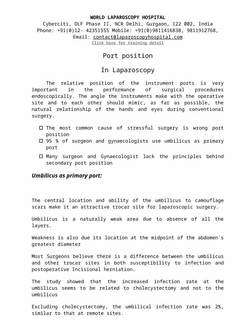
Base ball Diamond Concept of Port position
A satisfactory relationship includes:
• An angle of 60 degree between the two instruments tips
• Tangential approach to the site
• Appropriate working distance
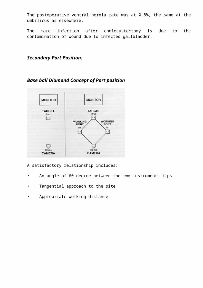
Target may be in supra-pubic region for LAVH, right iliac fossa for Appendicectomy, Right upper quadrant for Laparoscopic cholecystectomy or Left upper quadrant for Fundoplication.
First Decide the Target
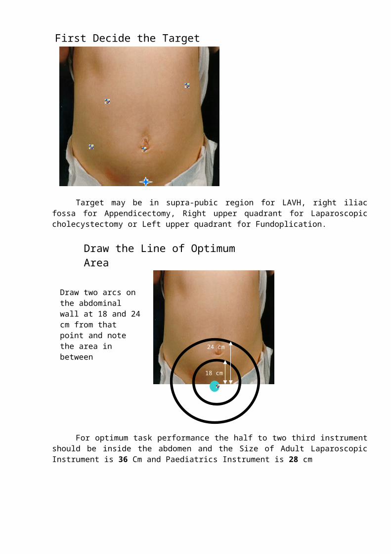
For optimum task performance the half to two third instrument should be inside the abdomen and the Size of Adult Laparoscopic Instrument is 36 Cm and Paediatrics Instrument is 28 cm
18 cm
24 cm
Draw the Line of Optimum Area
Draw two arcs on the abdominal wall at 18 and 24 cm from that point and note the area in between
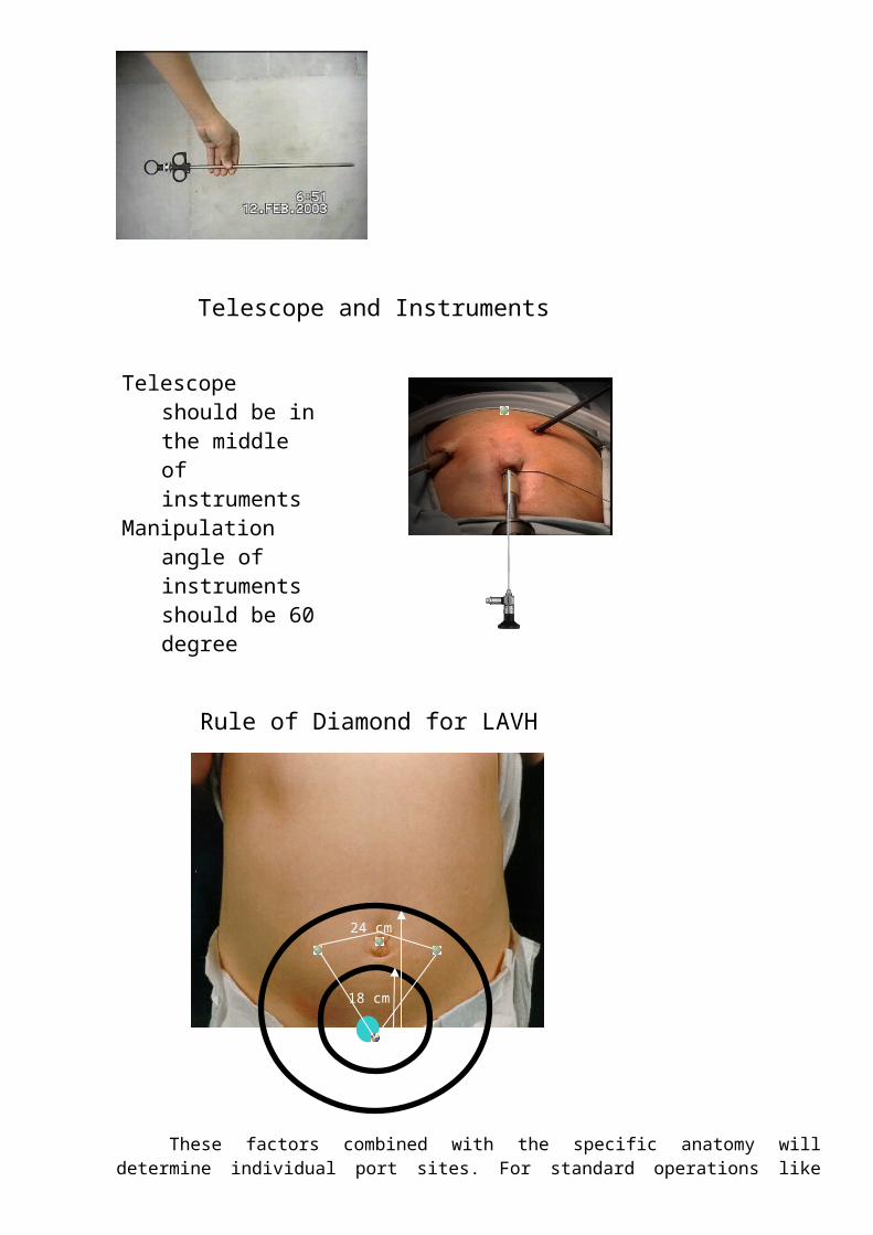
Telescope and Instruments
Telescope should be in the middle of instruments
Manipulation angle of instruments should be 60 degree
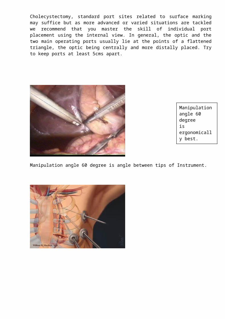
These factors combined with the specific anatomy will determine individual port sites. For standard operations like Cholecystectomy, standard port sites related to surface marking may suffice but as more advanced or varied situations are tackled we recommend that you master the skill of individual port placement using the internal view. In general, the optic and the two main operating ports usually lie at the points of a flattened triangle, the optic being centrally and more distally placed. Try to keep ports at least 5cms apart.
Manipulation angle 60 degree is angle between tips of Instrument.
Rule of Diamond for LAVH
18 cm
24 cm
Manipulation angle 60 degreeis ergonomically best.
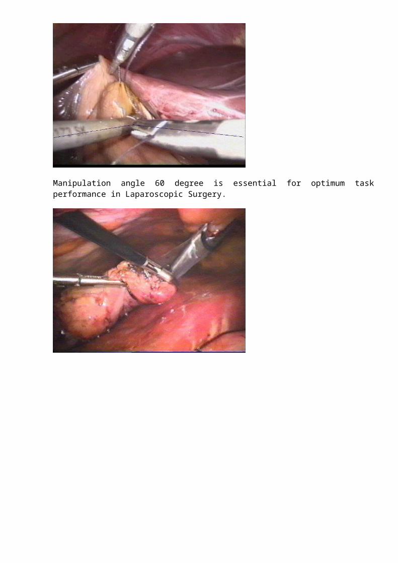
Manipulation angle 60 degree is essential for optimum task performance in Laparoscopic Surgery.
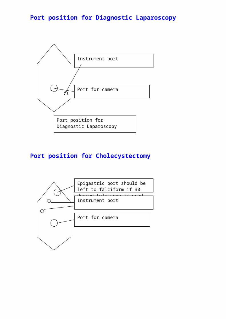
Port position for Diagnostic Laparoscopy
Port position for Cholecystectomy
Instrument port
Port for camera
Port position for Diagnostic Laparoscopy
Epigastric port should be left to falciform if 30 degree telescope is used
Port for camera
Instrument port
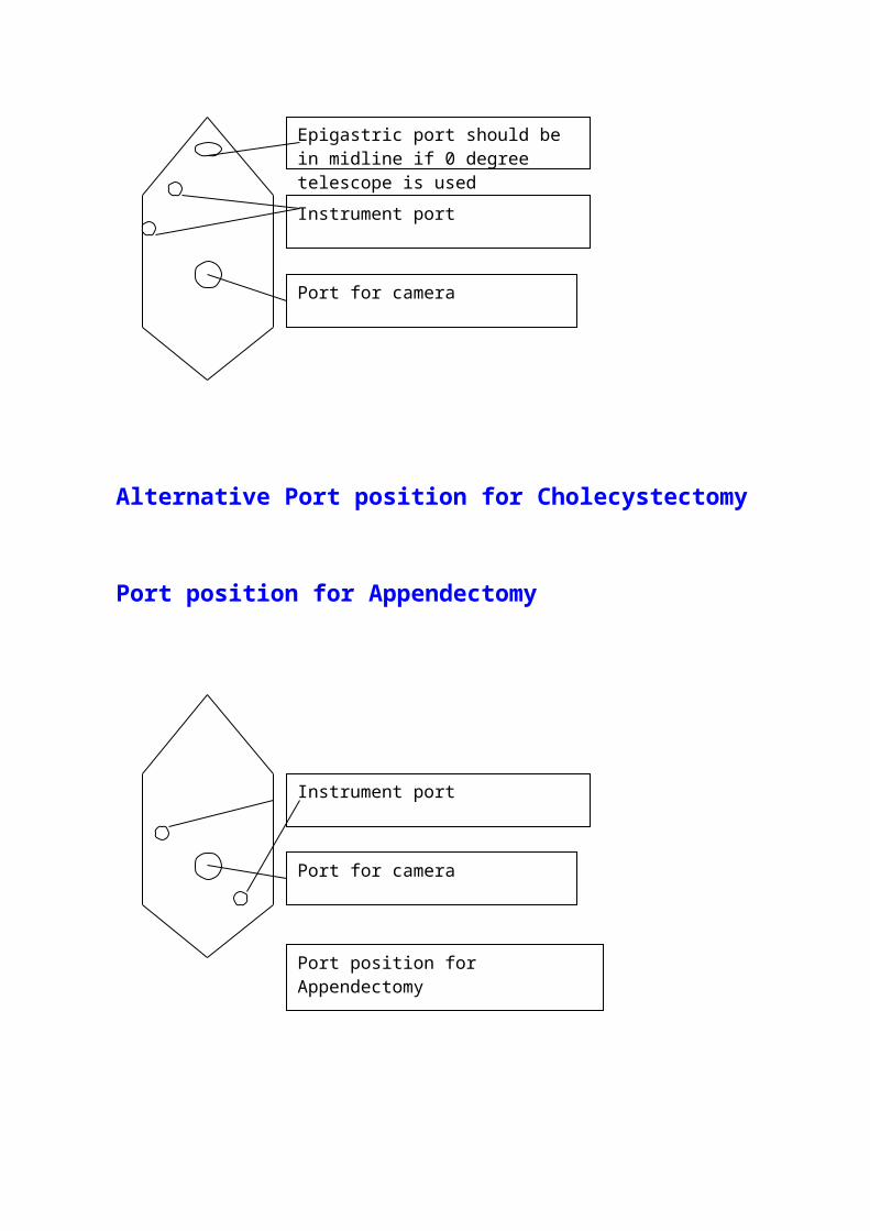
Alternative Port position for Cholecystectomy
Port position for Appendectomy
Epigastric port should be in midline if 0 degree telescope is used
Instrument port
Port for camera
Instrument port
Port for camera
Port position for Appendectomy
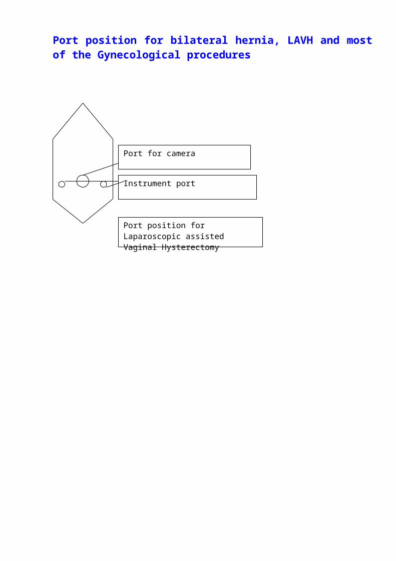
Port position for bilateral hernia, LAVH and most of the Gynecological procedures
Instrument port
Port for camera
Port position for Laparoscopic assisted Vaginal Hysterectomy
Problems due to incorrect port position:
Swording
Swording occurs when the telescope or the shaft of the assistant’s instrument obstruct the operator’s instruments. If this occurs you may need to consider:
Repositioning retracting instruments Rotation of an angled telescope allowing alteration of the position of the end of the
telescope
Withdrawal of the telescope
Transposition of the operator’s instruments
Additional port placement
changing the instruments to a different port
Complications of Access Technique:
Improper trocar insertion causes most of the operative complications of laparoscopic surgery. Examples are injury to the bowel, major vessels, bladder, inferior epigastric vessels, and subcutaneous emphysema. Other complications include thermal injury to the bowel, abdominal wall contusions, trocar-site herniation with possible bowel obstruction, and trocar-site tumor implants. Fortunately, the overall incidence of complications is relatively low (about 2%).
Visceral injuries
Hollow viscus
Stomach 0.02% Small bowel 2.7%
Large bowel 0.15
Bladder 0.5%
Solid organs
Liver Spleen
Vessel injury
Inferior Epigastric Omental
Mesenteric vessels
Aorta
Inferior Vena Cava
Other Complications
Gas embolism 1:10 000 to 1:60 000 but lethal
Pneumo-omentum,
Surgical emphysema,
Pneumo-mediastinum
Sudden collapse
The anesthetist will be checking for conditions such as drug reactions Pneumothorax, Gas embolism which may give rise to myocardial arrhythmias.
If Cardiac arrhythmia is found
Stop insufflation Withdraw instrument and remove Co2 by opening the valve but leave port in position.
Turn the patient to left
Correct hypoxia and resuscitate
Postpone surgery
If Severe hypotension Proceed to immediate laparotomy with all instruments left in situ.
Assume retroperitoneal bleeding to be the cause.
Mild to moderate hypotension
The surgeon should consider:
•Discontinuing gas insufflation immediately and reducing intra abdominal pressure to 8.0 mm Hg.
•Proceed immediately to 360° scan of the abdominal cavity for retroperitoneal bleeding.
If bleeding or expanding haematoma is seen, proceed immediately to long midline laparotomy and compression of the bleeding vessel. Aspirate blood, expose and control with vascular clamps. When necessary obtain assistance of a vascular surgeon.
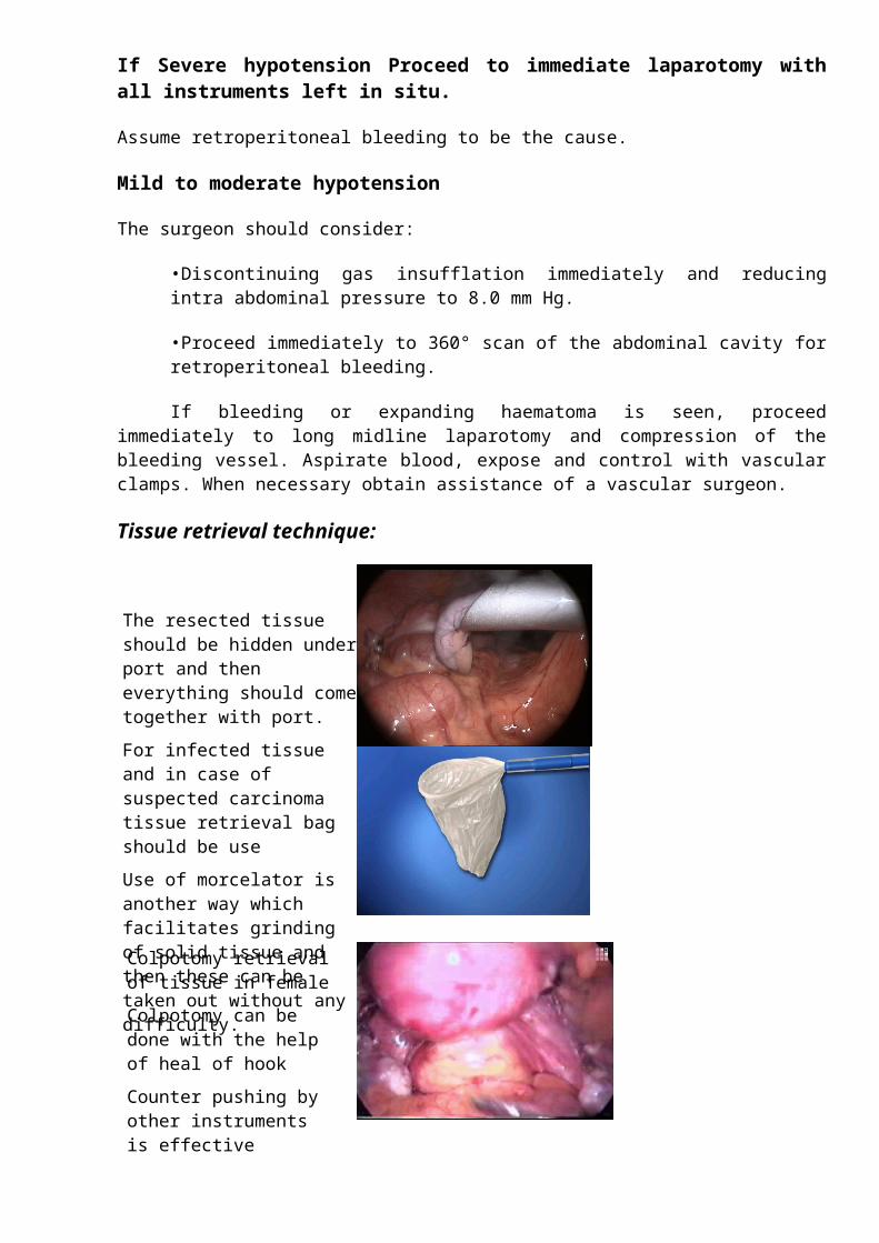
Tissue retrieval technique:
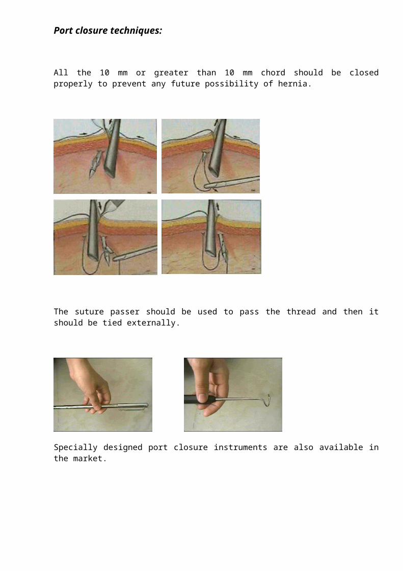
Port closure techniques:
All the 10 mm or greater than 10 mm chord should be closed properly to prevent any future possibility of hernia.
The resected tissue should be hidden under port and then everything should come together with port.
For infected tissue and in case of suspected carcinoma tissue retrieval bag should be use
Use of morcelator is another way which facilitates grinding of solid tissue and then these can be taken out without any difficulty.
Colpotomy retrieval of tissue in female
Colpotomy can be done with the help of heal of hook
Counter pushing by other instruments is effective
The suture passer should be used to pass the thread and then it should be tied externally.
Specially designed port closure instruments are also available in the market.
Precaution should be taken not taking port out suddenly without removing complete gas
If port is suddenly taken out the chance of port site hernia and adhesion is much higher. It is a good practice to insert some blunt instrument while removing the last port out to prevent entrapment of omentum or bowel content.
For More Information Contact:
Laparoscopy HospitalUnit of Shanti Hospital, 8/10 Tilak Nagar, New Delhi, 110018. India.Phone: +91(0)11- 25155202+91(0)9811416838, 9811912768
Email: [email protected]
Copyright © 2001 [Laparoscopyhospital.com]. All rights reserved.
Revised: .


