
Potential Neurological Emergencies Related to
COVID-19 InfectionARTHUR H.P. MAWUNTU
Bagian/KSM Neurologi FK Unsrat/RSUP Prof. dr. R.D. Kandou Manado

dr. Arthur H.P. Mawuntu, SpS(K)• TTL : Manado, 16 Januari 1980
• Pendidikan
• Dokter Umum FK Unsrat, Manado thn 2004
• PPDS1 Ilmu Penyakit Saraf FKUI, Jakarta thn 2011
• Fellow Neuroinfeksi, Neuroimunologi, dan Neuro-AIDS, FKUI Jakarta, thn 2014
• Brevet Konsultan Bidang Neuroinfeksi dari KNI thn 2018
• Pekerjaan
• Tenaga Pendidik di Bagian Neurologi FK Unsrat Manado 2006 – sekarang
• Staf KSM Neurologi RSUP Prof. dr. R.D. Kandou Manado 2005 – sekarang
• Jabatan
• Koord. Divisi Neurotraumatologi Bagian/KSM Neurologi FK Unsrat/RSUP Prof. Dr.
R.D. Kandou Manado 2011-15
• Koord. Divisi Neuroinfeksi, Neuroimunologi, dan Neuro-AIDS Bagian/KSM Neurologi
FK Unsrat/RSUP Prof. Dr. R.D. Kandou Manado 2015 – sekarang
• Koordinator Program Studi Program Pendidikan Dokter Spesialis Neurologi FK
Unsrat 2015-2019
• Plt. Koordinator Program Studi Program Pendidikan Dokter Spesialis Neurologi FK
Unsrat 2019 – sekarang
• Sekretaris dan Verifikator P2KB Perdossi Cabang Manado 2015 – sekarang
• Koordinator Subkomite Keselamatan Pasien Komite PMKP RSUP Prof. Dr. R.D.
Kandou Manado 2015-sekarang
CURRICULLUM VITAE
Bagian/KSM Neurologi FK Unsrat/RSUP Prof. dr. R.D. Kandou Manado

Bagian/KSM Neurologi FK Unsrat/RSUP Prof. dr. R.D. Kandou Manado
Current situation as access on 13 May 2020, 10.00 pm
https://covid19.go.id/peta-sebaranClick here for updates:
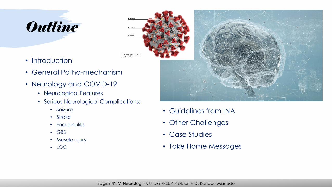
Outline
• Introduction
• General Patho-mechanism
• Neurology and COVID-19
• Neurological Features
• Serious Neurological Complications:
• Seizure
• Stroke
• Encephalitis
• GBS
• Muscle injury
• LOC
Bagian/KSM Neurologi FK Unsrat/RSUP Prof. dr. R.D. Kandou Manado
• Guidelines from INA
• Other Challenges
• Case Studies
• Take Home Messages

Introduction
• COVID-19 → a disease caused by a
new coronavirus (SARS-COV-2) that
mostly affects the lungs & airways
• Currently, nobody was immune
• Mode of transmission → droplet,
“aerosol”, etc.
• 80% patients → asymptomatic/mild
symptoms → could transmit the virus
• High risk: elderly, some comorbidities,
weakened immune system
Bagian/KSM Neurologi FK Unsrat/RSUP Prof. dr. R.D. Kandou Manado
• RNA virus, single-stranded
• The spike proteins :
• The main viral antigen
• Important role in virus attachment to the
cell membrane and viral entry (viral to viral
receptor interaction)
• A metallopeptidase, ACE2, acts as the
receptor for viral entry
https://encrypted-tbn0.gstatic.com/images?q=tbn%3AANd9GcQuCPOMnWnM1sQbVHVipG6jkiy0a5rEXYCmIzjaSWx5j2EPVRDX&usqp=CAU

30 Dec 19: 1-st case reported
7 Jan 20: The virus isolated
12 Jan 20: Genomic sequence released
21 Jan 20: Bats were reported as potential origin
21 Jan 20: Wuhan was
locked-down
30 Jan 20: Declared as pandemic
11 Feb 20: COVID-19 & SARS-CoV-2
terms were used
18 Feb 20: Ultrastructure of
human ACE2 reported
2 Mar 20: Indonesia
reported its first cases
14 Mar 20: North Sulawesi
reported its first case in Manado
19 Mar 20: WHO announced the
Solidarity Trial
1 May 20: PCR Lab for SARS-COV-2 was
operational in Manado
4 May 20: Eijkman reported the sequences from Indonesia
Bagian/KSM Neurologi FK Unsrat/RSUP Prof. dr. R.D. Kandou Manado

General Patho-mechanism
Bagian/KSM Neurologi FK Unsrat/RSUP Prof. dr. R.D. Kandou Manado
https://pbs.twimg.com/media/EW6_8siWAAAFsmn.jpg

Neurology & COVID-19
• Patients with particular neurological disease and/or receive certain treatments → at risk → MG, NMOSD, MS, Neuro-AIDS, myositis, muscular dystrophies, CNS vasculitis, CIDP, MNDs,
• Neurological features in COVID-19 infection
• Drugs interactions
• Direct infection to CNS or PNS → ??
• Serious neurological complication:
Bagian/KSM Neurologi FK Unsrat/RSUP Prof. dr. R.D. Kandou Manadoc
• Seizures
• Stroke
• Encephalitis
• GBS
• Muscle injury
• LOC
• Etc.
Sugiarto P, dkk. Panduan untuk pelayanan pasien dengan gangguan neurologis terkait infeksi COVID-19. Perdossi. Apri 2020

Bagian/KSM Neurologi FK Unsrat/RSUP Prof. dr. R.D. Kandou Manadoc

• Neurological symptoms were seen in 36.4% patients
• More common in patients with severe infection
Mao, et al. JAMA Neurol. April 2020. DOI: 10.1001/jamaneurol.2020.1127
Neurological features
Bagian/KSM Neurologi FK Unsrat/RSUP Prof. dr. R.D. Kandou Manado

Bagian/KSM Neurologi FK Unsrat/RSUP Prof. dr. R.D. Kandou Manado
*Hyposmia
*Hypogeusia
*Headache
*Dizziness
*Ataxia
*Visual problems
*Paresthesia-Hypesthesia
*Weakness
*Muscle pain
* Neuronal infiltration
* Mild-severe inflammation
* ↑ ICP
* Hematological complication
* ↓ brain perfusion
* Other mechanisms
*Not specific
* May present early in the disease course
*Always look for other manifestations
*Raise awareness for further COVID-19 work-ups during this pandemic
< cognition??
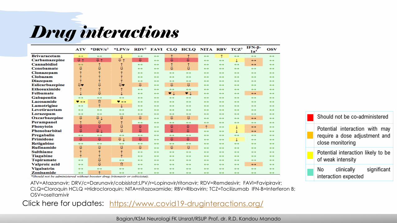
Should not be co-administered
Potential interaction with may
require a dose adjustment and
close monitoring
Potential interaction likely to be
of weak intensity
No clinically significant
interaction expectedATV=Atazanavir; DRV/c=Darunavir/cobisistat;LPV/r=Lopinavir/ritonavir; RDV=Remdesivir; FAVI=Favipiravir;
CLQ=Cloroquin HCLQ =Hidrocloroquin; NITA=nitazoxamide; RBV=Ribavirin; TCZ=Tocilizumab IFN-B=Interferon B;
OSV=oseltamivir
Bagian/KSM Neurologi FK Unsrat/RSUP Prof. dr. R.D. Kandou Manado
https://www.covid19-druginteractions.org/Click here for updates:
Drug interactions

Direct infection to CNS or PNS
• CNS direct infection → theoretically plausible:
• Previous reports in SARS & MERS
• ACE2 receptors are found in neuron
• Possible neural transmission from olfactory nerves or hematogenous transmission
• Autopsy of patients with COVID-19 → hyperemic & edematous brain tissue, degenerated neurons
• Animal model
• Up until now:
• Some reports about SARS-CoV-2 associated meningoencephalitis
• Mostly based on neurological deficits in confirmed COVID-19 cases + abnormal neuroimaging + non-specific abnormality of the CSF + no proof of other direct cause
• One case report → CSF (+)/NP swab (-)
• PNS direct infection → ??
Bagian/KSM Neurologi FK Unsrat/RSUP Prof. dr. R.D. Kandou Manado
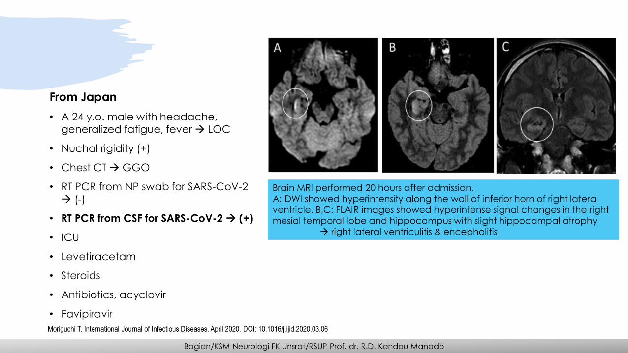
Brain MRI performed 20 hours after admission.
A: DWI showed hyperintensity along the wall of inferior horn of right lateral
ventricle. B,C: FLAIR images showed hyperintense signal changes in the right
mesial temporal lobe and hippocampus with slight hippocampal atrophy
→ right lateral ventriculitis & encephalitis
From Japan
• A 24 y.o. male with headache,
generalized fatigue, fever → LOC
• Nuchal rigidity (+)
• Chest CT → GGO
• RT PCR from NP swab for SARS-CoV-2
→ (-)
• RT PCR from CSF for SARS-CoV-2 → (+)
• ICU
• Levetiracetam
• Steroids
• Antibiotics, acyclovir
• Favipiravir
Moriguchi T. International Journal of Infectious Diseases. April 2020. DOI: 10.1016/j.ijid.2020.03.06
Bagian/KSM Neurologi FK Unsrat/RSUP Prof. dr. R.D. Kandou Manado

Serious Neurological Complications
• Examination are sometimes difficult to perform
• MRI and EEG → no specific findings
• Negative RT-PCR in the CSF
Neurological features of COVID-19 patients with ARDS
Bagian/KSM Neurologi FK Unsrat/RSUP Prof. dr. R.D. Kandou Manado
Helms, et al. NEJM. April 2020. DOI: 10.1056/NEJMc2008597

Stroke Seizure Encephalitis
GBS Muscle injury LOC
Bagian/KSM Neurologi FK Unsrat/RSUP Prof. dr. R.D. Kandou Manado

Stroke
Bagian/KSM Neurologi FK Unsrat/RSUP Prof. dr. R.D. Kandou Manado
• COVID-19 patients → high risk for
developing acute stroke especially if
multiple organ dysfunction presents
• A retrospective study from the COVID-19 outbreak in Wuhan→
stroke incidence in hospitalized
COVID-19 patients ≈ 5%
• Causes: Vasculitis or coagulopathy
& vascular endothelial dysfunction
• Treatment approach is similar but
with some considerations
• Coagulopathy & vascular
endothelial dysfunction
• Recent case series from New York
City reported 5 young patients with
COVID-19 & large-vessel stroke
• Large-vessel stroke was reported in
association with the 2004 SARS
outbreak in Singapore
Seizure
• Seizure is not considered a presenting
symptom of COVID-19
• During disease progression → release
of inflammatory cytokines → brain
damage → neuronal hyperexcitability
via glutamate receptor activation →
seizures
• Consider also other potential causes
(drug toxicity, metabolic changes,
direct brain infection, etc.)
• Seizure → poor outcome
• Risk of seizure ↑ in certain conditions →
especially a history of previous seizure
• AEDs selection: consider drug-drug interaction, side effects, and disease status
• Recognize subtle seizure or NCSE (exclude with EEG)
• Management:
• Treat seizure immediately
• Modification in seizure protocol →preferable to select AED with minimal interaction with other drugs (e.g. Levetiracetam)
• If unavailable → give classic AED with precaution
• Causative therapy
• Supportive therapy
Bagian/KSM Neurologi FK Unsrat/RSUP Prof. dr. R.D. Kandou Manado

Vira
l in
fec
tio
ns
Direct infection
Primary
Latent infection
Metabolic changes
Hypoglycemia
Sepsis
etc.Inflammation
Drug toxicity
Neuronal hyperexcitability via glutamate receptor
activation
Ac
ute
se
izu
re
Predisposing factors
Bagian/KSM Neurologi FK Unsrat/RSUP Prof. dr. R.D. Kandou Manado

Encephalitis
En
ce
ph
alit
is
Direct infection
Neural transmission
Hematogenous
Others
Inflammation
Overt inflammatory reaction due to the disease
Para-infectious syndrome
Toxic Drugs
Bagian/KSM Neurologi FK Unsrat/RSUP Prof. dr. R.D. Kandou Manado

From USA
• A female airline worker in her late fifties
presented with a 3-day history of cough,
fever, and altered mental status
• RT PCR from NP swab (+) for SARS-CoV-2
• RT PCR from CSF for HSV1, HSV2, VZV,
and WNV (-)
• Bacterial culture from CSF (-)
• RT PCR from CSF for SARS-CoV-2 → N/T
T2 FLAIR hyperintensity within the bilateral medial temporal lobes and
thalami (A, B, E, F) with evidence of hemorrhage indicated by hypointense
signal intensity on susceptibility-weighted images (C, G) and rim
enhancement on postcontrast images (D, H)
Poyiadji N, et al. Radiology. DOI: 10.1148/radiol.2020201187
Bagian/KSM Neurologi FK Unsrat/RSUP Prof. dr. R.D. Kandou Manado

GBS
• Para-infectious/post-infectious syndrome
• Autoimmune
• Mostly, targeting myelin and/or axons of the peripheral nerves
• Classic clinical findings: acute, symmetric, flaccid, ascending paralysis
• Requires CSF analysis & EMG for diagnosis
• >> Self-limiting disease; fatal outcomes in some patients
• Tx: IVIg, PE
• In COVID-19:• Already reported in case reports
• >> follow a para-infectious profile, instead of the classic postinfectious profile
• Could contribute to the respiratory problem
• Should be considered in patients with acute general weakness + areflexia, acute cranial nerve palsies
• Dysautonomia → ↑ mortality
Bagian/KSM Neurologi FK Unsrat/RSUP Prof. dr. R.D. Kandou Manado
Zhao, et al. The Lancet. April 2020. DOI: 10.1016/S1474-4422(20)30109-5
Muscle Injury
• Muscle injury: Skeletal muscle pain +
serum CK level >200U/L
• Coronavirus infections may be
associated with myopathies
• Myalgia or fatigue → 44%–70% of
hospitalized patients and ↑ CK→ 33%
of admitted patients
• Coronavirus may cause a viral myositis
• Very sick coronavirus patients → critical
illness myopathy or polyneuropathy
• Risk from treatment:
• Hydroxychloroquine and chloroquine
→ toxic neuropathy and myopathy
• Antiviral treatments
• Risk from vaccination
• Possible inflammatory neuropathy
Bagian/KSM Neurologi FK Unsrat/RSUP Prof. dr. R.D. Kandou Manado

Guidelines From INA
Bagian/KSM Neurologi FK Unsrat/RSUP Prof. dr. R.D. Kandou Manado

Other Challenges
• The non-COVID-19 patients
• Triage & screening in patients coming to the hospital
• Patients w/ chronic treatment
• Patients receiving immunosuppressive agents
• Telemedicine
• Feasibility
• Legal aspect
• CMEs
Bagian/KSM Neurologi FK Unsrat/RSUP Prof. dr. R.D. Kandou Manado
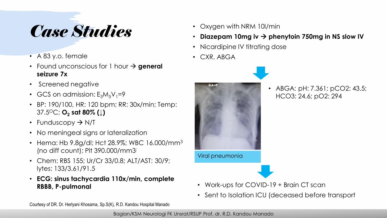
Case Studies• A 83 y.o. female
• Found unconscious for 1 hour → general
seizure 7x
• Screened negative
• GCS on admission: E3M5V1=9
• BP: 190/100, HR: 120 bpm; RR: 30x/min; Temp:
37.5OC; O2 sat 80% (↓)
• Funduscopy → N/T
• No meningeal signs or lateralization
• Hema: Hb 9.8g/dl; Hct 28.9%; WBC 16.000/mm3
(no diff count); Plt 390.000/mm3;
• Chem: RBS 155; Ur/Cr 33/0.8; ALT/AST: 30/9;
lytes: 133/3.61/91.5
• ECG: sinus tachycardia 110x/min, complete
RBBB, P-pulmonal
Bagian/KSM Neurologi FK Unsrat/RSUP Prof. dr. R.D. Kandou Manado
Courtesy of DR. Dr. Herlyani Khosama, Sp.S(K), R.D. Kandou Hospital Manado
• Oxygen with NRM 10l/min
• Diazepam 10mg iv → phenytoin 750mg in NS slow IV
• Nicardipine IV titrating dose
• CXR, ABGA
Viral pneumonia
• ABGA: pH: 7.361; pCO2: 43.5;
HCO3: 24.6; pO2: 294
• Work-ups for COVID-19 + Brain CT scan
• Sent to Isolation ICU (deceased before transport

Case Studies
Bagian/KSM Neurologi FK Unsrat/RSUP Prof. dr. R.D. Kandou Manado
• A 62 y.o. male with hypertension
• Headache + right-sided weakness for 4 days
• Decreased consciousness for 1 day → referred
• Screened negative on the first hospital
• GCS on admission: E2M4V1=7
• BP: 190/100, HR: 120 bpm; RR: 30x/min; Temp: 37.5OC; O2 sat 87% (↓)
• Rales +/+ (basal)
• Funduscopy → N/T
• No meningeal signs
• Right hemiparesis
• Hema: Hb 14.9g/dl; Hct 44.5%; WBC 18.000/mm3
(0/0/20/43/34/3); Plt 296.000/mm3;
• Chem: RBS 91; Ur/Cr 16/0.5; lytes: 137/2.76/100.4;
CT scan: not done
• Admitted to Isolation Ward
• No anti-thrombotics
• Mannitol
• Antibiotics
• Other treatments
Hemorrhagic stroke
based on clinical
score

Case Studies• A 37 y.o. female, no previously known VD
risk factor
• Sudden left-sided weakness for 18 hours +
swallowing difficulty + headache
• History of fever 1 month ago + cough → now
only cough + shortness of breath
• GCS on admission: 15
• BP: 110/70, HR: 80 bpm; RR: 24x/min; Temp:
36.5OC
• Swallowing test → not done
• Left hemiplegia with decreased tone
• Hema: Hb 13.6; Hct 37%; Plt 400.000 /mm3;
WBC 12.000/mm3 (5/1/3/56/28/7)
• Chem: Ur/Cr 16.6/0.72; lytes: 142/3.2/109
• Antiplatelets
• Statin
• High resolution Chest CT
Bilateral-posterior, mild subpleural
ground glass opacities & parenchymal
bands noted; Possible Covid-19, susp.
absorption stage, leaving/residual GGO
& fibrous – parenchymal bands
• Admitted to Isolation Ward
• Antibiotic
• Pharyngeal swabCourtesy of Dr. Mohammad Kurniawan, Sp.S(K), M.Sc(stroke med.), Cipto Mangunkusumo Hospital Jakarta
Bagian/KSM Neurologi FK Unsrat/RSUP Prof. dr. R.D. Kandou Manado
No bleeding

Case Studies
• A 23 y.o. ♂ with upper & lower facial weakness, became bilateral & complete within 2 days, mastoid
pain, loss of taste, and lower limb paresthesia.
• 10 days ago → fever & sore throat for 3 days, treated with AB for 5 days.
• Neuro exam: complete facial palsy, generalized areflexia, sensory ataxia.
• Brain MRI: focal contrast enhancement at the internal acoustic meatus.
• EMG (12th day after admission): axonal sensory-motor damage involving the lower limbs, with sural nerve
sparing, + ↓ facial nerve CMAP amplitude.
• Therapy: IVIg → mild improvement of the facial weakness + disappearance of limb paresthesia.
• Normal thorax imaging → pharyngeal swab (+) for SARS-CoV-2.
Bagian/KSM Neurologi FK Unsrat/RSUP Prof. dr. R.D. Kandou Manado
Toscano G, Palmerini F, Ravaglia S, et al. Guillain–Barré syndrome associated with SARS-CoV-2. N Engl J Med. DOI: 10.1056/NEJMc2009191

Bagian/KSM Neurologi FK Unsrat/RSUP Prof. dr. R.D. Kandou Manado
Take Home Messages• COVID-19 → potential serious neurological
complications
• COVID-19 patients: high-risk for stroke →
protected stroke code
• Seizures: poor prognosis → consider drug-drug
interaction & careful monitoring for treatment
• GBS → anticipate after the acute phase but
not too long
• CNS infection: needs further research but
based on known mechanism & previous CoVoutbreaks → plausible
• Anticipate other problems like the non-
COVID-19 patients who are also need our
help
LP equipment during COVID-
19 pandemic(consent has been granted from
the patient)


