Predicting Errors in Offset Templating for Total Hip ArthroplastyBy:
Del Schutte, M.D.,1,2 William R. Barfield Ph.D.1, Jeffrey Conrad, M.D.1, Neil Romero, M.D.1, Timothy McTighe, Dr. H.S. (hc)2 & Ed McPherson, M.D.2,3
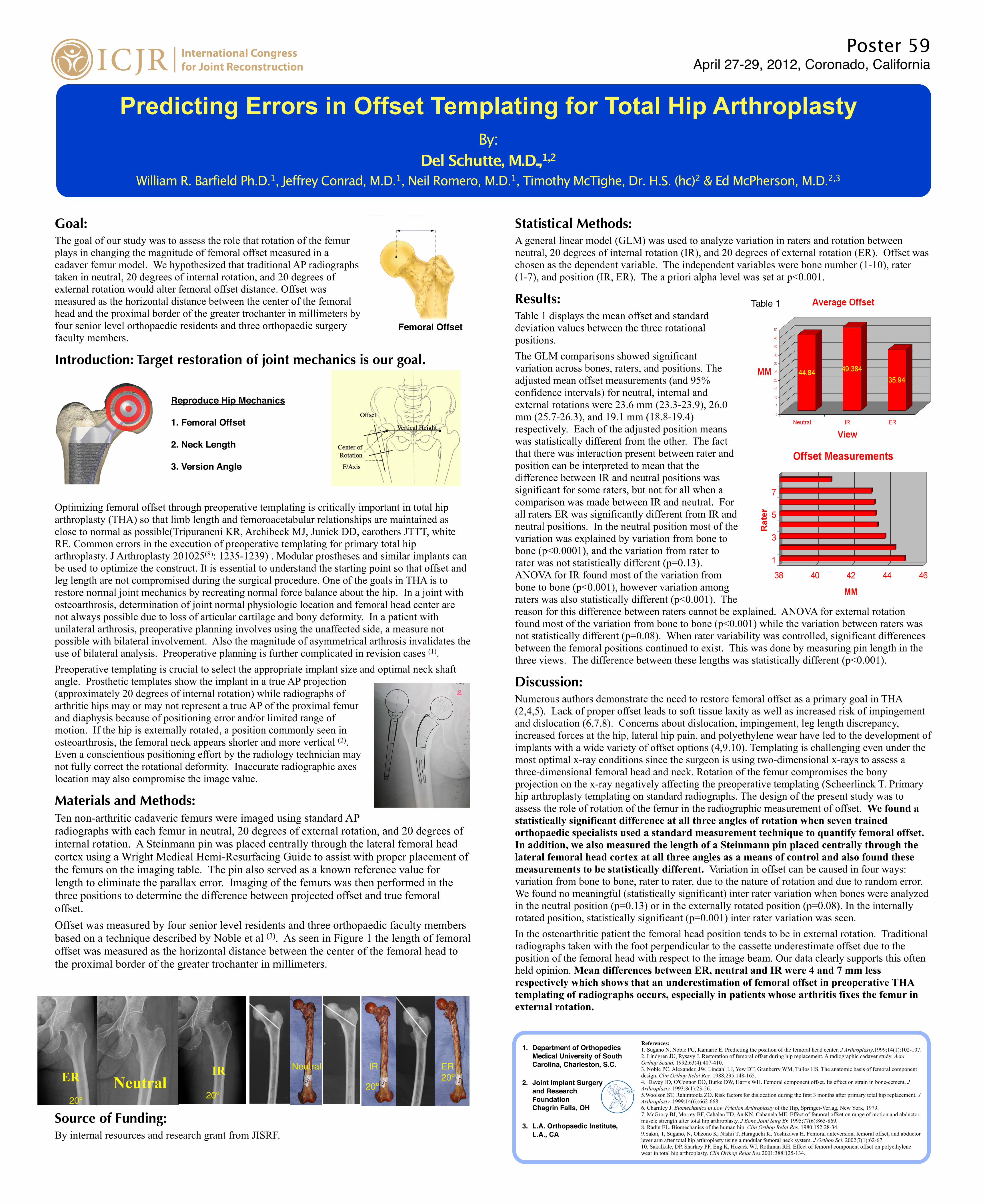
Goal: The goal of our study was to assess the role that rotation of the femur plays in changing the magnitude of femoral offset measured in a cadaver femur model. We hypothesized that traditional AP radiographs taken in neutral, 20 degrees of internal rotation, and 20 degrees of external rotation would alter femoral offset distance. Offset was measured as the horizontal distance between the center of the femoral head and the proximal border of the greater trochanter in millimeters by four senior level orthopaedic residents and three orthopaedic surgery faculty members.
Introduction: Target restoration of joint mechanics is our goal.
Optimizing femoral offset through preoperative templating is critically important in total hip arthroplasty (THA) so that limb length and femoroacetabular relationships are maintained as close to normal as possible(Tripuraneni KR, Archibeck MJ, Junick DD, carothers JTTT, white RE. Common errors in the execution of preoperative templating for primary total hip arthroplasty. J Arthroplasty 201025(8): 1235-1239) . Modular prostheses and similar implants can be used to optimize the construct. It is essential to understand the starting point so that offset and leg length are not compromised during the surgical procedure. One of the goals in THA is to restore normal joint mechanics by recreating normal force balance about the hip. In a joint with osteoarthrosis, determination of joint normal physiologic location and femoral head center are not always possible due to loss of articular cartilage and bony deformity. In a patient with unilateral arthrosis, preoperative planning involves using the unaffected side, a measure not possible with bilateral involvement. Also the magnitude of asymmetrical arthrosis invalidates the use of bilateral analysis. Preoperative planning is further complicated in revision cases (1).Preoperative templating is crucial to select the appropriate implant size and optimal neck shaft angle. Prosthetic templates show the implant in a true AP projection (approximately 20 degrees of internal rotation) while radiographs of arthritic hips may or may not represent a true AP of the proximal femur and diaphysis because of positioning error and/or limited range of motion. If the hip is externally rotated, a position commonly seen in osteoarthrosis, the femoral neck appears shorter and more vertical (2). Even a conscientious positioning effort by the radiology technician may not fully correct the rotational deformity. Inaccurate radiographic axes location may also compromise the image value.
Materials and Methods: Ten non-arthritic cadaveric femurs were imaged using standard AP radiographs with each femur in neutral, 20 degrees of external rotation, and 20 degrees of internal rotation. A Steinmann pin was placed centrally through the lateral femoral head cortex using a Wright Medical Hemi-Resurfacing Guide to assist with proper placement of the femurs on the imaging table. The pin also served as a known reference value for length to eliminate the parallax error. Imaging of the femurs was then performed in the three positions to determine the difference between projected offset and true femoral offset.Offset was measured by four senior level residents and three orthopaedic faculty members based on a technique described by Noble et al (3). As seen in Figure 1 the length of femoral offset was measured as the horizontal distance between the center of the femoral head to the proximal border of the greater trochanter in millimeters.
Source of Funding:By internal resources and research grant from JISRF.
Statistical Methods:A general linear model (GLM) was used to analyze variation in raters and rotation between neutral, 20 degrees of internal rotation (IR), and 20 degrees of external rotation (ER). Offset was chosen as the dependent variable. The independent variables were bone number (1-10), rater (1-7), and position (IR, ER). The a priori alpha level was set at p<0.001.
Results:Table 1 displays the mean offset and standard deviation values between the three rotational positions.The GLM comparisons showed significant variation across bones, raters, and positions. The adjusted mean offset measurements (and 95% confidence intervals) for neutral, internal and external rotations were 23.6 mm (23.3-23.9), 26.0 mm (25.7-26.3), and 19.1 mm (18.8-19.4) respectively. Each of the adjusted position means was statistically different from the other. The fact that there was interaction present between rater and position can be interpreted to mean that the difference between IR and neutral positions was significant for some raters, but not for all when a comparison was made between IR and neutral. For all raters ER was significantly different from IR and neutral positions. In the neutral position most of the variation was explained by variation from bone to bone (p<0.0001), and the variation from rater to rater was not statistically different (p=0.13). ANOVA for IR found most of the variation from bone to bone (p<0.001), however variation among raters was also statistically different (p<0.001). The reason for this difference between raters cannot be explained. ANOVA for external rotation found most of the variation from bone to bone (p<0.001) while the variation between raters was not statistically different (p=0.08). When rater variability was controlled, significant differences between the femoral positions continued to exist. This was done by measuring pin length in the three views. The difference between these lengths was statistically different (p<0.001).
Discussion:Numerous authors demonstrate the need to restore femoral offset as a primary goal in THA (2,4,5). Lack of proper offset leads to soft tissue laxity as well as increased risk of impingement and dislocation (6,7,8). Concerns about dislocation, impingement, leg length discrepancy, increased forces at the hip, lateral hip pain, and polyethylene wear have led to the development of implants with a wide variety of offset options (4,9.10). Templating is challenging even under the most optimal x-ray conditions since the surgeon is using two-dimensional x-rays to assess a three-dimensional femoral head and neck. Rotation of the femur compromises the bony projection on the x-ray negatively affecting the preoperative templating (Scheerlinck T. Primary hip arthroplasty templating on standard radiographs. The design of the present study was to assess the role of rotation of the femur in the radiographic measurement of offset. We found a statistically significant difference at all three angles of rotation when seven trained orthopaedic specialists used a standard measurement technique to quantify femoral offset. In addition, we also measured the length of a Steinmann pin placed centrally through the lateral femoral head cortex at all three angles as a means of control and also found these measurements to be statistically different. Variation in offset can be caused in four ways: variation from bone to bone, rater to rater, due to the nature of rotation and due to random error. We found no meaningful (statistically significant) inter rater variation when bones were analyzed in the neutral position (p=0.13) or in the externally rotated position (p=0.08). In the internally rotated position, statistically significant (p=0.001) inter rater variation was seen.In the osteoarthritic patient the femoral head position tends to be in external rotation. Traditional radiographs taken with the foot perpendicular to the cassette underestimate offset due to the position of the femoral head with respect to the image beam. Our data clearly supports this often held opinion. Mean differences between ER, neutral and IR were 4 and 7 mm less respectively which shows that an underestimation of femoral offset in preoperative THA templating of radiographs occurs, especially in patients whose arthritis fixes the femur in external rotation.
Poster 59April 27-29, 2012, Coronado, California
Femoral Offset
Reproduce Hip Mechanics
1. Femoral Offset
2. Neck Length
3. Version Angle
ER Neutral IR
20º20º20º
IR ER20º
Neutral
Table 1
References:1. Sugano N, Noble PC, Kamaric E. Predicting the position of the femoral head center. J Arthroplasty.1999;14(1):102-107.2. Lindgren JU, Rysavy J. Restoration of femoral offset during hip replacement. A radiographic cadaver study. Acta Orthop Scand. 1992;63(4):407-410.3. Noble PC, Alexander, JW, Lindahl LJ, Yew DT, Granberry WM, Tullos HS. The anatomic basis of femoral component design. Clin Orthop Relat Res. 1988;235:148-165.4. Davey JD, O'Connor DO, Burke DW, Harris WH. Femoral component offset. Its effect on strain in bone-cement. J Arthroplasty. 1993;8(1):23-26.5.Woolson ST, Rahimtoola ZO. Risk factors for dislocation during the first 3 months after primary total hip replacement. J Arthroplasty. 1999;14(6):662-668.6. Charnley J. Biomechanics in Low Friction Arthroplasty of the Hip, Springer-Verlag, New York, 1979.7. McGrory BJ, Morrey BF, Cahalan TD, An KN, Cabanela ME. Effect of femoral offset on range of motion and abductor muscle strength after total hip arthroplasty. J Bone Joint Surg Br. 1995;77(6):865-869.8. Radin EL. Biomechanics of the human hip. Clin Orthop Relat Res. 1980;152:28-34.9.Sakai, T, Sugano, N, Ohzono K, Nishii T, Haraguchi K, Yoshikawa H. Femoral anteversion, femoral offset, and abductor lever arm after total hip arthroplasty using a modular femoral neck system. J Orthop Sci. 2002;7(1):62-67.10. Sakalkale, DP, Sharkey PF, Eng K, Hozack WJ, Rothman RH. Effect of femoral component offset on polyethylene wear in total hip arthroplasty. Clin Orthop Relat Res.2001;388:125-134.
1. Department of Orthopedics Medical University of South Carolina, Charleston, S.C.
2. Joint Implant Surgery and Research Foundation Chagrin Falls, OH
3. L.A. Orthopaedic Institute, L.A., CA

![[T3CON12CA] TYPO3 Phoenix Templating Workshop](https://cdn.vdocuments.net/doc/165x107/5550fda4b4c90572478b4be3/t3con12ca-typo3-phoenix-templating-workshop.jpg)


