Protein Function
C483 Spring 2013
Function
• Transport (binding)• Structure• Motor• Catalysis (binding)• Immunity (binding)• Regulation (binding)• Signaling (binding)
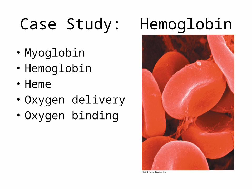
Case Study: Hemoglobin
• Myoglobin• Hemoglobin• Heme• Oxygen delivery• Oxygen binding
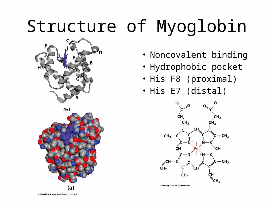
Structure of Myoglobin
• Noncovalent binding• Hydrophobic pocket• His F8 (proximal) • His E7 (distal)
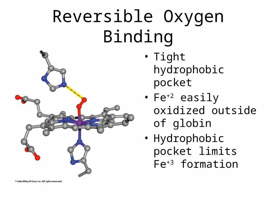
Reversible Oxygen Binding
• Tight hydrophobic pocket
• Fe+2 easily oxidized outside of globin
• Hydrophobic pocket limits Fe+3 formation
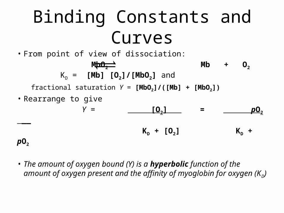
Binding Constants and Curves• From point of view of dissociation: MbO2 Mb + O2 KD = [Mb] [O2]/[MbO2] and
fractional saturation Y = [MbO2]/([Mb] + [MbO2])
• Rearrange to give Y = [O2] = pO2 __
KD + [O2] KD + pO2
• The amount of oxygen bound (Y) is a hyperbolic function of the amount of oxygen present and the affinity of myoglobin for oxygen (KD)
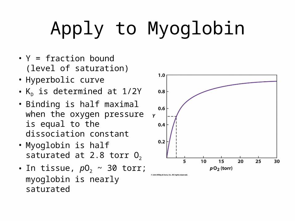
Apply to Myoglobin
• Y = fraction bound (level of saturation)
• Hyperbolic curve• KD is determined at 1/2Y• Binding is half maximal when
the oxygen pressure is equal to the dissociation constant
• Myoglobin is half saturated at 2.8 torr O2
• In tissue, pO2 ~ 30 torr; myoglobin is nearly saturated
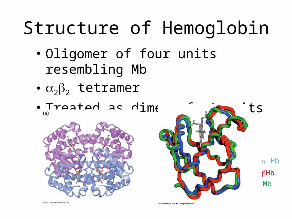
Structure of Hemoglobin• Oligomer of four units resembling Mb• a2b2 tetramer• Treated as dimer of ab units
bHb
a Hb
Mb
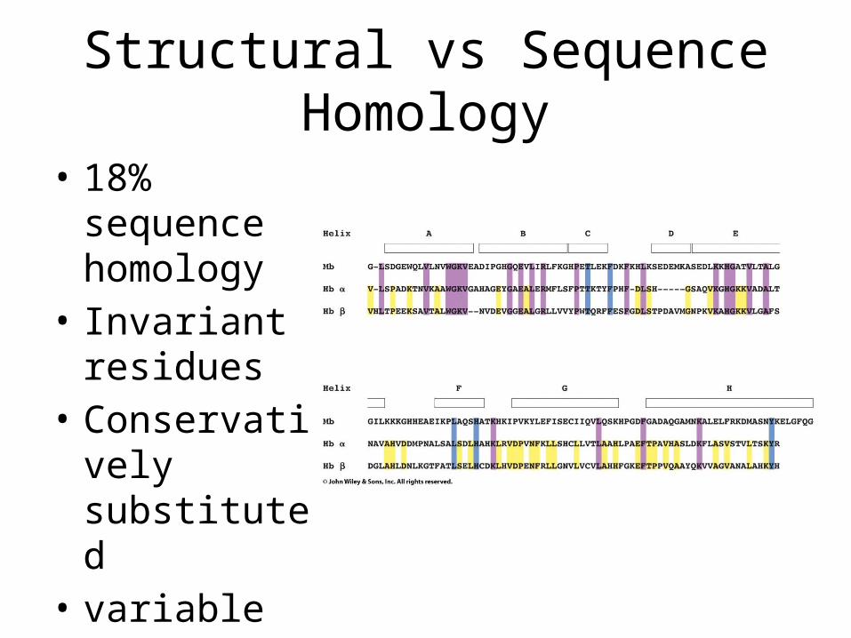
Structural vs Sequence Homology
• 18% sequence homology
• Invariant residues
• Conservatively substituted
• variable
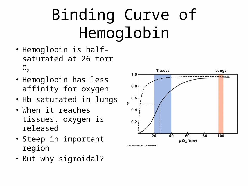
Binding Curve of Hemoglobin
• Hemoglobin is half-saturated at 26 torr O2
• Hemoglobin has less affinity for oxygen
• Hb saturated in lungs• When it reaches tissues,
oxygen is released• Steep in important
region• But why sigmoidal?
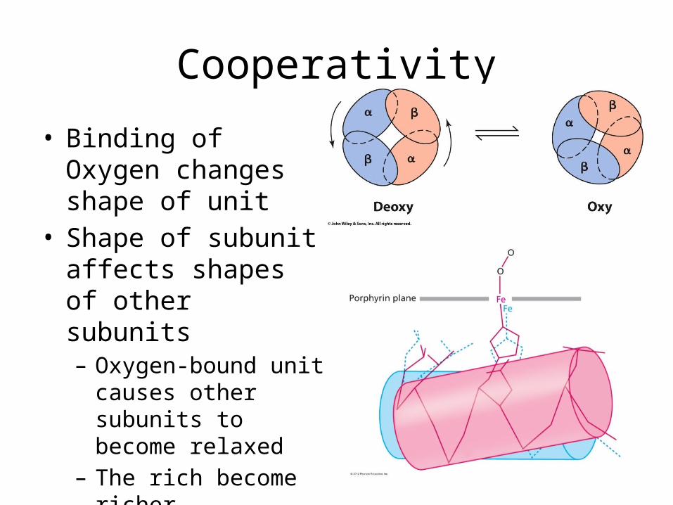
Cooperativity
• Binding of Oxygen changes shape of unit
• Shape of subunit affects shapes of other subunits– Oxygen-bound unit
causes other subunits to become relaxed
– The rich become richer– Cooperative binding
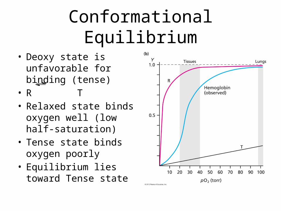
Conformational Equilibrium
• Deoxy state is unfavorable for binding (tense)
• R T• Relaxed state binds
oxygen well (low half-saturation)
• Tense state binds oxygen poorly
• Equilibrium lies toward Tense state
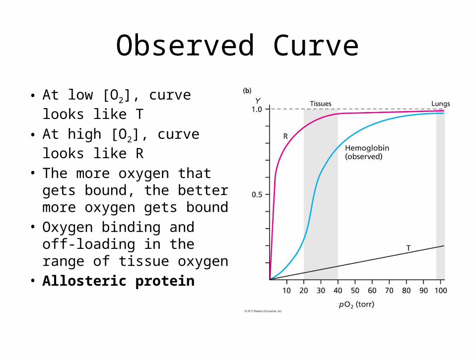
Observed Curve
• At low [O2], curve looks like T
• At high [O2], curve looks like R
• The more oxygen that gets bound, the better more oxygen gets bound
• Oxygen binding and off-loading in the range of tissue oxygen
• Allosteric protein
Why is Tight Favored?
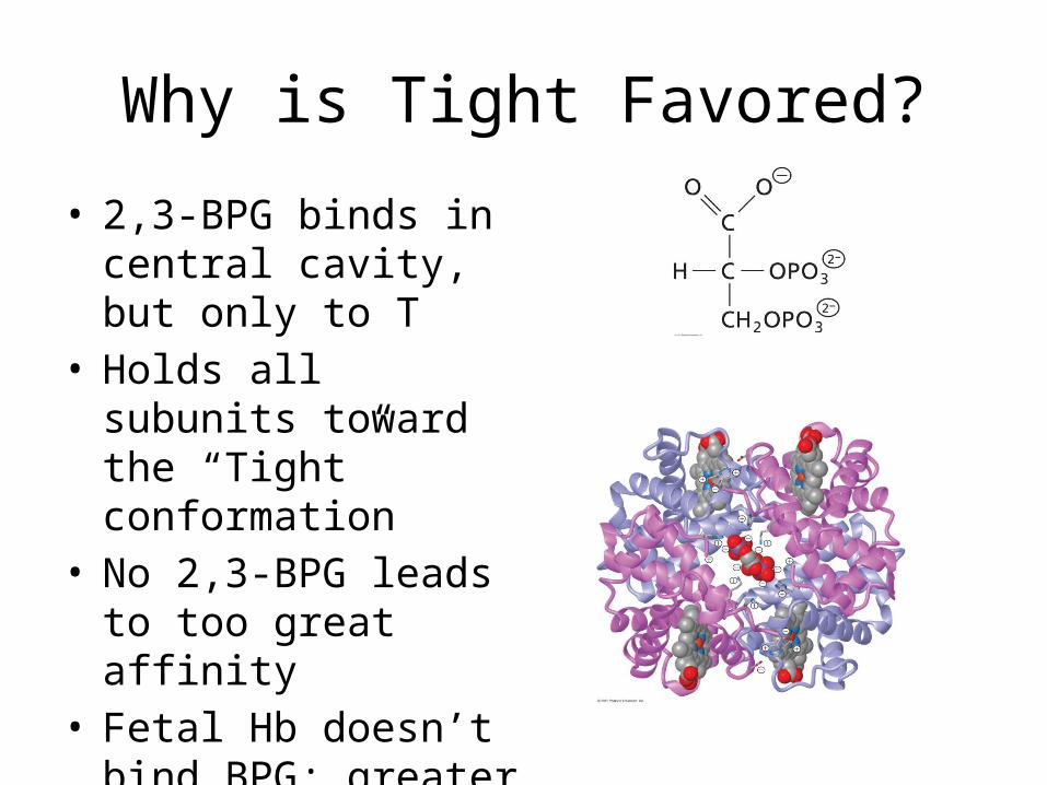
• 2,3-BPG binds in central cavity, but only to T
• Holds all subunits toward the “Tight” conformation
• No 2,3-BPG leads to too great affinity
• Fetal Hb doesn’t bind BPG; greater oxygen affinity
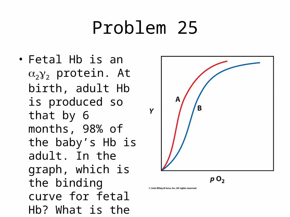
Problem 25
• Fetal Hb is an a2g2 protein. At birth, adult Hb is produced so that by 6 months, 98% of the baby’s Hb is adult. In the graph, which is the binding curve for fetal Hb? What is the physiological purpose?
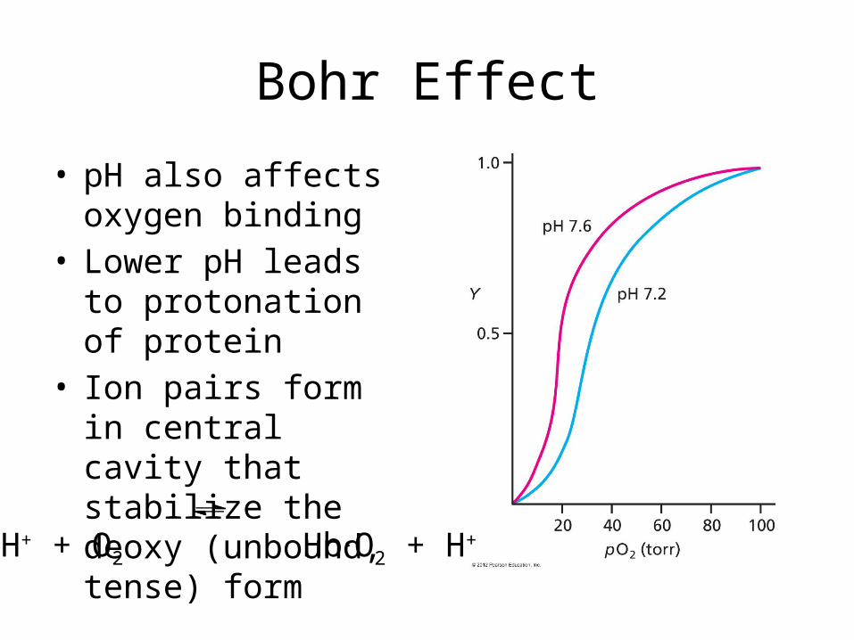
Bohr Effect
• pH also affects oxygen binding
• Lower pH leads to protonation of protein
• Ion pairs form in central cavity that stabilize the deoxy (unbound, tense) form
Hb.H+ + O2 Hb.O2 + H+
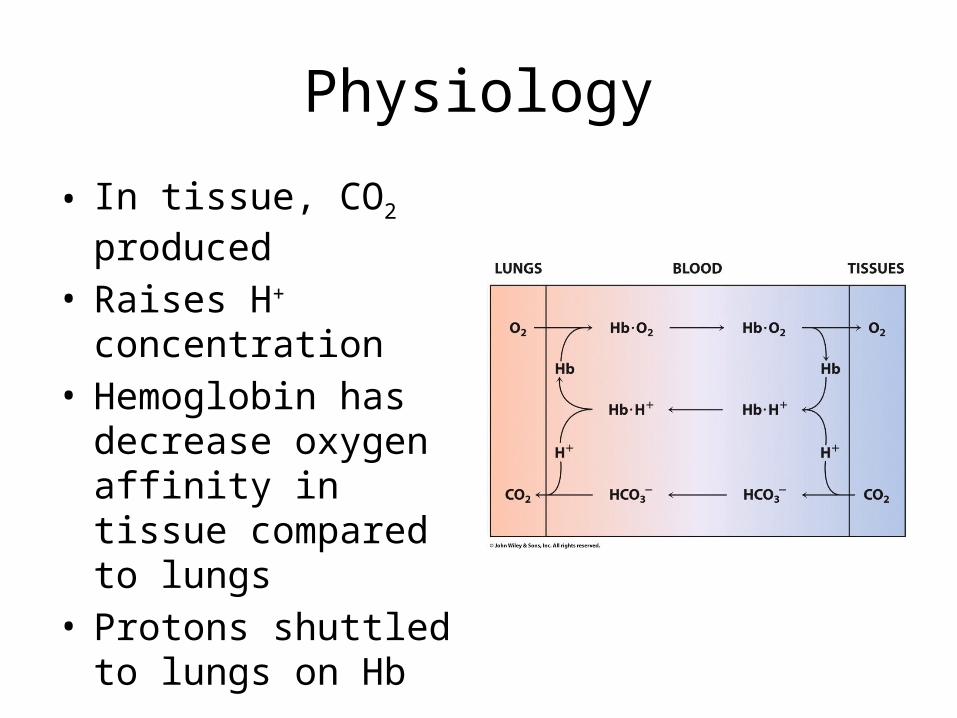
Physiology
• In tissue, CO2 produced• Raises H+ concentration• Hemoglobin has
decrease oxygen affinity in tissue compared to lungs
• Protons shuttled to lungs on Hb
Problem 31
• Propose a few explanations of how a KN mutation of a residue in the central cavity could lead to a mutant Hb with greater oxygen binding affinity.
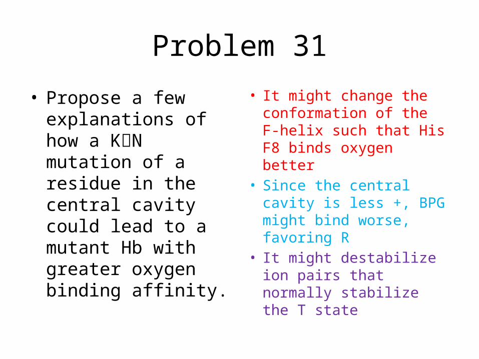
Problem 31
• Propose a few explanations of how a KN mutation of a residue in the central cavity could lead to a mutant Hb with greater oxygen binding affinity.
• It might change the conformation of the F-helix such that His F8 binds oxygen better
• Since the central cavity is less +, BPG might bind worse, favoring R
• It might destabilize ion pairs that normally stabilize the T state
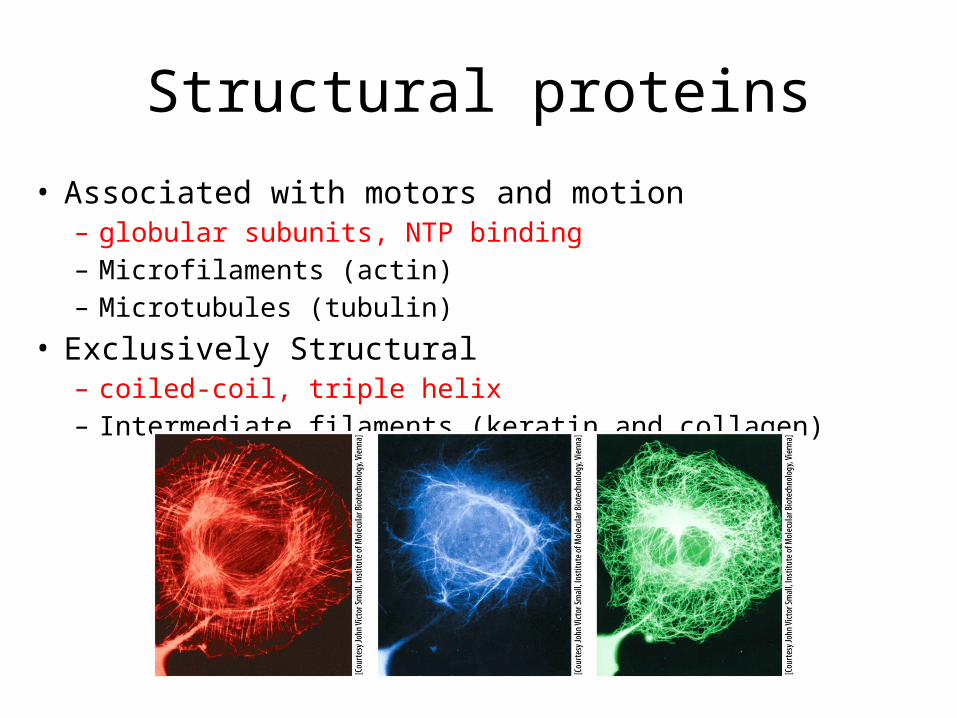
Structural proteins
• Associated with motors and motion– globular subunits, NTP binding– Microfilaments (actin)– Microtubules (tubulin)
• Exclusively Structural– coiled-coil, triple helix– Intermediate filaments (keratin and collagen)
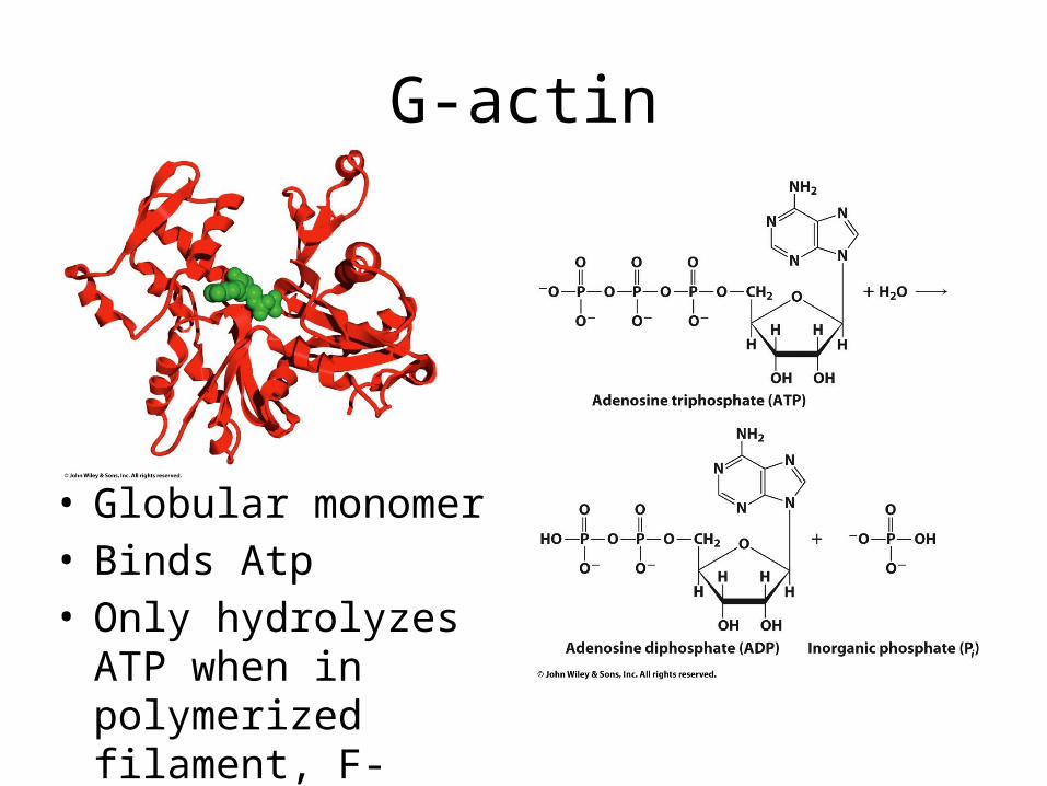
G-actin
• Globular monomer• Binds Atp• Only hydrolyzes ATP
when in polymerized filament, F-actin
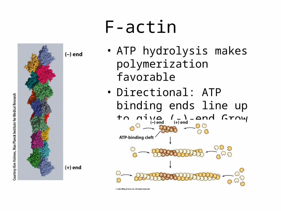
F-actin• ATP hydrolysis makes
polymerization favorable• Directional: ATP binding ends
line up to give (-)-end Grow faster at (+)-end
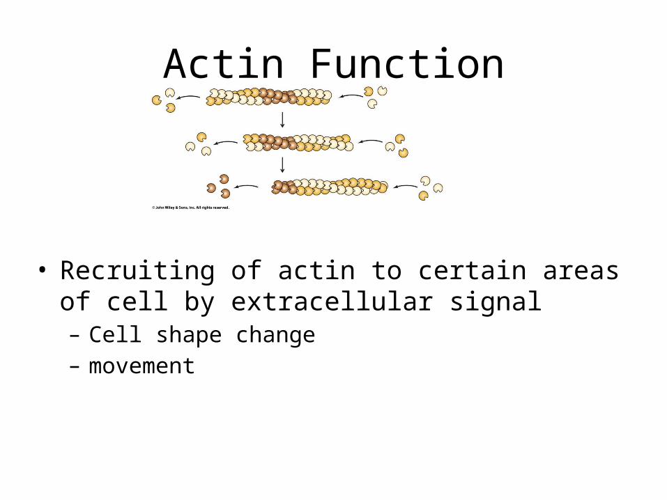
Actin Function
• Recruiting of actin to certain areas of cell by extracellular signal– Cell shape change– movement
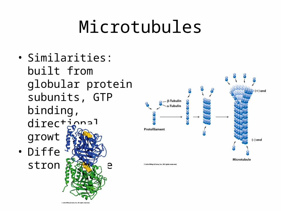
Microtubules
• Similarities: built from globular protein subunits, GTP binding, directional growth
• Differences: stronger tube
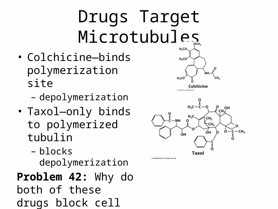
Drugs Target Microtubules
• Colchicine—binds polymerization site– depolymerization
• Taxol—only binds to polymerized tubulin– blocks depolymerization
Problem 42: Why do both of these drugs block cell division?
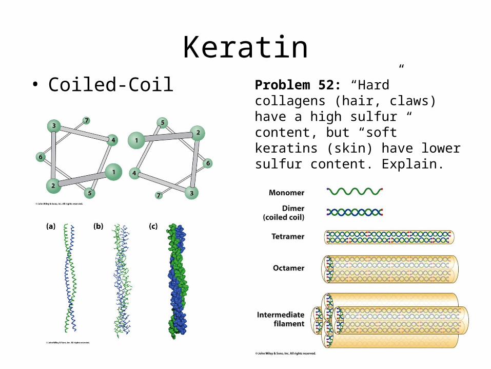
Keratin• Coiled-Coil Problem 52: “Hard” collagens
(hair, claws) have a high sulfur content, but “soft” keratins (skin) have lower sulfur content. Explain.
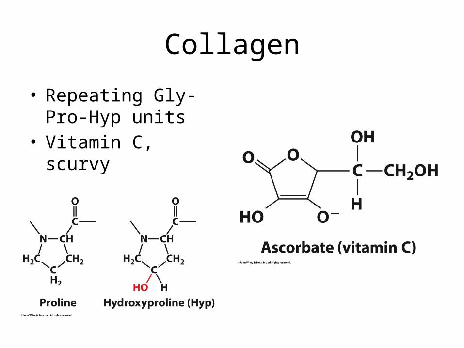
Collagen
• Repeating Gly-Pro-Hyp units
• Vitamin C, scurvy
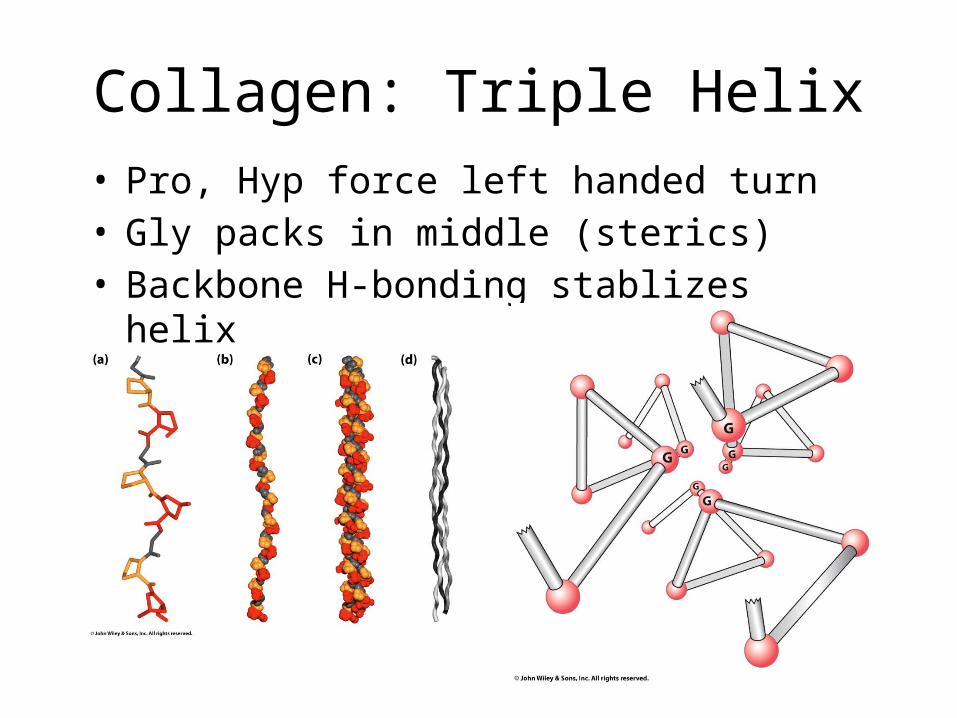
Collagen: Triple Helix• Pro, Hyp force left handed turn• Gly packs in middle (sterics)• Backbone H-bonding stablizes helix
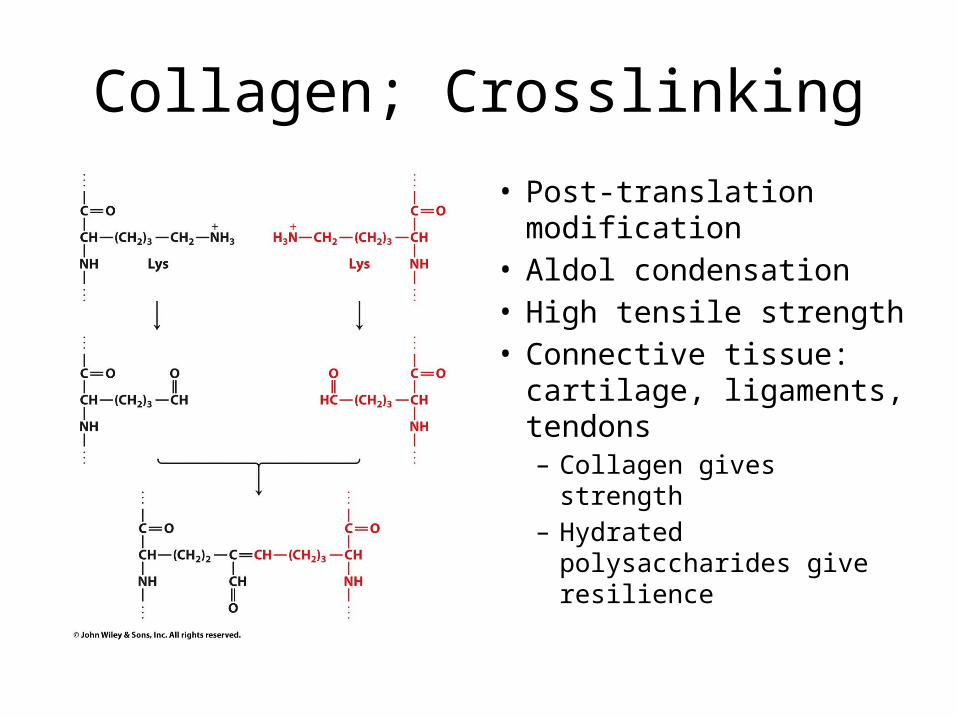
Collagen; Crosslinking
• Post-translation modification
• Aldol condensation• High tensile strength• Connective tissue:
cartilage, ligaments, tendons– Collagen gives strength– Hydrated polysaccharides
give resilience
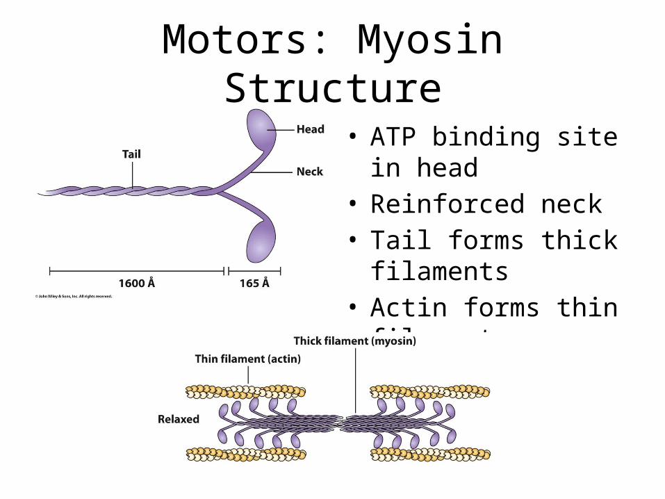
Motors: Myosin Structure
• ATP binding site in head• Reinforced neck• Tail forms thick
filaments• Actin forms thin
filament
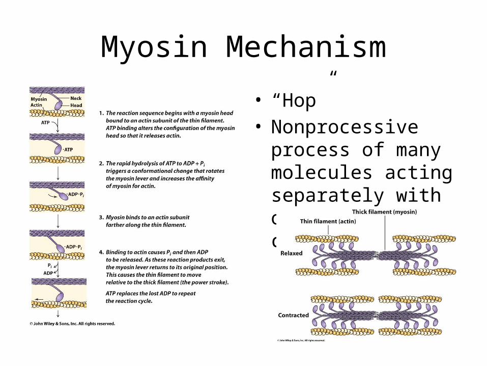
Myosin Mechanism
• “Hop”• Nonprocessive process
of many molecules acting separately with overall contraction
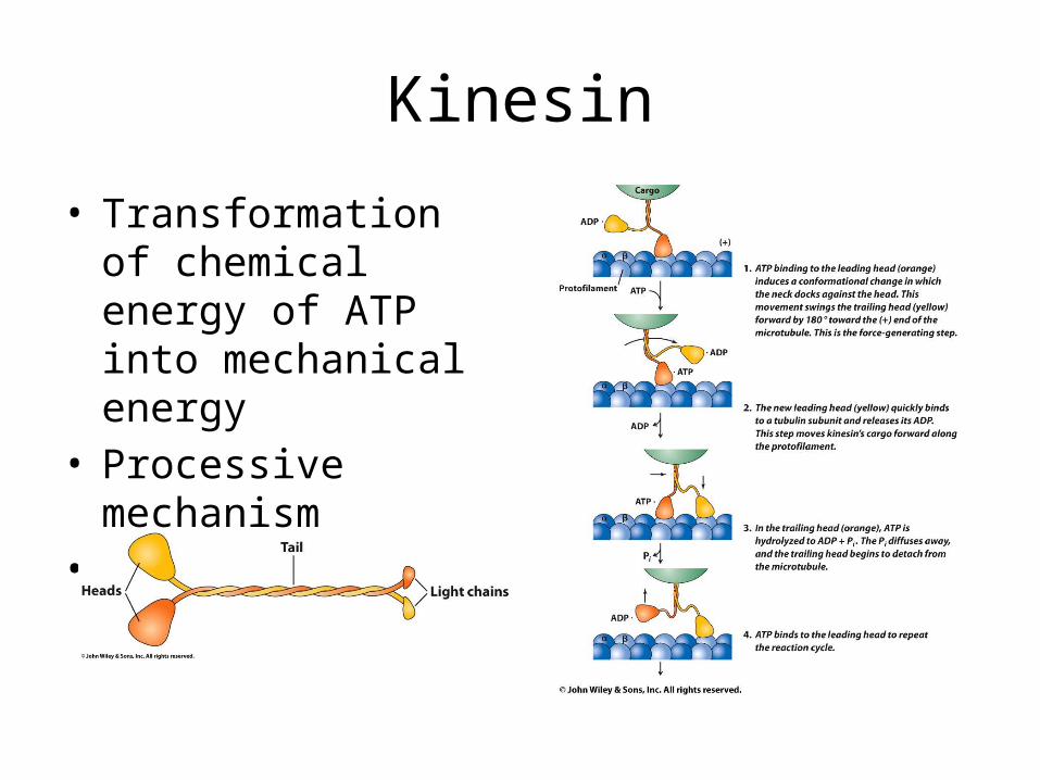
Kinesin
• Transformation of chemical energy of ATP into mechanical energy
• Processive mechanism• Allows carrying of
vesicles
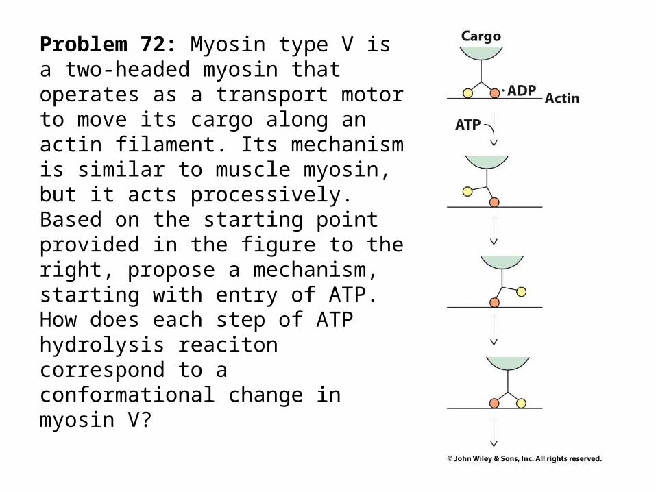
Problem 72: Myosin type V is a two-headed myosin that operates as a transport motor to move its cargo along an actin filament. Its mechanism is similar to muscle myosin, but it acts processively. Based on the starting point provided in the figure to the right, propose a mechanism, starting with entry of ATP. How does each step of ATP hydrolysis reaciton correspond to a conformational change in myosin V?
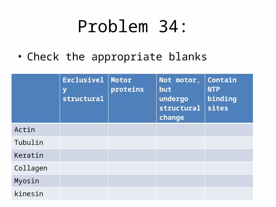
Problem 34:
• Check the appropriate blanks
Exclusively structural
Motor proteins
Not motor, but undergo structural change
Contain NTP binding sites
Actin
Tubulin
Keratin
Collagen
Myosin
kinesin


