Proteome Mapping by 2-D HPLC as a Tool for Veterinary Biologic Antigen Identification.
Timothy J. Barder, Ph.D.
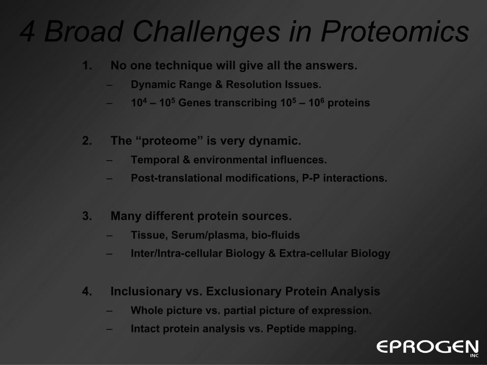
4 Broad Challenges in Proteomics1. No one technique will give all the answers.
– Dynamic Range & Resolution Issues.– 104 – 105 Genes transcribing 105 – 106 proteins
2. The “proteome” is very dynamic.– Temporal & environmental influences.– Post-translational modifications, P-P interactions.
3. Many different protein sources.– Tissue, Serum/plasma, bio-fluids– Inter/Intra-cellular Biology & Extra-cellular Biology
4. Inclusionary vs. Exclusionary Protein Analysis– Whole picture vs. partial picture of expression.– Intact protein analysis vs. Peptide mapping.
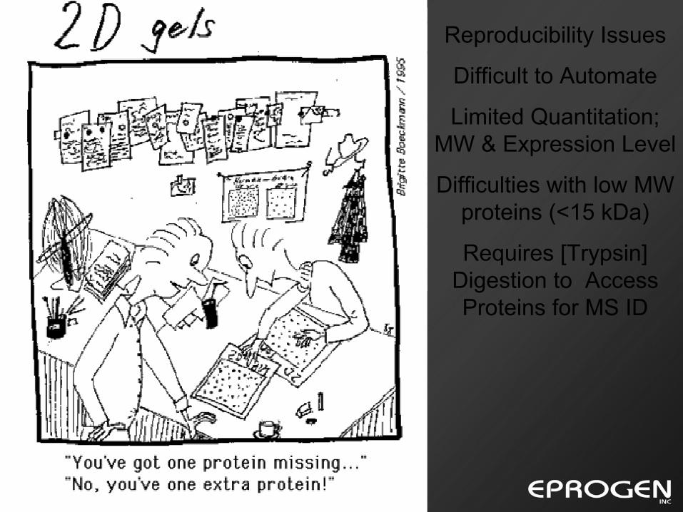
Reproducibility Issues
Difficult to Automate
Limited Quantitation; MW & Expression Level
Difficulties with low MW proteins (<15 kDa)
Requires [Trypsin] Digestion to Access Proteins for MS IDProtein pI
Pro
tein
MW
2D – Gels
Traditional means of resolving complex protein expression.
High resolution & mapping of protein expression in samples.
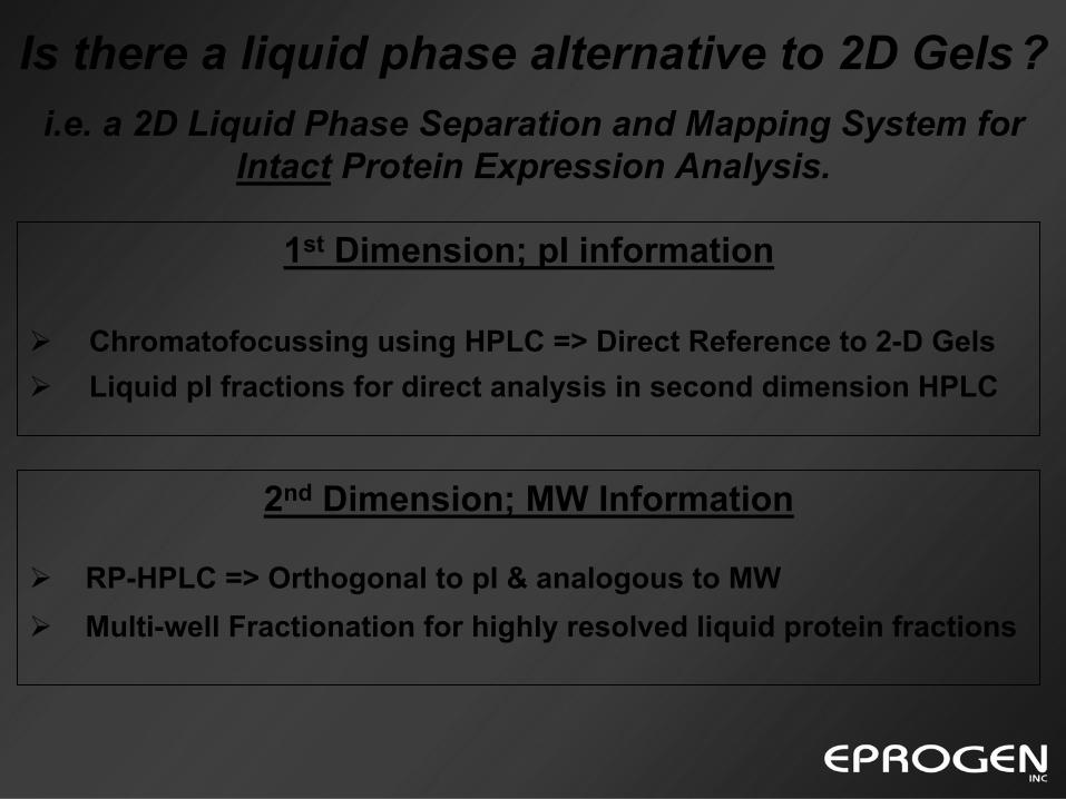
1st Dimension; pI information
Chromatofocussing using HPLC => Direct Reference to 2-D GelsLiquid pI fractions for direct analysis in second dimension HPLC
2nd Dimension; MW Information
RP-HPLC => Orthogonal to pI & analogous to MWMulti-well Fractionation for highly resolved liquid protein fractions
Is there a liquid phase alternative to 2D Gels?i.e. a 2D Liquid Phase Separation and Mapping System for
Intact Protein Expression Analysis.
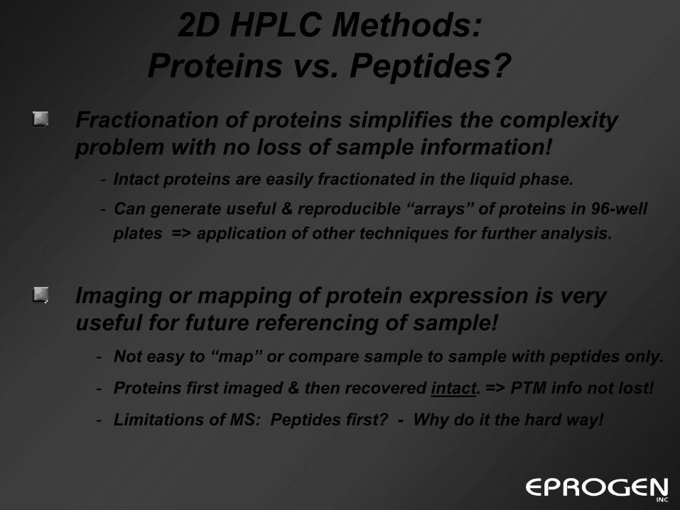
Imaging or mapping of protein expression is very useful for future referencing of sample!
- Not easy to “map” or compare sample to sample with peptides only.
- Proteins first imaged & then recovered intact. => PTM info not lost!
- Limitations of MS: Peptides first? - Why do it the hard way!
Fractionation of proteins simplifies the complexity problem with no loss of sample information!
- Intact proteins are easily fractionated in the liquid phase.
- Can generate useful & reproducible “arrays” of proteins in 96-well plates => application of other techniques for further analysis.
2D HPLC Methods: Proteins vs. Peptides?
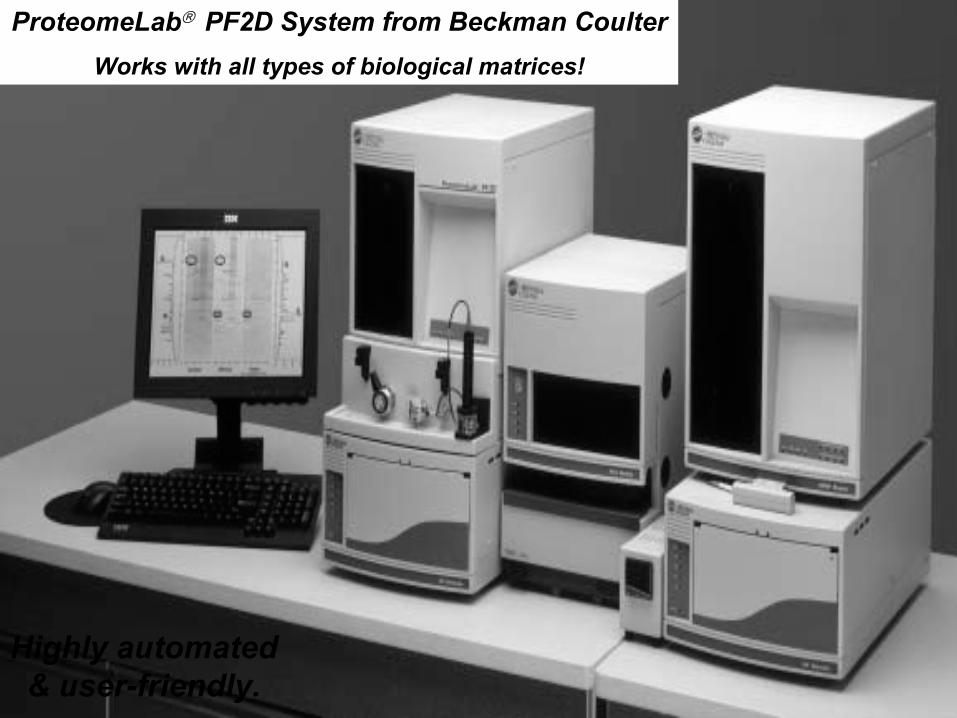
ProteomeLab PF2D System from Beckman CoulterWorks with all types of biological matrices!
Highly automated & user-friendly.
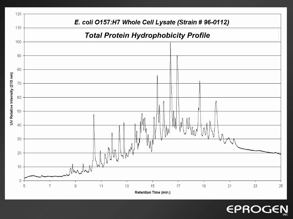
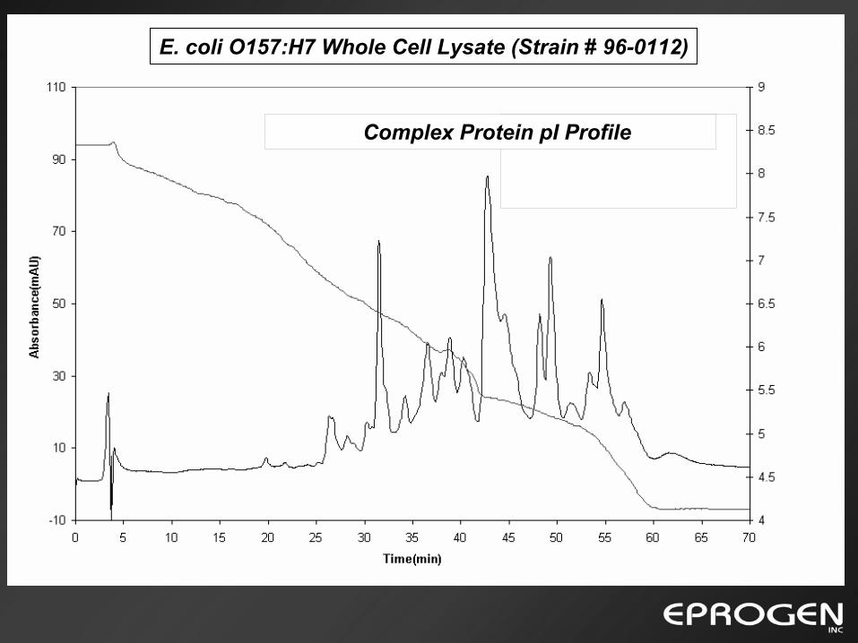
Total Protein Hydrophobicity Profile
E. coli O157:H7 Whole Cell Lysate (Strain # 96-0112)
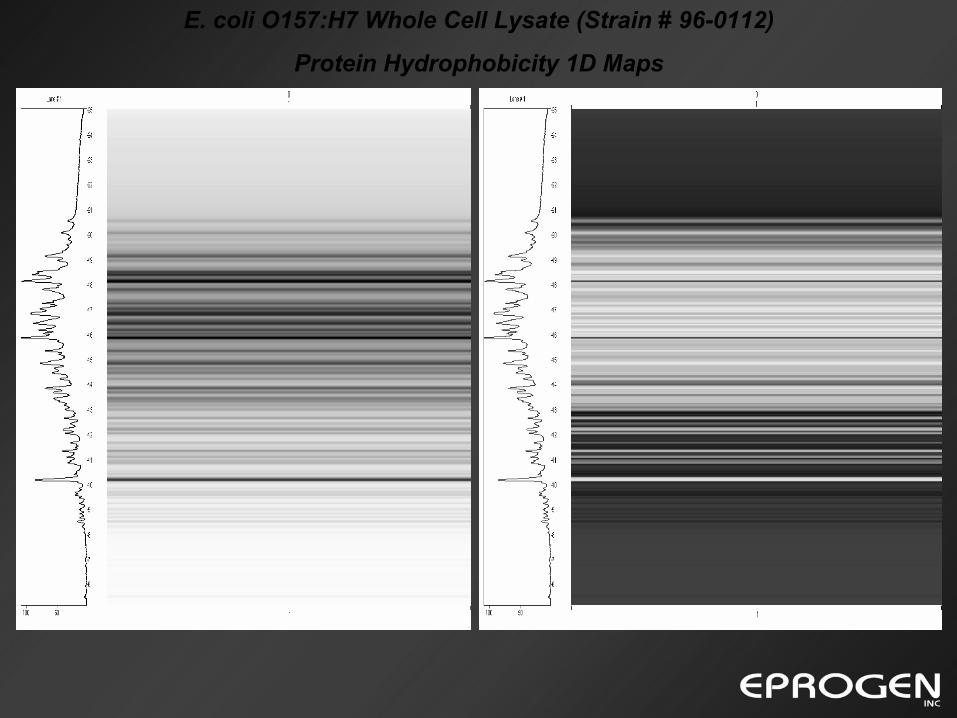
Protein Hydrophobicity 1D Maps
E. coli O157:H7 Whole Cell Lysate (Strain # 96-0112)
Complex Protein pI Profile
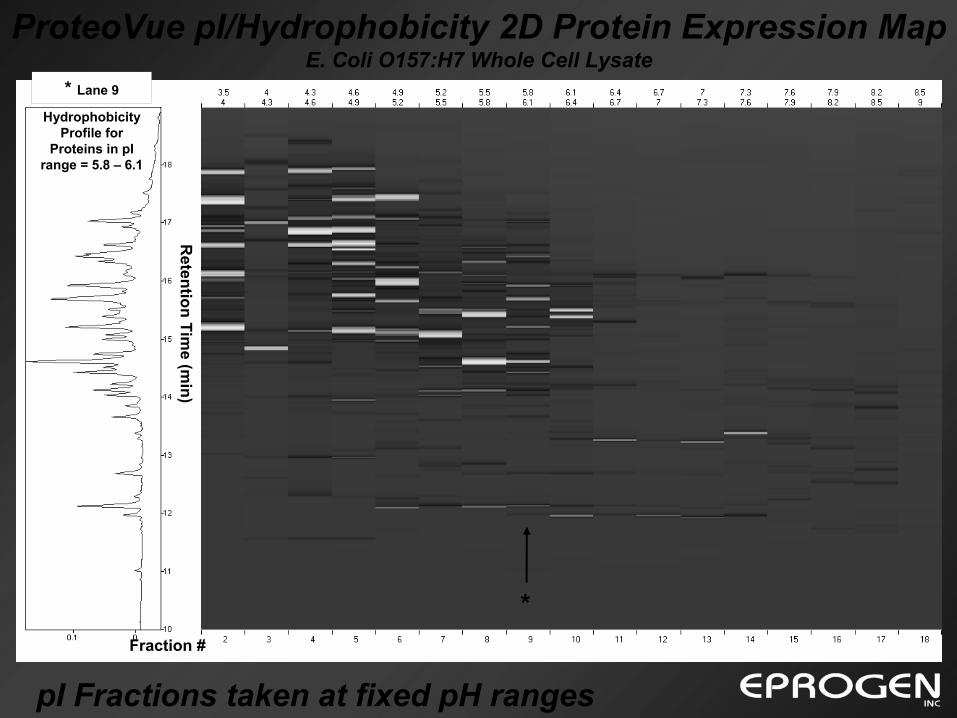
pI Fractions taken at fixed pH ranges
ProteoVue pI/Hydrophobicity 2D Protein Expression Map E. Coli O157:H7 Whole Cell Lysate
pI
Retention Tim
e (min)
Fraction #
Hydrophobicity Profile for
Proteins in pI range = 5.8 – 6.1
* Lane 9
*
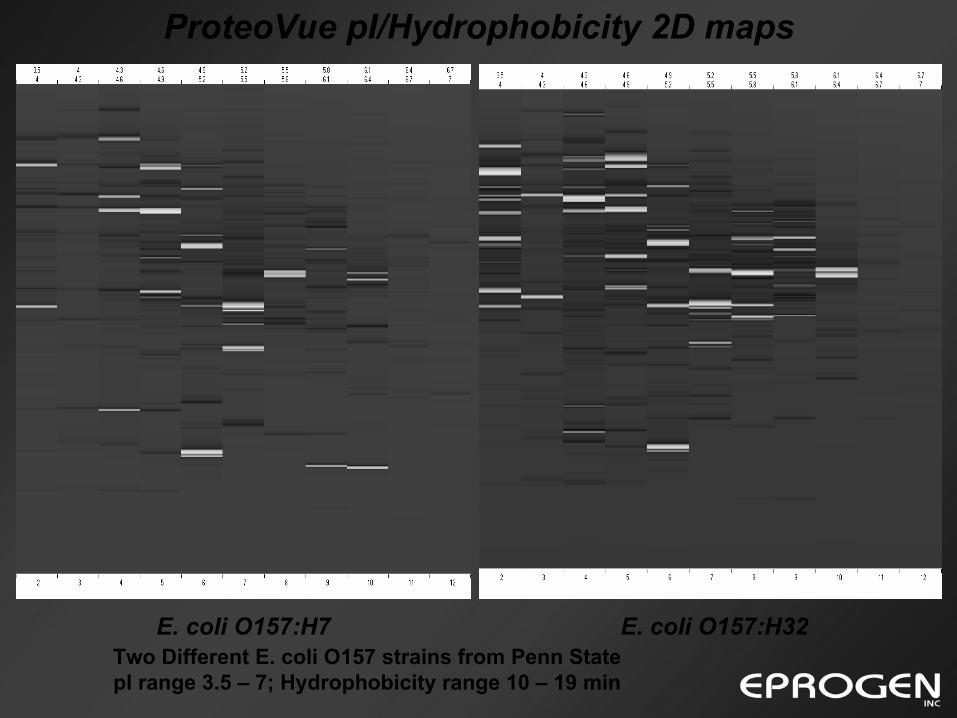
Two Different E. coli O157 strains from Penn StatepI range 3.5 – 7; Hydrophobicity range 10 – 19 min
ProteoVue pI/Hydrophobicity 2D maps
E. coli O157:H7 E. coli O157:H32
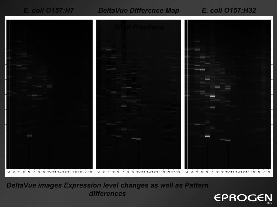
E. coli O157:H7 E. coli O157:H32DeltaVue Difference Map
All pI Fractions
DeltaVue images Expression level changes as well as Pattern differences
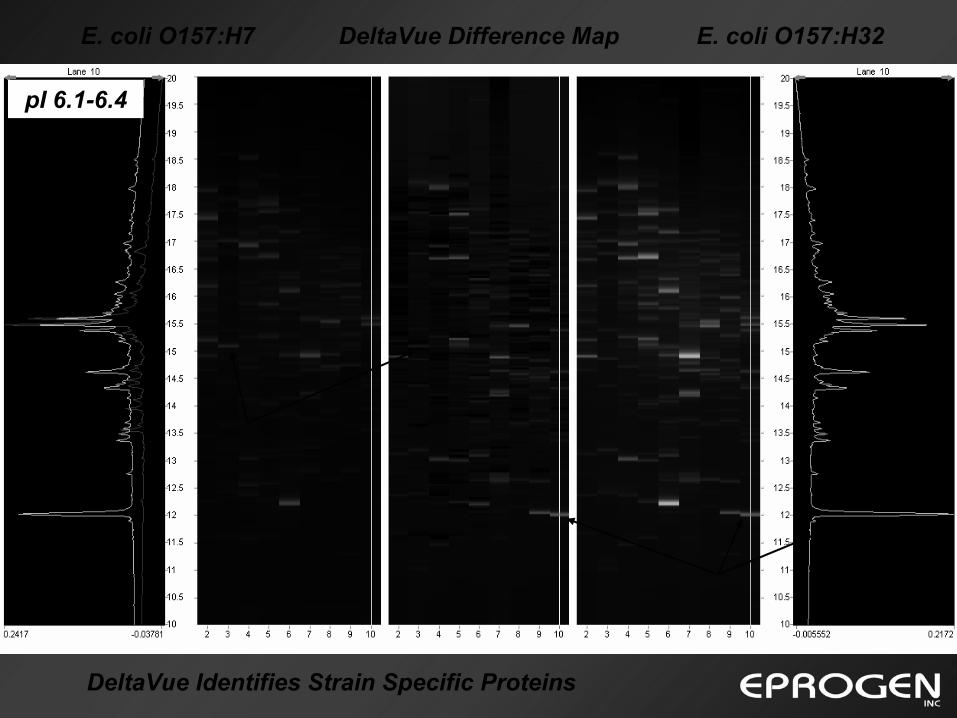
E. coli O157:H7 E. coli O157:H32DeltaVue Difference Map
pI 6.1-6.4
DeltaVue Identifies Strain Specific Proteins
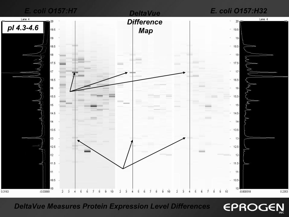
E. coli O157:H7 E. coli O157:H32
pI 4.3-4.6
DeltaVueDifference
Map
DeltaVue Measures Protein Expression Level Differences
H7/H32 = 1.96
H7/H32 = 0.45
Overlay Mode
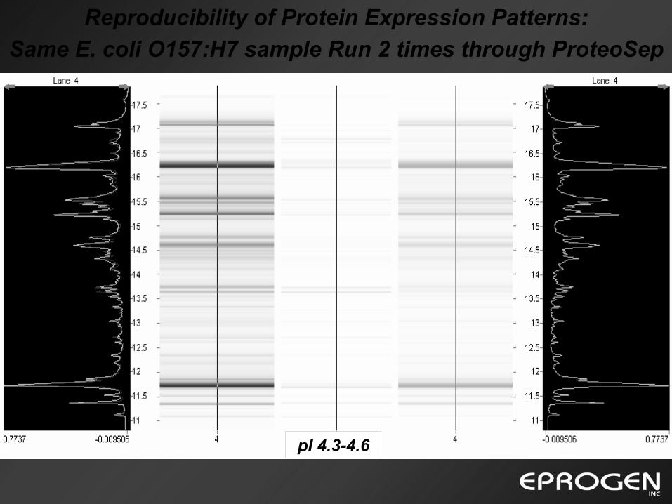
Reproducibility of Protein Expression Patterns:Same E. coli O157:H7 sample Run 2 times through ProteoSep
pI 4.3-4.6
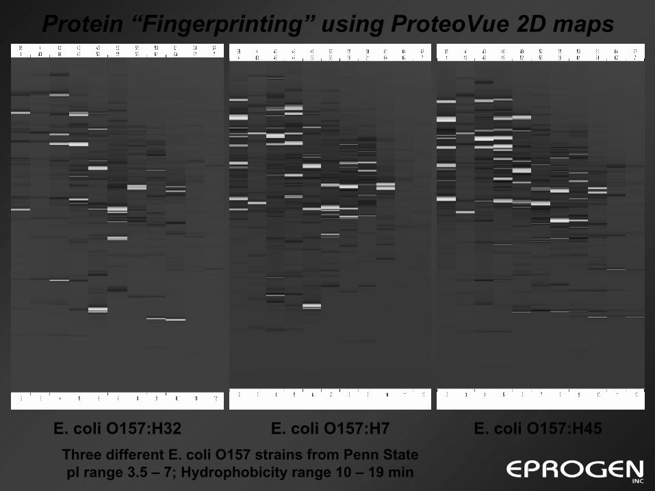
Protein “Fingerprinting” using ProteoVue 2D maps
E. coli O157:H7E. coli O157:H32 E. coli O157:H45Three different E. coli O157 strains from Penn StatepI range 3.5 – 7; Hydrophobicity range 10 – 19 min
2D Protein HPLC and MS:
Liquid Phase Intact Protein MW Analysis with MS
⇒Protein ID, Expression level & PTM information!!!
Insight into total biology of sample or organism from
gene – transcription – protein expression.
Peptide mapping only provides gene based Protein ID information.
Database MW values often differ from expressed MW values.
Numerous possible protein modification pathways.
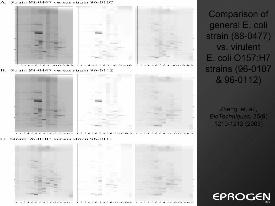
Comparison of general E. coli
strain (88-0477) vs. virulent
E. coli O157:H7 strains (96-0107
& 96-0112)
Zheng, et. al., BioTechniques, 35(6)
1210-1212 (2003)
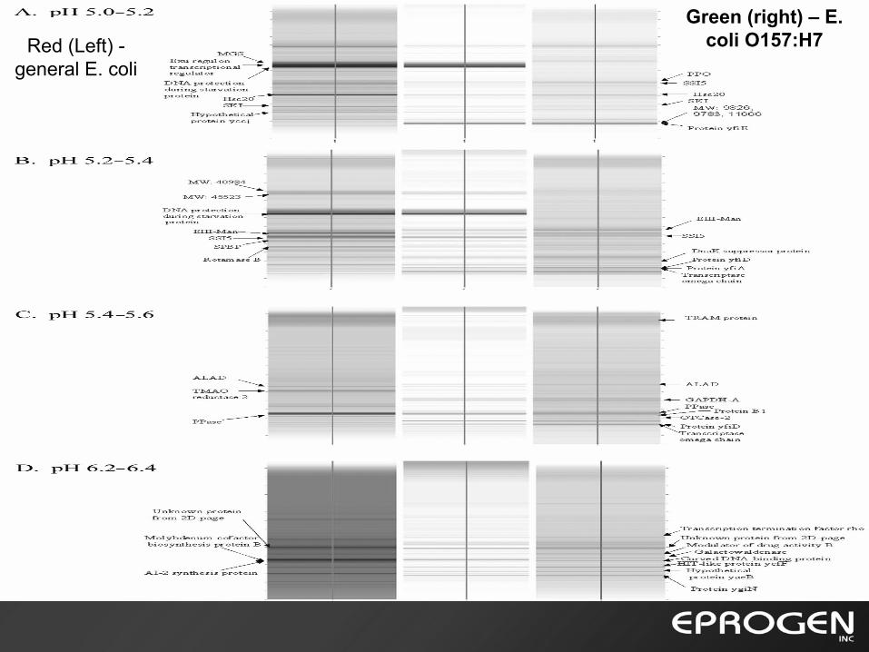
Green (right) – E. coli O157:H7Red (Left) -
general E. coli
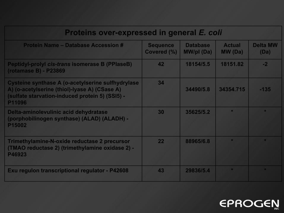
**29836/5.443Exu regulon transcriptional regulator - P42608
**88965/6.822Trimethylamine-N-oxide reductase 2 precursor (TMAO reductase 2) (trimethylamine oxidase 2) -P46923
**35625/5.230Delta-aminolevulinic acid dehydratase(porphobilinogen synthase) (ALAD) (ALADH) -P15002
-13534354.71534490/5.834Cysteine synthase A (o-acetylserine sulfhydrylase
A) (o-acetylserine (thiol)-lyase A) (CSase A) (sulfate starvation-induced protein 5) (SSI5) -P11096
-2 18151.8218154/5.542Peptidyl-prolyl cis-trans isomerase B (PPIaseB) (rotamase B) - P23869
Delta MW(Da)
Actual MW (Da)
Database MW/pI (Da)
Sequence Covered (%)
Protein Name – Database Accession #
Proteins over-expressed in general E. coli
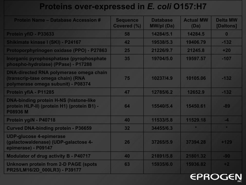
+215936.8215935/6.063Unknown protein from 2-D PAGE (spots PR25/LM16/2D_000LR3) - P39177
-9021801.3221891/5.840Modulator of drug activity B - P40717
+12937394.2837265/5.926UDP-glucose 4-epimerase (galactowaldenase) (UDP-galactose 4-epimerase) - P09147
**34455/6.332Curved DNA-binding protein - P36659-411529.1811533/5.840Protein ygiN - P40718
-8915450.6115540/5.464DNA-binding protein H-NS (histone-like protein HLP-II) (protein H1) (protein B1) -P08936 M
-13212652.912785/6.247Protein yfiA - P11285
-13210105.0610237/4.975DNA-directed RNA polymerase omega chain (transcrip-tase omega chain) (RNA polymerase omega subunit) - P08374
-10719597.5719704/5.035Inorganic pyrophosphatase (pyrophosphate phospho-hydrolase) (PPase) - P17288
+2021245.821226/9.725Protoporphyrinogen oxidase (PPO) - P27863 -13219406.7919538/5.342Shikimate kinase I (SKI) - P24167
014284.514284/5.158Protein yfiD - P33633
Delta MW[Daltons]
Actual MW (Da)
Database MW/pI (Da)
Sequence Covered (%)
Protein Name – Database Accession #
Proteins over-expressed in E. coli O157:H7
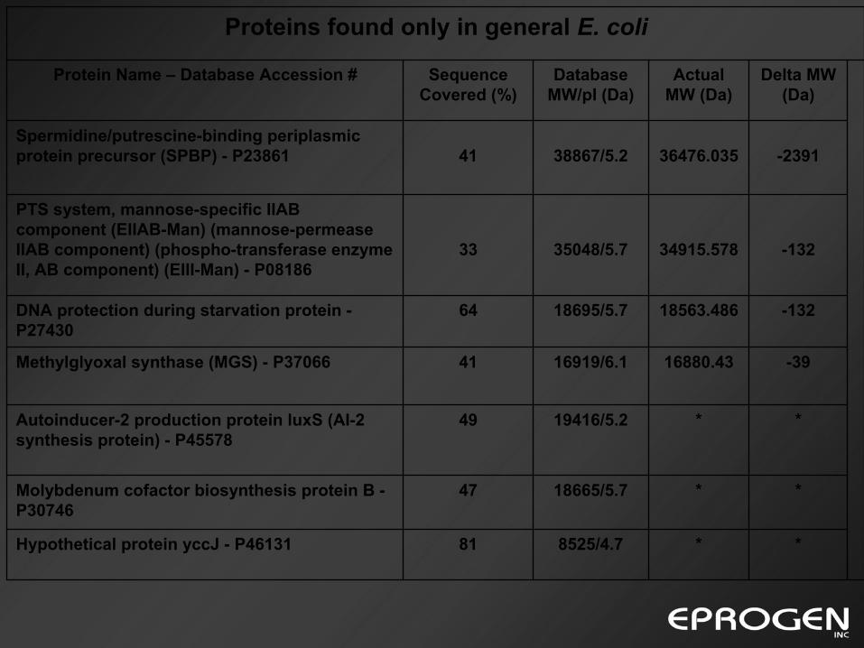
**8525/4.781Hypothetical protein yccJ - P46131
**18665/5.747Molybdenum cofactor biosynthesis protein B -P30746
**19416/5.249Autoinducer-2 production protein luxS (AI-2 synthesis protein) - P45578
-3916880.4316919/6.141Methylglyoxal synthase (MGS) - P37066
-13218563.48618695/5.764DNA protection during starvation protein -P27430
-13234915.57835048/5.733
PTS system, mannose-specific IIAB component (EIIAB-Man) (mannose-permeaseIIAB component) (phospho-transferase enzyme II, AB component) (EIII-Man) - P08186
-239136476.03538867/5.241Spermidine/putrescine-binding periplasmicprotein precursor (SPBP) - P23861
Delta MW (Da)
Actual MW (Da)
Database MW/pI (Da)
Sequence Covered (%)
Protein Name – Database Accession #
Proteins found only in general E. coli
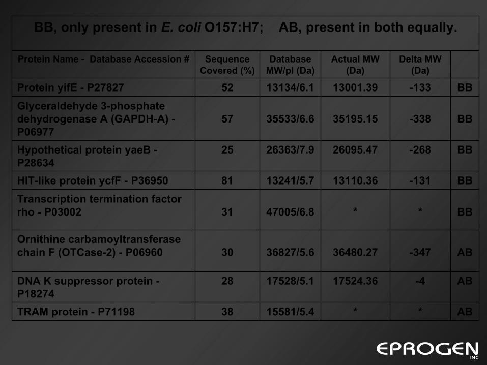
Delta MW (Da)
Actual MW (Da)
Database MW/pI (Da)
Sequence Covered (%)
Protein Name - Database Accession #
AB**15581/5.438TRAM protein - P71198
AB-417524.3617528/5.128DNA K suppressor protein -P18274
AB-34736480.2736827/5.630Ornithine carbamoyltransferasechain F (OTCase-2) - P06960
BB**47005/6.831Transcription termination factor rho - P03002
BB-13113110.3613241/5.781HIT-like protein ycfF - P36950
BB-26826095.4726363/7.925Hypothetical protein yaeB -P28634
BB-33835195.1535533/6.657Glyceraldehyde 3-phosphate dehydrogenase A (GAPDH-A) -P06977
BB-13313001.3913134/6.152Protein yifE - P27827
BB, only present in E. coli O157:H7; AB, present in both equally.
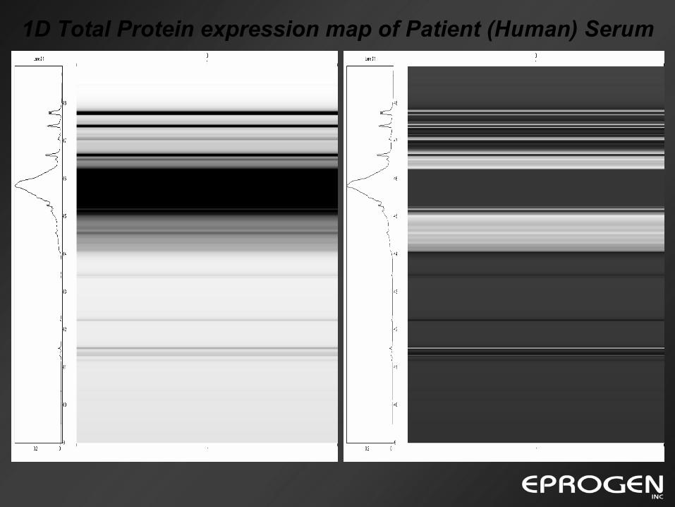
1D Total Protein expression map of Patient (Human) Serum
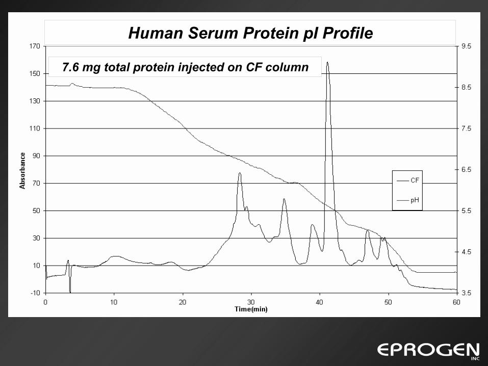
7.6 mg total protein injected on CF column
Human Serum Protein pI Profile
pI
Retention Tim
e (min)
Fraction #
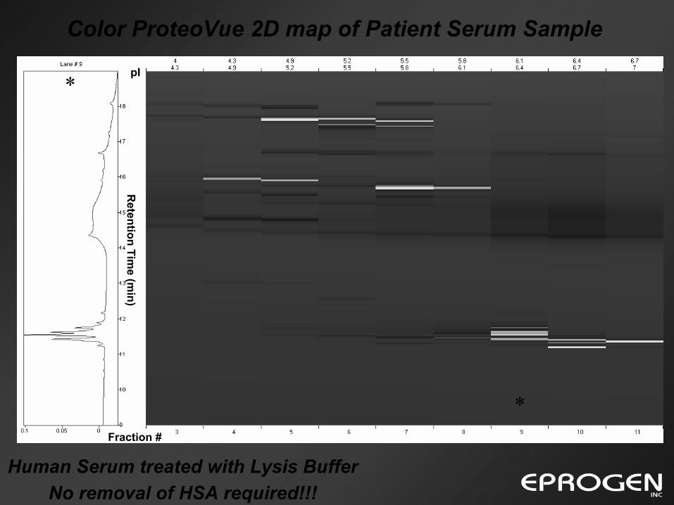
Color ProteoVue 2D map of Patient Serum Sample
Human Serum treated with Lysis Buffer No removal of HSA required!!!
*
*
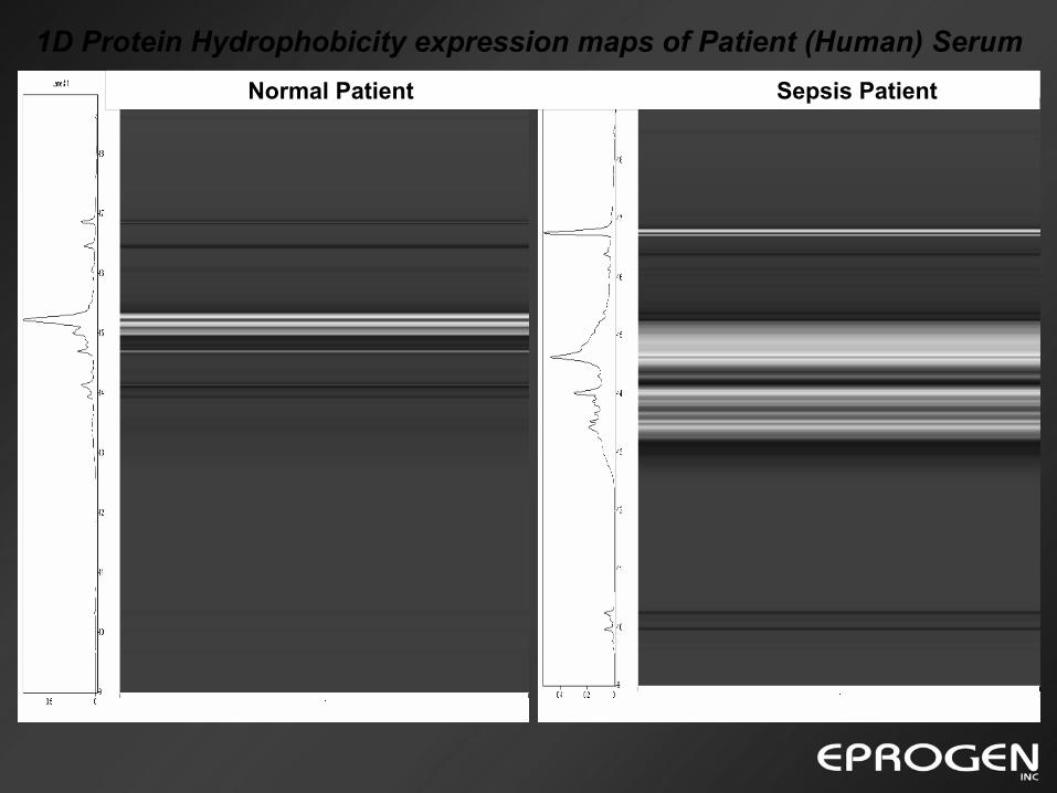
1D Protein Hydrophobicity expression maps of Patient (Human) SerumNormal Patient Sepsis Patient
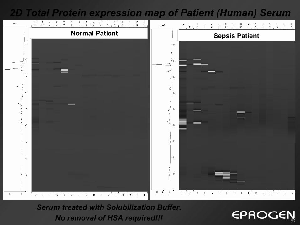
2D Total Protein expression map of Patient (Human) Serum
Serum treated with Solubilization Buffer.No removal of HSA required!!!
Normal Patient Sepsis Patient
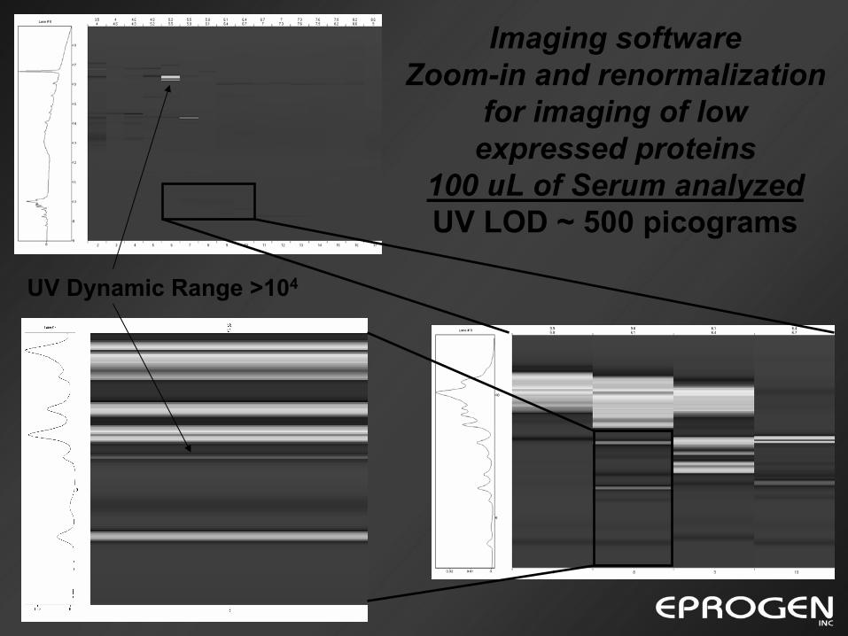
Imaging softwareZoom-in and renormalization
for imaging of low expressed proteins
100 uL of Serum analyzedUV LOD ~ 500 picograms
UV Dynamic Range >104
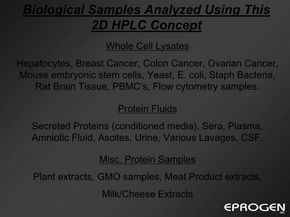
Biological Samples Analyzed Using This 2D HPLC Concept
Whole Cell Lysates
Hepatocytes, Breast Cancer, Colon Cancer, Ovarian Cancer, Mouse embryonic stem cells, Yeast, E. coli, Staph Bacteria,
Rat Brain Tissue, PBMC’s, Flow cytometry samples.
Protein Fluids
Secreted Proteins (conditioned media), Sera, Plasma, Amniotic Fluid, Ascites, Urine, Various Lavages, CSF.
Misc. Protein Samples
Plant extracts, GMO samples, Meat Product extracts,
Milk/Cheese Extracts
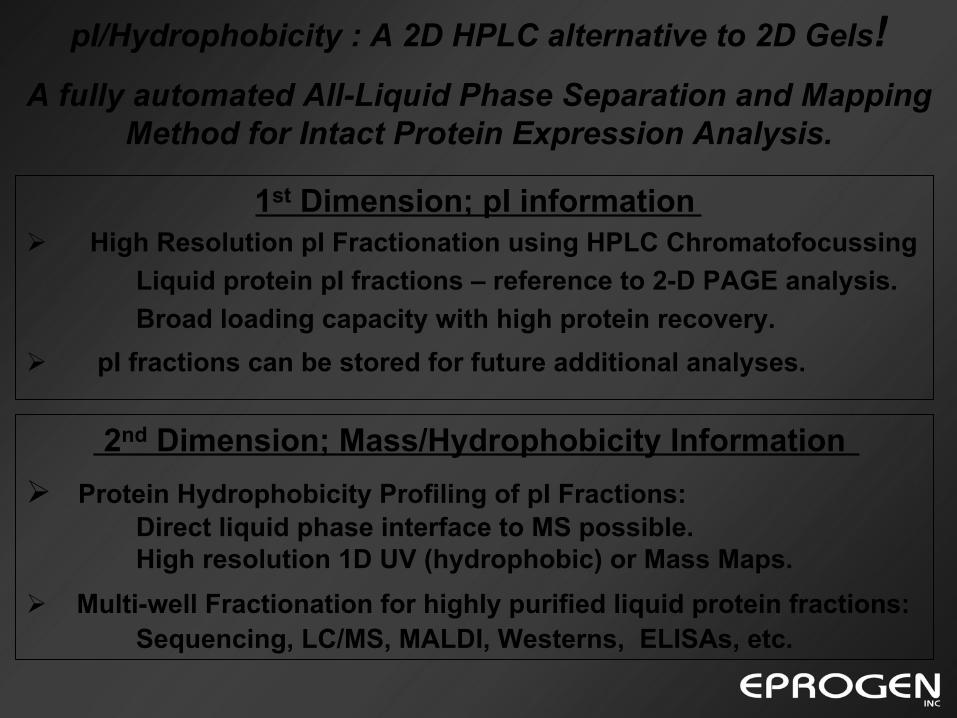
1st Dimension; pI informationHigh Resolution pI Fractionation using HPLC Chromatofocussing
Liquid protein pI fractions – reference to 2-D PAGE analysis.Broad loading capacity with high protein recovery.
pI fractions can be stored for future additional analyses.
2nd Dimension; Mass/Hydrophobicity InformationProtein Hydrophobicity Profiling of pI Fractions:
Direct liquid phase interface to MS possible.High resolution 1D UV (hydrophobic) or Mass Maps.
Multi-well Fractionation for highly purified liquid protein fractions:Sequencing, LC/MS, MALDI, Westerns, ELISAs, etc.
pI/Hydrophobicity : A 2D HPLC alternative to 2D Gels!A fully automated All-Liquid Phase Separation and Mapping
Method for Intact Protein Expression Analysis.
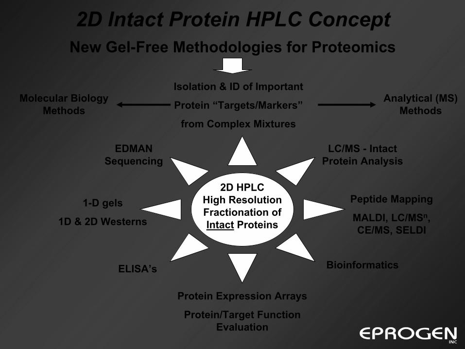
Analytical (MS) Methods
2D Intact Protein HPLC Concept
Peptide Mapping
MALDI, LC/MSn, CE/MS, SELDI
EDMAN Sequencing
Protein Expression Arrays
Protein/Target Function Evaluation
ELISA’s
Isolation & ID of Important
Protein “Targets/Markers”
from Complex Mixtures
Bioinformatics
1-D gels
1D & 2D Westerns
2D HPLC High Resolution Fractionation of Intact Proteins
LC/MS - Intact Protein Analysis
Molecular Biology Methods
New Gel-Free Methodologies for Proteomics
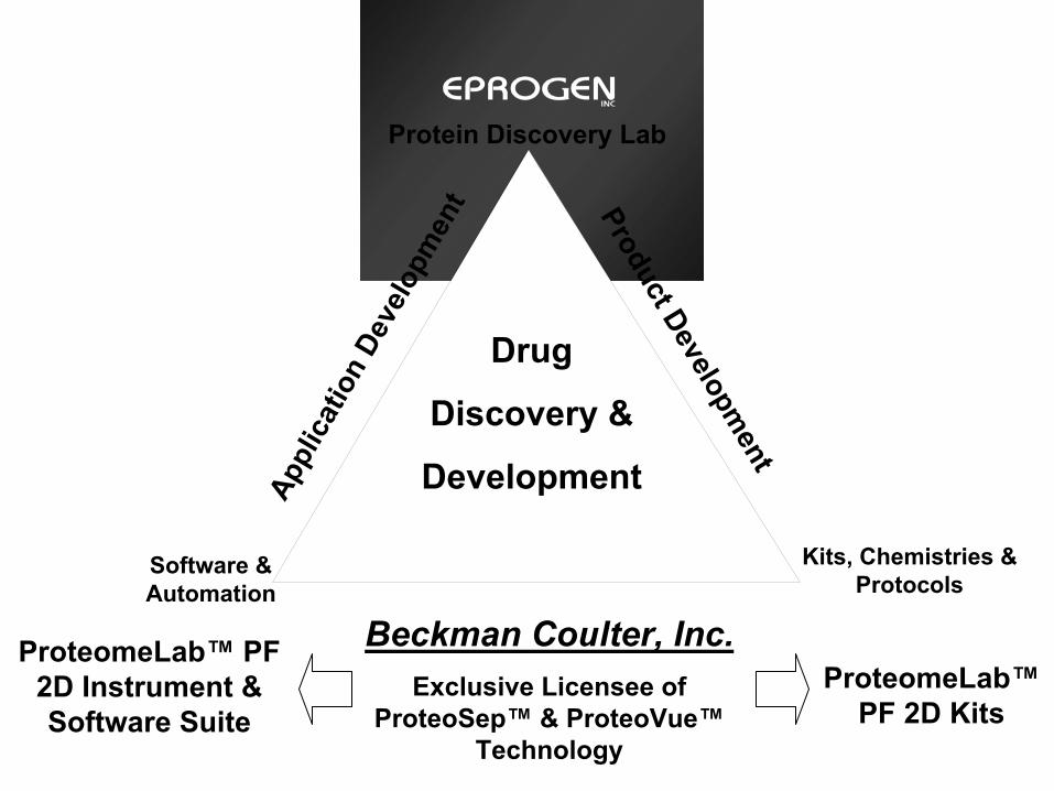
Protein Discovery Lab
Drug
Discovery &
DevelopmentProduct Developm
entAp
plic
atio
n De
velo
pmen
t
Kits, Chemistries & Protocols
Software & Automation
ProteomeLab™ PF 2D Instrument & Software Suite
ProteomeLab™PF 2D Kits
Beckman Coulter, Inc.Exclusive Licensee of
ProteoSep™ & ProteoVue™Technology

Acknowledgements
• Prof. David Lubman (U of Michigan)
• Suipeng Zheng (U of Michigan)
• Kim Schneider (U of Maryland)
• Dr. Steve Parus (U of Michigan)
• Ron Doucet (Eprogen)
• Linda Lin (Eprogen)
• Phil DuBois (Eprogen)
For more info visit www.eprogen.com or www.beckmancoulter.com


