
APPLIED MICROBIOLOGY, Feb. 1970, p. 207-213Copyright © 1970 American Society for Microbiology
Vol. 19, No. 2Printed in U.S.A.
Recovery of Anaerobic Microorganisms from ClinicalSpecimens in Prereduced Media Versus Recovery by
Routine Clinical Laboratory Methods1M. TALMAGE McMINN AND JAMES J. CRAWFORD
Dental Research Center and Department of Endodontics, School of Dentistry, University of North Carolina.Chapel Hill, North Carolina 27514
Received for publication 13 October 1969
Prereduced anaerobically sterilized culture media, used with rigid adherence tothe cultivation techniques described by Moore and his associates, were capable ofrecovering more than twice the number of anaerobic bacteria from clinical speci-mens than could be recovered by the conventional use of fluid thioglycolate mediumand of blood-agar plates incubated anaerobically with hydrogen generation packets.No loss of clinical isolates was encountered with the more sensitive methods; how-ever many of the isolates recovered only in prereduced media would not grow whenplaced into thioglycolate medium. A representative anaerobic isolate placed intoaerobic transport broth was unable to survive beyond 30 min. Methods employingprereduced media were not difficult to master and were feasible for clinical labora-tory use. Evidence implicating the gingival crevice flora as an important possiblesource of anaerobic bacteria that become involved in systemic infections was con-sidered.
There is ample evidence in the literature thatanaerobic bacteria form a distinctive part of theflora of many types of lesions (1, 6, 20, 21).
Severe and fatal infections attributed tobacteroides septicemia (12) and fusospirochetalinfections of the lungs (3, 19) are among theanaerobic infections for which present hospitallaboratory culture procedures may frequentlybe inadequate.Whereas the pathogenicity of indigenous
anaerobic bacteria is still in need of additionalstudy, improved ability to isolate and distinguishanaerobes and to determine their sensitivity toantibiotics, when the need is indicated, couldgreatly assist treatment of disease processes inwhich they are involved.Hungate (10) developed new techniques for
recovery of anaerobic bacteria from rumenspecimens based on preventing oxygen con-tamination of culture medium from the time ofits initial preparation. The Hungate procedureswere recently adapted to anaerobic mediapreparation, culture inoculation procedures (4,14), and improvements in classification (4, 13, 20)that should be applicable to diagnostic micro-biology laboratories.
I Taken in part from a thesis submitted by M. T. McMinn inpartial fulfillment of the requirements for a master of sciencedegree from the University of North Carolina.
Difficulty in utilizing these more sensitivetechniques for cultivation of anaerobic bacteriaresides mainly in the preparation of prereducedanaerobically sterilized media. The present com-mercial availability of these media should makewidespread uses of these media feasible forhospital laboratories.
This present study was conducted to comparethe feasibility and sensitivity of the newer pre-reduced medium techniques with conventionalthioglycolate medium and the Brewer-jar tech-niques for the cultivation of anaerobic micro-organisms from clinical specimens. An experimentwas performed to determine the recovery of acommon oxygen-sensitive anaerobe from asimulated clinical specimen handled withoutprotection from air.
MATERIALS AND METHODSSelection of specimens. Clinical specimens were
obtained from patients of the University of NorthCarolina Dental School and the Clinical Microbiol-ogy Laboratory of the North Carolina MemorialHospital at Chapel Hill, N. C.
Specimens included exudates from deep lesions,pleural fluids, fresh urines, joint fluids, exudates frompockets of inflammation in the throat and dentalmaterial from the gingival margin and crevices.Scrapings from skin and open lesions, bandage mate-rial and nasal and throat swabs were excluded, al-
207
Dow
nloa
ded
from
http
s://j
ourn
als.
asm
.org
/jour
nal/a
m o
n 16
Oct
ober
202
1 by
180
.70.
253.
165.

McMINN AND CRAWFORD
though some of these types of specimens containanaerobic organisms. Most specimens were sub-mitted aerobically.To determine the degree of resistance a strict
anaerobic bacterial strain had to atmospheric oxygen,an experiment was conducted with sterile dry cottonswabs placed in Trypticase Soy Broth. Cotton swabscontaining Propionibacterium propionicum isolatedfrom fresh pleural fluid in prereduced media wereplaced in ordinary culture tubes containing 4.0 ml ofthe broth prepared 6 days prior to the experiment.Swabs were removed at various times and inoculatedinto prereduced media for incubation at 37 C.
Media. For comparison purposes, standard 5%sheep blood-agar plates, deoxycholate-agar plates,and thioglycolate broth without indicator but with1% soluble starch were inoculated and incubatedaerobically. Sheep blood-agar plates for anaerobicincubation were also inoculated.
Most of the methods for preparation and inocula-tion of pre-reduced anaerobically sterilized media(PRAS) have been described by Hungate (10), Mooreand Cato (14) and Moore (13).PRAS media used in this study were prepared
commercially (Robbin Laboratories, Inc., ChapelHill, N.C.) in tubes closed with solid black rubberstoppers by the method of Cato et al. (4). Some mediawere also prepared in this laboratory including PRASmilk, bile, gelatin, nitrate, peptone-yeast, glucose,and glycerol. The pH of the media did not changeappreciably when the tubes were stoppered underoxygen-free carbon dioxide, although nitrogen isrecommended (4).
Commercial PRAS fluid "E" medium was usedfor primary isolation in most instances, which we dis-pensed under oxygen-free CO2 in 3-ml volumes in5-ml serum vials with elongated narrow necks (5 mm,internal diameter). Since the recommended butylrubber stoppers (4) were not available, the vials wereclosed with stoppers of red rubber. This reducedshelf life to 6 or 8 weeks before oxidation, so thevials were used almost immediately after their prep-aration, which would not be practical for routine use.PRAS milk was prepared from commercial ho-
mogenized pasteurized whole milk by the method ofCato et al. (4). The pH was adjusted to 6.9 to 7.2.Powdered skim milk formed undesirable precipitateswhen autoclaved.
Culture methodology. All inoculation needles usedin this study were of stainless steel. Cleaning wiressupplied with 5.5-inch (14.0 cm) 18-gauge fillingneedles (B-D; Becton, Dickinson and Co., Ruther-ford, N.J.) were satisfactory. Other metal needlestended to oxidize the media.
Oxygen-free gas used for media preparation and forinoculations was obtained by passing commercialcarbon dioxide through a glass tube (5 by 30 cm)filled with copper turnings maintained at 350 to 400 C.All connections were made with high quality gumrubber surgical tubing, completed with long 16-gaugestainless-steel filling needles preceded by a short lengthof glass tubing filled with sterile cotton. The needleswere flamed and inserted into the tubes to deliver asteady stream of gas into the culture tubes while
their stoppers were removed for inoculating or media-dispensing purposes.A mixture of commercial N2 or CO2 (97%) and
H2 (3%) can be purified of oxygen by passing thegas through a catalytic gas purifier ("Deoxo" Hydro-gen Purifier, Fisher Scientific Co., Pittsburgh, Pa.).The purifier will only reduce gas containing at least3% hydrogen. The C02-H2 mixture is preferred (4).PRAS agar slants and PRAS agar roll tubes werefound to be satisfactory for streak isolation of mixedcultures. A device to facilitate inoculation of rolltubes was obtained from Robbin Laboratories, Inc.,Chapel Hill, N.C.
Material from the deepest part of liquid specimenswas drawn into a syringe and inoculated into 5-mlserum vials containing 2.4 to 3.0 ml of "E" media or,occasionally, tubes of PRAS chopped meat. Freshmaterial from cotton swabs in small volumes ofliquid media was mixed well and drawn into a syringeflushed with "E" medium for ino_ulation into thevials. At all times, great care was maintained to pre-vent as much contamination with the air as possible.
All specimens were examined microscopically.Notation was made of material containing severaldifferent morphological types of organisms.
For comparison all specimens were also placed influid thioglycolate medium and on blood-agar platesand deoxycholate-agar plates in Brewer jars. Dispos-able hydrogen-carbon dioxide generators ("Gaspak,"BBL) were used to produce anaerobic conditions inthe Brewer jars similar to those in the clinical micro-biology laboratory (2). Blood-agar plates and de-oxycholate-agar plates were inoculated with thespecimens and incubated aerobically to help screenout aerobic and facultative species.
Specimens were incubated at 37 C and checkedeach day for turbidity. Microscopic examinationswere made at the time of noticing turbidity, or at theend of 72 hr and at the end of 10 days. Negative speci-mens were discarded at 3 weeks after microscopicobservation. Specimens on agar plates were examinedat 48 hr under a dissecting microscope.
Specimens exhibiting growth in the vials of "E"media were inoculated into chopped-meat media, andthe morphology of the organisms present was noted.
Anaerobes that grew in the prereduced recoverymedium were reinoculated into thioglycolate brothto determine their subsequent ability to grow in thatmedium.
Careful comparison was made between the micro-scopic and colonial morphology of the aerobic organ-isms that grew on the aerobically incubated agarplates and organisms that grew in the PRAS mediaso that facultative organisms growing in the PRASmedia could be distinguished from true anaerobes.An additional check consisted of inoculating organ-isms from the PRAS media onto aerobically incu-bated agar plates. Roll tubes were examined with theaid of a dissecting microscope for careful differentiat-ing and picking of colonies.
Identification was based on morphology and onthe biochemical reactions described by Cato et al.(4), Hare (8), and Smith and Holdeman (20). Theseincluded final pH obtained from growth in peptone
208 APPL. MICROBIOL.
Dow
nloa
ded
from
http
s://j
ourn
als.
asm
.org
/jour
nal/a
m o
n 16
Oct
ober
202
1 by
180
.70.
253.
165.

RECOVERY OF ANAEROBIC MICROORGANISMS
yeast broth, glucose, fructose, esculin, lactose, maltose,sucrose, and glycerol broth, motility, nitrate reduction,gelatin liquefaction, catalase production, reactions inmilk, and oxygen tolerance. Ambiguous results indi-cated the presence of more than one organism inevery instance in this study.
More critical identification of the species by gaschromatographic analysis of metabolic products asspecified by Cato et al. (4) was not utilized in thisinitial study.
RESULTSRecovery of anaerobic bacteria. The recovery of
49 organisms from 38 specimens collected byroutine methods and inoculated into PRAS mediaand thioglycolate broth and onto agar platesincubated in anaerobic jars is shown in Table 1.
TABLE 1. Recovery rate of 49 anaerobic bacteriafrom 38 clintical specimens inoculated into
various mediaa
Mediab
Inoculation Bot
1 2 3 .Both1 2 ~~~~2and 3
Initial inoculation 100% 6.1% 8.2% 28.6%Subsequent inocu- 100% 10.2% 24.5% 28.6%
lation from orig-inal PRAS cul-ture
a Columns 2 through 4 present recovery resultsobtained by the usual anaerobic culture techniquescompared with total recovery of anaerobes inPRAS media in column 1.bMedia: 1, PRAS media; 2, thioglycolate me-
dium; 3, agar plates in Brewer jar.
Time required for the anaerobic organisms toappear in the PRAS broth media ranged from 24hr to 7 days. All cultures were maintained at 37 Cfor 3 weeks, but no recovery was observed after7 days, although there were several instances ofincreased turbidity with time. There were no
instances of organisms being recovered from any
clinical specimen in thioglycollate broth or inthe anaerobic jars that did not demonstratebetter growth in the PRAS media. All recoveredanaerobes that did not appear in culture tubes in24 hr eventually developed a recognizableturbidity in the PRAS media before demon-strating any growth in the thioglycolate broth.
Inoculations of agar plates and thioglycolatebroth were occasionally made from vials ofPRAS "E" media that had been inoculated a fewhours prior to subculture. These vials had beenstored at refrigerator temperature, and there was
seldom any suggestion of bacterial growth at thetime of subculture.
All bacteria that had not originally grown inthioglycolate broth or on anaerobically incubatedagar plates were subsequently inoculated ontothese media from viable anaerobic culturesmaintained in PRAS media. These cultures were
usually taken from tubes of PRAS chopped-meatmedia or PRAS "E" media and rarely from PRASpeptone-yeast media. There was no significantdifference in subsequent growth from the inoculaof 0.1 ml on agar plates and 1.0 ml into thio-glycolate broth from the three different PRASmedia. The increase in organisms subsequentlygrowing in thioglycolate broth and in the anaero-
bic jars after initial recovery and culture in PRASmedia can also be seen in Table 1.
TABLE 2. Organisms isolated anid identified from 38 various clinical specimens
Abscess PoPleural_______ BloodWud Surgical Knee PoOrganisms fluid Urine culture Woundrain fluid lid
Sputum Ulcerfli
Bacteroides melaninogenicus.4 1 1 1 1Propionibacterium propionicum.2 1 1Peptostreptococcus anaerobius.1 3 2 1P. intermedius ...1 2 1Fusobacterium fusiforme.1 2 1 1Bacteroides oralis.1 1B. fragilis.2 1 1Veillonella alcalescens.2 1 2Peptococcus sp. (unable to identify).P. asaccharolyticus.1P. magnus ..2 1Propionibacterium acnes 1Eubacterium ventriosum 1Catenabacterium filamentosum 1Propionibacterium anaerobium 1
209VOL. 19,y 1970
Dow
nloa
ded
from
http
s://j
ourn
als.
asm
.org
/jour
nal/a
m o
n 16
Oct
ober
202
1 by
180
.70.
253.
165.
McMINN AND CRAWFORD
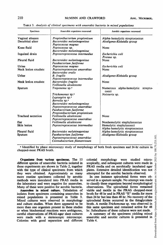
TABLE 3. Analysis of clinical specimens with anaerobic bacteria in mixed populations
Specimen Anaerobic organisms recovered Aerobic organisms recovered
Vaginal abscessDecubital ulcer
Knee fluid
Inguinal drain
Pleural fluid
UrineNeck lesion exudate
Urine
Neck lesion exudate
Sputum
Tracheal secretion
EarSkin lesion
Pleural fluidSputum
Propionibacterium propionicumBacteroides melaninogenicusPeptococcus magnusPeptococcus sp.Bacteroides melaninogenicusPeptostreptococcus intermedius
Bacteroides melaninogenicusFusobacterium fusiformePeptococcus magnusPeptostreptococcus anaerobiusBacteroides oralisB. fragilisPeptostreptococcus intermediusBacteroides fragilisVeillonella alcalescensTreponema sp.a
Trichomonas sp.aLeptospira sp.aBorrelia sp.aBacteroides melaninogenicusPeptostreptococcus anaerobiusFusobacterium fusiformePropionibacterium propionicumVeillonella alcalescensPeptostreptococcus anaerobiusVeillonella alcalescensPeptostreptococcus intermedius
Bacteroides melaninogenicusFusobacterium fusiformePeptostreptococcus anaerobiusCatenabacterium filamentosum
Alpha-hemolytic streptococcusAlcaligenes-Klebsiella group
None
Escherichia coliProteus sp.None
Escherichia coliNone
Alcaligenes-Klebsiella group
None
Numerous alpha-hemolytic strepto-cocci
Neisseria sp.
None
Nonhemolytic staphylococcusHemolytic staphylococciAlpha-hemolytic streptococcusAlpha-hemolytic streptococcusAlcaligenes-Klebsiella group
a Identified by phase microscopy study of morphology of both fresh specimen and 24-hr culture inchopped-meat PRAS broth.
Organisms from various specimens. The 15different species of anaerobic bacteria isolated inthese experiments are shown in Table 2, togetherwith the type of clinical specimens from whichthey were obtained. Approximately as manymore routine specimens collected by aerobicmethods were inoculated into PRAS media inthe laboratory and were negative for anaerobes.Many of these were positive for aerobic bacteria.
Anaerobes in mixed culture. Tabulation ofisolates from specimens containing anaerobes inmixed populations is presented in Table 3.Mixed cultures were observed in morphologyand culture studies. When there appeared to bemore than one organism present in these studiesor when biochemical reactions were ambiguous,careful observations of PRAS-agar slant cultureswere made with a stereoscopic microscope.Colonies with good separation and different
colonial morphology were studied micro-scopically, and subsequent cultures were made inPRAS media and on aerobically incubated agarplates. Extensive detailed identification was notattempted for the aerobic bacteria observed.
In one instance spirochetal forms were ob-served in a sputum sample. No attempt was madeto classify these organisms beyond morphologicalobservations. The spirochetal forms remainedviable and motile in the PRAS chopped-meatbroth for 48 hr and in PRAS "E" media for morethan 24 hr but less than 48 hr. No recovery of thespirochetal forms occurred in the thioglycolatebroth. A motile Trichomonas sp. was observed inthis same specimen and remained active for 48hr. Subcultures of these cultures were not made.A summary of the specimens yielding mixed
anaerobic and aerobic cultures is presented inTable 4.
210 APPL. MICROBIOL.
Dow
nloa
ded
from
http
s://j
ourn
als.
asm
.org
/jour
nal/a
m o
n 16
Oct
ober
202
1 by
180
.70.
253.
165.

RECOVERY OF ANAEROBIC MICROORGANISMS
TABLE 4. Occurrence of 49 anaerobic bacteria inpure and mixed cultures
Bacteria Occurrence
Anaerobe in pure culture............ 45.0Mixed, two anaerobes............... 20.4Mixed, an aerobe with an anaerobe... 12.2Mixed, aerobes and anaerobes ....... 22.4
TABLE 5. Recovery of Propionibacteriumpropionicum from aerobic media
iaeTime in minTrypticase before inocula- Growth at Growth atSoy Broth don of PRAS 24 hra 48 hra
tube media
1 0 4+ 4+2 5 4+ 4+3 10 4+ 4+4 15 4+ 4+5 20 3+ 4+6 25 2+ 4+7 30 1+ 2+8 35 ? NG9 45 ? NG10 50 NGb NG11 60 NG NG
a Key: 1+, bacteria detected only microscopically;2+, barely visible turbidity; 3+, marked turbidity;4+, dense turbidity with sediment.
b No growth.
Inhibitory effects of aerobic media. Manyspecimens with requests for anaerobic studieswere received at the laboratory on cotton swabsin regular culture tubes containing small volumesof well-aerated Trypticase Soy Broth. Only oneanaerobic species was ever recovered from a swabspecimen collected in this manner.An experiment was designed to determine what
effect such aerobic transport and storage wouldhave on the ability to recover an anaerobicbacterium. A sheep blood-agar plate was coveredwith 0.5 ml of a viable culture of Propionibac-terium propionicum originally isolated from adeep tube of pleural fluid and maintained inPRAS chopped-meat media. Cotton swabs wereused to pick up organisms from different areasof the agar surface and placed in 3 ml of Trypti-case Soy Broth that had been stored under cottonplugs for 1 week. Results of growth obtained fromthe inoculated swabs removed at different timesand used to inoculate PRAS "E" media are shownin Table 5. No recovery of this anaerobe wasobtained after it had been in plain aerobic brothfor 30 min.
Cultures of oral specimens. Specimens obtainedfrom the oral cavity of four individuals werecultured in PRAS media. Two of the specimens
were from diseased gingiva and the other twowere from healthy oral surfaces. Isolating purecultures of individual species from the originalextremely mixed population posed remarkabledifficulties. Several attempts at colony separationon PRAS agar slants with the aid of a dissectingmicroscope were required before a good separa-tion could be achieved. The extent to whichselective media and serial dilutions may lessenthese difficulties was not determined.
Table 6 shows the anaerobes isolated from theseoral specimens.A distinctively heavier population of similar
organisms in the cultures from diseased gingivaltissues was observed.
TABLE 6. Anaerobes isolated and identified fromoral specimensa
Patient Anaerobes observed
17-year-old female withclinically diagnosedacute periodontaldisease
47-year-old male withclinically diagnosedperiodontal disease
10-year-old male withexcellent oral health
33-year-old male withgood oral health
Fusobacterium fusiformeVeillonella parvulaPeptococcus magnusPeptococcus prevotiiPropionibacterium pro-pionicum
Trichomonas Sp.bTreponema Sp.bBorrelia Sp.b
Bacteroides melanino-genicus
Fusobacterium fusiformeVeillonella alcalescensPeptostreptococcus in-
termediusPropionibacterium pro-pionicum
Treponema Sp.bBorrelia Sp.b
Fusobacterium fusiformeBacteroides melanino-genicus
Veillonella alcalescensPeptostreptococcus in-
termediusBorrelia Sp.b
Bacteroides melanino-genicus
Bacteroides fragilisPropionibacterium an-aerobium
Treponema Sp.bBorrelia Sp.b
a All specimens contained numerous aerobicbacteria which were not identified.
b Tentative identification on the basis of mor-phological studies alone.
211VOL. 19, 1970
Dow
nloa
ded
from
http
s://j
ourn
als.
asm
.org
/jour
nal/a
m o
n 16
Oct
ober
202
1 by
180
.70.
253.
165.

McMINN AND CRAWFORD
The spirochetal forms were obviously viableand extremely active 48 hr after inoculation intoPRAS media. No differences were noted inmorphological studies of fresh specimens from theperiodontal patients and in the 48-hr PRAScultures. There was no recovery of viable spiro-chetal or trichomonal forms when either freshspecimens of diseased tissue or viable PRAScultures were inoculated into thioglycolate broth.
DISCUSSION
Prereduced media and the anaerobic cultureprocedures involved in their utilization werefound to be feasible for use in the clinical labora-tory. These procedures were at least twice aseffective for detecting anaerobic species in theclinical specimens studied as fluid thioglycolatemedium or aerobically stored agar plates incu-bated in a Brewer jar made anaerobic by use of acommercial hydrogen-generating packet. In-creased recovery outweighed any inconvenience inpassing a stream of oxygen-free gas into eachtube while inoculating or transferring culturesin the prereduced media. Technical mastery of theprocedures was not difficult to achieve.
Inability of an anaerobic isolate, P. propio-nicum, to survive beyond 30 min in a tube ofaerobic transport broth supported the assertionthat more anaerobic strains would be recoveredfrom clinical specimens if they were placeddirectly into prereduced media at the time of theircollection, as described by Cato et al. (4).Although the data presented were obtained
mainly from routine specimens collected andtransported to the laboratory by aerobic means,use of prereduced media under those limitingcircumstances still markedly increased the yieldof anaerobic strains over that obtained by theother commonly used methods.Each specimen that yielded growth of anaerobic
species produced noticeably more growth in ashorter period of time in prereduced media thanin the other media. Microscopic examination ofthioglycolate cultures that appeared negativeoften revealed the presence of anaerobic bacteria.All gave rise to rapid heavy growth when trans-ferred to prereduced media. No difficulty wasencountered in retaining viability of the anaerobicstrains for 1 to 2 weeks in "E" medium at incu-bator or room temperature, whereas in thio-glycolate medium few turbid cultures survivedfor up to a week.The superiority of PRAS media is attributable
to the reduced state of the medium componentsand an oxidation reduction potential of -100 mv(15). In contrast to the common method of usingthe Brewer jar with a chemical hydrogen-generat-
ing packet, more effective procedures can beemployed (4). These include use of agar platesstored anaerobically and repeated flushing of thetank with oxygen-free gas. The equipment neededis relatively immobile, and the specimen inoculummust still be exposed to some oxygen. Thesedisadvantages can be avoided by the directinoculation of prereduced rubber-stoppered tubesat the patient's bedside with only a syringe andneedle. A small lecture tank of 3% H2 in CO2with a deoxo catalyst can provide a mobilesource of gas for use in placing swabs or solidmaterial directly into prereduced tubes. In thelaboratory, direct streaking of specimens on solidmedia in prereduced roll tubes should improverecovery of anaerobes further by avoiding over-growth of anaerobes by facultative organisms.More detail about these procedures is availablein the manual published by the Virginia Poly-technic Institute Anaerobe Laboratory (4).
Incidence of detectable anaerobic species inclinical specimens of blood and exudates cannotbe estimated from the present study, which wasconcerned only with comparing methods ofcultivation. However, the data support the sug-gestions of other workers that anaerobic bacteriaare much more commonly involved in infectiousprocesses than past experience would indicate(15, 21). Studies of patients' specific immuneresponse to anaerobic isolates should reveal moreabout their pathogenic involvement.
Recent evidence indicates that anaerobicisolates have varied profiles of antibiotic sensi-tivities and some are resistant to broad-spectrumantibiotics (15). Improved ability to cultivatesuch anaerobes that become involved in persistentinfections, and to determine their sensitivity tospecific drugs should help to establish a scientificbasis for treatment of such infections. In acuteanaerobic infections such as bacteriodes septi-cemia (12) early diagnosis and treatment wouldappear essential and require use of sensitive andreliable culture techniques.The origin of anaerobic organisms found in
infections is also of clinical concern, and pertainsto the data presented. It was recently shown thatmany of the anaerobic organisms isolated frominfected tissues are common to the normalgastrointestinal tract and may derive from lesionsof the gastrointestinal mucosa (15). The presentinvestigation and literature on the oral flora (3,18) reveal that the vast majority of anaerobicspecies isolated in prereduced media fromsystemic lesions are also common to compara-tively healthy and diseased mouths. A number ofstudies have indicated that bacteremias commonlyresult from minor, even daily, manipulations ofthe oral gingival tissues (5, 7, 9, 11, 16, 17).
212 APPL. MICROBIOL.
Dow
nloa
ded
from
http
s://j
ourn
als.
asm
.org
/jour
nal/a
m o
n 16
Oct
ober
202
1 by
180
.70.
253.
165.

RECOVERY OF ANAEROBIC MICROORGANISMS
Chronic disease of the gingival tissues associatedwith large numbers of anaerobic bacteria isconsidered ubiquitous among adults (3), andshould not be overlooked as a possible source of*anaerobic organisms in systemic infections (9,19).The results presented indicate the significantly
greater sensitivity of prereduced media for culti-vation of anaerobic microorganisms from clinicalspecimens. They support the findings of otherworkers that use of prereduced media can providea useful clinical and research tool to aid diagnosisof infections, to investigate the involvement ofanaerobic organisms in many disease processes,and to facilitate improved approaches to theirtreatment.
ACKNOWLEDGMENTS
These studies were supported in part by Public Health Servicegrant FR 05333 from the Division of Research Facilities andResources and by a grant from the University of North CarolinaResearch Council.
LITERATURE CITED
1. Beerens, H., and M. Tahon-Castel. 1965. Infections humainesa bacteries anaerobies non toxigenes. Presses AcademiquesEuropeennes, Brussels.
2. Brewer, J. H., and D. L. Allgeier. 1966. Safe self-containedcarbon dioxide-hydrogen anaerobic system. Appl. Micro-biol. 14:985-988.
3. Burnett, G. W., and H. W. Scherp. 1968. Oral microbiologyand infectious disease, p. 273-442. Williams & Wilkins Co.,Baltimore.
/Y. Cato, E. P., C. S. Cummins, L. V. Holdeman, J. L. Johnson,W. E. C. Moore, R. M. Smibert, and L. DS. Smith. 1969.Outline of clinical methods in anaerobic bacteriology. TheVirginia Polytechnic Institute Anaerobic Laboratory,Blacksburg, Virginia.
5. Conner, H. D., S. Haberman, C. K Collings, and T. E. Win-ford. 1967. Bacteremia following periodontal scaling in
patients with healthy appearing gingiva. J. Periodont. 38:466-472.
6. Dubos, R. J., and J. G. Hirsch. 1965. Bacterial and mycoticinfections of man, 4th ed., p. 545-567. J. B. Lippincott Co.,Philadelphia.
7. Ernstene, A. C., C. J. McGarvey, and J. A. Ecker. 1951.Prophylaxis of subacute bacterial endocarditis. ClevelandClin. Quart. 18:1-5.
8. Hare, R. 1967. The anaerobic cocci, p. 284-317. In A. P.Waterson (ed.), Recent advances in medical microbiology.Little, Brown and Company, Boston.
9. Harvey, W. P., and M. A. Compone. 1961. Bacterial endo-carditis related to cleaning and filling of teeth with particu-lar emphasis to the inadequacy of present day knowledgeand practice of antibiotic prophylaxis for all dental proce-
dures. Amer. J. Cardiol. 7:793-798.10. Hungate, R. E. 1950. The anaerobic mesophilic cellulolytic
bacteria. Bacteriol. Rev. 14:1-63.11. Khairat, 0. 1966. The non-aerobes of post-extraction bac-
teremia. J. Dent. Res. 45:1191-1 197.12. McVay, L. V., and D. H. Sprunt. 1952. Bacteroides infections.
Ann. Intern. Med. 36:56-76.13. Moore, W. E. C. 1966. Techniques for routine culture ofY,
fastidious anaerobes. Int. J. Syst. Bacteriol. 16:173-190. /14. Moore, W. E. C., and E. P. Cato. 1965. Synonymy of Eubac-
terium limosum and Butyribacterium rettgeri: Butyribac-terium limosum comb. nov. Int. Bull. Bacteriol. Nomencl.Taxon. 15:69-80.
15. Moore, W. E. C., E. P. Cato and L. V. Holdeman. 1969.Anaerobic bacteria of the gastrointestinal flora and their 7
occurrence in clinical infections. J. Infec. Dis. 119:641-649./16. Murray, M., and F. Moosnick. 1941. Incidence of bacteremia
in patients with dental disease. J. Lab. Clin. Med. 26:801-802.
17. O'Kell, C. C. 1935. Bacteremia and oral sepsis, with specialreference to the etiology of subacute endocarditis. Lancet 2:869-872.
18. Rosebury, T. 1962. Microorganisms indigenous to man.
McGraw-Hill Book Co., Inc., New York.19. Smith, D. T., N. F. Conant, and H. P. Willett. 1968. Zinsser
Microbiology, p. 821-826. Appleton-Century-Crofts, NewYork.
20. Smith, L. DS., and L. V. Holdeman. 1968. The pathogenic'--anaerobic bacteria. Charles C Thomas. Publisher, Spring-field, Ill.
21. Stokes, E. J. 1958. Anaerobes in routine diagnostic cultures.Lancet 1:668-670.
VOL. 19, 1970 213
Dow
nloa
ded
from
http
s://j
ourn
als.
asm
.org
/jour
nal/a
m o
n 16
Oct
ober
202
1 by
180
.70.
253.
165.


