Replication fork dynamics and the DNA damageresponseJones, Rebecca; Petermann, Eva
DOI:10.1042/BJ20112100
Document VersionPeer reviewed version
Citation for published version (Harvard):Jones, R & Petermann, E 2012, 'Replication fork dynamics and the DNA damage response' BiochemicalJournal, vol 443, pp. 13-26. DOI: 10.1042/BJ20112100
Link to publication on Research at Birmingham portal
Publisher Rights Statement:Biochem. J. (2012) 443 (13–26) (Printed in Great Britain) doi:10.1042/BJ20112100. The final version of record is available athttp://www.biochemj.org/bj/443/bj4430013.htm
General rightsUnless a licence is specified above, all rights (including copyright and moral rights) in this document are retained by the authors and/or thecopyright holders. The express permission of the copyright holder must be obtained for any use of this material other than for purposespermitted by law.
•Users may freely distribute the URL that is used to identify this publication.•Users may download and/or print one copy of the publication from the University of Birmingham research portal for the purpose of privatestudy or non-commercial research.•User may use extracts from the document in line with the concept of ‘fair dealing’ under the Copyright, Designs and Patents Act 1988 (?)•Users may not further distribute the material nor use it for the purposes of commercial gain.
Where a licence is displayed above, please note the terms and conditions of the licence govern your use of this document.
When citing, please reference the published version.
Take down policyWhile the University of Birmingham exercises care and attention in making items available there are rare occasions when an item has beenuploaded in error or has been deemed to be commercially or otherwise sensitive.
If you believe that this is the case for this document, please contact [email protected] providing details and we will remove access tothe work immediately and investigate.
Download date: 11. Jun. 2018
1
Replication fork dynamics and the DNA damage response Rebecca M. Jones and Eva Petermann* School of Cancer Sciences, University of Birmingham, Edgbaston, Birmingham, B15 2TT, United Kingdom
*Correspondence to: Eva Petermann; School of Cancer Sciences, University of Birmingham, Edgbaston, Birmingham, B15 2TT, United Kingdom; Email: [email protected] Key words: cell cycle, checkpoint, DNA repair, DNA helicase, homologous recombination, translesion synthesis
2
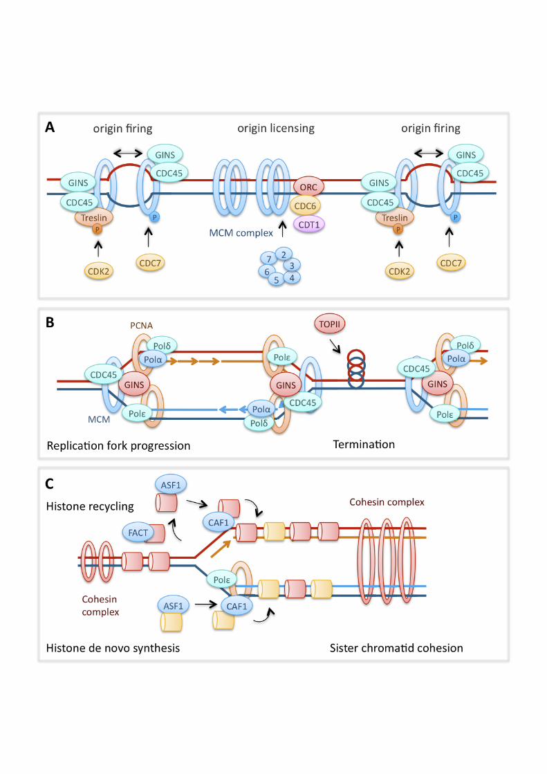
Synopsis Prevention and repair of DNA damage is essential for maintenance of genomic stability and cell survival. DNA replication during S phase can be a source of DNA damage if endogenous or exogenous stresses impair the progression of replication forks. It has become increasingly clear that DNA damage response pathways do not only respond to the presence of damaged DNA, but also modulate DNA replication dynamics to prevent DNA damage formation during S phase. Such observations may help explain the developmental defects or cancer predisposition caused by mutations in DNA damage response genes. This review focuses on molecular mechanisms by which DNA damage response pathways control and promote replication dynamics in vertebrate cells. In particular, DNA damage pathways contribute to proper replication by regulating replication initiation, stabilising transiently stalled forks, promoting replication restart and facilitating fork movement on difficult-to-replicate templates. If replication fork progression fails to be rescued, this may lead to DNA damage and genomic instability via nuclease processing of aberrant fork structures or incomplete sister chromatid separation during mitosis. Introduction In dividing cells, faithful and complete replication of the genome during S phase is essential for maintenance of genomic stability and cell survival. If the progression of DNA replication forks is impaired, this can lead to formation of aberrant DNA structures and generation of DNA damage including double strand breaks (DSBs). Spontaneous, replication-associated DNA damage may inhibit cell growth during development and promote genomic instability that leads to cancer. Consequently, cellular pathways that prevent and repair damage at replication forks are important during development and can act as tumour suppressors. Thanks to advances in the measurement of replication dynamics using DNA fibre approaches and Xenopus egg extracts [1, 2], it has become increasingly clear in recent years that DNA damage response and repair pathways do not only repair damage generated at replication forks, but also modulate the initiation and progression of DNA replication. Pathways that are activated by perturbed or stalled replication forks, such as checkpoint signalling and homologous recombination, modulate replication initiation and fork progression in response to DNA damaging treatments, but they also perform similar functions in the absence of exogenously induced damage. Replication dynamics and the incidence of replication-associated DNA damage are therefore often notably altered in cells with defects in these pathways, which may underlie the developmental and/or cancer-prone genetic disorders that can be associated with such defects. This review will discuss the molecular mechanisms by which the DNA damage response may regulate replication dynamics in vertebrate cells, based on findings from Xenopus, chicken DT40 and mammalian systems while also drawing on central findings from yeast and bacteria. We have aimed to concentrate on the most recent findings, as some of these topics have been covered in our earlier reviews [3, 4]. Eukaryotic DNA replication Replication initiation DNA replication initiates at genomic sites termed origins of replication. In higher eukaryotes, these are organised into initiation zones or clusters that are activated at different times during S phase [5, 6]. Initially, the DNA double helix is unwound to form a “replication bubble” before two replication complexes are assembled. Two individual replication forks (which form the replicon) then move bi-directionally
3
away from the origin, unless a replication fork barrier (RFB) is present. Origins are selected through binding of the origin recognition complex (ORC), a six-subunit complex consisting of ORC1-6 [7, 8]. Origin selection seems to be dependent on the ORC1 subunit [9, 10] and, at least in higher eukaryotes, is sequence-independent [11]. Factors influencing the spacing of origin firing include chromatin structure and epigenetics, nuclear organisation or –matrix, developmental stage and gene expression [12]. During G1 and prior to S phase, cell division cycle 6 (CDC6), chromatin licensing and DNA replication factor 1 (CDT1) and the mini-chromosome maintenance 2-7 (MCM) helicase complex are recruited to sites of ORC binding in a coordinated fashion to form the pre-replication complex (pre-RC) [13]. Replication is then initiated in early S phase as the pre-RC becomes specifically phosphorylated by the Cyclin E- cyclin-dependent kinase 2 (CDK2) and Dbf4/Drf1-dependent cell division cycle 7 (CDC7) kinase (DDK) [14, 15]. In budding yeast, the pre-RC components phosphorylated by CDK2 are Sld3 and Sld2 [16, 17], with the possible metazoan homologues of these proteins being (RecQ protein-like 4) RECQL4, geminin coiled-coil domain containing 1 (GEMC1) and Treslin [18-22]. DDK, on the other hand, phosphorylates the MCM2-7 complex, showing preference for the MCM2 subunit [14]. Phosphorylation of these pre-RC components promotes loading of cell division cycle 45 (CDC45) and the go-ichi-ni-san (GINS) complex, which are essential for the actual initiation of replication (origin firing) and fork progression [23-25]. Replication fork progression The MCM2-7 hexamer and CDC45 form the active replicative helicase required for DNA unwinding during replication. While the MCM complex unwinds the parental DNA in an ATP-dependent manner, the GINS complex maintains protein-protein interactions within the replication complex (replisome) [26-28]. The replisome also contains the clamp loader, replication factor C (RFC), which helps to load the sliding clamp, proliferating cell nuclear antigen (PCNA), onto primed DNA. PCNA tethers the DNA polymerases to the chromosome, enabling processive and high-speed DNA replication [29]. The replisome contains three DNA polymerases, the polymerase !-primase and the replicative polymerases " and !, which replicate the lagging and leading strands, respectively. RFC and PCNA regulate the switch from the POL!-primase to the replicative polymerases [30], and also act as a loading platform for flap endonuclease-1 (FEN-1) and DNA Ligase I, which process and seal Okazaki fragments. If replication forks encounter obstacles on the DNA template, they may stall or collapse. Such obstacles can include spontaneous DNA damage or secondary structures that may arise in repeated, A-T-rich or G-C-rich sequences [31, 32]. Replication fork stalling may also occur when the replisome collides with transcription machinery [33, 34] or when it meets an RFB, a DNA-bound protein or protein complex that actively promotes fork pausing or -stalling. Such RFBs promote mating type switching in fission yeast [35] or maintenance of ribosomal DNA repeat copy number from yeast to humans [36, 37]. A stalled replication fork is halted in its movement but able to resume progression when the blockade is removed. A collapsed fork, however, has become inactivated, for example by running into a single-strand break causing generation of a double-strand break or by dissociation of the replication machinery (reviewed in [4]). To stabilise forks, cells must maintain viable fork structures and assembly of the replication machinery until the replication block is removed, as will be discussed below. Replication forks that have been stalled for
4
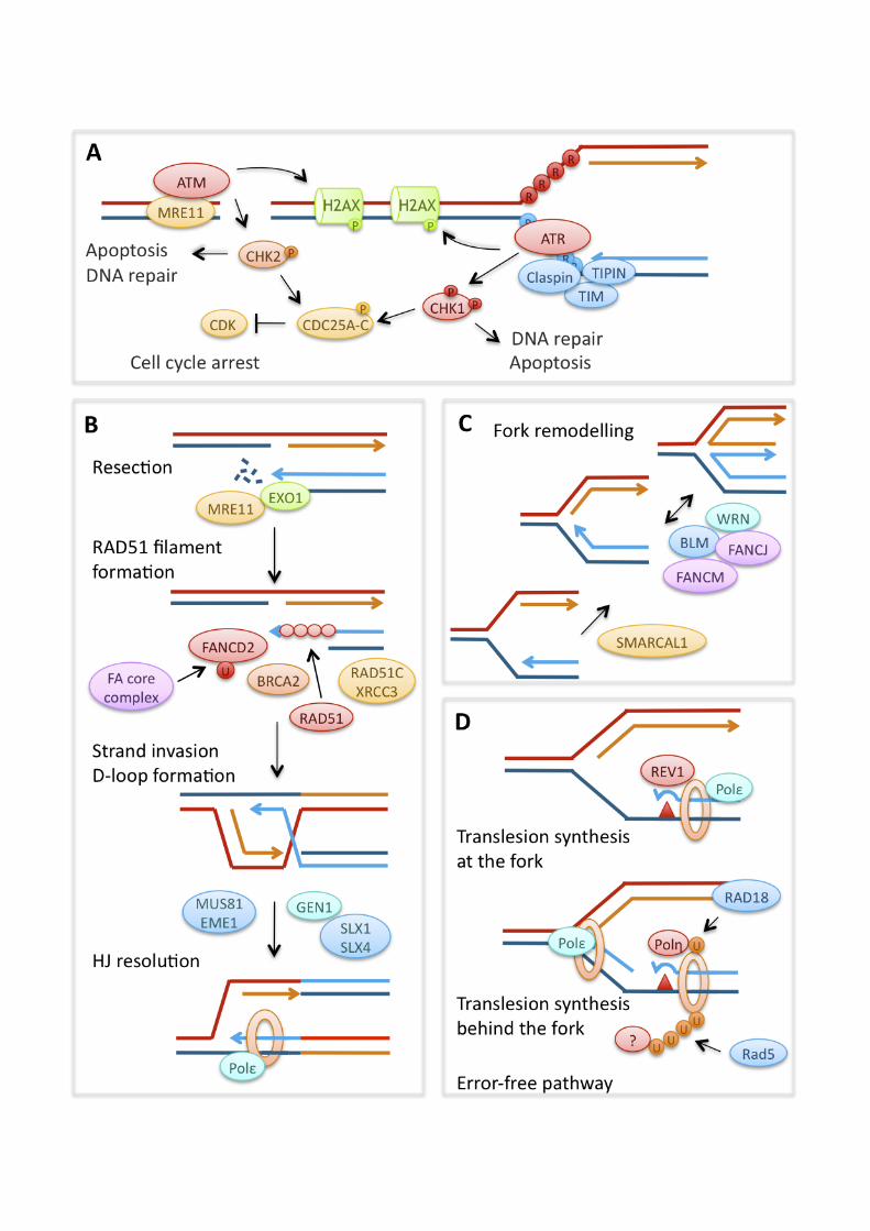
many hours become inactivated and are unable to restart. Under these circumstances, global replication restart is achieved by new origin firing [38]. Inactivation of replication forks also coincides with the generation of fork-associated DSBs by the structure-specific endonuclease complex of MUS81 endonuclease homolog (MUS81) and essential meiotic endonuclease 1 homolog 1 (EME1) [38, 39]. Replication termination Termination of DNA replication in eukaryotes occurs when two opposing replication forks converge. In bacteria, the circular chromosome contains a specific terminator site (TER) opposite to the origin of replication [40]. Efficient termination at these sites requires an RFB posed by the Tus protein, which blocks progression of the replicative helicase [41]. Less is known about replication termination in vertebrates but in Xenopus egg extracts at least, it appears that termination occurs at random sites [42]. A large amount of evidence from SV40 and yeast systems suggests that sister chromatids become intertwined (catenated) at replication termination sites and that the resolution of these structures, for successful completion of termination, requires DNA topoisomerase II (TOPII) [43-45]. Similar mechanisms are likely to operate in higher eukaryotes [46]. Chromatin remodelling during DNA replication Efficient replication fork progression requires that the replisome gain access to the DNA through remodelling of the chromatin structure. Nucleosomes are disassembled ahead of the replication fork, leading to eviction of parental histones, which are recycled onto the daughter strands behind the replication fork, combined with de novo deposition of newly synthesised histones. Histone eviction likely requires ATP-dependent chromatin-remodelling enzymes, while histone chaperones sequester the released histones to facilitate recycling [47]. Histone chaperones such as anti-silencing function 1 (ASF1) and the FACT complex interact with the replisome and act during replication. FACT may be involved in disrupting nucleosomes ahead of the fork, while ASF1 acts as acceptor of parental and newly synthesised histones and donor for the major histone loader, chromatin assembly factor 1 (CAF-1) [47-51]. CAF-1 deposits histones behind the replication fork, and its interaction with PCNA couples chromatin assembly with DNA replication [47, 52]. ASF1 and CAF-1 are required for S phase progression [53-55], and both the FACT subunit structure specific recognition protein 1 (SSRP1) and ASF1 promote efficient replication fork progression. [49, 50]. The newly replicated sister chromatids need to be tethered together to promote the proper segregation of chromatids during mitosis. This process, termed sister chromatid cohesion, requires Cohesin, a ring-shaped complex consisting of four structural maintenance of chromosome (SMC) subunits [56, 57]. The complex is loaded onto DNA in a CDC7-dependent manner during G1-S phase [58]. Its role in maintaining a close distance between chromosome arms is key in facilitating sister chromatid-dependent modes of DNA damage repair [59]. Recruitment of the complex to sites of DNA damage is checkpoint-dependent [60-62], while mutations in the complex lead to an increase in chromosomal aberrations [59]. DNA damage response pathways at replication forks Checkpoint signalling Obstacles on the DNA that specifically affect the progression of the DNA polymerases can lead to uncoupling of the replicative polymerase and helicase
5
activities, with the helicase continuing unwinding to generate excessively long stretches of single-stranded DNA (ssDNA) [63, 64]. In addition, exonuclease resection of damaged DNA structures such as DSBs can generate ssDNA, which occurs especially during S and G2 phase as resection is controlled by CDK activity [65-67]. This ssDNA is recognised as DNA damage by the cell cycle checkpoint machinery. Specifically, replication protein A (RPA)-coated ssDNA recruits and activates the checkpoint kinase ataxia telangiectasia and Rad3 related (ATR) and its interaction partner ATR interacting protein (ATRIP) [68]. While loss of ATR and other components of the ATR pathway is embryonic lethal, hypomorphic mutations in ATR and other genes of the pathway can cause growth and developmental disorders such as ATR-Seckel syndrome [69, 70]. Activated ATR phosphorylates a large number of downstream targets involved in checkpoint signalling, DNA repair and apoptosis, such as histone H2AX [71] and tumour protein p53 (p53) [72]. One important role of ATR is the phosphorylation and activation of the effector kinase checkpoint kinase 1 (CHK1) [73, 74]. Efficient phosphorylation of CHK1 by ATR is promoted by several other replication and checkpoint proteins, including Rad9 homolog (RAD9), Rad17 homolog (RAD17), topoisomerase (DNA) II binding protein 1 (TOPBP1), Claspin, and the complex of timeless homolog (TIM) and TIMELESS interacting protein (TIPIN) [75-80]. CHK1 in turn phosphorylates the cell cycle phosphatases cell division cycle 25 (CDC25) -A, -B and -C to prevent activation of CyclinE/A-CDK2 and CyclinB-CDK1. This slows progression through S phase and prevents mitotic entry, providing the cell with time to restart or repair the stalled replication forks [81-83]. In budding yeast, the functional homologue of CHK1, Rad53, suppresses late origin firing in response to DNA damage by phosphorylating Sld3 and Dbf4 [84-86]. Other substrates of CHK1 include p53 and DNA repair proteins such as RAD51 homolog (RAD51), breast cancer 2, early onset (BRCA2) and Fanconi Anaemia, complementation group E (FANCE) (see below) [87-90]. The ataxia telangiectasia mutated (ATM) checkpoint kinase, mutated in the neurodegenerative and cancer-prone genetic disorder Ataxia telangiectasia, is activated in response to DSBs. This requires the meiotic recombination 11/ Rad50 homolog/ nibrin (MRE11/RAD50/NBS1) complex in concert with mediator of DNA-damage checkpoint 1 (MDC1), tumour protein p53 binding protein 1 (53BP1) and a host of other signalling and mediator proteins. Its effector kinase is checkpoint kinase 2 (CHK2), which similarly to CHK1 targets CDKs and p53 (reviewed in [91]). ATM is considered to be less important in the response to replication blocks than ATR. However, ATM is upstream of ATR activation by promoting exonuclease processing of DSBs, and can itself be phosphorylated and activated by ATR in response to replication blocks [67, 92]. Indeed, ATM signalling also slows DNA replication in response to DNA damage, likely by inhibiting origin firing, and plays roles in the stabilisation and repair of damaged replication forks [93-96]. Homologous recombination Homologous recombination (HR) was initially identified as the pathway mediating genetic recombination in meiosis. It has become clear, however, that HR also repairs DNA double-strand breaks, single-strand gaps and damaged replication forks in mitotic cells, as well as having roles during normal DNA replication [97, 98]. HR repairs double-strand breaks by using homologous DNA sequences (the homologous chromosome, sister chromatid or homologous sequences elsewhere in the genome) as a template for DNA polymerase-mediated re-synthesis of the sequence containing the
6
break (reviewed in [99, 100]). Similar mechanisms are thought to be involved in the functions of HR during replication. In eukaryotes, HR activity is controlled by CDKs, restricting it to S and G2 phases of the cell cycle [65, 66, 101]. HR during S and G2 phases mainly uses the identical sister chromatids, produced by DNA replication, as homologous templates for repair, limiting the potential for large-scale genomic rearrangements. The central HR factor is RecA in bacteria and RAD51 in eukaryotes. RAD51 is recruited to DNA ends with 3’-single stranded overhangs and possibly single-stranded DNA gaps, where it forms protein-DNA filaments [102]. RAD51 filament formation and -stability is regulated by a large number of RAD51 interacting proteins, including the RAD51 paralogues X-ray repair complementing defective repair in Chinese hamster cells 2 (XRCC2), XRCC3, RAD51 homolog B (RAD51B), RAD51C, RAD51D and BRCA2, the protein product of the breast-cancer susceptibility gene [103-106]. The requirement for single-stranded DNA makes HR repair of double-strand breaks dependent on DNA end resection by nucleases such as MRE11 and exonuclease 1 (EXO1), a process that is controlled by CDK activity and therefore restricted to S and G2 phase of the cell cycle [65, 66, 107]. Once bound to the overhang, RAD51 catalyses homology search and recombines the 3’overhang into the homologous double-stranded DNA to form a D (displacement)-loop, which generates a Holliday Junction (HJ) [108]. Upon completion of DNA repair synthesis, the remaining HJ structures are removed by HJ resolution by HJ resolvases such as Gen homolog 1, endonuclease (GEN1), or HJ dissolution by a complex of Bloom syndrome, RecQ helicase-like (BLM), topoisomerase IIIa (TOPIIIa) and RecQ mediated genome instability 1, homolog (hRMI1) [109, 110]. Efficient HR requires the Fanconi Anaemia (FA) pathway, so called because its components are mutated in patients with the autosomal recessive disorder Fanconi Anaemia. Mutations in the FA pathway cause extreme sensitivity to DNA crosslinking agents, cancer predisposition, developmental defects and anaemia [111]. In the current understanding of the FA pathway, its central factors seem to be FANCD2, which is phosphorylated by ATR in response to replication stress, followed by ubiquitylation by the activated FA core complex [112], and the tumour suppressor BRCA2 (FANCD1). While the FA pathway seems to interact with both translesion synthesis (see below) and HR, its role during HR is characterised better; in particular, BRCA2 directly promotes RAD51 function by facilitating RAD51 filament formation and -stability [113]. BRCA2 also interacts with the FA protein FANCN (PALB2) and together they promote RAD51-mediated D-loop formation [114, 115]. Alternative helicases and polymerases In addition to the MCM2-7 replicative helicase, other DNA helicases play roles at replication forks encountering DNA damage. These helicases can remodel perturbed replication fork structures or difficult-to-replicate secondary structures to aid progression of the replisome. In humans these include two of the known RecQ helicases, Werner syndrome, RecQ helicase-like (WRN) and BLM. Individuals carrying non-functional copies of the BLM or WRN genes suffer from the rare autosomal recessive disorders Bloom and Werner syndrome, respectively, which are characterised by a strong predisposition to all types of cancer [116, 117]. Both BLM and WRN have functions in regulating HR; they can disrupt intermediate structures such as the HJ, thus preventing spontaneous HR and genomic instability at perturbed replication forks [118, 119], and whilst BLM also suppresses genetic rearrangements via its HJ dissolution activity [109]. The SWI/SNF related, matrix associated, actin
7
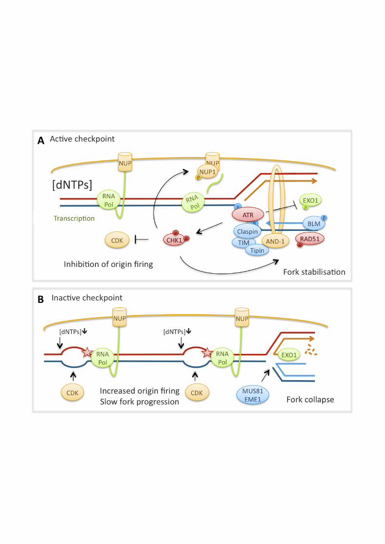
dependent regulator of chromatin, subfamily a-like 1 (SMARCAL1) helicase, mutated in the genetic disorder Schimke Immunoosseous dysplasia, is an unusual ATP-dependent ssDNA annealing helicase that can potentially reverse previous unwinding by the MCM complex [120]. A further two DNA helicases with roles at perturbed forks are the FA pathway components FANCJ/BRIP1 and FANCM [121-123]. Both can function similarly to BLM and WRN by remodelling stalled DNA replication forks [121, 123], and FANCJ can also by unwind secondary structures [122, 124]. FANCJ potentially works alongside specialised polymerases [125], which replace the conventional replicative polymerase in such circumstances; a process known as translesion synthesis (TLS). These polymerases can replicate past a variety of non-coding DNA lesions as their active sites can tolerate a distorted template (reviewed in [126, 127]). So far there are 5 known TLS polymerases: DNA Polymerases ", !, ", # (the latter comprised of two subunits REV3 and REV7) and REV1 homolog (REV1). REV1 interacts with all other known TLS polymerases via its C-terminus [128] and is believed to be a key regulator of lesion bypass. Because low-fidelity TLS polymerases may also increase the chance of mutations, TLS is only activated at sites of DNA damage through monoubiquitination of PCNA by ubiquitin-conjugating enzyme E2B (RAD6) and the E3 ubiquitin ligase RAD18 homolog (RAD18) [129, 130]. This allows switching from replicative to TLS polymerases as the latter have an increased affinity for monoubiquitinated PCNA through their ubiquitin-binding motifs [131]. Polyubiquitination of PCNA at the same residue by Rad5 in yeast, in contrast, activates not TLS but an alternative “error-free” damage avoidance mechanism thought to involve HR-like activities such as template switching (reviewed in [132]). Mammalian cells contain two Rad5 homologues, helicase-like transcription factor (HLTF) and SNF2 histone linker PHD RING helicase (SHPRH) [133-135]. Regulation of replication by the DNA damage response Checkpoint signalling controls replication initiation ATR and ATM signalling pathways down-regulate CDK activity, thereby reducing the overall levels of origin firing during normal S phase, and inhibition of these pathways increases origin firing [136]. This pathway is particularly well studied in the case of ATR-CHK1 signalling. Reduced CHK1 activity leads to accumulation of CDC25A in undamaged cells [82, 83]. This is accompanied by increased Cyclin E-CDK2 activity, increased loading of CDC45 onto chromatin and increased frequencies of origin firing [137-139]. CHK1 also regulates Cyclin A-CDK1 activity, which specifically promotes late origin firing [140]. It has been proposed that the ssDNA that is transiently generated during replication initiation causes low levels of checkpoint activation, thus generating a feedback loop to down-regulate further initiation [136]. In Xenopus egg extracts, checkpoint inhibition shortens the overall duration of S phase [136], although this is not necessarily the case in non-embryonic vertebrate cells, possibly due to the simultaneous fork slowing observed [141, 142]. Changes in CDK activity affect the activation of whole replication clusters more strongly than the activation of origins within clusters, and checkpoint inhibition therefore predominantly increases the number of simultaneously active clusters [143]. At the same time, other signalling pathways downstream of the checkpoint counteract the suppression of origin firing, possibly only within clusters. Polo-like kinase 1 (PLK1) is recruited during replication stress in an ATR-dependent manner and can phosphorylate ORC2 to counteract the checkpoint and allow some amount of origin firing even in presence of replication blocks [144, 145]. This is probably necessary
8
because activation of dormant origins within replication clusters serves as an important mechanism to allow completion of global replication and prevent genomic instability [146-149]. The above observations suggest that the checkpoint selectively allows origin firing within clusters but not activation of new clusters [143]. If, however, replication cluster activation is increased due to checkpoint inhibition, this causes DNA damage [139]. Checkpoint signalling controls replication fork progression In addition to increased replication initiation, vertebrate cells that are defective in ATR-CHK1 signalling display reduced average speeds of replication fork progression during an unperturbed S phase. This applies to cells with defects in ATR, CHK1, Claspin or TIM [78, 150-153]. These observations suggest that, just as the S phase checkpoint regulates origin firing in unperturbed cells, it also modulates the progression of replication forks during normal replication. Evidence from our own work suggests that replication initiation control by the checkpoint may underlie its promotion of replication fork progression. Preventing excessive origin firing in CHK1-defective cells by simultaneous inhibition of CDK2 using roscovitine, or siRNA depletion of CDC7, can restore normal speeds of replication fork progression [141]. This suggests that inhibition of CDK (and possibly DDK), to regulate origin firing, may be an important mechanism by which CHK1 promotes normal replication fork progression. In agreement with this, the generation of spontaneous DNA damage in CHK1-depleted cells depends on CDK2 and CDC25A [139, 154]. In another study, overexpression of active Cyclin A2-CDK1 increased origin firing and slowed replication fork progression, whilst loss of CDK1 increased speeds of fork progression [155]. Several mechanisms by which increased origin density might perturb replication fork progression can be envisaged. The increase in active replication forks may deplete replication factors such as nucleotides [156]. Changes in origin density could also interfere with the spatial coordination of replication initiation with transcription, which may increase conflicts between replication and transcription activities [157-160]. In addition to controlling origin firing, ATR-CHK1 signalling may facilitate replication fork progression by promoting fork stability via any of the pathways discussed below; reduced stability of spontaneously stalled forks is likely to result in slower average replication fork speeds. Intriguingly, evidence suggests that in the presence of DNA damage, the checkpoint can also act to slow down replication fork progression, which would be a useful mechanism to reduce collisions of the replication machinery with DNA lesions. CHK1 and TIPIN have been shown to slow replication fork progression in presence of the DNA damaging agents camptothecin and UV, respectively [78, 161]. The molecular mechanism of checkpoint-dependent replication fork slowing is not yet understood, but it could involve phosphorylation of replication proteins such as the MCM complex [162]. The checkpoint controls replication fork stability Checkpoint signalling stabilises replication forks i.e. it prevents the accumulation of aberrant fork structures or DNA damage during replication, both in cells treated with replication inhibitors and during unperturbed S phase. Components of both the ATR and the ATM pathways have been shown to stabilise replication forks stalled by replication inhibitors: ATR, ATM, MRE11, CHK1, Claspin, TIM and TIPIN [96, 163-165]. ATR, ATM, MRE11 and CHK1 also protect from DNA breakage during an
9
unperturbed S phase, possibly by stabilising spontaneously stalled forks in addition to promoting DNA repair [96, 139, 166, 167]. Checkpoint signalling might stabilise replication forks through a variety of downstream mechanisms. Checkpoint kinases phosphorylate the MCM complex [162] and it has been observed that the checkpoint prevents replication proteins from dissociating from stalled or collapsed forks [96]. Other checkpoint targets that promote replication fork stability include the BLM and WRN helicases [168-170]. Further, checkpoint kinases can potentially regulate HR by phosphorylating RAD51, BRCA2, FANCE and especially FANCD2 [87, 89, 90, 112]. In our hands, RAD51 depletion and CHK1 inhibition had an additive effect on inhibiting replication fork restart, suggesting that CHK1 activity stabilises stalled replication forks through mechanisms other than promoting HR [38]. However, the replication fork restart assay cannot distinguish between effects on replication fork progression and –stability [4]. The checkpoint also down-regulates the activity of nucleases that could otherwise inactivate stalled replication forks by aberrant resection or processing into DSBs: the structure-specific endonuclease Mus81-Eme1 in fission yeast and EXO1 in mammalian cells [171, 172]. Spontaneous DSB formation during replication in mammalian cells deficient in CHK1 or WEE1 (a checkpoint kinase that acts downstream of CHK1) is MUS81-dependent, but a direct regulation of MUS81-EME1 by checkpoint kinases has not yet been found [173, 174]. Another target of the checkpoint is the transcription machinery. In budding yeast, the functional homologue of CHK1, Rad53, prevents stalled forks from reversing into X-shaped structures representing HJ formation [175]. This fork reversal is thought to indicate fork collapse and has been explained by topological strains on stalled replication forks that result from the tethering of transcription to nuclear pores. In response to replication stress, checkpoint signalling targets nuclear pore components to disrupt this tethering and stabilise stalled forks [176]. The checkpoint may also stabilise stalled forks by promoting cohesion [60-62], or regulating histone dynamics at forks. The histone chaperone ASF1 is potentially regulated by ATM and CHK1, which inactivate the tousled-like kinases (TLK1 and TLK2), leading to dephosphorylation of ASF1 [177, 178]. During replication inhibition, ASF1 buffers excess histones so that a pool of histones is readily available for deposition once the block has been removed and replication forks restart [48, 51]. Changes in the regulation of ASF1 could potentially affect its promotion of replication fork progression [50], and evidence from yeast suggests that proper histone supply promotes the stability of replication forks [179]. Claspin, TIM and TIPIN may promote replication independently of ATR signalling Certain components of the ATR pathway may promote replication fork progression independently of ATR signalling itself. While Claspin promotes replication fork progression in a manner similar to CHK1, it does not appear to control origin firing via CDC25 or CDK and seems to work in a pathway parallel to CHK1 [152, 164]. Accordingly, down-regulating origin firing in Claspin-deficient cells has only a small effect on increasing replication fork speeds [180]. Claspin, as well as TIM and TIPIN, may instead promote replication fork progression through their direct interactions with the replication machinery [181-183]. These proteins are orthologues of budding yeast Mrc1 (Claspin), Tof1 (TIM) and Csm3 (TIPIN), which form a complex that promotes both replication fork progression and –stability [184-186]. One way in which the Mrc1/Tof1/Csm3 complex is thought to achieve this is by helping to couple polymerase and helicase activities to prevent excessive unwinding and ssDNA
10
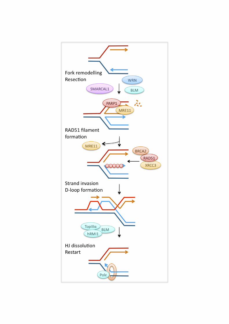
formation [186, 187], and a similar function has been proposed for its vertebrate homologues. TIM depletion increases spontaneous formation of ssDNA, supporting the idea that TIM-TIPIN could also counteract polymerase-helicase uncoupling [188]. This function appeared to be independent of ATR signalling, just as some functions of Tof1 and Csm3 are independent of the ATR homologue Mec1 [189]. Current evidence suggests that TIM and TIPIN may promote replication fork progression and -stability by promoting proper sister chromatid cohesion through their interaction with the cohesion factor WD repeat and HMG-box DNA binding protein 1 (AND-1) [165, 190-192]. TIPIN and AND-1 also promote loading of DNA polymerase !, which could facilitate replication fork restart by re-priming downstream of a lesion [190]. Homologous recombination promotes replication fork stability and restart Our models of HR-dependent pathways of replication fork restart are largely informed by observations from E. coli [193, 194]. HR-dependent fork restart involves HJ and D-loop intermediates, which can be formed either directly from a stalled fork structure or after generation of a DSB at the fork. This process allows loading of the replication machinery in E. coli and budding yeast [97]. Similarly, HR plays an important role in fork stabilisation and restart in vertebrate cells. Defects in the HR proteins BRCA2, RAD51, MRE11, FANCA, or FANCD2 lead to accumulation of spontaneous and replication inhibitor-induced DSBs, suggesting that these proteins stabilise stalled replication forks [166, 167, 195-197]. The HR proteins MRE11, RAD51 and XRCC3 have been reported to facilitate efficient replication fork restart after treatment with replication inhibitors [38, 96, 198]. MRE11 is recruited to stalled replication forks [199-201] and may perform end processing so as to generate the lagging strand gap, or 3’-overhang, that would be required for RAD51 loading. In addition, MRE11 could perform a number of other functions. It interacts with BLM and WRN and might collaborate with these helicases in functions other than resection (see below) [199, 202]. Budding yeast Mre11 has been suggested to promote fork restart by facilitating sister chromatid cohesion [203]. Once the correct single-stranded overhangs are available, XRCC3 could then promote RAD51 loading for D-loop formation and fork restart. RAD51-mediated fork restart seems to differ from HR-mediated DSB repair in that it does not involve RAD51 foci formation, which would be indicative of very long RAD51 filaments, or detectable long patch recombination in a reporter construct [38]. Recombination-free restart could be supported by the BLM-TOPIIIa- hRMI1 complex, which resolves double HJ in a process that avoids crossing over [109]. If stalled forks collapse and are processed into DSBs after longer replication blocks, RAD51-dependent HR is required for the repair of the breaks [38, 170]. The role of HR at stalled replication forks in higher eukaryotes is complicated by the many additional factors that play roles during HR, such as breast cancer 1, early onset (BRCA1), BRCA2, poly (ADP-ribose) polymerase 1 (PARP-1) and the FA complex. BRCA2 and PARP-1 promote replication fork stabilisation or restart, which is attributed to roles in the recruitment and regulation of MRE11 and/or RAD51. PARP-1, absent in yeast but the major poly(ADP-ribose) polymerase enzyme in vertebrate cells, is activated by DNA damaged structures including ssDNA gaps and has a variety of signalling and recruitment functions in the DNA damage response. PARP-1 interacts with MRE11 [198, 204] and, in response to replication inhibitor treatment, it promotes MRE11 foci formation, ssDNA generation and replication fork restart, suggesting that PARP-1 promotes replication fork restart through MRE11-dependent DNA resection [198].
11
Supporting the idea that MRE11 may promote resection at stalled replication forks, the tumour suppressor BRCA2 was found to prevent excessive MRE11-dependent resection at stalled forks [205]. RAD51 itself has a similar preventive effect, and BRCA2 is thought to prevent resection by regulating RAD51 loading and the stability of RAD51 filaments [205, 206]. Absence of BRCA2 has not been found to impair replication fork restart, but it leads to fork collapse and increased genomic rearrangements after replication inhibitor treatment [195, 205]. The effects of HR proteins on the speed of replication fork progression are still poorly understood. There is evidence that HR-deficient cells display decreased fork progression during normal S phase [205, 207]. This could result from a requirement for HR to stabilise or restart forks that stall when encountering endogenous obstacles. On the other hand, the presence of RAD51 and its paralogues XRCC3 and XRCC2 actively slow fork progression on templates containing bulky lesions as induced by cisplatin. RAD54, which is involved in later stages of HR, was not required for this fork slowing. It therefore seems that it is RAD51 loading onto the damaged template that slows fork progression directly or indirectly [208]. This suggests that RAD51 activity does not serve to promote fork progression in this situation but rather the opposite, although HR is important for survival of cisplatin damage through its repair function [106]. Fork-remodelling helicases promote replication fork progression and restart The WRN, BLM, FANCM and SMARCAL1 DNA helicases have all been implicated in replication fork progression and -restart [209-213]. Deficiency in both BLM and WRN causes slow fork progression in untreated cells, as well as defects in the restart of replication forks stalled by replication inhibitors or DNA damage [211-215]. BLM and WRN have the ability to unwind difficult-to-replicate secondary structures in concert with FANCJ, which may aid replisome progression [125, 216, 217]. In addition, BLM has been suggested to promote fork progression by indirectly modulating nucleotide pools. BLM-deficient cells display decreased levels of cytidine deaminase (CDA), which leads to a relative increase in cellular dCTP levels. Re-balancing of pyrimidine pools by expression of CDA or addition of deoxyuridine improved replication fork speeds, but not spontaneous fork stalling in BLM-deficient cells. This suggests that slow fork progression and replication fork restart defect in the absence of BLM may be two separate phenotypes [218]. BLM-dependent replication fork restart is assisted by RAP1 interacting factor homolog (RIF1), a DNA-binding protein of unknown biochemical function [219]. BLM and WRN might both promote fork restart after replication blocks through their modulation of HR. Both helicases could promote D-loop-mediated restart by facilitating fork regression to form a HJ [220, 221]. BLM is also involved in one of the two MRE11-dependent pathways of DNA resection, which may promote RAD51 loading during fork restart [107]. While promoting RAD51 function appears important for replication fork restart, BLM and WRN also possess anti-recombinogenic activities that could promote fork restart if HR activities are detrimental for restart under some circumstances (see above). BLM and WRN could prevent HR by reversing fork regression through their HJ migration activity and repressing aberrant formation of RAD51 filaments at stalled or collapsed forks [118, 119, 222]. Interestingly, ATR and ATM differentially phosphorylate WRN in response to replication blocks: ATR phosphorylation of WRN stabilises stalled forks early during replication blocks, while ATM phosphorylation promotes HR repair of collapsed forks after long replication blocks [170]. This could
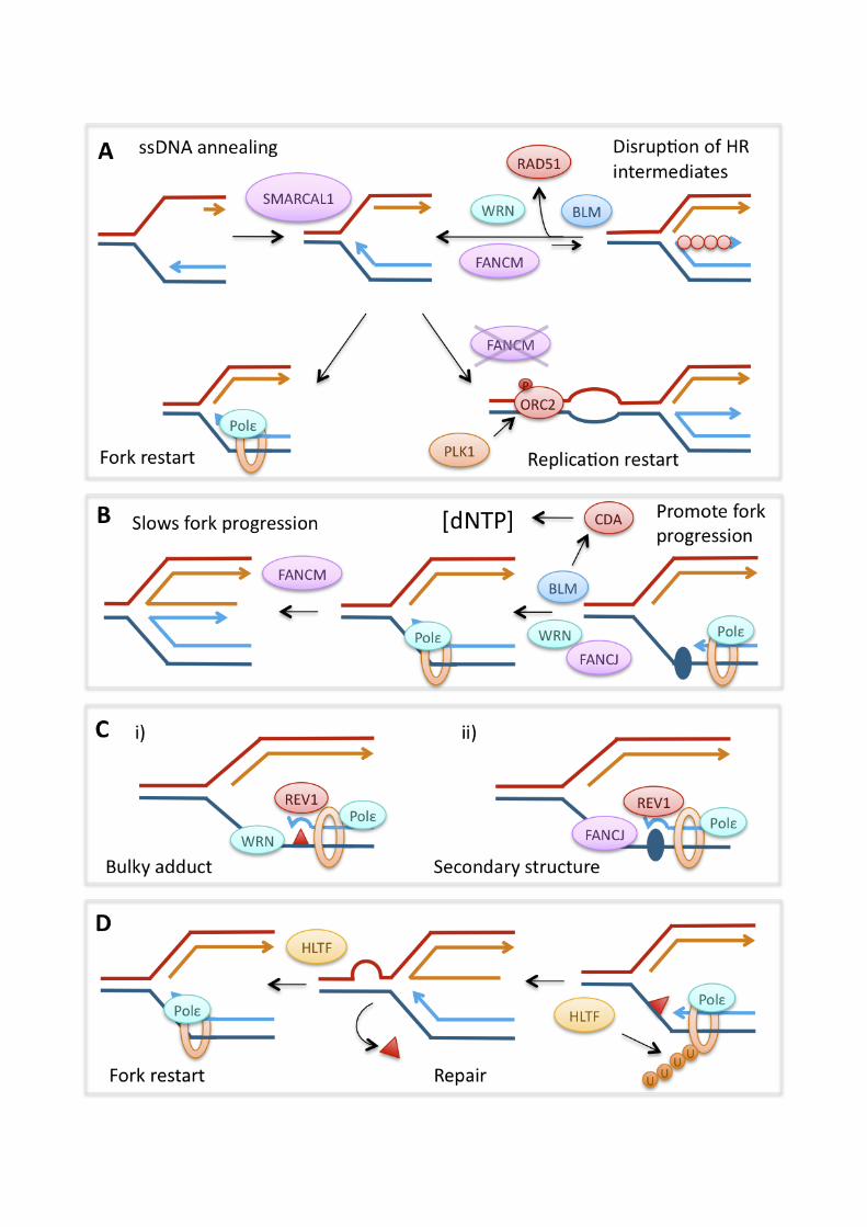
12
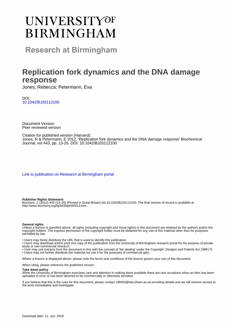
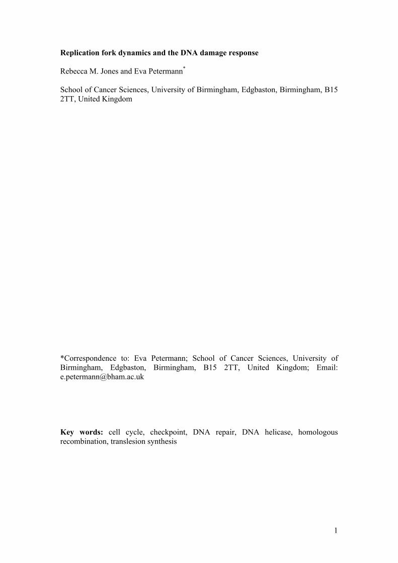
suggest that WRN has an anti-recombinogenic function at stalled forks, and raises the possibility that preventing HR can be a requirement for fork stability under some circumstances. BLM- and WRN-deficient cells treated with replication inhibitors display increased RAD51 foci formation, which could be interpreted as elevated, unscheduled RAD51 filament formation [223]. Alternatively however, increased RAD51 foci formation could indicate an increased need for the repair of forks collapsed into DSB, as MUS81-dependent DSB formation at forks was observed in WRN-deficient cells [213], or a failure to complete HR. When remodelling replication forks, BLM and WRN may work together with the annealing helicase, SMARCAL1. SMARCAL1 is recruited to ssDNA-containing lesions such as stalled replication forks, and its absence leads to both ssDNA accumulation and defective replication fork restart, supporting the idea that SMARCAL1 may promote fork stability and -restart by re-annealing long stretches of ssDNA generated at stalled forks [210, 224]. The FANCM helicase seems to have particularly intriguing effects on replication. DT40 cells deficient in FANCM (but not other FA proteins) displayed not decreased, but increased replication fork restart after release from camptothecin that was dependent on ATR-, CDK- and PLK-activity [225]. FANCM-deficient cells also displayed increased ATR-dependent phosphorylation of MCM2 and increased origin firing. This suggests that the ATR- and PLK1-dependent pathway of promoting origin firing during replication stress becomes activated in absence of FANCM to allow replication restart in the vicinity of stalled forks [144, 225]. Human cells lacking FANCM, or FANCM ATPase activity, displayed increased replication fork speeds and reduced accumulation of ssDNA during unperturbed S phase and in the presence of replication inhibitors, suggesting that FANCM helicase activity slows replication fork progression [209]. In contrast to the first report, however, this group found that absence of FANCM reduced replication fork restart when cells were treated with camptothecin [209]. One potential reason for this discrepancy could be that new origin firing was able to rescue replication in DT40, but not human (HeLa) cells. REV1 and HLTF facilitate replication fork progression and restart The TLS pathway can maintain replication fork progression on templates containing bulky adducts by enabling lesion bypass directly at the fork (early response), but it also performs post-replicative filling of ssDNA gaps left behind the replication fork (late response) [226]. The REV1 polymerase in particular has been implicated in performing TLS directly at the fork, as it promotes replication fork progression on templates containing bulky adducts in chicken DT40 cells and mouse embryonic fibroblasts [226, 227]. It seems that, in DT40 cells at least, this REV1-mediated mode of fork-associated lesion bypass works independently of RAD18 and PCNA ubiquitination, which instead promote post-replicative gap filling by TLS [227]. This process is aided by the WRN helicase [228]. REV1 can also promote fork progression through secondary structures in the DNA, together with FANCJ [125, 229]. In REV1-deficient cells, which are unable to bypass such lesions directly and rely on post-replicative gap filling instead, histone deposition at the site of lesions becomes uncoupled from replication fork progression. In this instance, specific silencing histone modifications are lost, which, should the lesion be located around a promoter region, can result in loss of gene silencing [125, 229]. The RAD5 homologue HLTF, which may be involved in the alternative “error-free” mechanism, has also been implicated in replication fork progression. Like RAD5, HLTF is a RING-domain ubiquitin ligase and SWI/SNF helicase. Via their helicase
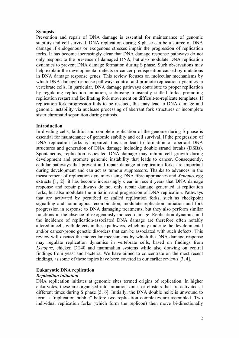
13
activity, Rad5 and HLTF can branch-migrate HJ and reverse replication forks in vitro, which has been suggested to enable template switching during the alternative pathway [230, 231]. Indeed, the HLTF helicase activity promotes timely replication fork restart after release from the methylating agent MMS [231]. In apparent contradiction to this, MMS treatment also leads to degradation of HLTF, which in turn promotes the activity of the second Rad5 homologue SHPRH to suppress MMS-induced mutagenesis [133]. Since promotion of fork restart by HLTF was observed at earlier time points than HLTF degradation [133, 231], it seems possible that HLTF and SHPRH may be involved in early and late responses to MMS damage at forks, such as fork progression and post-replication gap filling, respectively. Resolution of termination intermediates prevents genomic instability Proper replication termination, and resolution of catenated DNA structures that may arise at termination sites, is ultimately required for the correct separation of sister chromatids during mitosis. Incomplete sister chromatid separation can be observed as DNA bridges that connect the daughter nuclei during anaphase. Especially the late-replicating centromeres frequently form such anaphase bridges even during normal cell growth, though the bridges disappear towards telophase, suggesting that replication intermediates are still resolved during mitosis [232]. In addition to TOPII, the complex of BLM, TOPIII! and hRMI1 is required for the resolution of anaphase bridges [232]. It was suggested that the latter complex helps resolve termination structures by a similar mechanism to its dissolution of double HJ structures during HR [232, 233]. Interestingly, there is increasing evidence that additional anaphase bridges can arise from impaired replication fork progression or a failure to activate dormant origins [147, 233, 234]. This presumably occurs because cells enter mitosis without having completed replication. Anaphase bridges resulting from replication inhibition do not localise to centromeric regions and are marked by foci of FANCD2 and FANCI, suggesting that the FA pathway is activated by the perturbed replication structures that give rise to these bridges [233, 234]. The FA pathway seems to promote BLM recruitment to help resolve non-centromeric bridges [234]. Increased levels of unresolved replication intermediates and anaphase bridges have been implicated in the generation of DNA damage and genomic instability. Inhibition of replication fork progression can cause symmetrical DNA lesions during the following mitosis and G1 phase [235]. Breakage of anaphase bridges can lead to the exclusion of chromosome fragments from the daughter nuclei to form micronuclei, resulting in loss of genetic material [147, 234]. That this could be a common mechanism underlying genomic instability is supported by the observation that anaphase bridges and micronuclei localise to common fragile sites, late-replicating genomic loci that frequently display instability in cells treated with replication inhibitors and are often re-arranged in FA, Bloom’s syndrome and cancer cells [233, 234, 236]. Conclusions Our insights into the effects of DNA damage response factors on the initiation and progression of DNA replication have greatly increased in recent years, mainly due to advances in measuring replication dynamics. We are also starting to gain a better understanding of how changes to replication dynamics cause DNA damage and genomic instability. The emerging picture suggests that DNA damage response pathways are activated during normal S phase by both endogenous DNA lesions and ssDNA intermediates at replication forks. The DNA damage response then promotes
14
the proper progression of replication forks by i) regulating aspects of replication such as initiation, stability of the replication complex and sister chromatid cohesion, ii) stabilising transiently stalled forks by regulating a variety of cellular pathways such as transcription to preventing aberrant re-arrangements and processing of these forks, iii) promoting replication restart by re-modelling of damaged replication forks and promoting firing of dormant origins and iv) taking over from the standard replication machinery to facilitate fork movement on difficult-to-replicate templates. If fork progression is impaired, this can lead to increased generation of DSBs at forks through the action of fork-processing nucleases. In addition, prevention of normal replication termination due to impaired fork progression and –restart can lead to incomplete sister chromatid separation followed by generation of DNA damage and genomic instability during mitosis. However, while we can observe the effects of the DNA damage response on replication dynamics, the exact nature of the molecular transactions at forks that underlie these observations are still largely subject to speculation, and further investigations and improved methodologies will be needed to test these speculative models. One major issue that remains to be addressed is the nature of the DNA structures formed at perturbed replication forks in vivo, for example whether transactions such as fork regression occur in human cells. Finding ways to answer these questions would constitute a major breakthrough. Further open questions include whether the DNA damage response promotes proper DNA replication by coordinating and integrating it with other cellular mechanisms such as transcription or cell signalling. What are mechanisms and consequences of replication-associated DNA damage in stem cells and during development? How do genetic defects in factors that promote replication fork progression lead to phenotypes from growth failure (e.g. ATR-Seckel syndrome) to cancer predisposition (e.g. Bloom syndrome)? An interesting new light has been shed on the latter question by recent reports that activation of oncogenes can cause perturbed replication fork progression, which may be important for genomic instability during cancer development [237-239]. This suggests that studying replication-associated DNA damage will advance our understanding of cancer development and may open up new possibilities for cancer therapy. Acknowledgements The authors thank Cancer Research UK and the Royal Society for financial support. We would like to apologise to all authors whose work we could not cite due to space restrictions.
Conflict of interest The authors declare no conflict of interest.
15
References 1 Schwab, R. A. and Niedzwiedz, W. (2011) Visualization of DNA Replication in the Vertebrate Model System DT40 using the DNA Fiber Technique. J Vis Exp pii: 3255. doi: 10.3791/3255.
2 Garner, E. and Costanzo, V. (2009) Studying the DNA damage response using in vitro model systems. DNA Repair (Amst). 8, 1025-1037
3 Petermann, E. and Caldecott, K. W. (2006) Evidence that the ATR/Chk1 pathway maintains normal replication fork progression during unperturbed S phase. Cell Cycle. 5, 2203-2209
4 Petermann, E. and Helleday, T. (2010) Pathways of mammalian replication fork restart. Nat Rev Mol Cell Biol. 11, 683-687
5 Gilbert, D. M. (2007) Replication origin plasticity, Taylor-made: inhibition vs recruitment of origins under conditions of replication stress. Chromosoma. 116, 341-347
6 Jackson, D. A. and Pombo, A. (1998) Replicon clusters are stable units of chromosome structure: evidence that nuclear organization contributes to the efficient activation and propagation of S phase in human cells. J Cell Biol. 140, 1285-1295
7 Bell, S. P. and Stillman, B. (1992) ATP-dependent recognition of eukaryotic origins of DNA replication by a multiprotein complex. Nature. 357, 128-134
8 Gavin, K. A., Hidaka, M. and Stillman, B. (1995) Conserved initiator proteins in eukaryotes. Science. 270, 1667-1671
9 Chesnokov, I., Remus, D. and Botchan, M. (2001) Functional analysis of mutant and wild-type Drosophila origin recognition complex. Proc Natl Acad Sci U S A. 98, 11997-12002
10 Ohta, S., Tatsumi, Y., Fujita, M., Tsurimoto, T. and Obuse, C. (2003) The ORC1 cycle in human cells: II. Dynamic changes in the human ORC complex during the cell cycle. J Biol Chem. 278, 41535-41540
11 Vashee, S., Cvetic, C., Lu, W., Simancek, P., Kelly, T. J. and Walter, J. C. (2003) Sequence-independent DNA binding and replication initiation by the human origin recognition complex. Genes Dev. 17, 1894-1908
12 Mechali, M. (2010) Eukaryotic DNA replication origins: many choices for appropriate answers. Nat Rev Mol Cell Biol. 11, 728-738
13 Mendez, J. and Stillman, B. (2003) Perpetuating the double helix: molecular machines at eukaryotic DNA replication origins. Bioessays. 25, 1158-1167
14 Lei, M., Kawasaki, Y., Young, M. R., Kihara, M., Sugino, A. and Tye, B. K. (1997) Mcm2 is a target of regulation by Cdc7-Dbf4 during the initiation of DNA synthesis. Genes Dev. 11, 3365-3374
16
15 Krude, T., Jackman, M., Pines, J. and Laskey, R. A. (1997) Cyclin/Cdk-dependent initiation of DNA replication in a human cell-free system. Cell. 88, 109-119
16 Tanaka, S., Umemori, T., Hirai, K., Muramatsu, S., Kamimura, Y. and Araki, H. (2007) CDK-dependent phosphorylation of Sld2 and Sld3 initiates DNA replication in budding yeast. Nature. 445, 328-332
17 Zegerman, P. and Diffley, J. F. (2007) Phosphorylation of Sld2 and Sld3 by cyclin-dependent kinases promotes DNA replication in budding yeast. Nature. 445, 281-285
18 Boos, D., Sanchez-Pulido, L., Rappas, M., Pearl, L. H., Oliver, A. W., Ponting, C. P. and Diffley, J. F. (2011) Regulation of DNA replication through Sld3-Dpb11 interaction is conserved from yeast to humans. Curr Biol. 21, 1152-1157
19 Balestrini, A., Cosentino, C., Errico, A., Garner, E. and Costanzo, V. (2010) GEMC1 is a TopBP1-interacting protein required for chromosomal DNA replication. Nat Cell Biol. 12, 484-491
20 Kumagai, A., Shevchenko, A. and Dunphy, W. G. (2010) Treslin collaborates with TopBP1 in triggering the initiation of DNA replication. Cell. 140, 349-359
21 Sangrithi, M. N., Bernal, J. A., Madine, M., Philpott, A., Lee, J., Dunphy, W. G. and Venkitaraman, A. R. (2005) Initiation of DNA replication requires the RECQL4 protein mutated in Rothmund-Thomson syndrome. Cell. 121, 887-898
22 Sansam, C. L., Cruz, N. M., Danielian, P. S., Amsterdam, A., Lau, M. L., Hopkins, N. and Lees, J. A. (2010) A vertebrate gene, ticrr, is an essential checkpoint and replication regulator. Genes Dev. 24, 183-194
23 Mimura, S. and Takisawa, H. (1998) Xenopus Cdc45-dependent loading of DNA polymerase alpha onto chromatin under the control of S-phase Cdk. EMBO J. 17, 5699-5707
24 Takayama, Y., Kamimura, Y., Okawa, M., Muramatsu, S., Sugino, A. and Araki, H. (2003) GINS, a novel multiprotein complex required for chromosomal DNA replication in budding yeast. Genes Dev. 17, 1153-1165
25 Ilves, I., Petojevic, T., Pesavento, J. J. and Botchan, M. R. (2010) Activation of the MCM2-7 helicase by association with Cdc45 and GINS proteins. Mol Cell. 37, 247-258
26 Gambus, A., Jones, R. C., Sanchez-Diaz, A., Kanemaki, M., van Deursen, F., Edmondson, R. D. and Labib, K. (2006) GINS maintains association of Cdc45 with MCM in replisome progression complexes at eukaryotic DNA replication forks. Nat Cell Biol. 8, 358-366
27 Pacek, M. and Walter, J. C. (2004) A requirement for MCM7 and Cdc45 in chromosome unwinding during eukaryotic DNA replication. EMBO J. 23, 3667-3676
17
28 Labib, K., Tercero, J. A. and Diffley, J. F. (2000) Uninterrupted MCM2-7 function required for DNA replication fork progression. Science. 288, 1643-1647
29 Stukenberg, P. T., Studwell-Vaughan, P. S. and O'Donnell, M. (1991) Mechanism of the sliding beta-clamp of DNA polymerase III holoenzyme. J Biol Chem. 266, 11328-11334
30 Stukenberg, P. T., Turner, J. and O'Donnell, M. (1994) An explanation for lagging strand replication: polymerase hopping among DNA sliding clamps. Cell. 78, 877-887
31 Woynarowski, J. M. (2004) AT islands - their nature and potential for anticancer strategies. Curr Cancer Drug Targets. 4, 219-234
32 Lobachev, K. S., Rattray, A. and Narayanan, V. (2007) Hairpin- and cruciform-mediated chromosome breakage: causes and consequences in eukaryotic cells. Front Biosci. 12, 4208-4220
33 Deshpande, A. M. and Newlon, C. S. (1996) DNA replication fork pause sites dependent on transcription. Science. 272, 1030-1033
34 Little, R. D., Platt, T. H. and Schildkraut, C. L. (1993) Initiation and termination of DNA replication in human rRNA genes. Mol Cell Biol. 13, 6600-6613
35 Dalgaard, J. Z. and Klar, A. J. (2000) swi1 and swi3 perform imprinting, pausing, and termination of DNA replication in S. pombe. Cell. 102, 745-751
36 Kobayashi, T., Heck, D. J., Nomura, M. and Horiuchi, T. (1998) Expansion and contraction of ribosomal DNA repeats in Saccharomyces cerevisiae: requirement of replication fork blocking (Fob1) protein and the role of RNA polymerase I. Genes Dev. 12, 3821-3830
37 Gerber, J. K., Gogel, E., Berger, C., Wallisch, M., Muller, F., Grummt, I. and Grummt, F. (1997) Termination of mammalian rDNA replication: polar arrest of replication fork movement by transcription termination factor TTF-I. Cell. 90, 559-567
38 Petermann, E., Orta, M. L., Issaeva, N., Schultz, N. and Helleday, T. (2010) Hydroxyurea-stalled replication forks become progressively inactivated and require two different RAD51-mediated pathways for restart and repair. Mol Cell. 37, 492-502
39 Hanada, K., Budzowska, M., Davies, S. L., van Drunen, E., Onizawa, H., Beverloo, H. B., Maas, A., Essers, J., Hickson, I. D. and Kanaar, R. (2007) The structure-specific endonuclease Mus81 contributes to replication restart by generating double-strand DNA breaks. Nat Struct Mol Biol. 14, 1096-1104
40 Kuempel, P. L., Duerr, S. A. and Seeley, N. R. (1977) Terminus region of the chromosome in Escherichia coli inhibits replication forks. Proc Natl Acad Sci U S A. 74, 3927-3931
18
41 Hill, T. M., Tecklenburg, M. L., Pelletier, A. J. and Kuempel, P. L. (1989) tus, the trans-acting gene required for termination of DNA replication in Escherichia coli, encodes a DNA-binding protein. Proc Natl Acad Sci U S A. 86, 1593-1597
42 Santamaria, D., Viguera, E., Martinez-Robles, M. L., Hyrien, O., Hernandez, P., Krimer, D. B. and Schvartzman, J. B. (2000) Bi-directional replication and random termination. Nucleic Acids Res. 28, 2099-2107
43 DiNardo, S., Voelkel, K. and Sternglanz, R. (1984) DNA topoisomerase II mutant of Saccharomyces cerevisiae: topoisomerase II is required for segregation of daughter molecules at the termination of DNA replication. Proc Natl Acad Sci U S A. 81, 2616-2620
44 Fields-Berry, S. C. and DePamphilis, M. L. (1989) Sequences that promote formation of catenated intertwines during termination of DNA replication. Nucleic Acids Res. 17, 3261-3273
45 Sundin, O. and Varshavsky, A. (1980) Terminal stages of SV40 DNA replication proceed via multiply intertwined catenated dimers. Cell. 21, 103-114
46 Cuvier, O., Stanojcic, S., Lemaitre, J. M. and Mechali, M. (2008) A topoisomerase II-dependent mechanism for resetting replicons at the S-M-phase transition. Genes Dev. 22, 860-865
47 Groth, A., Rocha, W., Verreault, A. and Almouzni, G. (2007) Chromatin challenges during DNA replication and repair. Cell. 128, 721-733
48 Groth, A., Ray-Gallet, D., Quivy, J. P., Lukas, J., Bartek, J. and Almouzni, G. (2005) Human Asf1 regulates the flow of S phase histones during replicational stress. Mol Cell. 17, 301-311
49 Abe, T., Sugimura, K., Hosono, Y., Takami, Y., Akita, M., Yoshimura, A., Tada, S., Nakayama, T., Murofushi, H., Okumura, K., Takeda, S., Horikoshi, M., Seki, M. and Enomoto, T. (2011) The histone chaperone facilitates chromatin transcription (FACT) protein maintains normal replication fork rates. J Biol Chem. 286, 30504-30512
50 Groth, A., Corpet, A., Cook, A. J., Roche, D., Bartek, J., Lukas, J. and Almouzni, G. (2007) Regulation of replication fork progression through histone supply and demand. Science. 318, 1928-1931
51 Jasencakova, Z., Scharf, A. N., Ask, K., Corpet, A., Imhof, A., Almouzni, G. and Groth, A. (2010) Replication stress interferes with histone recycling and predeposition marking of new histones. Mol Cell. 37, 736-743
52 Shibahara, K. and Stillman, B. (1999) Replication-dependent marking of DNA by PCNA facilitates CAF-1-coupled inheritance of chromatin. Cell. 96, 575-585
53 Hoek, M. and Stillman, B. (2003) Chromatin assembly factor 1 is essential and couples chromatin assembly to DNA replication in vivo. Proc Natl Acad Sci U S A. 100, 12183-12188
19
54 Schulz, L. L. and Tyler, J. K. (2006) The histone chaperone ASF1 localizes to active DNA replication forks to mediate efficient DNA replication. Faseb J. 20, 488-490
55 Sanematsu, F., Takami, Y., Barman, H. K., Fukagawa, T., Ono, T., Shibahara, K. and Nakayama, T. (2006) Asf1 is required for viability and chromatin assembly during DNA replication in vertebrate cells. J Biol Chem. 281, 13817-13827
56 Michaelis, C., Ciosk, R. and Nasmyth, K. (1997) Cohesins: chromosomal proteins that prevent premature separation of sister chromatids. Cell. 91, 35-45
57 Losada, A., Hirano, M. and Hirano, T. (1998) Identification of Xenopus SMC protein complexes required for sister chromatid cohesion. Genes Dev. 12, 1986-1997
58 Takahashi, T. S., Basu, A., Bermudez, V., Hurwitz, J. and Walter, J. C. (2008) Cdc7-Drf1 kinase links chromosome cohesion to the initiation of DNA replication in Xenopus egg extracts. Genes Dev. 22, 1894-1905
59 Sonoda, E., Matsusaka, T., Morrison, C., Vagnarelli, P., Hoshi, O., Ushiki, T., Nojima, K., Fukagawa, T., Waizenegger, I. C., Peters, J. M., Earnshaw, W. C. and Takeda, S. (2001) Scc1/Rad21/Mcd1 is required for sister chromatid cohesion and kinetochore function in vertebrate cells. Dev Cell. 1, 759-770
60 Kim, J. S., Krasieva, T. B., LaMorte, V., Taylor, A. M. and Yokomori, K. (2002) Specific recruitment of human cohesin to laser-induced DNA damage. J Biol Chem. 277, 45149-45153
61 Unal, E., Arbel-Eden, A., Sattler, U., Shroff, R., Lichten, M., Haber, J. E. and Koshland, D. (2004) DNA damage response pathway uses histone modification to assemble a double-strand break-specific cohesin domain. Mol Cell. 16, 991-1002
62 Kim, B. J., Li, Y., Zhang, J., Xi, Y., Yang, T., Jung, S. Y., Pan, X., Chen, R., Li, W., Wang, Y. and Qin, J. (2010) Genome-wide reinforcement of cohesin binding at pre-existing cohesin sites in response to ionizing radiation in human cells. J Biol Chem. 285, 22784-22792
63 Byun, T. S., Pacek, M., Yee, M. C., Walter, J. C. and Cimprich, K. A. (2005) Functional uncoupling of MCM helicase and DNA polymerase activities activates the ATR-dependent checkpoint. Genes Dev. 19, 1040-1052
64 Walter, J. and Newport, J. (2000) Initiation of eukaryotic DNA replication: origin unwinding and sequential chromatin association of Cdc45, RPA, and DNA polymerase alpha. Mol Cell. 5, 617-627
65 Aylon, Y., Liefshitz, B. and Kupiec, M. (2004) The CDK regulates repair of double-strand breaks by homologous recombination during the cell cycle. Embo J. 23, 4868-4875
66 Ira, G., Pellicioli, A., Balijja, A., Wang, X., Fiorani, S., Carotenuto, W., Liberi, G., Bressan, D., Wan, L., Hollingsworth, N. M., Haber, J. E. and Foiani, M. (2004) DNA end resection, homologous recombination and DNA damage checkpoint activation require CDK1. Nature. 431, 1011-1017
20
67 Jazayeri, A., Falck, J., Lukas, C., Bartek, J., Smith, G. C., Lukas, J. and Jackson, S. P. (2006) ATM- and cell cycle-dependent regulation of ATR in response to DNA double-strand breaks. Nat Cell Biol. 8, 37-45
68 Zou, L. and Elledge, S. J. (2003) Sensing DNA damage through ATRIP recognition of RPA-ssDNA complexes. Science. 300, 1542-1548
69 O'Driscoll, M., Ruiz-Perez, V. L., Woods, C. G., Jeggo, P. A. and Goodship, J. A. (2003) A splicing mutation affecting expression of ataxia-telangiectasia and Rad3-related protein (ATR) results in Seckel syndrome. Nat Genet. 33, 497-501
70 O'Driscoll, M., Dobyns, W. B., van Hagen, J. M. and Jeggo, P. A. (2007) Cellular and clinical impact of haploinsufficiency for genes involved in ATR signaling. Am J Hum Genet. 81, 77-86
71 Ward, I. M. and Chen, J. (2001) Histone H2AX is phosphorylated in an ATR-dependent manner in response to replicational stress. J Biol Chem. 276, 47759-47762
72 Tibbetts, R. S., Brumbaugh, K. M., Williams, J. M., Sarkaria, J. N., Cliby, W. A., Shieh, S. Y., Taya, Y., Prives, C. and Abraham, R. T. (1999) A role for ATR in the DNA damage-induced phosphorylation of p53. Genes Dev. 13, 152-157
73 Guo, Z., Kumagai, A., Wang, S. X. and Dunphy, W. G. (2000) Requirement for Atr in phosphorylation of Chk1 and cell cycle regulation in response to DNA replication blocks and UV-damaged DNA in Xenopus egg extracts. Genes Dev. 14, 2745-2756
74 Liu, Q., Guntuku, S., Cui, X. S., Matsuoka, S., Cortez, D., Tamai, K., Luo, G., Carattini-Rivera, S., DeMayo, F., Bradley, A., Donehower, L. A. and Elledge, S. J. (2000) Chk1 is an essential kinase that is regulated by Atr and required for the G(2)/M DNA damage checkpoint. Genes Dev. 14, 1448-1459
75 Furuya, K., Poitelea, M., Guo, L., Caspari, T. and Carr, A. M. (2004) Chk1 activation requires Rad9 S/TQ-site phosphorylation to promote association with C-terminal BRCT domains of Rad4TOPBP1. Genes Dev. 18, 1154-1164
76 Kumagai, A. and Dunphy, W. G. (2000) Claspin, a novel protein required for the activation of Chk1 during a DNA replication checkpoint response in Xenopus egg extracts. Mol Cell. 6, 839-849
77 Unsal-Kacmaz, K., Mullen, T. E., Kaufmann, W. K. and Sancar, A. (2005) Coupling of human circadian and cell cycles by the timeless protein. Mol Cell Biol. 25, 3109-3116
78 Unsal-Kacmaz, K., Chastain, P. D., Qu, P. P., Minoo, P., Cordeiro-Stone, M., Sancar, A. and Kaufmann, W. K. (2007) The human Tim/Tipin complex coordinates an Intra-S checkpoint response to UV that slows replication fork displacement. Mol Cell Biol. 27, 3131-3142
79 Yoshizawa-Sugata, N. and Masai, H. (2007) Human Tim/Timeless-interacting protein, Tipin, is required for efficient progression of S phase and DNA replication checkpoint. J Biol Chem. 282, 2729-2740
21
80 Zou, L., Cortez, D. and Elledge, S. J. (2002) Regulation of ATR substrate selection by Rad17-dependent loading of Rad9 complexes onto chromatin. Genes Dev. 16, 198-208
81 Sanchez, Y., Wong, C., Thoma, R. S., Richman, R., Wu, Z., Piwnica-Worms, H. and Elledge, S. J. (1997) Conservation of the Chk1 checkpoint pathway in mammals: linkage of DNA damage to Cdk regulation through Cdc25. Science. 277, 1497-1501
82 Sorensen, C. S., Syljuasen, R. G., Falck, J., Schroeder, T., Ronnstrand, L., Khanna, K. K., Zhou, B. B., Bartek, J. and Lukas, J. (2003) Chk1 regulates the S phase checkpoint by coupling the physiological turnover and ionizing radiation-induced accelerated proteolysis of Cdc25A. Cancer Cell. 3, 247-258
83 Zhao, H., Watkins, J. L. and Piwnica-Worms, H. (2002) Disruption of the checkpoint kinase 1/cell division cycle 25A pathway abrogates ionizing radiation-induced S and G2 checkpoints. Proc Natl Acad Sci U S A. 99, 14795-14800
84 Shirahige, K., Hori, Y., Shiraishi, K., Yamashita, M., Takahashi, K., Obuse, C., Tsurimoto, T. and Yoshikawa, H. (1998) Regulation of DNA-replication origins during cell-cycle progression. Nature. 395, 618-621
85 Zegerman, P. and Diffley, J. F. (2010) Checkpoint-dependent inhibition of DNA replication initiation by Sld3 and Dbf4 phosphorylation. Nature. 467, 474-478
86 Santocanale, C. and Diffley, J. F. (1998) A Mec1- and Rad53-dependent checkpoint controls late-firing origins of DNA replication. Nature. 395, 615-618
87 Bahassi, E. M., Ovesen, J. L., Riesenberg, A. L., Bernstein, W. Z., Hasty, P. E. and Stambrook, P. J. (2008) The checkpoint kinases Chk1 and Chk2 regulate the functional associations between hBRCA2 and Rad51 in response to DNA damage. Oncogene. 27, 3977-3985
88 Shieh, S. Y., Ahn, J., Tamai, K., Taya, Y. and Prives, C. (2000) The human homologs of checkpoint kinases Chk1 and Cds1 (Chk2) phosphorylate p53 at multiple DNA damage-inducible sites. Genes Dev. 14, 289-300
89 Sorensen, C. S., Hansen, L. T., Dziegielewski, J., Syljuasen, R. G., Lundin, C., Bartek, J. and Helleday, T. (2005) The cell-cycle checkpoint kinase Chk1 is required for mammalian homologous recombination repair. Nat Cell Biol. 7, 195-201
90 Wang, X., Kennedy, R. D., Ray, K., Stuckert, P., Ellenberger, T. and D'Andrea, A. D. (2007) Chk1-mediated phosphorylation of FANCE is required for the Fanconi anemia/BRCA pathway. Mol Cell Biol. 27, 3098-3108
91 Lavin, M. F. (2008) Ataxia-telangiectasia: from a rare disorder to a paradigm for cell signalling and cancer. Nat Rev Mol Cell Biol. 9, 759-769
92 Stiff, T., Walker, S. A., Cerosaletti, K., Goodarzi, A. A., Petermann, E., Concannon, P., O'Driscoll, M. and Jeggo, P. A. (2006) ATR-dependent phosphorylation and activation of ATM in response to UV treatment or replication fork stalling. Embo J. 25, 5775-5782
22
93 Costanzo, V., Robertson, K., Ying, C. Y., Kim, E., Avvedimento, E., Gottesman, M., Grieco, D. and Gautier, J. (2000) Reconstitution of an ATM-dependent checkpoint that inhibits chromosomal DNA replication following DNA damage. Mol Cell. 6, 649-659
94 Falck, J., Mailand, N., Syljuasen, R. G., Bartek, J. and Lukas, J. (2001) The ATM-Chk2-Cdc25A checkpoint pathway guards against radioresistant DNA synthesis. Nature. 410, 842-847
95 Merrick, C. J., Jackson, D. and Diffley, J. F. (2004) Visualization of altered replication dynamics after DNA damage in human cells. J Biol Chem. 279, 20067-20075
96 Trenz, K., Smith, E., Smith, S. and Costanzo, V. (2006) ATM and ATR promote Mre11 dependent restart of collapsed replication forks and prevent accumulation of DNA breaks. Embo J. 25, 1764-1774
97 Liu, J., Xu, L., Sandler, S. J. and Marians, K. J. (1999) Replication fork assembly at recombination intermediates is required for bacterial growth. Proc Natl Acad Sci U S A. 96, 3552-3555
98 Saintigny, Y., Delacote, F., Vares, G., Petitot, F., Lambert, S., Averbeck, D. and Lopez, B. S. (2001) Characterization of homologous recombination induced by replication inhibition in mammalian cells. Embo J. 20, 3861-3870
99 Li, X. and Heyer, W. D. (2008) Homologous recombination in DNA repair and DNA damage tolerance. Cell Res. 18, 99-113
100 Moynahan, M. E. and Jasin, M. (2010) Mitotic homologous recombination maintains genomic stability and suppresses tumorigenesis. Nat Rev Mol Cell Biol. 11, 196-207
101 Esashi, F., Christ, N., Gannon, J., Liu, Y., Hunt, T., Jasin, M. and West, S. C. (2005) CDK-dependent phosphorylation of BRCA2 as a regulatory mechanism for recombinational repair. Nature. 434, 598-604
102 Ogawa, T., Yu, X., Shinohara, A. and Egelman, E. H. (1993) Similarity of the yeast RAD51 filament to the bacterial RecA filament. Science. 259, 1896-1899
103 Bishop, D. K., Ear, U., Bhattacharyya, A., Calderone, C., Beckett, M., Weichselbaum, R. R. and Shinohara, A. (1998) Xrcc3 is required for assembly of Rad51 complexes in vivo. J Biol Chem. 273, 21482-21488
104 Sigurdsson, S., Van Komen, S., Bussen, W., Schild, D., Albala, J. S. and Sung, P. (2001) Mediator function of the human Rad51B-Rad51C complex in Rad51/RPA-catalyzed DNA strand exchange. Genes Dev. 15, 3308-3318
105 O'Regan, P., Wilson, C., Townsend, S. and Thacker, J. (2001) XRCC2 is a nuclear RAD51-like protein required for damage-dependent RAD51 focus formation without the need for ATP binding. J Biol Chem. 276, 22148-22153
23
106 Takata, M., Sasaki, M. S., Tachiiri, S., Fukushima, T., Sonoda, E., Schild, D., Thompson, L. H. and Takeda, S. (2001) Chromosome instability and defective recombinational repair in knockout mutants of the five Rad51 paralogs. Mol Cell Biol. 21, 2858-2866
107 Nimonkar, A. V., Genschel, J., Kinoshita, E., Polaczek, P., Campbell, J. L., Wyman, C., Modrich, P. and Kowalczykowski, S. C. (2011) BLM-DNA2-RPA-MRN and EXO1-BLM-RPA-MRN constitute two DNA end resection machineries for human DNA break repair. Genes Dev. 25, 350-362
108 Baumann, P., Benson, F. E. and West, S. C. (1996) Human Rad51 protein promotes ATP-dependent homologous pairing and strand transfer reactions in vitro. Cell. 87, 757-766.
109 Wu, L. and Hickson, I. D. (2003) The Bloom's syndrome helicase suppresses crossing over during homologous recombination. Nature. 426, 870-874.
110 Ip, S. C., Rass, U., Blanco, M. G., Flynn, H. R., Skehel, J. M. and West, S. C. (2008) Identification of Holliday junction resolvases from humans and yeast. Nature. 456, 357-361
111 Taniguchi, T. and D'Andrea, A. D. (2006) Molecular pathogenesis of Fanconi anemia: recent progress. Blood. 107, 4223-4233
112 Andreassen, P. R., D'Andrea, A. D. and Taniguchi, T. (2004) ATR couples FANCD2 monoubiquitination to the DNA-damage response. Genes Dev. 18, 1958-1963
113 Moynahan, M. E., Pierce, A. J. and Jasin, M. (2001) BRCA2 is required for homology-directed repair of chromosomal breaks. Mol Cell. 7, 263-272
114 Buisson, R., Dion-Cote, A. M., Coulombe, Y., Launay, H., Cai, H., Stasiak, A. Z., Stasiak, A., Xia, B. and Masson, J. Y. (2010) Cooperation of breast cancer proteins PALB2 and piccolo BRCA2 in stimulating homologous recombination. Nat Struct Mol Biol. 17, 1247-1254
115 Dray, E., Etchin, J., Wiese, C., Saro, D., Williams, G. J., Hammel, M., Yu, X., Galkin, V. E., Liu, D., Tsai, M. S., Sy, S. M., Schild, D., Egelman, E., Chen, J. and Sung, P. (2010) Enhancement of RAD51 recombinase activity by the tumor suppressor PALB2. Nat Struct Mol Biol. 17, 1255-1259
116 Rossi, M. L., Ghosh, A. K. and Bohr, V. A. (2010) Roles of Werner syndrome protein in protection of genome integrity. DNA Repair (Amst). 9, 331-344
117 Chu, W. K. and Hickson, I. D. (2009) RecQ helicases: multifunctional genome caretakers. Nat Rev Cancer. 9, 644-654
118 Constantinou, A., Tarsounas, M., Karow, J. K., Brosh, R. M., Bohr, V. A., Hickson, I. D. and West, S. C. (2000) Werner's syndrome protein (WRN) migrates Holliday junctions and co-localizes with RPA upon replication arrest. EMBO Rep. 1, 80-84
24
119 Karow, J. K., Constantinou, A., Li, J. L., West, S. C. and Hickson, I. D. (2000) The Bloom's syndrome gene product promotes branch migration of holliday junctions. Proc Natl Acad Sci U S A. 97, 6504-6508
120 Yusufzai, T. and Kadonaga, J. T. (2008) HARP is an ATP-driven annealing helicase. Science. 322, 748-750
121 Gari, K., Decaillet, C., Stasiak, A. Z., Stasiak, A. and Constantinou, A. (2008) The Fanconi anemia protein FANCM can promote branch migration of Holliday junctions and replication forks. Mol Cell. 29, 141-148
122 Wu, Y., Shin-ya, K. and Brosh, R. M., Jr. (2008) FANCJ helicase defective in Fanconia anemia and breast cancer unwinds G-quadruplex DNA to defend genomic stability. Mol Cell Biol. 28, 4116-4128
123 Yan, Z., Delannoy, M., Ling, C., Daee, D., Osman, F., Muniandy, P. A., Shen, X., Oostra, A. B., Du, H., Steltenpool, J., Lin, T., Schuster, B., Decaillet, C., Stasiak, A., Stasiak, A. Z., Stone, S., Hoatlin, M. E., Schindler, D., Woodcock, C. L., Joenje, H., Sen, R., de Winter, J. P., Li, L., Seidman, M. M., Whitby, M. C., Myung, K., Constantinou, A. and Wang, W. (2010) A histone-fold complex and FANCM form a conserved DNA-remodeling complex to maintain genome stability. Mol Cell. 37, 865-878
124 London, T. B., Barber, L. J., Mosedale, G., Kelly, G. P., Balasubramanian, S., Hickson, I. D., Boulton, S. J. and Hiom, K. (2008) FANCJ is a structure-specific DNA helicase associated with the maintenance of genomic G/C tracts. J Biol Chem. 283, 36132-36139
125 Sarkies, P., Murat, P., Phillips, L. G., Patel, K. J., Balasubramanian, S. and Sale, J. E. (2011) FANCJ coordinates two pathways that maintain epigenetic stability at G-quadruplex DNA. Nucleic Acids Res
126 Lehmann, A. R., Niimi, A., Ogi, T., Brown, S., Sabbioneda, S., Wing, J. F., Kannouche, P. L. and Green, C. M. (2007) Translesion synthesis: Y-family polymerases and the polymerase switch. DNA Repair (Amst). 6, 891-899
127 Prakash, S., Johnson, R. E. and Prakash, L. (2005) Eukaryotic translesion synthesis DNA polymerases: specificity of structure and function. Annu Rev Biochem. 74, 317-353
128 Guo, C., Fischhaber, P. L., Luk-Paszyc, M. J., Masuda, Y., Zhou, J., Kamiya, K., Kisker, C. and Friedberg, E. C. (2003) Mouse Rev1 protein interacts with multiple DNA polymerases involved in translesion DNA synthesis. EMBO J. 22, 6621-6630
129 Hoege, C., Pfander, B., Moldovan, G. L., Pyrowolakis, G. and Jentsch, S. (2002) RAD6-dependent DNA repair is linked to modification of PCNA by ubiquitin and SUMO. Nature. 419, 135-141
130 Stelter, P. and Ulrich, H. D. (2003) Control of spontaneous and damage-induced mutagenesis by SUMO and ubiquitin conjugation. Nature. 425, 188-191
25
131 Bienko, M., Green, C. M., Crosetto, N., Rudolf, F., Zapart, G., Coull, B., Kannouche, P., Wider, G., Peter, M., Lehmann, A. R., Hofmann, K. and Dikic, I. (2005) Ubiquitin-binding domains in Y-family polymerases regulate translesion synthesis. Science. 310, 1821-1824
132 Ulrich, H. D. and Walden, H. (2010) Ubiquitin signalling in DNA replication and repair. Nat Rev Mol Cell Biol. 11, 479-489
133 Lin, J. R., Zeman, M. K., Chen, J. Y., Yee, M. C. and Cimprich, K. A. (2011) SHPRH and HLTF act in a damage-specific manner to coordinate different forms of postreplication repair and prevent mutagenesis. Mol Cell. 42, 237-249
134 Unk, I., Hajdu, I., Fatyol, K., Hurwitz, J., Yoon, J. H., Prakash, L., Prakash, S. and Haracska, L. (2008) Human HLTF functions as a ubiquitin ligase for proliferating cell nuclear antigen polyubiquitination. Proc Natl Acad Sci U S A. 105, 3768-3773
135 Motegi, A., Liaw, H. J., Lee, K. Y., Roest, H. P., Maas, A., Wu, X., Moinova, H., Markowitz, S. D., Ding, H., Hoeijmakers, J. H. and Myung, K. (2008) Polyubiquitination of proliferating cell nuclear antigen by HLTF and SHPRH prevents genomic instability from stalled replication forks. Proc Natl Acad Sci U S A. 105, 12411-12416
136 Shechter, D., Costanzo, V. and Gautier, J. (2004) ATR and ATM regulate the timing of DNA replication origin firing. Nat Cell Biol. 6, 648-655
137 Sorensen, C. S., Syljuasen, R. G., Lukas, J. and Bartek, J. (2004) ATR, Claspin and the Rad9-Rad1-Hus1 Complex Regulate Chk1 and Cdc25A in the Absence of DNA Damage. Cell Cycle. 3
138 Miao, H., Seiler, J. A. and Burhans, W. C. (2003) Regulation of cellular and SV40 virus origins of replication by Chk1-dependent intrinsic and UVC radiation-induced checkpoints. J Biol Chem. 278, 4295-4304
139 Syljuasen, R. G., Sorensen, C. S., Hansen, L. T., Fugger, K., Lundin, C., Johansson, F., Helleday, T., Sehested, M., Lukas, J. and Bartek, J. (2005) Inhibition of human Chk1 causes increased initiation of DNA replication, phosphorylation of ATR targets, and DNA breakage. Mol Cell Biol. 25, 3553-3562
140 Nakanishi, M., Katsuno, Y., Niida, H., Murakami, H. and Shimada, M. (2010) Chk1-cyclin A/Cdk1 axis regulates origin firing programs in mammals. Chromosome Res. 18, 103-113
141 Petermann, E., Woodcock, M. and Helleday, T. (2010) Chk1 promotes replication fork progression by controlling replication initiation. Proc Natl Acad Sci U S A. 107, 16090-16095
142 Zachos, G., Rainey, M. D. and Gillespie, D. A. (2003) Chk1-deficient tumour cells are viable but exhibit multiple checkpoint and survival defects. Embo J. 22, 713-723
143 Ge, X. Q. and Blow, J. J. (2010) Chk1 inhibits replication factory activation but allows dormant origin firing in existing factories. J Cell Biol. 191, 1285-1297
26
144 Trenz, K., Errico, A. and Costanzo, V. (2008) Plx1 is required for chromosomal DNA replication under stressful conditions. Embo J. 27, 876-885
145 Song, B., Liu, X. S., Davis, K. and Liu, X. (2011) Plk1 Phosphorylation of Orc2 Promotes DNA Replication under Conditions of Stress. Mol Cell Biol. 31, 4844-4856
146 Ge, X. Q., Jackson, D. A. and Blow, J. J. (2007) Dormant origins licensed by excess Mcm2-7 are required for human cells to survive replicative stress. Genes Dev. 21, 3331-3341
147 Kawabata, T., Luebben, S. W., Yamaguchi, S., Ilves, I., Matise, I., Buske, T., Botchan, M. R. and Shima, N. (2011) Stalled fork rescue via dormant replication origins in unchallenged S phase promotes proper chromosome segregation and tumor suppression. Mol Cell. 41, 543-553
148 Letessier, A., Millot, G. A., Koundrioukoff, S., Lachages, A. M., Vogt, N., Hansen, R. S., Malfoy, B., Brison, O. and Debatisse, M. (2011) Cell-type-specific replication initiation programs set fragility of the FRA3B fragile site. Nature. 470, 120-123
149 Ozeri-Galai, E., Lebofsky, R., Rahat, A., Bester, A. C., Bensimon, A. and Kerem, B. (2011) Failure of origin activation in response to fork stalling leads to chromosomal instability at fragile sites. Mol Cell. 43, 122-131
150 Maya-Mendoza, A., Petermann, E., Gillespie, D. A., Caldecott, K. W. and Jackson, D. A. (2007) Chk1 regulates the density of active replication origins during the vertebrate S phase. Embo J. 26, 2719-2731
151 Petermann, E., Maya-Mendoza, A., Zachos, G., Gillespie, D. A., Jackson, D. A. and Caldecott, K. W. (2006) Chk1 Requirement for High Global Rates of Replication Fork Progression during Normal Vertebrate S Phase. Mol Cell Biol. 26, 3319-3326
152 Petermann, E., Helleday, T. and Caldecott, K. W. (2008) Claspin promotes normal replication fork rates in human cells. Mol Biol Cell. 19, 2373-2378
153 Wilsker, D., Petermann, E., Helleday, T. and Bunz, F. (2008) Essential function of Chk1 can be uncoupled from DNA damage checkpoint and replication control. Proc Natl Acad Sci U S A. 105, 20752-20757
154 Beck, H., Nahse, V., Larsen, M. S., Groth, P., Clancy, T., Lees, M., Jorgensen, M., Helleday, T., Syljuasen, R. G. and Sorensen, C. S. (2010) Regulators of cyclin-dependent kinases are crucial for maintaining genome integrity in S phase. J Cell Biol. 188, 629-638
155 Katsuno, Y., Suzuki, A., Sugimura, K., Okumura, K., Zineldeen, D. H., Shimada, M., Niida, H., Mizuno, T., Hanaoka, F. and Nakanishi, M. (2009) Cyclin A-Cdk1 regulates the origin firing program in mammalian cells. Proc Natl Acad Sci U S A. 106, 3184-3189
27
156 Bester, A. C., Roniger, M., Oren, Y. S., Im, M. M., Sarni, D., Chaoat, M., Bensimon, A., Zamir, G., Shewach, D. S. and Kerem, B. (2011) Nucleotide deficiency promotes genomic instability in early stages of cancer development. Cell. 145, 435-446
157 Cadoret, J. C., Meisch, F., Hassan-Zadeh, V., Luyten, I., Guillet, C., Duret, L., Quesneville, H. and Prioleau, M. N. (2008) Genome-wide studies highlight indirect links between human replication origins and gene regulation. Proc Natl Acad Sci U S A. 105, 15837-15842
158 Sequeira-Mendes, J., Diaz-Uriarte, R., Apedaile, A., Huntley, D., Brockdorff, N. and Gomez, M. (2009) Transcription initiation activity sets replication origin efficiency in mammalian cells. PLoS Genet. 5, e1000446
159 Tuduri, S., Crabbe, L., Tourriere, H., Coquelle, A. and Pasero, P. (2010) Does interference between replication and transcription contribute to genomic instability in cancer cells? Cell Cycle. 9
160 Gan, W., Guan, Z., Liu, J., Gui, T., Shen, K., Manley, J. L. and Li, X. (2011) R-loop-mediated genomic instability is caused by impairment of replication fork progression. Genes Dev. 25, 2041-2056
161 Seiler, J. A., Conti, C., Syed, A., Aladjem, M. I. and Pommier, Y. (2007) The intra-S-phase checkpoint affects both DNA replication initiation and elongation: single-cell and -DNA fiber analyses. Mol Cell Biol. 27, 5806-5818
162 Cortez, D., Glick, G. and Elledge, S. J. (2004) Minichromosome maintenance proteins are direct targets of the ATM and ATR checkpoint kinases. Proc Natl Acad Sci U S A. 101, 10078-10083
163 Errico, A., Costanzo, V. and Hunt, T. (2007) Tipin is required for stalled replication forks to resume DNA replication after removal of aphidicolin in Xenopus egg extracts. Proc Natl Acad Sci U S A. 104, 14929-14934
164 Scorah, J. and McGowan, C. H. (2009) Claspin and Chk1 regulate replication fork stability by different mechanisms. Cell Cycle. 8, 1036-1043
165 Leman, A. R., Noguchi, C., Lee, C. Y. and Noguchi, E. (2010) Human Timeless and Tipin stabilize replication forks and facilitate sister-chromatid cohesion. J Cell Sci. 123, 660-670
166 Costanzo, V., Robertson, K., Bibikova, M., Kim, E., Grieco, D., Gottesman, M., Carroll, D. and Gautier, J. (2001) Mre11 protein complex prevents double-strand break accumulation during chromosomal DNA replication. Mol Cell. 8, 137-147
167 Yamaguchi-Iwai, Y., Sonoda, E., Sasaki, M. S., Morrison, C., Haraguchi, T., Hiraoka, Y., Yamashita, Y. M., Yagi, T., Takata, M., Price, C., Kakazu, N. and Takeda, S. (1999) Mre11 is essential for the maintenance of chromosomal DNA in vertebrate cells. Embo J. 18, 6619-6629
28
168 Davies, S. L., North, P. S., Dart, A., Lakin, N. D. and Hickson, I. D. (2004) Phosphorylation of the Bloom's syndrome helicase and its role in recovery from S-phase arrest. Mol Cell Biol. 24, 1279-1291
169 Pichierri, P., Rosselli, F. and Franchitto, A. (2003) Werner's syndrome protein is phosphorylated in an ATR/ATM-dependent manner following replication arrest and DNA damage induced during the S phase of the cell cycle. Oncogene. 22, 1491-1500
170 Ammazzalorso, F., Pirzio, L. M., Bignami, M., Franchitto, A. and Pichierri, P. (2010) ATR and ATM differently regulate WRN to prevent DSBs at stalled replication forks and promote replication fork recovery. EMBO J. 29, 3156-3169
171 Kai, M., Boddy, M. N., Russell, P. and Wang, T. S. (2005) Replication checkpoint kinase Cds1 regulates Mus81 to preserve genome integrity during replication stress. Genes Dev. 19, 919-932
172 El-Shemerly, M., Hess, D., Pyakurel, A. K., Moselhy, S. and Ferrari, S. (2008) ATR-dependent pathways control hEXO1 stability in response to stalled forks. Nucleic Acids Res. 36, 511-519
173 Forment, J. V., Blasius, M., Guerini, I. and Jackson, S. P. (2011) Structure-specific DNA endonuclease mus81/eme1 generates DNA damage caused by chk1 inactivation. PLoS One. 6, e23517
174 Dominguez-Kelly, R., Martin, Y., Koundrioukoff, S., Tanenbaum, M. E., Smits, V. A., Medema, R. H., Debatisse, M. and Freire, R. (2011) Wee1 controls genomic stability during replication by regulating the Mus81-Eme1 endonuclease. J Cell Biol. 194, 567-579
175 Sogo, J. M., Lopes, M. and Foiani, M. (2002) Fork reversal and ssDNA accumulation at stalled replication forks owing to checkpoint defects. Science. 297, 599-602
176 Bermejo, R., Capra, T., Jossen, R., Colosio, A., Frattini, C., Carotenuto, W., Cocito, A., Doksani, Y., Klein, H., Gomez-Gonzalez, B., Aguilera, A., Katou, Y., Shirahige, K. and Foiani, M. (2011) The replication checkpoint protects fork stability by releasing transcribed genes from nuclear pores. Cell. 146, 233-246
177 Sillje, H. H. and Nigg, E. A. (2001) Identification of human Asf1 chromatin assembly factors as substrates of Tousled-like kinases. Curr Biol. 11, 1068-1073
178 Groth, A., Lukas, J., Nigg, E. A., Sillje, H. H., Wernstedt, C., Bartek, J. and Hansen, K. (2003) Human Tousled like kinases are targeted by an ATM- and Chk1-dependent DNA damage checkpoint. EMBO J. 22, 1676-1687
179 Clemente-Ruiz, M. and Prado, F. (2009) Chromatin assembly controls replication fork stability. EMBO Rep. 10, 790-796
180 Izawa, N., Wu, W., Sato, K., Nishikawa, H., Kato, A., Boku, N., Itoh, F. and Ohta, T. (2011) HERC2 Interacts with Claspin and regulates DNA origin firing and replication fork progression. Cancer Res. 71, 5621-5625
29
181 Gotter, A. L., Suppa, C. and Emanuel, B. S. (2007) Mammalian TIMELESS and Tipin are evolutionarily conserved replication fork-associated factors. J Mol Biol. 366, 36-52
182 Lee, J., Kumagai, A. and Dunphy, W. G. (2003) Claspin, a Chk1-regulatory protein, monitors DNA replication on chromatin independently of RPA, ATR, and Rad17. Mol Cell. 11, 329-340
183 Sar, F., Lindsey-Boltz, L. A., Subramanian, D., Croteau, D. L., Hutsell, S. Q., Griffith, J. D. and Sancar, A. (2004) Human claspin is a ring-shaped DNA-binding protein with high affinity to branched DNA structures. J Biol Chem. 279, 39289-39295
184 Szyjka, S. J., Viggiani, C. J. and Aparicio, O. M. (2005) Mrc1 is required for normal progression of replication forks throughout chromatin in S. cerevisiae. Mol Cell. 19, 691-697
185 Tourriere, H., Versini, G., Cordon-Preciado, V., Alabert, C. and Pasero, P. (2005) Mrc1 and Tof1 promote replication fork progression and recovery independently of Rad53. Mol Cell. 19, 699-706
186 Katou, Y., Kanoh, Y., Bando, M., Noguchi, H., Tanaka, H., Ashikari, T., Sugimoto, K. and Shirahige, K. (2003) S-phase checkpoint proteins Tof1 and Mrc1 form a stable replication-pausing complex. Nature. 424, 1078-1083
187 Nedelcheva, M. N., Roguev, A., Dolapchiev, L. B., Shevchenko, A., Taskov, H. B., Stewart, A. F. and Stoynov, S. S. (2005) Uncoupling of unwinding from DNA synthesis implies regulation of MCM helicase by Tof1/Mrc1/Csm3 checkpoint complex. J Mol Biol. 347, 509-521
188 Smith, K. D., Fu, M. A. and Brown, E. J. (2009) Tim-Tipin dysfunction creates an indispensible reliance on the ATR-Chk1 pathway for continued DNA synthesis. J Cell Biol. 187, 15-23
189 Calzada, A., Hodgson, B., Kanemaki, M., Bueno, A. and Labib, K. (2005) Molecular anatomy and regulation of a stable replisome at a paused eukaryotic DNA replication fork. Genes Dev. 19, 1905-1919
190 Errico, A., Cosentino, C., Rivera, T., Losada, A., Schwob, E., Hunt, T. and Costanzo, V. (2009) Tipin/Tim1/And1 protein complex promotes Pol alpha chromatin binding and sister chromatid cohesion. EMBO J. 28, 3681-3692
191 Tanaka, H., Kubota, Y., Tsujimura, T., Kumano, M., Masai, H. and Takisawa, H. (2009) Replisome progression complex links DNA replication to sister chromatid cohesion in Xenopus egg extracts. Genes Cells. 14, 949-963
192 Bermudez, V. P., Farina, A., Tappin, I. and Hurwitz, J. (2010) Influence of the human cohesion establishment factor Ctf4/AND-1 on DNA replication. J Biol Chem. 285, 9493-9505
193 Sandler, S. J. (2000) Multiple genetic pathways for restarting DNA replication forks in Escherichia coli K-12. Genetics. 155, 487-497

30
194 Seigneur, M., Bidnenko, V., Ehrlich, S. D. and Michel, B. (1998) RuvAB acts at arrested replication forks. Cell. 95, 419-430
195 Lomonosov, M., Anand, S., Sangrithi, M., Davies, R. and Venkitaraman, A. R. (2003) Stabilization of stalled DNA replication forks by the BRCA2 breast cancer susceptibility protein. Genes Dev. 17, 3017-3022
196 Sobeck, A., Stone, S., Costanzo, V., de Graaf, B., Reuter, T., de Winter, J., Wallisch, M., Akkari, Y., Olson, S., Wang, W., Joenje, H., Christian, J. L., Lupardus, P. J., Cimprich, K. A., Gautier, J. and Hoatlin, M. E. (2006) Fanconi anemia proteins are required to prevent accumulation of replication-associated DNA double-strand breaks. Mol Cell Biol. 26, 425-437
197 Sonoda, E., Sasaki, M. S., Buerstedde, J. M., Bezzubova, O., Shinohara, A., Ogawa, H., Takata, M., Yamaguchi-Iwai, Y. and Takeda, S. (1998) Rad51-deficient vertebrate cells accumulate chromosomal breaks prior to cell death. Embo J. 17, 598-608
198 Bryant, H. E., Petermann, E., Schultz, N., Jemth, A. S., Loseva, O., Issaeva, N., Johansson, F., Fernandez, S., McGlynn, P. and Helleday, T. (2009) PARP is activated at stalled forks to mediate Mre11-dependent replication restart and recombination. Embo J. 28, 2601-2615
199 Franchitto, A. and Pichierri, P. (2002) Bloom's syndrome protein is required for correct relocalization of RAD50/MRE11/NBS1 complex after replication fork arrest. J Cell Biol. 157, 19-30
200 Mirzoeva, O. K. and Petrini, J. H. (2003) DNA replication-dependent nuclear dynamics of the Mre11 complex. Mol Cancer Res. 1, 207-218
201 Robison, J. G., Elliott, J., Dixon, K. and Oakley, G. G. (2004) Replication protein A and the Mre11.Rad50.Nbs1 complex co-localize and interact at sites of stalled replication forks. J Biol Chem. 279, 34802-34810
202 Franchitto, A. and Pichierri, P. (2004) Werner syndrome protein and the MRE11 complex are involved in a common pathway of replication fork recovery. Cell Cycle. 3, 1331-1339
203 Tittel-Elmer, M., Alabert, C., Pasero, P. and Cobb, J. A. (2009) The MRX complex stabilizes the replisome independently of the S phase checkpoint during replication stress. Embo J. 28, 1142-1156
204 Haince, J. F., McDonald, D., Rodrigue, A., Dery, U., Masson, J. Y., Hendzel, M. J. and Poirier, G. G. (2008) PARP1-dependent kinetics of recruitment of MRE11 and NBS1 proteins to multiple DNA damage sites. J Biol Chem. 283, 1197-1208
205 Schlacher, K., Christ, N., Siaud, N., Egashira, A., Wu, H. and Jasin, M. (2011) Double-Strand Break Repair-Independent Role for BRCA2 in Blocking Stalled Replication Fork Degradation by MRE11. Cell. 145, 529-542
31
206 Hashimoto, Y., Chaudhuri, A. R., Lopes, M. and Costanzo, V. (2010) Rad51 protects nascent DNA from Mre11-dependent degradation and promotes continuous DNA synthesis. Nat Struct Mol Biol. 17, 1305-1311
207 Daboussi, F., Courbet, S., Benhamou, S., Kannouche, P., Zdzienicka, M. Z., Debatisse, M. and Lopez, B. S. (2008) A homologous recombination defect affects replication-fork progression in mammalian cells. J Cell Sci. 121, 162-166
208 Henry-Mowatt, J., Jackson, D., Masson, J. Y., Johnson, P. A., Clements, P. M., Benson, F. E., Thompson, L. H., Takeda, S., West, S. C. and Caldecott, K. W. (2003) XRCC3 and Rad51 modulate replication fork progression on damaged vertebrate chromosomes. Mol Cell. 11, 1109-1117
209 Luke-Glaser, S., Luke, B., Grossi, S. and Constantinou, A. (2009) FANCM regulates DNA chain elongation and is stabilized by S-phase checkpoint signalling. Embo J
210 Ciccia, A., Bredemeyer, A. L., Sowa, M. E., Terret, M. E., Jallepalli, P. V., Harper, J. W. and Elledge, S. J. (2009) The SIOD disorder protein SMARCAL1 is an RPA-interacting protein involved in replication fork restart. Genes Dev. 23, 2415-2425
211 Davies, S. L., North, P. S. and Hickson, I. D. (2007) Role for BLM in replication-fork restart and suppression of origin firing after replicative stress. Nat Struct Mol Biol. 14, 677-679
212 Sidorova, J. M., Li, N., Folch, A. and Monnat, R. J., Jr. (2008) The RecQ helicase WRN is required for normal replication fork progression after DNA damage or replication fork arrest. Cell Cycle. 7, 796-807
213 Franchitto, A., Pirzio, L. M., Prosperi, E., Sapora, O., Bignami, M. and Pichierri, P. (2008) Replication fork stalling in WRN-deficient cells is overcome by prompt activation of a MUS81-dependent pathway. J Cell Biol. 183, 241-252
214 Rao, V. A., Conti, C., Guirouilh-Barbat, J., Nakamura, A., Miao, Z. H., Davies, S. L., Sacca, B., Hickson, I. D., Bensimon, A. and Pommier, Y. (2007) Endogenous {gamma}-H2AX-ATM-Chk2 checkpoint activation in Bloom's syndrome helicase deficient cells is related to DNA replication arrested forks. Mol Cancer Res. 5, 713-724
215 Rodriguez-Lopez, A. M., Jackson, D. A., Iborra, F. and Cox, L. S. (2002) Asymmetry of DNA replication fork progression in Werner's syndrome. Aging Cell. 1, 30-39
216 Fry, M. and Loeb, L. A. (1999) Human werner syndrome DNA helicase unwinds tetrahelical structures of the fragile X syndrome repeat sequence d(CGG)n. J Biol Chem. 274, 12797-12802
217 Sun, H., Karow, J. K., Hickson, I. D. and Maizels, N. (1998) The Bloom's syndrome helicase unwinds G4 DNA. J Biol Chem. 273, 27587-27592
32
218 Chabosseau, P., Buhagiar-Labarchede, G., Onclercq-Delic, R., Lambert, S., Debatisse, M., Brison, O. and Amor-Gueret, M. (2011) Pyrimidine pool imbalance induced by BLM helicase deficiency contributes to genetic instability in Bloom syndrome. Nat Commun. 2, 368
219 Xu, D., Muniandy, P., Leo, E., Yin, J., Thangavel, S., Shen, X., Ii, M., Agama, K., Guo, R., Fox, D., 3rd, Meetei, A. R., Wilson, L., Nguyen, H., Weng, N. P., Brill, S. J., Li, L., Vindigni, A., Pommier, Y., Seidman, M. and Wang, W. (2010) Rif1 provides a new DNA-binding interface for the Bloom syndrome complex to maintain normal replication. EMBO J. 29, 3140-3155
220 Machwe, A., Xiao, L., Groden, J. and Orren, D. K. (2006) The Werner and Bloom syndrome proteins catalyze regression of a model replication fork. Biochemistry. 45, 13939-13946
221 Ralf, C., Hickson, I. D. and Wu, L. (2006) The Bloom's syndrome helicase can promote the regression of a model replication fork. J Biol Chem. 281, 22839-22846
222 Bugreev, D. V., Yu, X., Egelman, E. H. and Mazin, A. V. (2007) Novel pro- and anti-recombination activities of the Bloom's syndrome helicase. Genes Dev. 21, 3085-3094
223 Rassool, F. V., North, P. S., Mufti, G. J. and Hickson, I. D. (2003) Constitutive DNA damage is linked to DNA replication abnormalities in Bloom's syndrome cells. Oncogene. 22, 8749-8757
224 Bansbach, C. E., Betous, R., Lovejoy, C. A., Glick, G. G. and Cortez, D. (2009) The annealing helicase SMARCAL1 maintains genome integrity at stalled replication forks. Genes Dev. 23, 2405-2414
225 Schwab, R. A., Blackford, A. N. and Niedzwiedz, W. (2010) ATR activation and replication fork restart are defective in FANCM-deficient cells. EMBO J. 29, 806-818
226 Jansen, J. G., Tsaalbi-Shtylik, A., Hendriks, G., Gali, H., Hendel, A., Johansson, F., Erixon, K., Livneh, Z., Mullenders, L. H., Haracska, L. and de Wind, N. (2009) Separate domains of Rev1 mediate two modes of DNA damage bypass in mammalian cells. Mol Cell Biol. 29, 3113-3123
227 Edmunds, C. E., Simpson, L. J. and Sale, J. E. (2008) PCNA ubiquitination and REV1 define temporally distinct mechanisms for controlling translesion synthesis in the avian cell line DT40. Mol Cell. 30, 519-529
228 Phillips, L. G. and Sale, J. E. (2010) The Werner's Syndrome protein collaborates with REV1 to promote replication fork progression on damaged DNA. DNA Repair (Amst). 9, 1064-1072
229 Sarkies, P., Reams, C., Simpson, L. J. and Sale, J. E. (2010) Epigenetic instability due to defective replication of structured DNA. Mol Cell. 40, 703-713
33
230 Blastyak, A., Pinter, L., Unk, I., Prakash, L., Prakash, S. and Haracska, L. (2007) Yeast Rad5 protein required for postreplication repair has a DNA helicase activity specific for replication fork regression. Mol Cell. 28, 167-175
231 Blastyak, A., Hajdu, I., Unk, I. and Haracska, L. (2010) Role of double-stranded DNA translocase activity of human HLTF in replication of damaged DNA. Mol Cell Biol. 30, 684-693
232 Chan, K. L., North, P. S. and Hickson, I. D. (2007) BLM is required for faithful chromosome segregation and its localization defines a class of ultrafine anaphase bridges. Embo J. 26, 3397-3409
233 Chan, K. L., Palmai-Pallag, T., Ying, S. and Hickson, I. D. (2009) Replication stress induces sister-chromatid bridging at fragile site loci in mitosis. Nat Cell Biol. 11, 753-760
234 Naim, V. and Rosselli, F. (2009) The FANC pathway and BLM collaborate during mitosis to prevent micro-nucleation and chromosome abnormalities. Nat Cell Biol. 11, 761-768
235 Lukas, C., Savic, V., Bekker-Jensen, S., Doil, C., Neumann, B., Pedersen, R. S., Grofte, M., Chan, K. L., Hickson, I. D., Bartek, J. and Lukas, J. (2011) 53BP1 nuclear bodies form around DNA lesions generated by mitotic transmission of chromosomes under replication stress. Nat Cell Biol. 13, 243-253
236 Bignell, G. R., Greenman, C. D., Davies, H., Butler, A. P., Edkins, S., Andrews, J. M., Buck, G., Chen, L., Beare, D., Latimer, C., Widaa, S., Hinton, J., Fahey, C., Fu, B., Swamy, S., Dalgliesh, G. L., Teh, B. T., Deloukas, P., Yang, F., Campbell, P. J., Futreal, P. A. and Stratton, M. R. (2010) Signatures of mutation and selection in the cancer genome. Nature. 463, 893-898
237 Bartkova, J., Rezaei, N., Liontos, M., Karakaidos, P., Kletsas, D., Issaeva, N., Vassiliou, L. V., Kolettas, E., Niforou, K., Zoumpourlis, V. C., Takaoka, M., Nakagawa, H., Tort, F., Fugger, K., Johansson, F., Sehested, M., Andersen, C. L., Dyrskjot, L., Orntoft, T., Lukas, J., Kittas, C., Helleday, T., Halazonetis, T. D., Bartek, J. and Gorgoulis, V. G. (2006) Oncogene-induced senescence is part of the tumorigenesis barrier imposed by DNA damage checkpoints. Nature. 444, 633-637
238 Bester, A. C., Roniger, M., Oren, Y. S., Im, M. M., Sarni, D., Chaoat, M., Bensimon, A., Zamir, G., Shewach, D. S. and Kerem, B. (2011) Nucleotide deficiency promotes genomic instability in early stages of cancer development. Cell. 145, 435-446
239 Di Micco, R., Fumagalli, M., Cicalese, A., Piccinin, S., Gasparini, P., Luise, C., Schurra, C., Garre, M., Nuciforo, P. G., Bensimon, A., Maestro, R., Pelicci, P. G. and d'Adda di Fagagna, F. (2006) Oncogene-induced senescence is a DNA damage response triggered by DNA hyper-replication. Nature. 444, 638-642
34
Figure 1. Overview of vertebrate DNA replication A) Origins of replications are organised into clusters that are activated at different times, and not all origins within a cluster are activated. Origins from during G1 phase, when ORC, CDC6 and CDT1 recruit the MCM2-7 helicase to chromatin (origin licensing). Origins are activated in S phase by phosphorylation of MCM2 by CDC7 and another protein, likely Treslin, by CDK2. This promotes loading of CDC45 and the GINS complex, which are essential for DNA unwinding and replisome loading to initiate replication (origin firing). B) The GINS complex maintains interactions within the replisome, which contains the MCM2-7 and CDC45 replicative helicase, the sliding clamp PCNA, and DNA polymerases $ and !, which replicate the lagging and leading strands, respectively. During termination of DNA replication, sister chromatids become intertwined (catenated), and these structures are resolved by the DNA topoisomerase TOPII. C) Histones are evicted ahead of the fork and recycled onto the daughter strands, together with newly synthesised histones. The histone chaperones FACT may disrupt nucleosomes ahead of the fork, while ASF1 binds parental and new histones and transfers them to the histone loader CAF-1. The newly replicated sister chromatids are tethered together by the Cohesin complex, which is loaded onto DNA during G1-S phase. Figure 2. DNA damage response pathways at replication forks A) Stalled or damaged replication forks recruit and activate ATR, and double-strand breaks (DSBs) activate ATM. ATR and ATM phosphorylate substrates such as histone H2AX and CHK1 or CHK2, respectively. A number of checkpoint factors such as Claspin, TIM, TIPIN or MRE11 promote ATR and ATM activity. Activated CHK1 and CHK2 phosphorylate CDC25A–C, thus preventing activation of CDKs, leading to cell cycle arrest, and also promote other pathways such as DNA repair and apoptosis. B) DSBs at collapsed replication forks require homologous recombination for repair. MRE11 and EXO1 resect the DNA end to generate a 5’ overhang for RAD51 loading, which is assisted by the RAD51 paralogues such as RAD51C and XRCC3, and the FA pathway including FANCD2 and BRCA2. RAD51 catalyses homology search and D-loop formation, which may allow re-establishing of a replication fork and DSB repair. The resulting Holliday junction (HJ) may be resolved by MUS81-EME1, GEN1 or SLX1-SLX4. C) The WRN, BLM and FANCJ helicases can remodel perturbed replication fork structures. This includes reversal HJ formation by regression of the fork, which may prevent spontaneous HR. SMARCAL1 helicase is an ssDNA annealing helicase that can potentially reverse unwinding by the MCM complex. D) Bulky adducts that block the replicative polymerase (Pol #) activate translesion synthesis (TLS) by polymerases such as REV1 and Pol " that can replicate a distorted template. In most cases, TLS is activated through monoubiquitination of PCNA by RAD18, causing polymerase switching. Polyubiquitination of PCNA by Rad5 (yeast) activates an alternative “error-free” damage avoidance mechanism thought to involve template switching. Figure 3: Control of replication fork progression and –stability by checkpoint factors ATR signalling down-regulates CDK activity, thereby reducing origin firing which may keep nucleotide (dNTP) levels high for optimal fork progression and prevent conflicts with transcription (RNA Pol). Checkpoint factors such as ATR, CHK1, Claspin, TIM and TIPIN stabilise replication forks through a variety of potential mechanisms such as stabilisation of the replisome, activation of BLM helicase and
35
RAD51 to promote homologous recombination and inhibition of nucleases such as EXO1. The checkpoint may also stabilise stalled forks by phosphorylation of nuclear pore components (NUP1) to disrupt this tethering of the transcription machinery and by promoting sister chromatid cohesion (AND-1). B) In absence of checkpoint activity, origin firing is increased and replication fork progression is decreased, possibly because high origin firing causes nucleotide (dNTP) depletion and conflicts with transcription complexes. These factors, as well as low stability of the replisome or insufficient cohesion, may lead to replication fork stalling. Topological strains from transcription complexes may promote reversal of stalled forks, which then become prone to processing by nucleases such as MUS81-EME1 and EXO1, resulting in DNA breakage. Figure 4: Replication fork restart by homologous recombination In this model, a stalled and excessively unwound replication fork is re-annealed by SMARCAL1 and reversed into a HJ by BLM and WRN. MRE11 is recruited by PARP-1 to generate 3’-overhang for RAD51 loading. BRCA2, which together with XRCC3 loads RAD51 and stabilises the RAD51 filament, prevents further resection by MRE11. RAD51 promotes D-loop formation and fork restart. Recombination-free restart could be supported by the BLM, which resolves double HJ in a process that avoids crossing over. Figure 5: DNA helicases and REV1 promote replication fork progression and restart A) BLM, WRN and FANCM could promote restart by facilitating fork regression to form a HJ or prevent HR by reversing fork regression and repressing aberrant formation of RAD51 filaments. SMARCAL1 may promote fork stability and -restart by re-annealing long stretches of ssDNA generated at stalled forks. In chicken DT40 cells lacking FANCM, replication restarts by an alternative pathway mediated by PLK1, which phosphorylates ORC2 to promote new origin firing. B) BLM and WRN promote fork progression, possibly by unwinding difficult-to-replicate secondary structures together with FANCJ. BLM may also promote fork progression by modulating nucleotide pools via controlling cytidine deaminase (CDA). In contrast, FANCM was found to slow fork progression, possibly by remodelling fork structures. C) REV1 maintains replication fork progression on templates containing i) bulky adducts or ii) secondary structures, which is aided by WRN or FANCJ, respectively. D) The HLTF ubiquitin ligase and DNA helicase is implicated in PCNA polyubiquitination and in the “error-free” DNA damage tolerance mechanism. HLTF might help reverse replication forks stalled by a methylating agent to promote fork restart, possibly after removal of the damage by a DNA repair mechanism.