Subscriber access provided by UNIV OF CALIFORNIA CDL ACQUISITIONS
Journal of Medicinal Chemistry is published by the American Chemical Society. 1155Sixteenth Street N.W., Washington, DC 20036Published by American Chemical Society. Copyright © American Chemical Society.However, no copyright claim is made to original U.S. Government works, or worksproduced by employees of any Commonwealth realm Crown government in the courseof their duties.
Article
Novel cGMP efflux inhibitors – Identified by virtual ligandscreening (VLS) and confirmed by experimental studies
Georg Sager, Elin Orvoll, Roy Lysaa, Irina Kufareva, Ruben Abagyan, and Aina Westrheim RavnaJ. Med. Chem., Just Accepted Manuscript • DOI: 10.1021/jm2014666 • Publication Date (Web): 01 Mar 2012
Downloaded from http://pubs.acs.org on March 9, 2012
Just Accepted
“Just Accepted” manuscripts have been peer-reviewed and accepted for publication. They are postedonline prior to technical editing, formatting for publication and author proofing. The American ChemicalSociety provides “Just Accepted” as a free service to the research community to expedite thedissemination of scientific material as soon as possible after acceptance. “Just Accepted” manuscriptsappear in full in PDF format accompanied by an HTML abstract. “Just Accepted” manuscripts have beenfully peer reviewed, but should not be considered the official version of record. They are accessible to allreaders and citable by the Digital Object Identifier (DOI®). “Just Accepted” is an optional service offeredto authors. Therefore, the “Just Accepted” Web site may not include all articles that will be publishedin the journal. After a manuscript is technically edited and formatted, it will be removed from the “JustAccepted” Web site and published as an ASAP article. Note that technical editing may introduce minorchanges to the manuscript text and/or graphics which could affect content, and all legal disclaimersand ethical guidelines that apply to the journal pertain. ACS cannot be held responsible for errorsor consequences arising from the use of information contained in these “Just Accepted” manuscripts.
1
Novel cGMP efflux inhibitors – Identified by virtual
ligand screening (VLS) and confirmed by experimental
studies
Georg Sager1, Elin Ø. Ørvoll1, Roy A. Lysaa1, Irina Kufareva2, Ruben Abagyan2, and Aina W. Ravna1*
1Medical Pharmacology and Toxicology, Department of Medical Biology, Faculty of Health Sciences,
University of Tromsø, 9037 Tromsø, Norway
2UCSD Skagg´s School of Pharmacy & Pharmaceutical Sciences, 9500 Gilman Drive, La Jolla, CA
92093, USA
Running title: Novel cGMP efflux inhibitors identified
* Corresponding author:
Dr. Aina Westrheim Ravna
Medical Pharmacology and Toxicology
Department of Medical Biology
Faculty of Health Sciences
University of Tromsø
N-9037 Tromsø
Norway
Page 1 of 36
ACS Paragon Plus Environment
Journal of Medicinal Chemistry
123456789101112131415161718192021222324252627282930313233343536373839404142434445464748495051525354555657585960

2
email: [email protected]
phone: +4777644706
Abstract
Elevated intracellular levels of cyclic guanosine monophosphate (cGMP) may induce apoptosis, and at
least some cancer cells seem to escape this effect by increased efflux of cGMP, as clinical studies have
shown that extracellular cGMP levels are elevated in various types of cancer. The human ATP binding
cassette (ABC) transporter ABCC5 transports cGMP out of cells, and inhibition of ABCC5 may have
cytotoxic effects. Sildenafil inhibits cGMP efflux by binding to ABCC5, and in order to search for
potential novel ABCC5 inhibitors, we have identified sildenafil derivates using structural and
computational guidance and tested them for the cGMP efflux effect. Eleven compounds from virtual
ligand screening (VLS) were tested in vitro, using inside-out vesicles (IOV), for inhibition of cGMP
efflux. 7 of 11 compounds predicted by VLS to bind to ABCC5 were more potent than sildenafil, and
the two most potent showed Ki-values of 50 -100 nM.
RECEIVED DATE (to be automatically inserted after your manuscript is accepted if required
according to the journal that you are submitting your paper to)
Nonstandard abbreviations:
ABC, ATP binding cassette; cGMP, cyclic guanosine monophosphate; GMP, guanosine
monophosphate; ICM, Internal Coordinate Mechanics; ICM-VLS, The virtual ligand screening (VLS)
add-on to the Internal Coordinate Mechanics (ICM) program; IOV, inside-out vesicles; MDR, multidrug
Page 2 of 36
ACS Paragon Plus Environment
Journal of Medicinal Chemistry
123456789101112131415161718192021222324252627282930313233343536373839404142434445464748495051525354555657585960

3
resistance; MTS, methanethiosulphonate; NBD, nucleotide binding domain; PDE5, phosphodiesterase
5; SAVES, Stuctural Analysis and Verification Server; TMD, transmembrane domain; TMH,
transmembrane helix; VLS, virtual ligand screening; WDI, World Drug Index
Page 3 of 36
ACS Paragon Plus Environment
Journal of Medicinal Chemistry
123456789101112131415161718192021222324252627282930313233343536373839404142434445464748495051525354555657585960

4
Introduction
Clinical studies have shown that extracellular cyclic guanosine monophosphate (cGMP) levels are
elevated in various types of cancer.1-4 The human ATP binding cassette (ABC) transporter ABCC5
transports cGMP out of cells,5 and the increased cGMP efflux from cancer cells may be caused by an up
regulation of ABCC5. cGMP regulates several apoptosis-associated genes, and intracellular elevation of
cGMP concentration by inhibition of ABCC5 efflux may give cytotoxic effects.6
ABCC5 belong to the ABC transporters, which are structurally related membrane proteins featuring
intracellular motifs that exhibit ATPase activity.7 This nucleotide binding domain (NBD), which
contains the Walker A and Walker B motifs, cleaves ATP's terminal phosphate to energize the transport
of substrate molecules against a concentration gradient. ABC genes are highly conserved within species,
indicating that these genes may have been present since the beginning of eukaryotic evolution.7, 8 The
overall topology of ABCC5 is divided into transmembrane domain (TMD) 1-NBD1-TMD2-NBD2.
No X-ray crystal structure of ABCC5 has been reported, but molecular models of ABCC5 may be
constructed by homology using a known 3D crystal structure of an evolutionary related protein as a
template. Docking, which is a method that suggests the preferred orientation of a drug molecule in the
binding site of a drug target, can be used to predict the binding poses of substrates, known inhibitors and
drug candidates to ABCC5, while the subsequent process of scoring can help rank-order them. In virtual
ligand screening (VLS), drug candidates from compound databases can be selected using computer
programs to theoretically predict whether they bind to ABCC5 using an ABCC5 model.
We have previously presented models of ABCB1,9, 10 ABCC410, 11 and ABCC510, 12 in outward-facing
conformations and inward-facing conformations based on the Staphyllococcus aureus ABC transporter
Sav1866,13 which has been crystallized in an outward-facing ATP-bound state, and on the Escherichia
coli MsbA, which has been crystallized in a wide open inward-facing conformation.14 Here we present
a homology model of ABCC5 based on the X-ray crystal structure of the Mus musculus ABCB1 in a
Page 4 of 36
ACS Paragon Plus Environment
Journal of Medicinal Chemistry
123456789101112131415161718192021222324252627282930313233343536373839404142434445464748495051525354555657585960
5
drug-bound conformation.15 ABCC5 efflux is inhibited by the phosphodiesterase 5 (PDE5) inhibitor
sildenafil,5 and in order to search for potential ABCC5 inhibitors, the ABCC5 model was used for VLS
of sildenafil derivates. The template resolution was low (4.4 A), and the amino acid sequence identity
between Mus musculus ABCB1 and the human ABCC5 is 24%, clearly implying that the ABCC5
homology model has elements of uncertainty.
The aim of this study was to use structural and computational guidance to create chemical diversity
around sildenafil and test it for the cGMP efflux effect. The methodologies of ligand-based drug design,
which relies on knowledge of other molecules that bind to the biological target of interest, and structure-
based drug design, which is based on knowledge about the three dimensional structure of the biological
target, were combined, searching for sildenafil analogues in databases and using the ABCC5 model as
an additional filter to select compounds to test for binding to ABCC5 in vitro. Eleven compounds from
the VLS, that were selected based on score and drug likeness, were tested in vitro for a modulation of
ABCC5 activity.
Page 5 of 36
ACS Paragon Plus Environment
Journal of Medicinal Chemistry
123456789101112131415161718192021222324252627282930313233343536373839404142434445464748495051525354555657585960
6
Results
In silico. The energy minimized ABCC5 model is shown in Figure 1. The model was in an inward-
facing conformation with the NBDs separated by approximately 18 Å. A large internal cavity open to
the cytoplasm was formed by two transmembrane helix (TMH) bundles; TMHs 1, 2, 3, 6, 10, 11 and
TMHs 4, 5, 7, 8, 9, 12. Key amino acid residues located in the large internal putative drug binding
cavity included Gln190 (TMH1), Val411 (TMH5), Asn441, Thr444 and Lys448 (TMH6), Ser872
(TMH7), and Gln1138 (TMH12). The Walker A motifs consisted of a coiled loop and a short α-helix
(P-loop), and the Walker B motifs were in β-sheet conformation and localized in the NBD's
hydrophobic cores, which were constituted of 5 parallel β-sheets and 1 anti-parallel β-sheet. The loop
connecting NBD1 and TMD2 of the model featured 3 α-helices in the region between His767-Val817,
while the region between Lys818-Val841 it was in an extended conformation. However, it should be
kept in mind that this loop was not present in the template, and that modeling loops of this lengths is
relatively inaccurate and consequently the modeled loop structures must be regarded as uncertain. The
loop is approximately 40 Å from the binding pocket, and accordingly, the inclusion of this loop may not
be necessary for the purpose of this study.
The Errat option of the Stuctural Analysis and Verification Server (SAVES)
http://nihserver.mbi.ucla.edu/SAVES/ reported that the overall quality factor of the ABCC5 model was
91.7, and a value above 90 indicates a good model. According to the Ramachandran plot provided by
the Procheck option, 80.1% of the ABCC5 residues were in the most favored regions, 14.8% were in
additional allowed regions, 2.5% were in generously allowed regions, and 2.6% were in disallowed
regions. The summary of the Whatcheck option reported that the ABCC5 model was satisfactory.
The best docking score of the known binders was -29.5 kcal/mol, and this value was used as a
threshold score for the VLS. Figure 2 shows sildenafil docked into the binding site of ABCC5. 30
compounds in the VLS had a better score than the threshold score of -29.5 kcal/mol, the best one being -
Page 6 of 36
ACS Paragon Plus Environment
Journal of Medicinal Chemistry
123456789101112131415161718192021222324252627282930313233343536373839404142434445464748495051525354555657585960
7
37.9 kcal/mol. Table 1 shows the potential ABCC5 inhibitors from the VLS that were ordered from
Ambinter. Docking revealed a tendency where the guanine-like moiety of the ligands interacted with
Gln190 (TMH1) of ABCC5. Of the three binding site conformations included in the 4D docking
procedure, the two conformations with the lowest energies were the conformations generally preferred
by the ligands.
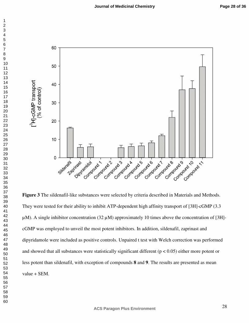
In vitro. A single concentration (32 µM) of the compounds shown in Figure 3 were tested for their
ability to inhibit [3H]-cGMP uptake into inside-out vesicles (IOV). Sildenafil was introduced as a
reference inhibitor, and the effect of the well-known PDE5 inhibitors (zaprinast and dipyridamole) were
also determined. Figure 3 shows that sildenafil inhibited 85% of cGMP uptake. The corresponding
values for zaprinast and dipyridamole were approximately 95%. However, several of the compounds
brought forward by VLS showed even higher potency than sildenafil. Compounds 1 and 2 blocked the
transport completely, whereas compounds 3, 4, 5, 6 and 7 inhibited transport more effectively than the
starting point molecule sildenafil.
Ki-values of the most potent inhibitors of cGMP transport where calculated, and while Sildenafil,
which was used as reference inhibitor, showed a Ki-value of 1200 ± 170 nM, the corresponding values
of compounds 1 and 2 were 75.3 ± 3.1 nM and 65.3 ± 6.4 nM, respectively.
Discussion
In this study, molecular modeling techniques were used for construction of an ABCC5 model, and for
identification of its interactions with sildenafil derivates. Molecular modeling and docking may help to
elucidate molecular interactions between drugs and drug targets, aiding the search to understand the
intermolecular forces involved in determining the potency and the specificity of the drug. However,
homology modeling of transporters must be regarded with caution; there are few templates available, if
any, and they often have low resolution, and homology between the target transporter and the template
may also be low. The sequence identity between target and template in the present study was relatively
Page 7 of 36
ACS Paragon Plus Environment
Journal of Medicinal Chemistry
123456789101112131415161718192021222324252627282930313233343536373839404142434445464748495051525354555657585960
8
low (24%), so even though a multiple sequence alignment, which highlights evolutionary relationships
and increases the probability that corresponding sequence positions are correctly aligned, was used to
guide the homology modeling procedure, the low homology gives elements of uncertainty to the
resulting model. Homology modeling of membrane transporters often implies low homology; the
sequence identity between the frequently used template Aquifex aeolicus leucine tranporter LeuTAa and
modeled monoamine transporters is ~20%,16 and the sequence identities in the transmembrane regions
between the G-protein coupled receptors and Bacteriorhodopsin are 6–11%, even though their TMH
arrangement is the same.17 The quality of the template must also be considered, both in regard to low
resolution, and in regard to the fact that the amphiphilic nature of membrane proteins causes difficulties
in experimental structure determination. Thus, even when crystallization is successful, the protein is no
longer in its natural environment, and conformational disruption of the transporter due to the presence of
detergent molecules during crystallization can not be excluded. Interestingly, recent cysteine cross-
linking studies, linking residues in TMH3 and TMH9 of ABCB1,18 and between residues in the C-
terminal ends of the two NBDs of ABCB1,19 have shown that ABCB1 still functions when covalently
cross-linked. This indicates that the Sav1866 X-ray crystal structure,13 which is in an outward-facing
conformation, also may serve as a template for modeling and running VLS on ABCC5, and also might
yield interesting inhibitors.
Structural flexibility was taken into account when performing docking and VLS on the ABCC5
model. A crystal structure of a transporter may not be a realistic representation of the transporter in its
native form, and transporters may undergo substantial conformational changes during the transport
cycle. Large ranges of motion, changing the accessibility of the transporter from a cytoplasmic (inward)
facing to an extracellular (outward) facing conformation, have been revealed from X-ray crystal
structures of the bacterial ABC transporter lipid flippase, MsbA, trapped in different conformations.14
Induced-fit, which has been demonstrated in a cysteine-scanning mutagenesis and oxidative cross-
linking study of substrate-induced changes in ABCB1,20 and conformational changes due to transport,
Page 8 of 36
ACS Paragon Plus Environment
Journal of Medicinal Chemistry
123456789101112131415161718192021222324252627282930313233343536373839404142434445464748495051525354555657585960
9
may be an important part of ligand recognition. The energy-based torsional sampling ("fumigation")
generated additional conformations of the ligand binding area of ABCC5, with lower energies than the
starting model. Of the three conformations used for docking and VLS, the two lowest energy
conformations were preferred by the ligands, indicating that these conformations were more
"druggable". The 4D docking, where the ligands are free to "choose" their preferred binding site
conformation, may be viewed as a theoretical model of how the ligand is attracted to the drug target in a
relatively more active conformation. Insight into structural changes of the drug target for yielding a
lower energy drug - drug target complex may elucidate how the conformation of the binding site
contributes to the adoption of an energetically favorable complex. Ideally, these observations can aid to
predict how a designed drug will fit into the drug target.
In an interdisciplinary approach combining in silico studies (theoretical molecular modeling), and in
vitro studies (experimental pharmacology), the ABC transporter model may be considered as a working
tool for generating hypotheses and designing further experimental studies related to ABC transporter
structure and function and their ligand interactions. In vitro studies and transporter modeling are
complementary to each other in an iterating process towards a better understanding of the structure and
function of these proteins. The VLS add-on to the Internal Coordinate Mechanics (ICM) program21
(ICM-VLS) has previously been applied to identify new leads for a number of targets.22, 23 In the present
study, the VLS docking correctly predicted seven ligands as having a higher binding affinity to ABCC5
than sildenafil. The most potent compounds, compounds 1 and 2, showed Ki-values of 50 -100 nM. As
far as we know, these are the most potent inhibitors described for ABCC5.
Both compounds 1 and 2 features a salicylic acid moiety, and the pKa of salicylic acid is 2.97. At
physiological pH, the carboxylic acid moiety of salicylic acids tend to be negatively charged, and
docking indicated that these compounds oriented the negatively charged COO- group towards Lys448
(TMH6). This ionic interaction may explain why these two compounds were the most potent ABCC5
inhibitors. The characteristic interaction of the guanine-like ring of the ligands with Gln190 (TMH1) of
Page 9 of 36
ACS Paragon Plus Environment
Journal of Medicinal Chemistry
123456789101112131415161718192021222324252627282930313233343536373839404142434445464748495051525354555657585960

10
ABCC5, which was found in most predicted complexes, has also been observed in X-ray crystal
structures of PDE5 complexed with either guanosine monophosphate (GMP) or sildenafil. In these
complexes, the guanine ring of GMP, or the guanine-like ring of sildenafil, has hydrogen bond
interactions with a glutamine (Gln817).24
The drug binding pocket of the ABCC5 model featured several polar amino acids (Gln190 (TMH1),
Asn441 and Thr444 (TMH6), and Ser872 (TMH7)) and a positively charged amino acid (Lys448
(TMH6)), contributing to a molecular environment favorable for transport of organic anions. ABCC5
has been shown to transport cGMP with high affinity (Km-value 2.1 µM ),5 and we have previously
restored ATP-dependent cGMP transport into proteoliposomes by membrane proteins from human
erythrocytes25 and cGMP-induced ATPase activity.25 At physiological intracellular cGMP concentration
there are a number of leads that indicate ABCC5 as a high affinity and selective transporter for cGMP.26
In the present study, the observed cGMP Km-value of 2.2 µM is in agreement with the Km-value of 2.1
µM reported earlier.5 The observation from the binding affinity studies of order of potency of the known
ABCC5 inhibitors sildenafil, zaprinast and dipyridamole, with zaprinast and dipyridamole being equally
potent, and both being more potent than sildenafil, were also in agreement with previous observations.27
In the present VLS study, it was observed that each ligand had a tendency to prefer two main
orientations, one towards Gln190 (in TMH1, corresponding to Leu65 (ABCB1)), and one towards
Val253 (In TMH5, corresponding to Ile306 (ABCB1). The differences in binding energy between
different orientations of the same ligand did not differ significantly. This is in accordance with a study
on ABCB1 where cysteine-scanning mutagenesis and reaction with an methanethiosulphonate (MTS)
thiol-reactive analogue of verapamil (MTS-verapamil) showed that mutants Leu65Cys (TMH1) and
Ile306Cys (TMH5) modified with MTS-verapamil have slightly different characteristics, indicating that
the bound verapamil molecules in these mutants have different orientations and that the protein can
function quite well with the substrate bound in different orientations.28 Theoretically, even though ten
different conformations of each ligand were evaluated by ICM during VLS, focusing on only the ligand
Page 10 of 36
ACS Paragon Plus Environment
Journal of Medicinal Chemistry
123456789101112131415161718192021222324252627282930313233343536373839404142434445464748495051525354555657585960

11
orientation with best score may lead to missing out better inhibitors oriented in a pose yielding a poorer
score.
Despite more than 30 years of research and development of drugs for use in co-therapy to
chemotherapeutic drugs to overcome multidrug resistance (MDR), these drugs has come no further than
to late-phase clinical trials.29 The possible roles of PDE5 inhibitors in overcoming MDR is currently
gaining attention as emerging evidence indicates that sildenafil and sildenafil-like drugs may enhance
the sensitivity of certain tumor cells to chemotherapeutic drugs. It has also been demonstrated that
sildenafil also inhibits the activity of ABC transporters such as ABCB1 and ABCG2, reversing MDR in
cancer cells mediated by these transporters.30 In the present study, we chose to use a sildenafil
substructure in order to achieve compounds for VLS, instead of using substructures of the slightly more
potent zaprinast and dipyridamole. Zaprinast has been unsuccessful in clinical trials, and both zaprinast
and dipyridamole are less selective than sildenafil.31 Very close analogs of sildenafil were considered in
this study, and even though the search pattern used for finding sildenafil analogs was the ring system in
Figure 5A, the 11 compounds that were selected for testing all have much more overlap with sildenafil
than the ring system in figure 5A. Vendors presumably have generated libraries around the drug, and the
additional drug-likeness filter could have biased the tested compounds even more towards close analogs
of sildenafil. The ABCC5 model could also have been used to search for molecules that could not be
trivially found from the starting sildenafil substructure, possibly discovering entirely new chemical
scaffolds, but since sildenafil is a very interesting molecule in regard to MDR, the goal in this study was
to create chemical diversity around sildenafil.
The IOV method is very time consuming, so using the ABCC5 model as a an additional filter to select
compounds to test for binding to ABCC5 in vitro, instead of testing all 105 compounds, may have saved
time and lab resources. The structural and computational guidance used in the present study aided the
identification of 7 compounds having higher effect on cGMP efflux effect than sildenafil. The
sildenafil-like hits compounds from the VLS in the present study may represent candidates for lead
Page 11 of 36
ACS Paragon Plus Environment
Journal of Medicinal Chemistry
123456789101112131415161718192021222324252627282930313233343536373839404142434445464748495051525354555657585960
12
optimization in the search for novel drugs that can be used to overcome MDR. Such drugs may also be
used to inhibit cGMP efflux from cancer cells, possibly enhancing the body’s own defense mechanism
of increasing cGMP levels.
Coordinates of the ABCC5 model are available upon request.
Experimental section
Software. The ICM program21 version 3.6-1e was used for homology modeling, compound docking
and sildenafil substructure search. The ICM program package included the ICM VLS add-on and access
to Molcart, a database of chemical structures for ~4M of commercially available compounds.
Chemicals. cGMP (Sigma Aldrich, St. Louis, MO, USA), 8[3H]-cGMP (Perkin Elmer, Boston, MA,
USA), Sildenafil (a kind gift from Pfizer Inc., NY, USA), 11 compounds based on VLS were purchased
from Ambinter (c/o Greenpharma SAS, 3, allée du titane, 45100 Orléans, France). Other chemicals of
analytical grade.
Homology modeling. A multiple sequence alignment of mouse ABCB1 (SWISS-PROT accession
number P06795), human ABCB1 (SWISS-PROT accession number P08183), human ABCC4 (SWISS-
PROT accession number O15439), human ABCC5 (SWISS-PROT accession number O15440), human
ABCC11 (SWISS-PROT accession number Q9BX80), Escherichia coli MsbA (SWISS-PROT
accession number P60752) and Vibrio cholerae MsbA (Q9KQW9) was generated using T-COFFEE32
version 4.71 (http://tcoffee.vital-it.ch/cgi-bin/Tcoffee/tcoffee_cgi/index.cgi) and used to guide the
input alignment (Figure 4) for the homology modeling.
The ABCC5 model was constructed using the X-ray crystal structure of the Mus musculus ABCB115
(PDB code 3G60), complexed with the ligand cyclic-tris-(R)-valineselenazole (QZ59-RRR), as a
template. The ICM homology modeling module constructs the molecular model by homology from core
sections defined by the average C-alpha atom positions in conserved regions. Loops were automatically
Page 12 of 36
ACS Paragon Plus Environment
Journal of Medicinal Chemistry
123456789101112131415161718192021222324252627282930313233343536373839404142434445464748495051525354555657585960

13
constructed in the ICM homology module by selecting the best-fitting loop based on calculating maps
around loops that were retrieved from the PDB databank33 and matched in regard to sequence similarity
and sterical interactions with the surroundings of the model, and scoring of their relative energies. The
loop connecting NBD1 and TMD2 in the template, which is missing in the template, was included in the
loop search procedure.
The ABCC5 model was refined by globally optimizing side-chain positions and annealing of the
backbone using the RefineModel macro of ICM. The RefineModel macro includes (1) a side-chain
conformational sampling using 'Montecarlo fast',34 (2) 5 iterative annealings of the backbone with
tethers, and (3) a second side-chain conformational sampling using 'Montecarlo fast'. In 'Montecarlo
fast', the conformational space of a molecule is sampled with the ICM global optimization procedure.
An iteration of the procedure, which consists of a random move followed by a local energy
minimization, and calculation of the complete energy, is accepted or rejected based on energy and
temperature. Tethers in the iterative annealings of the backbone are harmonic restraints pulling an atom
in the model to a static point in space represented by a corresponding atom in the template. The
RefineModel macro was followed by the “Regul” option of ICM. “Regul”, which denotes
“Regularization”, is a procedure for fitting a protein model with the ideal covalent geometry of the
residues.
The refined ABCC5 model was energy minimized using the leaprc.ff03 force field of the AMBER 9.0
program package.35 Two energy minimizations were performed; (1) with restrained backbone by 500
cycles of the steepest descent minimization followed by 500 steps of conjugate gradient minimization,
and (2) with no restraints by 1000 cycles of the steepest descent minimization followed by 1500 steps of
conjugate gradient minimization. A 10 Å cutoff radius for non-bonded interactions and a dielectric
multiplicative constant of 1.0 for the electrostatic interactions were used in the molecular mechanics
calculations.
The stereochemical quality of the ABCC5 model was checked using the SAVES Metaserver for
Page 13 of 36
ACS Paragon Plus Environment
Journal of Medicinal Chemistry
123456789101112131415161718192021222324252627282930313233343536373839404142434445464748495051525354555657585960
14
analyzing and validating protein structures http://nihserver.mbi.ucla.edu/SAVES/. Programs run were
Procheck,36 What_check,37 and Errat.38
4D VLS docking. In order to investigate putative ligand binding modes in the highly flexible
transporter protein, energy-based torsional sampling was used to generate additional conformations of
the ligand binding area of ABCC5. This computational technique called “fumigation”39 is aimed at
generating more “druggable” conformations of ligand binding pockets. Fumigation is based on torsional
sampling of the binding pocket side chains in the presence of a repulsive density representing a generic
ligand, using the ICM biased probability Monte Carlo sampling procedure. The pocket used for
torsional sampling was defined using the ligand skin mesh of QZ59-RRR from the template.15
In order to get a threshold score for the VLS, known binders to ABCC5 were docked using 4D
docking. Thus, the VLS threshold was based scores calculated by docking sildenafil, MK571,
glibenclamide, sulfinpyrazone, trequinsin, benzbromarone, verapamil, zaprinast, dipyridamole,
probenecid and cGMP. Ligands were prepared in the ICM ligand editor and converted to 3D when
setting up the ligand during the docking session. Charges were also assigned in this step. A 4D docking
procedure was used employing the binding pocket conformational ensembles. In this approach, the
pocket ensemble conformations is used as an extra, fourth dimension of the ligand sampling space,
allowing ligand docking to the multiple binding pocket conformations in a single docking simulation.40
The 3 binding pocket conformations with lowest energy were used in the 4D docking procedure.
The Molcart chemical management system, featuring a number of compound databases (including
Ambinter, Chembrigde, Lifechemicals, etc.) that can be analyzed and searched using ICM
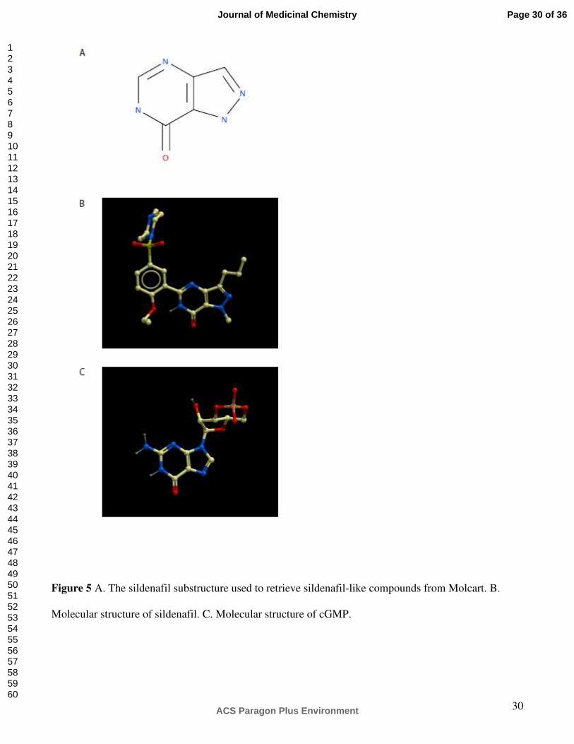
cheminformatic tools, was used to retrieve compounds with a common substructure as in sildenafil
(Figure 5). This substructure is a guanine-like moiety, resembling the guanine part of cGMP. A database
of 105 sildenafil-like compounds was obtained and used for a 4D VLS docking into the ABCC5
transporter. ICM-VLS, which is a combination of internal coordinate docking methodology and global
optimization scheme, provides empirically adjusted scoring functions and accurate and fast potentials,
Page 14 of 36
ACS Paragon Plus Environment
Journal of Medicinal Chemistry
123456789101112131415161718192021222324252627282930313233343536373839404142434445464748495051525354555657585960

15
yielding efficient virtual screening methodology in which ligands are fully and continuously flexible. 11
hits with scores above the score threshold were selected based on the drug-likeness score and ordered
from Ambinter (http://www.ambinter.com/) for in vitro testing. The ICM drug-likeness score is
predicted based on 5000 marketed drugs from the World Drug Index (WDI) (positives) and 10000 non-
drug compounds (negatives).
Preparation of IOV. In the present study, a modified version of the Steck IOV preparation41 was
used. Fresh human EDTA blood was used to produce IOVs from human erythrocytes. All steps after
collecting the blood were performed at 0-4ºC. The cells were sedimented by centrifugation 2300g for 15
minutes. Plasma and buffycoat were discarded, and the red blood cells were washed 3 times by
centrifugation at 1000g 5 mM Tris•HCl, 113 mM KCl, pH=8.1. Cells were lysed in 10 volumes of 5
mM TrisHCl, 0.5 mM EGTA, 4 mM KCl, pH=8.1 and washed by repeated centrifugation at 20000g for
20 minutes and resuspension in the same buffer until ghosts were milky white. Vesiculation was
initiated by adding 39 volumes of 500 nM TrisHCl, pH=8.2 to one volume of cell suspension. The
vesiculation was completed by homogenization of vesicles and unsealed ghosts by passing the
suspension five times through a 27G cannula. IOVs, right-side out vesicles and unsealed vesicles and
ghosts were separated by ultracentrifugation (100.000 x g) over night using a density gradient from 1,048
g/ml to 1,146 g/ml Histodenz (Sigma-Aldrich, St. Louis, MO, USA) in 5 mM Tris, 3 mM KCl, 0.3 mM
EGTA. The uppermost band was collected, washed and resuspended in 1.47 mM KH2PO4, 81 mM K2HPO4
and 140 mM KCl, pH 7.6. Sidedness was verified using acetylcholinesterase accessibility.
Transport assay. cGMP is transported out of cells via ABCC5 with a Km value of 2.1 µM.5 In the
present study cGMP uptake into IOVs was determined for an inhibitor concentration range of 0 - 320
µM, and the [3H]-cGMP transport was characterized with a Km value of 2.2 ± 0.35 µM. IOVs were
incubated for 60 minutes with or without 2.0 mM ATP in a mixture containing 20 mM Tris•HCl, 10
mM MgCl2, 1 mM EGTA, 2 µM [3H]-labeled cGMP, 121 mM KCl, pH=8.0 at 37º, and inhibitor in a
concentration range from 0 µM to 316 µM. The transport process was stopped with addition of ice-cold
Page 15 of 36
ACS Paragon Plus Environment
Journal of Medicinal Chemistry
123456789101112131415161718192021222324252627282930313233343536373839404142434445464748495051525354555657585960

16
1.47 mM KH2PO4, 8,1 mM K2HPO4 and 140 mM KCl, pH 7.6. The IOVs were separated from the
incubation medium by filtration (nitrocellulose membrane, 0.22 µm GSWP, Millipore, Billerica, MA, USA).
The radioactivity on the filters was quantified by liquid scintillation (Ultima Gold XR, Packard, Groningen,
The Netherlands) in a Packard 1900 TR Liquid Scintillation analyzer. DMSO was needed to dissolve some
of the inhibitors and was added to a similar concentration in the control samples (without inhibitors).
Determination of Ki-values. The IC50-values were determined and transformed to Ki-values
according to Cheng and Prusoff.42
Statistics. The results are presented as mean value ± SEM. Unpaired two-tailed t test with Welch
correction was performed using GraphPad InStat version 3.06, GraphPad Software, San Diego,
California, USA.
Acknowledgements
The gift of sildenafil from Pfizer Ltd is acknowledged. We thank Ms. Natalia Smaglyukova for
excellent technical assistance.
References
(1) Luesley, D.M.; Blackledge, G.R.; Chan, K.K.; Newton, J.R. Random urinary cyclic 3',5'
guanosine monophosphate in epithelial ovarian cancer: relation to other prognostic variables and to
survival. Br J Obstet Gynaecol. 1986, 93, 380-385.
(2) Orbo, A.; Jaeger, R.; Sager, G. Urinary levels of cyclic guanosine monophosphate
(cGMP) in patients with cancer of the uterine cervix: a valuable prognostic factor of clinical outcome?
Eur J Cancer. 1998, 34, 1460-1462.
(3) Peracchi, M.; Bamonti-Catena, F.; Lombardi, L.; Toschi, V.; Bareggi, B.; Cortelezzi, A.;
Page 16 of 36
ACS Paragon Plus Environment
Journal of Medicinal Chemistry
123456789101112131415161718192021222324252627282930313233343536373839404142434445464748495051525354555657585960

17
Maiolo, A.T.; Polli, E.E. Plasma cyclic nucleotide levels in monitoring acute leukemia patients. Cancer
Detect Prev. 1985, 8, 291-295.
(4) Turner, G.A.; Greggi, S.; Guthrie, D.; Benedetti Panici, P.; Ellis, R.D.; Scambia, G.;
Mancuso, S. Monitoring ovarian cancer using urine cyclic GMP. A two-centre study. Eur J Gynaecol
Oncol. 1990, 11, 421-427.
(5) Jedlitschky, G.; Burchell, B.; Keppler, D. The multidrug resistance protein 5 functions as
an ATP-dependent export pump for cyclic nucleotides. J Biol Chem. 2000, 275, 30069-30074.
(6) Pilz, R.B.; Broderick, K.E. Role of cyclic GMP in gene regulation. Frontiers in
bioscience : a journal and virtual library. 2005, 10, 1239-1268.
(7) Saier, M.H., Jr. A functional-phylogenetic classification system for transmembrane
solute transporters. Microbiol Mol Biol Rev. 2000, 64, 354-411.
(8) Dean, M.; Rzhetsky, A.; Allikmets, R. The human ATP-binding cassette (ABC)
transporter superfamily. Genome Res. 2001, 11, 1156-1166.
(9) Ravna, A.W.; Sylte, I.; Sager, G. Molecular model of the outward facing state of the
human P-glycoprotein (ABCB1), and comparison to a model of the human MRP5 (ABCC5). Theor Biol
Med Model. 2007, 4, 33.
(10) Ravna, A.W.; Sylte, I.; Sager, G. Binding site of ABC transporter homology models
confirmed by ABCB1 crystal structure. Theor Biol Med Model. 2009, 6, 20.
(11) Ravna, A.W.; Sager, G. Molecular model of the outward facing state of the human
multidrug resistance protein 4 (MRP4/ABCC4). Bioorg Med Chem Lett. 2008, 18, 3481-3483.
(12) Ravna, A.W.; Sylte, I.; Sager, G. A molecular model of a putative substrate releasing
conformation of multidrug resistance protein 5 (MRP5). Eur J Med Chem. 2008, 43, 2557-2567.
(13) Dawson, R.J.; Locher, K.P. Structure of a bacterial multidrug ABC transporter. Nature.
2006, 443, 180-185.
(14) Ward, A.; Reyes, C.L.; Yu, J.; Roth, C.B.; Chang, G. Flexibility in the ABC transporter
Page 17 of 36
ACS Paragon Plus Environment
Journal of Medicinal Chemistry
123456789101112131415161718192021222324252627282930313233343536373839404142434445464748495051525354555657585960
18
MsbA: Alternating access with a twist. Proc Natl Acad Sci U S A. 2007, 104, 19005-19010.
(15) Aller, S.G.; Yu, J.; Ward, A.; Weng, Y.; Chittaboina, S.; Zhuo, R.; Harrell, P.M.; Trinh,
Y.T.; Zhang, Q.; Urbatsch, I.L.; Chang, G. Structure of P-glycoprotein reveals a molecular basis for
poly-specific drug binding. Science. 2009, 323, 1718-1722.
(16) Beuming, T.; Shi, L.; Javitch, J.A.; Weinstein, H. A comprehensive structure-based
alignment of prokaryotic and eukaryotic neurotransmitter/Na+ symporters (NSS) aids in the use of the
LeuT structure to probe NSS structure and function. Mol Pharmacol. 2006, 70, :1630-1642.
(17) Hibert, M.F.; Trumpp-Kallmeyer, S.; Bruinvels, A.; Hoflack, J. Three-dimensional
models of neurotransmitter G-binding protein-coupled receptors. Mol Pharmacol. 1991, 40, 8-15.
(18) Loo, T.W.; Bartlett, M.C.; Clarke, D.M. Human P-glycoprotein is active when the two
halves are clamped together in the closed conformation. Biochem Biophys Res Commun. 2010, 395,
436-440.
(19) Verhalen, B.; Wilkens, S. P-glycoprotein retains drug-stimulated ATPase activity upon
covalent linkage of the two nucleotide binding domains at their C-terminal ends. J Biol Chem. 2011,
286, 10476-10482.
(20) Loo, T.W.; Bartlett, M.C.; Clarke, D.M. Substrate-induced conformational changes in the
transmembrane segments of human P-glycoprotein. Direct evidence for the substrate-induced fit
mechanism for drug binding. J Biol Chem. 2003, 278, 13603-13606.
(21) Abagyan, R.; Totrov, M.; Kuznetsov, D.N. ICM - a new method for protein modeling
and design. Applications to docking and structure prediction from the distorted native comformation. J
Comp Chem. 1994, 15, 488-506.
(22) Cavasotto, C.N.; Orry, A.J.; Murgolo, N.J.; Czarniecki, M.F.; Kocsi, S.A.; Hawes, B.E.;
O'Neill, K.A.; Hine, H.; Burton, M.S.; Voigt, J.H.; Abagyan, R.A.; Bayne, M.L.; Monsma, F.J., Jr.
Discovery of novel chemotypes to a G-protein-coupled receptor through ligand-steered homology
modeling and structure-based virtual screening. J Med Chem. 2008, 51, 581-588.
Page 18 of 36
ACS Paragon Plus Environment
Journal of Medicinal Chemistry
123456789101112131415161718192021222324252627282930313233343536373839404142434445464748495051525354555657585960

19
(23) Katritch, V.; Byrd, C.M.; Tseitin, V.; Dai, D.; Raush, E.; Totrov, M.; Abagyan, R.;
Jordan, R.; Hruby, D.E. Discovery of small molecule inhibitors of ubiquitin-like poxvirus proteinase
I7L using homology modeling and covalent docking approaches. J Comput Aided Mol Des. 2007, 21,
549-558.
(24) Zhang, K.Y.; Card, G.L.; Suzuki, Y.; Artis, D.R.; Fong, D.; Gillette, S.; Hsieh, D.;
Neiman, J.; West, B.L.; Zhang, C.; Milburn, M.V.; Kim, S.H.; Schlessinger, J.; Bollag, G. A glutamine
switch mechanism for nucleotide selectivity by phosphodiesterases. Mol Cell. 2004, 15, 279-286.
(25) Boadu, E.; Sager, G. ATPase activity and transport by a cGMP transporter in human
erythrocyte ghosts and proteoliposome-reconstituted membrane extracts. Biochim Biophys Acta. 2000,
1509, 467-474.
(26) Sager, G.; Ravna, A.W. Cellular efflux of cAMP and cGMP - a question about
selectivity. Mini Rev Med Chem. 2009, 9, 1009-1013.
(27) Sundkvist, E.; Jaeger, R.; Sager, G. Pharmacological characterization of the ATP-
dependent low K(m) guanosine 3',5'-cyclic monophosphate (cGMP) transporter in human erythrocytes.
Biochem Pharmacol. 2002, 63, 945-949.
(28) Loo, T.W.; Bartlett, M.C.; Clarke, D.M. Transmembrane segment 1 of human P-
glycoprotein contributes to the drug-binding pocket. Biochem J. 2006, 396, 537-545.
(29) Wu, C.P.; Calcagno, A.M.; Ambudkar, S.V. Reversal of ABC drug transporter-mediated
multidrug resistance in cancer cells: evaluation of current strategies. Current molecular pharmacology.
2008, 1, 93-105.
(30) Shi, Z.; Tiwari, A.K.; Patel, A.S.; Fu, L.W.; Chen, Z.S. Roles of sildenafil in enhancing
drug sensitivity in cancer. Cancer research. 2011, 71, 3735-3738.
(31) Kulkarni, S.K.; Patil, C.S. Phosphodiesterase 5 enzyme and its inhibitors: update on
pharmacological and therapeutical aspects. Methods and findings in experimental and clinical
pharmacology. 2004, 26, 789-799.
Page 19 of 36
ACS Paragon Plus Environment
Journal of Medicinal Chemistry
123456789101112131415161718192021222324252627282930313233343536373839404142434445464748495051525354555657585960

20
(32) Notredame, C.; Higgins, D.G.; Heringa, J. T-Coffee: A novel method for fast and
accurate multiple sequence alignment. J Mol Biol. 2000, 302, 205-217.
(33) Berman, H.M.; Westbrook, J.; Feng, Z.; Gilliland, G.; Bhat, T.N.; Weissig, H.;
Shindyalov, I.N.; Bourne, P.E. The Protein Data Bank. Nucleic Acids Res. 2000, 28, 235-242.
(34) Abagyan, R.; Totrov, M. Biased probability Monte Carlo conformational searches and
electrostatic calculations for peptides and proteins. J Mol Biol. 1994, 235, 983-1002.
(35) Case, D.A.; Darden, T.A.; Cheatham III, T.E.; Simmerling, C.L.; Wang, J.; Duke, R.E.;
Luo, R.; Merz, K.M.; Pearlman, D.A.; Crowley, M.; Walker, R.C.; Zhang, W.; Wang, B.; Hayik, A.;
Roitberg, A.; Seabara, G.; K.F., W.; Paesani, F.; Wu, X.; Brozell, S.; Tsui, V.; Gohlke, H.; Yang, L.;
Tan, C.; Mongan, J.; Hornak, V.; Cui, G.; Beroza, P.; Mathews, D.H.; Schafmeister, C.; Ross, W.S.;
Kollman, P.A., AMBER 9, 2006, University of California: San Francisco.
(36) Laskoswki, R.A.; MacArthur, M.W.; Moss, D.S.; Thorton, J.M. PROCHECK: a program
to check the stereochemical quality of protein structures. J Appl Cryst. 1993, 26, 283-291.
(37) Hooft, R.W.; Vriend, G.; Sander, C.; Abola, E.E. Errors in protein structures. Nature.
1996, 381, 272.
(38) Colovos, C.; Yeates, T.O. Verification of protein structures: patterns of nonbonded
atomic interactions. Protein Sci. 1993, 2, 1511-1519.
(39) Abagyan, R.; Kufareva, I. The flexible pocketome engine for structural chemogenomics.
Methods Mol Biol. 2009, 575, 249-279.
(40) Bottegoni, G.; Kufareva, I.; Totrov, M.; Abagyan, R. Four-dimensional docking: a fast
and accurate account of discrete receptor flexibility in ligand docking. Journal of medicinal chemistry.
2009, 52, 397-406.
(41) Steck, T.L.; Weinstein, R.S.; Straus, J.H.; Wallach, D.F. Inside-out red cell membrane
vesicles: preparation and purification. Science. 1970, 168, 255-257.
(42) Cheng, Y.; Prusoff, W.H. Relationship between the inhibition constant (K1) and the
Page 20 of 36
ACS Paragon Plus Environment
Journal of Medicinal Chemistry
123456789101112131415161718192021222324252627282930313233343536373839404142434445464748495051525354555657585960
21
concentration of inhibitor which causes 50 per cent inhibition (I50) of an enzymatic reaction.
Biochemical pharmacology. 1973, 22, 3099-3108.
Page 21 of 36
ACS Paragon Plus Environment
Journal of Medicinal Chemistry
123456789101112131415161718192021222324252627282930313233343536373839404142434445464748495051525354555657585960
22
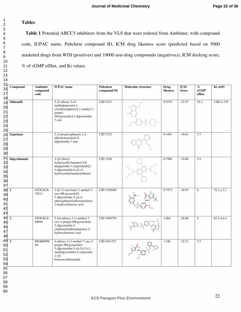
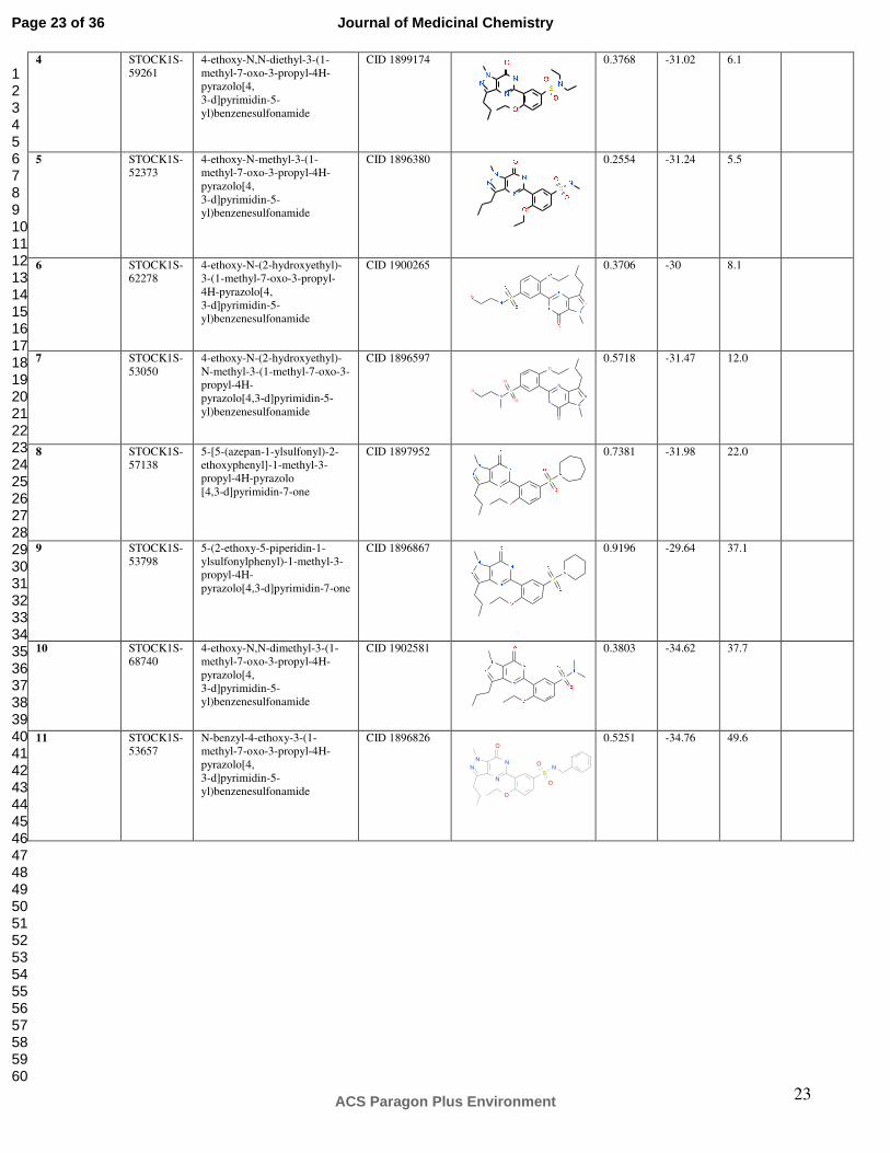
Tables
Table 1 Potential ABCC5 inhibitors from the VLS that were ordered from Ambinter, with compound
code, IUPAC name, Pubchem compound ID, ICM drug likeness score (predicted based on 5000
marketed drugs from WDI (positives) and 10000 non-drug compounds (negatives)), ICM docking score,
% of cGMP efflux, and Ki values.
Compound Ambinter
compound
code
IUPAC name Pubchem
compound ID
Molecular structure Drug
likeness
ICM
Score
%
cGMP
efllux
Ki (nM)
Sildenafil 5-[2-ethoxy-5-(4-methylpiperazin-1-yl)sulfonylphenyl]-1-methyl-3-propyl- 4H-pyrazolo[4,3-d]pyrimidin-7-one
CID 5212
N
N
O
N
N
O S
O
O
N N
0.9335 -23.55 16.2 1200 ± 170
Zaprinast 5-(2-propoxyphenyl)-2,3-dihydrotriazolo[4,5-d]pyrimidin-7-one
CID 5722
0.4367 -19.61 5.7
Dipyridamole 2-[[2-[bis(2-hydroxyethyl)amino]-4,8-di(piperidin-1-yl)pyrimido[5, 4-d]pyrimidin-6-yl]-(2-hydroxyethyl)amino]ethanol
CID 3108
0.7904 -19.08 5.9
1 STOCK3S-39213
5-[[3-(3-tert-butyl-1-methyl-7-oxo-4H-pyrazolo[4, 3-d]pyrimidin-5-yl)-4-ethoxyphenyl]sulfonylamino]-2-hydroxybenzoic acid
CID 1598490 0.7573
-30.93
0 75.3 ± 3.1
2 STOCK1S-60049
5-[[4-ethoxy-3-(1-methyl-7-oxo-3-propyl-4H-pyrazolo[4, 3-d]pyrimidin-5-yl)phenyl]sulfonylamino]-2-hydroxybenzoic acid
CID 1899750
1.064
-29.06
0 65.3 ± 6.4
3 PHAR099048
4-ethoxy-3-(1-methyl-7-oxo-3-propyl-4H-pyrazolo[4, 3-d]pyrimidin-5-yl)-N-[3-(1-methylpyrrolidin-2-yl)pyridin-2-yl] benzenesulfonamide
CID 4921527 1.246
-32.33
5.5
Page 22 of 36
ACS Paragon Plus Environment
Journal of Medicinal Chemistry
123456789101112131415161718192021222324252627282930313233343536373839404142434445464748495051525354555657585960
23
4 STOCK1S-59261
4-ethoxy-N,N-diethyl-3-(1-methyl-7-oxo-3-propyl-4H-pyrazolo[4, 3-d]pyrimidin-5-yl)benzenesulfonamide
CID 1899174
0.3768
-31.02
6.1
5 STOCK1S-52373
4-ethoxy-N-methyl-3-(1-methyl-7-oxo-3-propyl-4H-pyrazolo[4, 3-d]pyrimidin-5-yl)benzenesulfonamide
CID 1896380
0.2554
-31.24
5.5
6 STOCK1S-62278
4-ethoxy-N-(2-hydroxyethyl)-3-(1-methyl-7-oxo-3-propyl-4H-pyrazolo[4, 3-d]pyrimidin-5-yl)benzenesulfonamide
CID 1900265
0.3706
-30
8.1
7 STOCK1S-53050
4-ethoxy-N-(2-hydroxyethyl)-N-methyl-3-(1-methyl-7-oxo-3-propyl-4H- pyrazolo[4,3-d]pyrimidin-5-yl)benzenesulfonamide
CID 1896597
N
S
N
N
NN
O
O
O
O
O
0.5718
-31.47
12.0
8 STOCK1S-57138
5-[5-(azepan-1-ylsulfonyl)-2-ethoxyphenyl]-1-methyl-3-propyl-4H-pyrazolo [4,3-d]pyrimidin-7-one
CID 1897952
0.7381
-31.98
22.0
9 STOCK1S-53798
5-(2-ethoxy-5-piperidin-1-ylsulfonylphenyl)-1-methyl-3-propyl-4H- pyrazolo[4,3-d]pyrimidin-7-one
CID 1896867 0.9196
-29.64
37.1
10 STOCK1S-68740
4-ethoxy-N,N-dimethyl-3-(1-methyl-7-oxo-3-propyl-4H-pyrazolo[4, 3-d]pyrimidin-5-yl)benzenesulfonamide
CID 1902581
0.3803
-34.62
37.7
11 STOCK1S-53657
N-benzyl-4-ethoxy-3-(1-methyl-7-oxo-3-propyl-4H-pyrazolo[4, 3-d]pyrimidin-5-yl)benzenesulfonamide
CID 1896826 0.5251
-34.76
49.6
N N
S
NN N
O
O
O
O
Page 23 of 36
ACS Paragon Plus Environment
Journal of Medicinal Chemistry
123456789101112131415161718192021222324252627282930313233343536373839404142434445464748495051525354555657585960

24
Legends for Figures
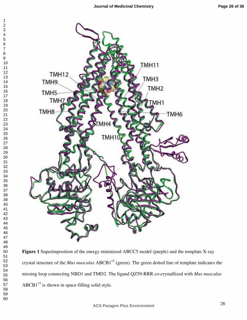
Figure 1 Superimposition of the energy minimized ABCC5 model (purple) and the template X-ray
crystal structure of the Mus musculus ABCB115 (green). The green dotted line of template indicates the
missing loop connecting NBD1 and TMD2. The ligand QZ59-RRR co-crystallized with Mus musculus
ABCB115 is shown in space-filling solid style.
Figure 2 Sildenafil docked into the binding site of ABCC5 viewed from the extracellular side. Amino
acids with hydrogen bond interactions with sildenafil are displayed as sticks colored according to atom
type (C = light yellow; H = grey; O = red; N = blue; sulfur = yellow); Gln190 (TMH1), Asn441
(TMH6), and Gln1138 (TMH12).
Figure 3 The sildenafil-like substances were selected by criteria described in Materials and Methods.
They were tested for their ability to inhibit ATP-dependent high affinity transport of [3H]-cGMP (3.3
µM). A single inhibitor concentration (32 µM) approximately 10 times above the concentration of [3H]-
cGMP was employed to unveil the most potent inhibitors. In addition, sildenafil, zaprinast and
dipyridamole were included as positive controls. Unpaired t test with Welch correction was performed
and showed that all substances were statistically significant different (p < 0.05) either more potent or
less potent than sildenafil, with exception of compounds 8 and 9. The results are presented as mean
value + SEM.
Figure 4 Alignment of Mouse ABCB1 and human ABCC5 used as input alignment for the ICM
homology modeling module. TMHs are indicated as boxes. Secondary structure of template is indicated:
Alpha helix = red cylinder; 310 helix = purple cylinder; beta sheet = green arrow.
Page 24 of 36
ACS Paragon Plus Environment
Journal of Medicinal Chemistry
123456789101112131415161718192021222324252627282930313233343536373839404142434445464748495051525354555657585960

25
Figure 5 A. The sildenafil substructure used to retrieve sildenafil-like compounds from Molcart. B.
Molecular structure of sildenafil. C. Molecular structure of cGMP.
Page 25 of 36
ACS Paragon Plus Environment
Journal of Medicinal Chemistry
123456789101112131415161718192021222324252627282930313233343536373839404142434445464748495051525354555657585960
26
Figure 1 Superimposition of the energy minimized ABCC5 model (purple) and the template X-ray
crystal structure of the Mus musculus ABCB115 (green). The green dotted line of template indicates the
missing loop connecting NBD1 and TMD2. The ligand QZ59-RRR co-crystallized with Mus musculus
ABCB115 is shown in space-filling solid style.
Page 26 of 36
ACS Paragon Plus Environment
Journal of Medicinal Chemistry
123456789101112131415161718192021222324252627282930313233343536373839404142434445464748495051525354555657585960

27
Figure 2 Sildenafil docked into the binding site of ABCC5 viewed from the extracellular side. Amino
acids with hydrogen bond interactions with sildenafil are displayed as sticks colored according to atom
type (C = light yellow; H = grey; O = red; N = blue; sulfur = yellow); Gln190 (TMH1), Asn441
(TMH6), and Gln1138 (TMH12).
Page 27 of 36
ACS Paragon Plus Environment
Journal of Medicinal Chemistry
123456789101112131415161718192021222324252627282930313233343536373839404142434445464748495051525354555657585960
28
Figure 3 The sildenafil-like substances were selected by criteria described in Materials and Methods.
They were tested for their ability to inhibit ATP-dependent high affinity transport of [3H]-cGMP (3.3
µM). A single inhibitor concentration (32 µM) approximately 10 times above the concentration of [3H]-
cGMP was employed to unveil the most potent inhibitors. In addition, sildenafil, zaprinast and
dipyridamole were included as positive controls. Unpaired t test with Welch correction was performed
and showed that all substances were statistically significant different (p < 0.05) either more potent or
less potent than sildenafil, with exception of compounds 8 and 9. The results are presented as mean
value + SEM.
Page 28 of 36
ACS Paragon Plus Environment
Journal of Medicinal Chemistry
123456789101112131415161718192021222324252627282930313233343536373839404142434445464748495051525354555657585960

29
Figure 4 Alignment of Mouse ABCB1 and human ABCC5 used as input alignment for the ICM
homology modeling module. TMHs are indicated as boxes. Secondary structure of template is indicated:
Alpha helix = red cylinder; 310 helix = purple cylinder; beta sheet = green arrow.
Page 29 of 36
ACS Paragon Plus Environment
Journal of Medicinal Chemistry
123456789101112131415161718192021222324252627282930313233343536373839404142434445464748495051525354555657585960
30
Figure 5 A. The sildenafil substructure used to retrieve sildenafil-like compounds from Molcart. B.
Molecular structure of sildenafil. C. Molecular structure of cGMP.
Page 30 of 36
ACS Paragon Plus Environment
Journal of Medicinal Chemistry
123456789101112131415161718192021222324252627282930313233343536373839404142434445464748495051525354555657585960
31
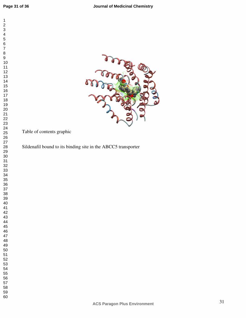
Table of contents graphic
Sildenafil bound to its binding site in the ABCC5 transporter
Page 31 of 36
ACS Paragon Plus Environment
Journal of Medicinal Chemistry
123456789101112131415161718192021222324252627282930313233343536373839404142434445464748495051525354555657585960

Figure 1 Superimposition of the energy minimized ABCC5 model (purple) and the template X-ray crystal structure of the Mus musculus ABCB115 (green). The green dotted line of template indicates the missing loop connecting NBD1 and TMD2. The ligand QZ59-RRR co-crystallized with Mus musculus ABCB115 is
shown in space-filling solid style. 101x127mm (300 x 300 DPI)
Page 32 of 36
ACS Paragon Plus Environment
Journal of Medicinal Chemistry
123456789101112131415161718192021222324252627282930313233343536373839404142434445464748495051525354555657585960
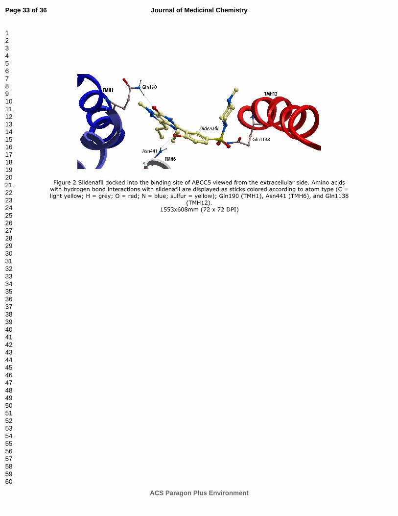
Figure 2 Sildenafil docked into the binding site of ABCC5 viewed from the extracellular side. Amino acids with hydrogen bond interactions with sildenafil are displayed as sticks colored according to atom type (C = light yellow; H = grey; O = red; N = blue; sulfur = yellow); Gln190 (TMH1), Asn441 (TMH6), and Gln1138
(TMH12). 1553x608mm (72 x 72 DPI)
Page 33 of 36
ACS Paragon Plus Environment
Journal of Medicinal Chemistry
123456789101112131415161718192021222324252627282930313233343536373839404142434445464748495051525354555657585960
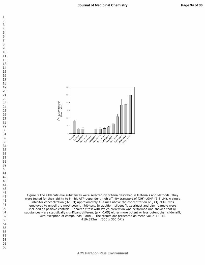
Figure 3 The sildenafil-like substances were selected by criteria described in Materials and Methods. They were tested for their ability to inhibit ATP-dependent high affinity transport of [3H]-cGMP (3.3 µM). A single
inhibitor concentration (32 µM) approximately 10 times above the concentration of [3H]-cGMP was
employed to unveil the most potent inhibitors. In addition, sildenafil, zaprinast and dipyridamole were included as positive controls. Unpaired t test with Welch correction was performed and showed that all
substances were statistically significant different (p < 0.05) either more potent or less potent than sildenafil, with exception of compounds 8 and 9. The results are presented as mean value + SEM.
419x593mm (300 x 300 DPI)
Page 34 of 36
ACS Paragon Plus Environment
Journal of Medicinal Chemistry
123456789101112131415161718192021222324252627282930313233343536373839404142434445464748495051525354555657585960

Figure 4 Alignment of Mouse ABCB1 and human ABCC5 used as input alignment for the ICM homology modeling module. TMHs are indicated as boxes. Secondary structure of template is indicated: Alpha helix =
red cylinder; 310 helix = purple cylinder; beta sheet = green arrow.
915x1168mm (72 x 72 DPI)
Page 35 of 36
ACS Paragon Plus Environment
Journal of Medicinal Chemistry
123456789101112131415161718192021222324252627282930313233343536373839404142434445464748495051525354555657585960

Figure 5 A. The sildenafil substructure used to retrieve sildenafil-like compounds from Molcart. B. Molecular structure of sildenafil. C. Molecular structure of cGMP.
191x214mm (300 x 300 DPI)
Page 36 of 36
ACS Paragon Plus Environment
Journal of Medicinal Chemistry
123456789101112131415161718192021222324252627282930313233343536373839404142434445464748495051525354555657585960


