
Seton Hall UniversityeRepository @ Seton HallSeton Hall University Dissertations and Theses(ETDs) Seton Hall University Dissertations and Theses
Fall 12-18-2018
Side Chain Modifications To Improve PeptideStructure–Activity RelationsNeelam LahankarSeton Hall University, [email protected]
Follow this and additional works at: https://scholarship.shu.edu/dissertations
Part of the Chemistry Commons
Recommended CitationLahankar, Neelam, "Side Chain Modifications To Improve Peptide Structure–Activity Relations" (2018). Seton Hall UniversityDissertations and Theses (ETDs). 2598.https://scholarship.shu.edu/dissertations/2598

i
SIDE CHAIN MODIFICATIONS TO IMPROVE PEPTIDE
STRUCTURE–ACTIVITY RELATIONSHIPS
A dissertation submitted to Seton Hall University in partial fulfillment for the
Doctor of Philosophy Degree
By:
Neelam Lahankar
December 2018
Department of Chemistry and Biochemistry
Seton Hall University
South Orange, NJ 07079
USA

ii
©2018 Neelam Lahankar

iii
Seton Hall University

iv
I am dedicating this dissertation to my beloved parents, Vasant and Subhadra and
my husband, Dr. Nandkumar. Though they are no longer part of this world, their
beautiful memories continue to shape up my life.
Also, I want to dedicate this dissertation to my daughter, Radhika and my son,
Kaushal for the numerous ways they have supported and encouraged me lovingly.

v
ABSTRACT
While natural peptides, are ideal starting points for peptide-based drug design and development,
they suffer from high conformational instability, which results in susceptibility to proteolytic degradation
and poor bioavailability. Peptidomimetics in recent years has helped circumvent these shortcomings by
improving the pharmacological properties of polypeptides. Peptidomimetics contain essential elements
(pharmacophores) that mimic a natural peptide or protein in 3D space and retain the ability to interact
with the biological target producing the same biological effect. In contrast, they offer conformationally
restricted structures, potentially minimizing cross-target interactions, which leads to better transport
properties through biologic membranes and resistance to immune responses.
Over the years, a wide variety of side chain modified polypeptides have been developed. These
modifications have been found to affect both functional as well as conformational properties of
polypeptides. This important application has proven to be the motivating force behind the two research
projects described in this thesis.
The site selective cleavage of peptide bonds is an essential complementary tool in protein
sequencing and various bioanalytical and biotherapeutic applications of peptides and proteins. In order
to cleave unreactive peptide bonds, mild and metal free, a glutamic acid selective cleavage methodology
with a broad substrate scope has been developed as reported in Chapter 2 of this thesis. This methodology
involves activation of side-chain carboxylate groups of glutamic acid residues followed by nucleophilic
attack of the backbone amide nitrogen resulting in the formation of a cyclic pyroglutamate imide
intermediate. The latter renders the scissile peptide bond susceptible to cleavage under neutral aqueous
conditions. Most importantly, the strategy provides an efficient tool for peptidolysis in a wide range of
peptide sequences, including Pro-Glu, disulfide bonding sites and at unnatural amino acid residues such
as D-amino acids in mutated peptides. The latter provides a chemical tool for cleaving peptidomimetics

vi
that are unsuitable substrates for proteases and may be potentially applicable for determining the
mutations responsible for various age-related disorders.
Though conformational instability of peptides resulting in reduced bioavailability limits their use as
promising drugs, constraining peptides by stapling (or cyclizing the side chain components in peptides)
improves their pharmacological performance by imparting structural stability and increased
bioavailability. In this regard, a biocompatible, mild and metal free stapling methodology with a broad
substrate scope is reported in Chapter 3 of this thesis. In this method, the nucleophilic side chain of lysine
is modified by reaction with an electrophilic bifunctional carbonylating agent to form urea stapled
peptides with increased α-helicity and improved proteolytic stability. Additionally, the stapling strategy
demonstrated the ability to synthesize bicyclic peptides with potential applications in peptide-based drug
design. Most importantly, the urea moiety is anticipated to form strong H-bonds, which may be useful in
catalyzing organic asymmetric transformations.
In summary, the research reported in this thesis describes design, development of peptidomimetics
through side-chain modifications of glutamic acid and lysine residues and their significant potential
applications in peptide based synthetic chemistry and drug design.

vii
AKNOWLEDGMENTS
“Coming together is a beginning; keeping together is progress; working together is success.”
-Edward Everett Hale
Success is the consistent and constructive collaboration and cooperation of multiple minds that
culminates into the fruitful accomplishment of an aim or purpose. In this vein, pursuing my Ph.D. was an
arduous task, and I am indebted to so many of you!
I could not have imagined having a better research adviser and mentor for my PhD study than Dr.
Monika Raj. Her strategic research vision offered many opportunities to challenge myself and
acknowledge my potential. She encouraged me towards diligent perseverance, which I will take with me
going forward. I would like to express my heartfelt gratitude to her for her boundless patience in
supporting me, motivating me, imparting immense knowledge and advising in my thesis writing.
I would also like to thank Dr. David Sabatino for the tremendous help throughout my research.
Though I was not part of his research group, he was always supportive and offered encouragement
whenever I needed it. Not only did his invaluable advice help me in my research and my presentations,
the completion of my thesis could not have been accomplished without his judicious comments and
mentorship of my thesis.
Additionally, my sincerest gratitude goes to Dr. Cecilia Marzabadi for being supportive while
serving on my dissertation committee and helping me prepare for my seminar. I am grateful to other
members of my matriculation committee as well, including Dr. Yuri Kazakevich and Dr. James Hanson. I
deeply appreciate additional insights into my proposed research that I received from them. I am grateful
for the solid foundation I received through the course, “Theoretical Organic Chemistry” by Dr. Hanson at
viii
the beginning of my PhD and the timely assistance by Dr. Yuri Kazakevich in fixing different instruments
with technical problems I encountered along the way.
I cannot express enough words of gratitude to the Department of Chemistry and Biochemistry for
encouraging me throughout my education. In particular, I would like to highlight the generosity of the
Department in granting me the Robert DeSimone Graduate Fellowship and nominating me as an
outstanding teaching assistant, which helped boost my self-confidence. I offer my sincere appreciation
to Dr. Kelty, Dr. Snow, Fr. Gerry, Dr. Fadeev, Dr. Gorun, Dr. Murphy, Dr. Antonacci and Dr. Khan for sharing
their knowledge and guiding me at different stages of my PhD. Also, my sincere gratitude to Maureen.
Not only was she instrumental in helping me enter the Department, but she also was a tremendous source
of help in many difficult situations.
I would be remiss not to recognize the consistent assistance from the following colleagues: Tiauna
Howard, Ryan Cohen, Zilma Muneeswaran, Lyssa Buisserth, Yonnette Sim, Anumeha Muthal, Marius
Pelmus, Mario T Da Costa and others; without your continuous support in my studies, this task would have
been difficult to achieve.
My loving, caring family is my pillar of strength. Without their continuous support, I wouldn’t be
where I am now. I am forever indebted to my late parents, Vasant and Subhadra and late husband, Dr,
Nandkumar for instilling a vision for higher education in me and inspiring me to achieve it. My daughter,
Radhika is my constant source of inspiration, who helped me persevere through my toughest adversity,
and was there when I needed someone the most. My son, Kaushal, with his calm demeanor helped to put
me at ease and move through my PhD with confident energy. He is always ready to help with my
difficulties, no matter how frivolous they may be! The same is true about my son-in-law, Bharat and
daughter-in-law, Pooja. How can I thank my sister Shobha, who has been there with me during all the
highs and lows of childhood and even into adulthood as a confidant and friend? Our strong bond over

ix
these years has resulted in a constant source of energy and mutual support. I also deeply appreciate the
emotional support offered to me by my brother-in- law, Shekhar. Finally, I would not have been able to
endure the stress of my studies without my sweet, cute grandchildren, Aaditya and Isha. How they kept
me entertained!

x
TABLE OF CONTENTS
DEDICATION ..................................................................................................................................... iv
ABSTRACT ......................................................................................................................................... v
ACKNOWLEDGMENTS ..................................................................................................................... vii
TABLE OF CONTENTS .......................................................................................................................... x
ABBREVIATIONS AND SYMBOLS ....................................................................................................... xii
LIST OF FIGURES ............................................................................................................................... xv
LIST OF SCHEMES ........................................................................................................................... xvii
LIST OF TABLES ........................................................................................................................................xviii
CHAPTER 1: GENERAL OVERVIEW OF PEPTIDE AND PEPTIDOMIMETIC BIOCHEMISTRY, METHODS FOR CHEMICAL SYNTHESIS, CHARACTERIZATION AND APPLICATIONS
1.1 INTRODUCTION TO AMINO ACID AND PEPTIDE STRUCTURE ................................................................ 1
1.2 IMPORTANCE OF PEPTIDES IN BIOLOGY ................................................................................................ 3
1.3 PEPTIDE SYNTHESIS OVERVIEW ............................................................................................................. 5
1.3.1 SOLID PHASE PEPTIDE SYNTHESIS ................................................................................................... 8
1.3.2 PROTECTING GROUP STRATEGIES ................................................................................................ 10
1.3.3 SOLID SUPPORTS .......................................................................................................................... 11
1.3.4 COUPLING REAGENTS ................................................................................................................... 13
1.3.5 PEPTIDE CLEAVAGE FROM SOLID SUPPORT ................................................................................ 15
1.4 PEPTIDOMIMETICS ............................................................................................................................... 15
1.5 THESIS OBJECTIVES ............................................................................................................................... 17
1.6 REFERENCES ........................................................................................................................................ 19
CHAPTER 2: PYROGLUTAMATE - A CYCLIC BACKBONE INTERMEDIATE FOR SELECTIVE CHEMICAL CLEAVAGE OF PEPTIDE BONDS 2.1 ABSTRACT ............................................................................................................................................. 23
2.2 CHAPTER OBJECTIVES ........................................................................................................................... 23
2.3 GRAPHICAL ABSTRACT ......................................................................................................................... 25
2.4 INTRODUCTION .................................................................................................................................... 26
2.5 RATIONAL DESIGN OF CYCLIC PYROGLUTAMYL (pGLU) IMIDE MOIETY CONTAINING PEPTIDES ........ 32
2.6 RESULTS AND DISCUSSION ................................................................................................................... 33

xi
2.7 CONCLUSIONS ..................................................................................................................................... 43
2.8 EXPERIMENTAL SECTION ..................................................................................................................... 44
2.9 REFERENCES ......................................................................................................................................... 47
CHAPTER 3: PEPTIDE STAPLING VIA UREA BRIDGING- CHEMICAL INSIGHTS INTO CYCLIC PEPTIDE
SYNTHESIS
3.1 ABSTRACT ............................................................................................................................................. 50
3.2 GRAPHICAL ABSTRACT ......................................................................................................................... 50
3.3 INTRODUCTION ..................................................................................................................................... 50
3.3.1 BIOACTIVE CYCLIC PEPTIDES ......................................................................................................... 51
3.3.2 SYNTHESIS OF CYCLIC PEPTIDES .................................................................................................... 52
3.4 CHAPTER OBJECTIVES .......................................................................................................................... 54
3.5 RESULTS AND DISCUSSION ................................................................................................................... 54
3.5.1 PRELIMINARY RESULTS .................................................................................................................. 54
3.5.2. PROTEOLYTIC STABILITY OF THE STAPLED PEPTIDE .................................................................... 63
3.5.3 SECONDARY STRUCTURE ANALYSIS OF THE STAPLED PEPTIDE .................................................... 66
3.5.4 SYNTHESIS OF A BICYCLIC PEPTIDE ................................................................................................ 66
3.6 CONCLUSIONS ..................................................................................................................................... 71
3.7. EXPERIMENTAL SECTION ................................................................................................................... 72
3.8 REFERENCES ........................................................................................................................................ 77
CHAPTER 4: CONTRIBUTIONS TO KNOWLEDGE AND FUTURE DIRECTIONS
4.1 CONTRIBUTIONS TO KNOWLEDGE MADE IN THIS THESIS .................................................................. 79
4.1.1 PYROGLUTAMATE - A CYCLIC BACKBONE INTERMEDIATE FOR SELECTIVE CHEMICAL CLEAVAGE
OF PEPTIDE BONDS ....................................................................................................................... 79
4.1.2 PEPTIDE STAPLING VIA UREA BRIDGING-CHEMICAL INSIGHTS INTO CYCLIC PEPTIDE
SYNTHESIS ..................................................................................................................................... 81
4.1.3 REFERENCES ............................................................................................................................... 83
4.2 PUBLICATIONS, CONFERENCE PRESENTATIONS, AND AWARDS ......................................................... 83
BIBLIOGRAPHY ............................................................................................................................................ 85
xii
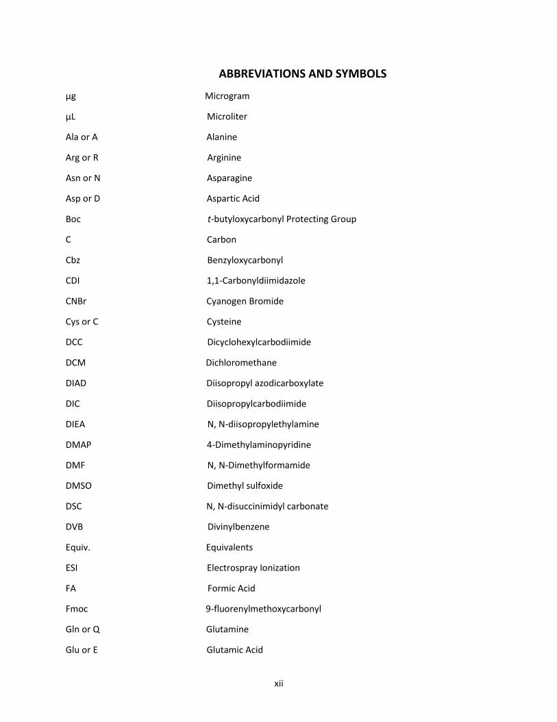
ABBREVIATIONS AND SYMBOLS
µg Microgram
µL Microliter
Ala or A Alanine
Arg or R Arginine
Asn or N Asparagine
Asp or D Aspartic Acid
Boc t-butyloxycarbonyl Protecting Group
C Carbon
Cbz Benzyloxycarbonyl
CDI 1,1-Carbonyldiimidazole
CNBr Cyanogen Bromide
Cys or C Cysteine
DCC Dicyclohexylcarbodiimide
DCM Dichloromethane
DIAD Diisopropyl azodicarboxylate
DIC Diisopropylcarbodiimide
DIEA N, N-diisopropylethylamine
DMAP 4-Dimethylaminopyridine
DMF N, N-Dimethylformamide
DMSO Dimethyl sulfoxide
DSC N, N-disuccinimidyl carbonate
DVB Divinylbenzene
Equiv. Equivalents
ESI Electrospray Ionization
FA Formic Acid
Fmoc 9-fluorenylmethoxycarbonyl
Gln or Q Glutamine
Glu or E Glutamic Acid

xiii
Gly or G Glycine
H or hr Hour
H2O Water
HATU O-(7-azabenzotriazol-1-yl)-N, N,N'N'-tetramethyluronium
hexafluorophosphate
HBTU O-benzotriazol-1-yl-N,N,N',N'-tetramethyluronium
hexafluorophosphate
HCTU O-(6-Chlorobenzotriazol-1-yl)-N, N,N',N'-tetramethyluronium
hexafluorophosphate
HF Hydrogen fluoride or Hydrofluoric Acid
His or H Histidine
HMBS Heteronuclear Multiple Bond Correlation
HOAt 1-hydroxy-7-azabenzotriazole
HOBt 1-hydroxy-benzotriazole
HPLC High Performance Liquid Chromatography
HRMS High Resolution Mass Spectrometry
Ile or I Isoleucine
IMS-MS Ion Mobility Spectrometry- Mass Spectrometry
LC Liquid Chromatography
LC-MS Liquid Chromatography-Mass Spectrometry
Leu or L Leucine
Lys or K Lysine
MeOH Methanol
Met or M Methionine
mg Milligram
Min Minute
mL Milliliter
mmol Millimole
MS Mass Spectrometer

xiv
MS/MS Tandem Mass Spectrometry
N2 Nitrogen
NCL Native Chemical Ligation
NH2 Amino group
nm Nanometer Wavelength
NMR Nuclear Magnetic Resonance
NPCF 4-nitrophenyl chloroformate
0 Degree
OAc Acetoxy
Pd (PPh3)4 Tetrakis (triphenylphosphine)palladium (0)
PEG Polyethylene glycol
Pg Protecting Group
pGlu Pyro-Glutamyl
Phe or F Phenylalanine
Pro or P Proline
PS Polystyrene
PyBroP Bromo-tris-pyrrolidino-phosphonium hexafluorophosphate
RCM Ring-closing metathesis RP HPLC Reverse Phase High Performance Liquid Chromatography
RT Room Temperature
Ser or S Serine
SPPS Solid Phase Peptide Synthesis
tBu Tert-butyl group
THF Tetrahydrofuran
Thr or T Threonine
TIPS Triisopropylsilane
tR Retention Time
Trp or W Tryptophan
Trt Trityl group

xv
Tyr or Y Tyrosine
UV-Vis Ultraviolet-Visible Spectroscopy
Val or V Valine
LIST OF FIGURES
CHAPTER 1
Figure 1.1 General amino acid structure 1
Figure 1.2 Venn diagram of amino acid properties 2
Figure 1.3 Bioactive peptides found in nature 5
Figure 1.4 Solid support for peptide synthesis 12
Figure 1.5 Functionalized resins for t-Boc/Bzl and Fmoc/tBu SPPS 13
Figure 1.6 Peptide coupling reagents 14
Figure 1.7 Peptidomimetcs: different methodologies 16
Figure 1.8 Amino acid side chain modifications 17
Figure 1.9 Structures of cyclic and bicylic compounds
synthesized in the study 19
CHAPTER 2
Figure 2.1 Graphical abstract for Glutamic Acid Selective Chemical Cleavage
Of Peptide Bonds 25
Figure 2.2 Protease digestion of a protein 26
Figure 2.3 Pyrrolide side product formation
Figure 2.4 13C NMR and 1H/15N HSQC NMR for modified Fmoc-Gly-pGlu 36
Figure 2.5 Comparison of NMR chemical shifts for cyclization of Glu: Five-
membered ring versus a six- membered ring 37
Figure 2.6 HPLC chromatogram of pGlu imide formation using optimized
reaction conditions 38
Figure 2.7 Reactivity of Asp vs Glu toward the backbone activation 41

xvi
CHAPTER 3
Figure 3.1 General method for the synthesis of urea-bridged cyclic peptides
Figure 3.2 Bioactive cyclic peptides 50
Figure 3.3 One-component and two-component stapling methodology 52
Figure 3.4 H/D exchange, MS, MS/MS, HRMS, NMR of stapled Ac-Phe-Arg-Lys-
Gly-Lys-Ala-CONH2 56
Figure 3.5 HPLC trace and MS for stapled Ac-Phe-Lys-Gly-Ala-Lys-Phe-NH2 at
250 C after 24 h 58
Figure 3.6 HPLC trace and MS for stapled Ac-Arg-Lys-Leu-Gly-Ala-Lys-Tyr-NH2
at 250C after 24 h 60
Figure 3.7 HPLC trace and MS Ac-Arg-Lys-Ala-Leu-Gly-Ala-Lys-Phe-NH2 at 250C
after 24 h 60
Figure 3.8 Selectivity of stapling: Stapled peptides Ac-Glu-Asn-Pro-Glu-Lys-Ile-
Leu-Asp-Lys-His-Val-Gln-Arg-Val-Met-NH2 and Ac-Thr-Ser-Phe-Lys-
Glu-Tyr-Trp-His-Leu-Leu-Ser-NH2 61
Figure 3.9 HPLC traces of stapled Ac-Phe-Lys-Gly-Lys-Leu-NH2 and Ac-Phe-Lys-
Phe-Lys-Leu-NH2 62
Figure 3.10 Expected cleavage sites and actual fragments for linear and stapled
peptide Ac-Glu-Asn-Pro-Glu-Lys-Ile-Leu-Asp-Lys-His-Val-Gln-Arg-Val-
Met-NH2 after exposure to trypsin 63
Figure 3.11 MS traces- Proteolytic stability of linear and urea stapled Ac-Glu-
Asn-Pro-Glu-Lys-Ile-Leu-Asp- Lys-His-Val-Gln-Arg-Val-Met-NH2 65
Figure 3.12 Helicity of linear and urea stapled Ac-Glu-Asn-Pro-Glu-Lys-Ile-Leu-Asp-
Lys-His-Val-Gln-Arg-Val-Met-NH2 66
Figure 3.13 HRMS and 1H NMR for monocyclic peptide Phe-Lys-Leu-Gly-Ala-Lys-
Phe-Glu-CONH2 68
Figure 3.14 MS and 1H NMR of bicyclic peptide Phe-Lys-Leu-Gly-Ala-Lys-Phe-Glu-

xvii
NH2 69
Figure 3.15 Extracted MS and Ion Mobility Resolved MS/MS (Overlay)
Conformations for Bicyclic Peptide Phe-Lys-Leu-Gly-Ala-Lys-Phe-
Glu-NH2 70
Figure 3.16 HPLC trace of two conformational diastereomers of the bicyclic
Peptide Phe-Lys-Leu-Gly-Ala-Lys-Phe-Glu-NH2 71
CHAPTER 4
Figure 4.1 Graphical abstract for Glutamic Acid Selective Chemical Cleavage of
Peptide Bond 79
Figure 4.2 Peptide sequencing with glutamic acid selective chemical cleavage 81
Figure 4.3 Mechanism for the urea stapling reaction 82
Figure 4.4 Urea stapled peptides as efficient organocatalysts 83
LIST OF SCHEMES
CHAPTER 1
Scheme 1.1 Peptide bond formation 3
Scheme 1.2 The first synthesis of a dipeptide by E. Fischer 6
Scheme 1.3 Solution-phase peptide synthesis of Oxytocin 7
Scheme 1.4 SPPS procedure followed by Merrifield 8.
Scheme 1.5 General solid phase peptide synthesis protocol 9
Scheme 1.6 Amide bond formation using DIC/HOBt 15
Scheme 1.7 Cyclization and cleavage of peptide bonds by formation of reactive
pGlu imide intermediate 18
Scheme 1.8 Synthesis of urea-bridged peptides 18
CHAPTER 2
Scheme 2.1 Dabsyl chloride N-terminal amino acid analysis 27

xviii
Scheme 2.2 Edman degradation 28
Scheme 2.3 Cyanogen bromide mediated cleavage of a peptide bond at
methionine residue 29
Scheme 2.4 o-iodosobenzoic acid mediated cleavage of a peptide bond at
tryptophan residue 30
Scheme 2.5 Copper (II)- organoradical conjugate mediated cleavage of a peptide
bond at serine residue 31
Scheme 2.6 Rationale for the glutamic acid-selective modification and peptide
cleavage in neutral aqueous solution 32
CHAPTER 3
Scheme 3.1 Proposed mechanism for the urea stapling reaction 54
Scheme 3.2 Synthesis of a bicyclic compound: stapling strategy 67
LIST OF TABLES
CHAPTER 1
Table 1.1 Commonly used protecting groups for the side chains in tBoc/Bzl
and Fmoc/tBu strategies 10
CHAPTER 2
Table 2.1 Acylation reagents screening for formation of pGlu imide moiety 34
Table 2.2 Glu selective amide bond cleavage of Fmoc-Val-X-Glu-Arg-Phe-Ala-
NH2 39
Table 2.3 Substrate scope of Glu-selective amide bond hydrolysis 42
Table 2.4 Glu-Selective cleavage of bioactive peptides 43
CHAPTER 3
Table 3.1 Reagent screening for the urea stapling reaction 57
Table 3.2 Optimization of conditions for the urea stapling reaction: Time 57
xix
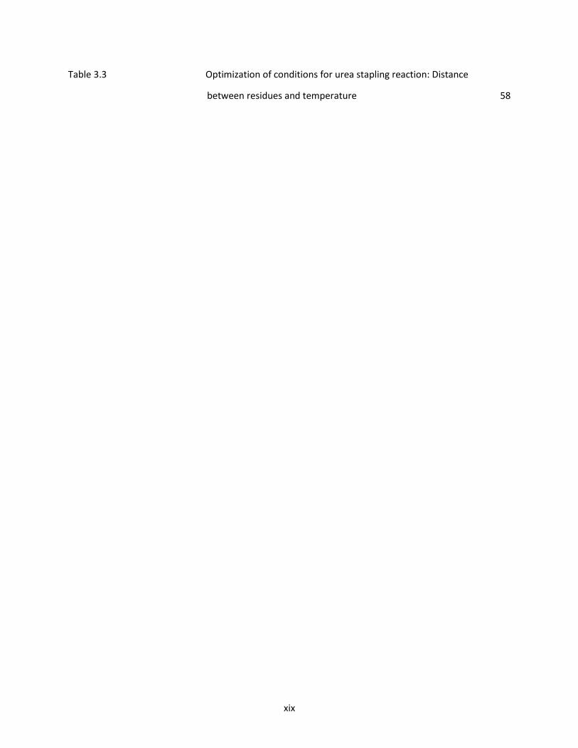
Table 3.3 Optimization of conditions for urea stapling reaction: Distance
between residues and temperature 58

1
CHAPTER 1: GENERAL OVERVIEW OF PEPTIDE AND PEPTIDOMIMETIC BIOCHEMISTRY, METHODS FOR
CHEMICAL SYNTHESIS, CHARACTERIZATION AND APPLICATIONS
1.1 INTRODUCTION TO AMINO ACID AND PEPTIDE STRUCTURE
Amino acids are critical to life as they are the building blocks of peptides and proteins.1 The twenty
naturally occurring amino acids that are found within proteins contain versatile chemical structures and
function in biological systems.2 In terms of its structure, each amino acid contains a central carbon atom,
called the α-carbon, to which both a basic amino and an acidic carboxylic acid group is bound. A hydrogen
atom and R group chemically bound to the central α-carbon satisfy the remaining tetravalent bonding
arrangement for the tetrahedral amino acid structure (Figure 1.1). At physiological pH (about 7–7.4),
amino acids exist largely as dipolar ions or zwitterions effectively making them neutral in the absence of
a charged side chain group (Figure 1.1).3 As the carbons of standard amino acids, except glycine, are
attached to four different groups, they exist as two enantiomers, D(R) and L(S). Though only L-amino acids
are common, mutations, chemical and enzymatic modifications can give rise to some D-amino acids.4
Figure 1.1 General amino acid structure.
Distinguishing the naturally occurring amino acids is the composition of the side chain R group,
which gives each amino acid its unique chemical, structural and biophysical properties. Based on the

2
nature of the R group, amino acids can be classified into the following four categories: hydrophobic
(nonpolar side chains), polar (uncharged residues with hydrophilic character), acidic (side chains with a
negatively charged carboxylate groups), and basic (side chains with a positively charged nitrogen) (Figure
1.2).2
Figure 1.2. Venn diagram of amino acid properties. (https://commons.wikimedia.org/wiki/File:Amino_Acids_Venn_Diagram.png)
Amino acids are joined by an amide or peptide bond to form peptides through condensation
reactions. In this reaction, an amino group of one amino acid reacts with the carboxylic acid group
of the other amino acid to form the peptide bond with the concomitant elimination of a water
molecule (Scheme 1.1).1 Peptides are formed with shorter (<50) amino acid sequences, whereas
peptides >50 amino acids are classified as proteins. The different amino acids in a peptide and the
order in which they are found from the N->C terminus is referred to as the primary peptide
structure.5

3
Scheme 1.1 Peptide bond formation.
Peptides have their unique structural and biochemical characteristics, such as an isoelectric pH
(pI) and ionization behavior depending on the quantity and type of each amino acid within the peptide
sequence. For example, the acid-base behavior of a peptide is defined by the zwitterionic nature of the
amino acid residues within the chain as well as the ionic structure of the N and C-terminal groups. All
these characteristics influences peptide structure and function in a biological system.
1.2 IMPORTANCE OF PEPTIDES IN BIOLOGY
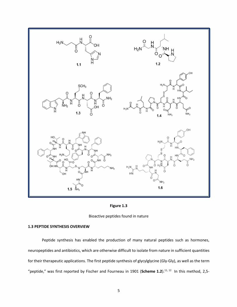
Some 7,000 naturally-occurring peptides are present in all organisms with size varying from two
to twenty amino acid residues, with biological and physiological functions that impacts bodily functions
and human health.6,7 Some noteworthy examples (Figure 1.3) include Carnosine, an antiglycating
dipeptide found in muscle tissue promoting longevity and general well-being (1.1),8 Melanostatin, a
tripeptide hormone that inhibits the release of melanocyte-stimulating hormone (1.2),9 Cholecystokinin,

4
a tetrapeptide hormone acting primarily in the brain as an anxiolytic (1.3),8 and Oxytocin, a nonapeptide
which plays a role in social bonding, childbirth and milk production (1.4).8 Peptides like Somatostatin and
Vasopressin have important roles in physiological functions.8 The tetradecapeptide Somatostatin not
only controls the production of several hormones such as the growth hormones, thyroid stimulating
hormones, insulin, glucagon and gastrointestinal hormones, but also regulates the rapid reproduction of
normal and tumor cells and acts as a neurotransmitter in the nervous system (1.5).8 Similarly, the
nonapeptide Vasopressin, an antidiuretic hormone helps water retention in the body for proper cellular
function and maintains proper flow of urine from the kidneys (1.6).8 Thus, peptides have evolved to
exhibit diverse biological roles, most prominently as signaling/regulatory molecules in a broad variety of
physiological processes, including defense, immunity, stress, growth, homeostasis, and reproduction.10
Their diverse biological functions has enabled the development of medicinal chemistry programs to
improve their therapeutic potential towards a variety of diseases and disorders.
5
Figure 1.3
Bioactive peptides found in nature
1.3 PEPTIDE SYNTHESIS OVERVIEW
Peptide synthesis has enabled the production of many natural peptides such as hormones,
neuropeptides and antibiotics, which are otherwise difficult to isolate from nature in sufficient quantities
for their therapeutic applications. The first peptide synthesis of glycylglycine (Gly-Gly), as well as the term
“peptide,” was first reported by Fischer and Fourneau in 1901 (Scheme 1.2).11, 12 In this method, 2,5-

6
diketopiperzaine was effectively hydrolyzed to Gly-Gly using acid catalyzed conditions. These harsh
conditions are not applicable to other peptides containing functional side chain groups and lengthier
sequences.
Scheme 1.2 The first synthesis of a dipeptide by E. Fischer12
In order to overcome the synthetic challenges associated with peptide synthesis, a removable
amino-protecting group was developed by M. Bergmann and L. Zervas by use of the carbobenzoxy (Cbz)
group.
In addition to the transient protecting group strategies, carbodiimide-based coupling strategies
developed in 1955 by J. C. Sheehan, G. P. Hess14 and H. G. Khorana15 gave the ability to form peptide bonds
more rapidly and efficiently. Therefore, the combination of blocking/deblocking the N-terminus followed
by activation and coupling of C-terminal amino acids enabled the production of a solution phase synthesis
approach (Scheme 1.3).13-15 This resulted in synthesis of an active hormone, the octapeptide Oxytocin
(Figure 1.3, peptide 1.4) leading to a new era of peptide synthesis, for which du Vigneaud was later
awarded the Nobel Prize.13
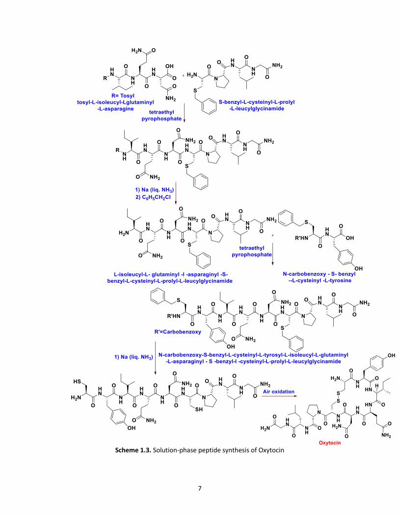
7
Scheme 1.3. Solution-phase peptide synthesis of Oxytocin
8
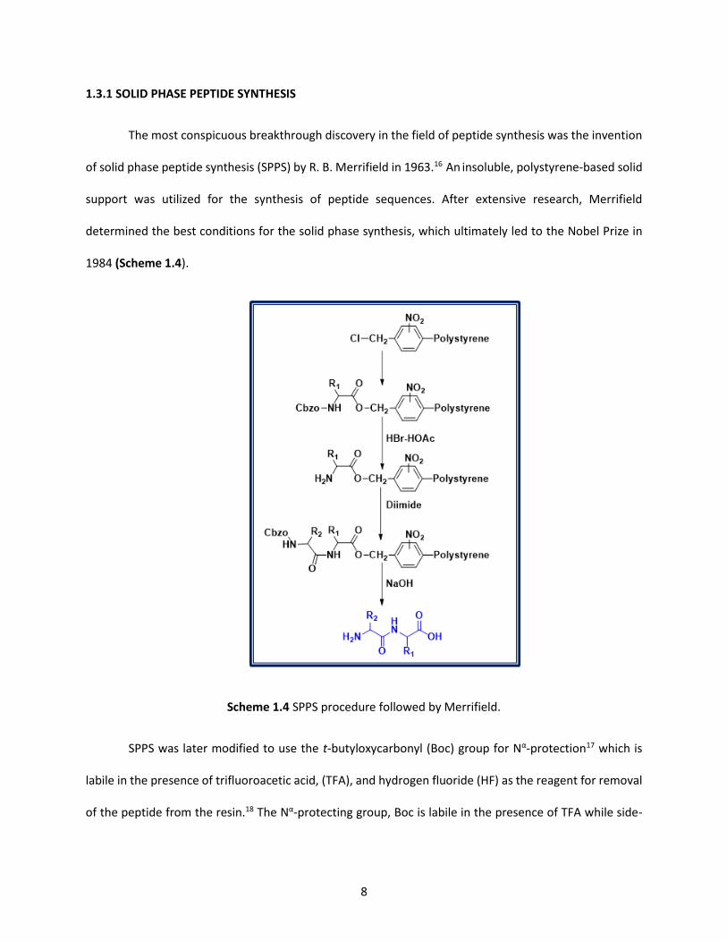
1.3.1 SOLID PHASE PEPTIDE SYNTHESIS
The most conspicuous breakthrough discovery in the field of peptide synthesis was the invention
of solid phase peptide synthesis (SPPS) by R. B. Merrifield in 1963.16 An insoluble, polystyrene-based solid
support was utilized for the synthesis of peptide sequences. After extensive research, Merrifield
determined the best conditions for the solid phase synthesis, which ultimately led to the Nobel Prize in
1984 (Scheme 1.4).
Scheme 1.4 SPPS procedure followed by Merrifield.
SPPS was later modified to use the t-butyloxycarbonyl (Boc) group for Nα-protection17 which is
labile in the presence of trifluoroacetic acid, (TFA), and hydrogen fluoride (HF) as the reagent for removal
of the peptide from the resin.18 The Nα-protecting group, Boc is labile in the presence of TFA while side-
9
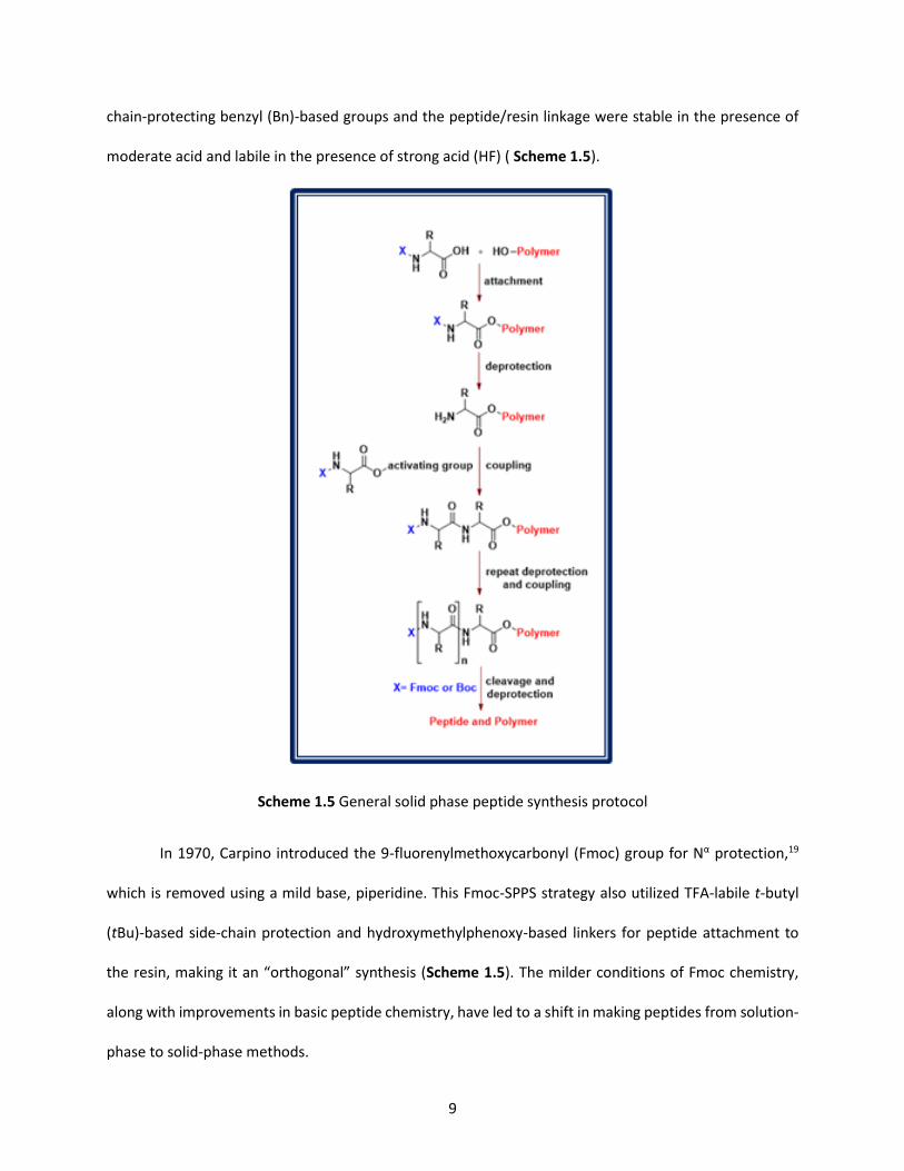
chain-protecting benzyl (Bn)-based groups and the peptide/resin linkage were stable in the presence of
moderate acid and labile in the presence of strong acid (HF) ( Scheme 1.5).
Scheme 1.5 General solid phase peptide synthesis protocol
In 1970, Carpino introduced the 9-fluorenylmethoxycarbonyl (Fmoc) group for Nα protection,19
which is removed using a mild base, piperidine. This Fmoc-SPPS strategy also utilized TFA-labile t-butyl
(tBu)-based side-chain protection and hydroxymethylphenoxy-based linkers for peptide attachment to
the resin, making it an “orthogonal” synthesis (Scheme 1.5). The milder conditions of Fmoc chemistry,
along with improvements in basic peptide chemistry, have led to a shift in making peptides from solution-
phase to solid-phase methods.

10
Solid-phase synthesis has many advantages over the classical solution-phase method, although
there are a few drawbacks associated with peptide purification following cleavage and deprotection from
the solid support and the concomitant cleavage of byproducts accumulated during synthesis.20 Desirably,
the solid-phase synthesis method can be automated and the problem of solubilization of the peptide no
longer exists since it remains attached to the solid matrix. With significant advances in the development
of polymeric carriers and linkers, reversible protective groups21 and methods for the activation of peptide
bond formation,2 SPPS has contributed as a powerful tool for advancement of protein and peptide
research.
1.3.2 PROTECTING GROUP STRATEGIES
In the last few years, more than 250 protecting groups have been found suitable for peptide
synthesis.23 Two main protecting groups strategies have been adopted for SPPS, the t-Boc/Bzl and
Fmoc/tBu strategies24. In t-Boc/Bzl, the t-Boc (tert-butoxycarbonyl) group is used for the protection of the
Nα amino group and a benzyl or cyclohexyl for the side chains of several amino acids. In Fmoc/tBu, the
Fmoc (9-fluorenyl methoxycarbonyl) group is used for the protection of the Nα amino group and the tert-
butyl group for the side chains of different amino acids in the peptide (Scheme 1.5 and Table 1.1).25
Table 1.1 Commonly used protecting groups for the side chains in t-Boc/Bzl and Fmoc/t-Bu trategies.26
Amino acid Side chain group
Protecting group
Fmoc strategy t-Boc strategy
serine R-OH t-butyl benzyl (Bzl)
threonine R-OH t-butyl benzyl (Bzl)
tyrosine Ph-OH t-butyl 2-Br-benzyloxycarbonyl
cysteine R-SH trityl p-methylbenzyl
aspartic acid R-COOH t-butyl benzyl

11
glutamic acid R-COOH t-butyl benzyl
lysine R-NH2 t-butyloxycarbonyl 2-Cl-benzyloxycarbonyl
arginine R-NH-C(=NH)-NH2 2,2,5,7,8-pentamethyl-
croman-6-sulfonyl
tosyl
histidine
trityl tosyl
2.4-dinitrophenyl
tryptophan
t-butyloxycarbonyl formyl
1.3.3 SOLID SUPPORTS
Solid supports for SPPS have to meet several requirements. Notably, the resin should be of
conventional and uniform size, mechanically robust, easily filterable, chemically inert, stable under the
conditions of synthesis and highly accessible to the solvents, which should allow the penetration of the
reagents and the lengthening of the peptide chain. They must not interact physically with the peptide
being synthesized and should be capable of being functionalized by a starting amino acid residue.
Many solid supports for SPPS include composite polymer materials and functionalized surfaces.
As homogeneous gel resins, such as those belonging to poly(ethylene) glycol (PEG) have shown optimal
performance during solid-phase peptide synthesis. Their loading, physical and chemical properties can be
varied easily, making these mechanically stable, beaded, homogeneous gel resins preferred for SPPS.27,28
Also, optimal properties have been obtained by radical polymerization of end group acryloylated long-
chain PEG.29 However, as the reactivity of radicals, carbenes, carbanions, carbenium ions, or strong Lewis

12
acids have to be considered for general organic synthesis, polystyrene resins have been found to be more
suited (Figure 1.4).29
Two types of polystyrene resins- uncrosslinked and crosslinked are available, where uncrosslinked
resins dissolve in hydrophobic solvents. Moreover, they precipitate in protic solvents. So polystyrene
supports used in solid phase chemistry contain 1% or 2% divinylbenzene (DVB) as a crosslinking agent and
are insoluble in all common organic solvents. Typically, these resins are utilized as small, spherical beads
and are functionalized with reactive linker groups such as amine or hydroxyl groups, on which peptide
chains can be built.30 The peptide remains covalently attached to the support throughout the synthesis.
Figure 1.4 Solid support for peptide synthesis.
Polystyrene beads are available in sizes ranging from less than a micron to 750 microns in
diameter. Reaction kinetics are faster on smaller beads due to the higher surface area to volume
ratio. Beads in the range of 75 to 150 microns in diameter offer a good balance of reaction kinetics versus
reliability. Bead size is reported in Tyler Mesh size. Two commonly used resin sizes are 100-200 mesh and
200-400 mesh. 31
Many polymeric supports are now available and can be derivatized with functional groups to
produce highly stable linkages 32 and peptides with different functionalities at the terminal carboxyl group
including amide, acid and thioester groups.33 Some examples are the p-methoxybenzhydrylamine
(MBHA), 4-hydroxymethyl-phenylacetamidomethyl (PAM) and hydroxymethyl functionalized resins used

13
for t-Boc/Bzl and 4-(2',4’-dimethoxyphenyl-aminomethyl)-phenoxymethyl-polystyrene (Rink), 2-
chlorotrityl chloride, and diphenyldiazomethane functionalized resins used for Fmoc/t-Bu (Figure 1.5).
Figure 1.5 Functionalized resins for t-Boc/Bzl and Fmoc/t-Bu SPPS
1.3.4 COUPLING REAGENTS
Amide bond formation is initiated by nucleophilic attack of the N-terminus amino group of an
amino acid on the electrophilic, activated carboxyl group of the neighboring amino acid (Scheme 1.5).
Coupling reagents such as the carbodiimides, dicyclohexylcarbodiimide (DCC) and
diisopropylcarbodiimide (DIC) have been frequently used as 'activators' to lower the activation energy
barrier associated with amide bond forming reactions (Figure 1.6).

14
Figure 1.6 Peptide coupling reagents
Considering carbodiimide-based coupling conditions can lead to racemization and epimerization,
additives such as 1-Hydroxybenzotriazole (HOBt) and 1-Hydroxyazabenzotriazole (HOAt) have been also
added to the reaction mixture to minimize epimerization via the formation of a more reactive 'active
ester' species in situ (Scheme 1.6).36 Towards this effect, more reactive coupling reagents such as the
phosphonium and uronium based reagents have been more commonly used in SPPS with minimal effects
on peptide racemization and epimerization.

15
Scheme 1.6 Amide bond formation using DIC/HOBt
1.3.5 PEPTIDE CLEAVAGE FROM SOLID SUPPORT
Following SPPS, the crude peptide is cleaved from the solid support while the protecting groups
are also removed using strong acids such as trifluoroacetic acid or a nucleophile, such as reactive thiols or
amines.37 The crude peptide is subsequently precipitated in diethyl ether to remove organic soluble
impurities and purified by reverse-phase (RP) HPLC.38
1.4 PEPTIDOMIMETICS
In spite of their biological activity, native peptides are prone to proteolytic degradation, limited
cell permeability, non-specific and low affinity binding to receptor targets which impedes their duration
of action. 39, 40 These drawbacks limit their use as drugs.41 These limitations can be circumvented by
modifying the peptides to improve their pharmacokinetic properties, which forms the basis for the field
of peptidomimetics.42
Peptidomimetics are synthetic or biological molecules whose essential elements
(pharmacophores) mimic a natural peptide in 3D space and retain the ability to interact with the biological

16
target and produce the same biological effect as the native peptide.42 In the rational design of
peptidomimetics, different important factors such as binding site optimal fit, polar or hydrophobic regions
and conformational stabilization43 must be considered. These structure-activity relationships (SAR) define
a minimal active sequence or major pharmacophore elements responsible for the biological effect. The 3-
D arrangement of the binding site key residues are used to re-assemble these critical elements on a
modified scaffold.42
Several different strategies including the cleavage of endogenous precursor peptides,44,45 L to D
amino acid substitution,46 unnatural amino acids substitution46 or cyclization involving side chains have
been explored in peptidomimetcs (Figure 1.7).
Figure 1.7 Peptidomimetics: different methodologies
Different synthetic methodologies for making cyclic peptidomimetics include side chain-to-side
chain, head-to-side chain, side chain-to-tail cyclization and backbone cyclization,4747 peptide stapling,48

17
and native chemical ligation.49 Cyclic peptidomimetics are widely used in the clinic, where chemically
stable bonds, such as amide, lactone, ether, thioether, or disulfide bonds are used to confer a stable
peptide backbone for improved therapeutic effects in biological systems (Figure 1.8).
B
Figure 1.8 Amino acid side chain modifications.
1.5 THESIS OBJECTIVES
Over the years, a wide variety of side chain modified polypeptides have been developed to affect
both conformational as well as functional properties.50 In this thesis, the effect of cyclic conformational
constraint on the peptide backbone and side chain geometry are explored as chemical tools for improving
peptide SARs against biological targets.
For example, Chapter 2 of this thesis describes the activation of the side-chain carboxylate group
in Glutamic acid (Glu) to form a cyclic backbone intermediate, pyroglutamate, pGlu imide moiety by
nucleophilic attack of the amide nitrogen, making the C−N bond prone to hydrolysis (Scheme 1.7). Taking
advantage of the susceptibility of pGlu imide moiety towards hydrolysis, we developed a new mild and
metal-free methodology which cleaves the unreactive peptide bonds specifically at Glu in native as well
as in mutated peptides, containing amide bonds unsuitable for enzymatic degradation.

18
Scheme 1.7. Cyclization and cleavage of peptide bonds by formation of reactive pGlu imide intermediate
Cyclized peptides have many advantages such as increased cell permeability,6 resistance towards
proteolytic degradation, enhanced bioactivity and enhanced binding to target molecules.7 Chapter 3 of
this thesis describes a new methodology for making cyclic peptides by virtue of a urea based methodology
for stapling the cyclic peptide (Scheme 1.8). In this methodology, the Lysine (Lys) ε-amine group is
intrinsically nucleophilic51 with pKa values of 10.4 in model compounds.52 Amino-acid nucleophiles have
been reacted with electrophilic groups to create drugs.51 In this application, the nucleophilic ε-amine
group of the Lys side chain is modified by a carbonyl donor to activate the peptide side chain for cyclization
reaction. 53, 54, 55
Scheme 1.8. Synthesis of urea-bridged peptides

19
In this manner, a carbonylating agent behaves as an electrophilic bifunctional linker providing
structural rigidity and conformational preorganization to the peptide scaffold. This method provides a
new mild, metal-free stapling strategy to build cyclic peptides with improved pharmacological properties
such as increased proteolytic stability and alpha helicity. Moreover, we have adapted the methodology to
build bicyclic compounds, as bicyclic compounds are capable of creation of a new generation of
biotherapeutics (Figure 1.9 b).5631
Figure 1.9. Structures of cyclic and bicylic compound synthesized in this study (R may be same or
different)
1.6 REFERENCES
1. Russell, P. J. iGenetics: A Molecular Approach, 3rd ed.; Pearson Education, 2010.
2. Berg, J. M.; Tymoczko, J. L.; Stryer, L. Protein Structure and Function, In Biochemistry, 5th edition. W
H Freeman: New York, 2002.
3. Eaton L.; Rogers K. The Building Blocks of Life: Examining Basic Chemical Molecules; 1st ed.;
Britannica Digital Learning: New York, 2018; p. 5.

20
4. Maeda, H.; Takata, T.; Fujii, N.; Sakaue, H.; Nirasawa, S.; Takahashi, S.; Sasaki, H.; Fujii, N. Anal. Chem.
2015, 87, 561.
5. Shemyakin, M. M. Pure Appl. Chem. 1968, 17, 313.
6. Meisel, H.; FitzGerald, R. J. Curr. Pharm. Des. 2003, 9, 1289.
7. Kitts, D. D.; Weiler, K. Curr. Pharm. Des. 2003, 9, 1309.
8. Hamley, I. W. Chem. Rev., 2017, 117, 14015.
9. Chartrel, N.; Conlon, J. M.; Danger, J. M.; Fournier, A.; Tonon, M. C.; Vaudry, H. Proc. Natl. Acad.
Sci. U.S.A. 1991, 88, 3862.
10. Hancock, R. E.; Sahl H. G. Nat. Biotechnol. 2006, 24, 1551.
11. Fischer, E.; Fourneau, E. Ber. dtsch. Chem. Ges. 1901, 34, 2868.
12. Kimmerlin, T.; Seebach, D. J. Peptide Res. 2005, 65, 229.
13. du Vigneau, V.; Ressler, B.; Swan, J. M.; Roberts, J. M.; Katsoyannis, C. W. J. Am. Chem. Soc. 1954, 76,
3115.
14. Sheehan, J. C.; Hess, G. P. J. Am. Chem. Soc. 1955, 77, 1067.
15. Khorana, H. G. Chem. Ind. (London) 1955, 33, 1087.
16. Merrifield, R. B. J. Am. Chem. Soc. 1963, 85, 2149.
17. Merrifield, R. B. Recent Prog. Horm. Res. 1967, 23, 451.
18. Sakakibara, S.; Shimonishi, Y.; Kishida, Y.; Okada, M.; Sugihara, H. Bull Chem. Soc. Jpn. 1967, 40, 2164.
19. Carpino, L. A.; Han, G.Y. J. Am. Chem. Soc. 1970, 92, 5748.
20. Andersson, L.; Blomberg, L.; FlegeL, M.; Lepsa, L.; Nilsson, B.; Verlander, M. Biopolymers, 2000, 55,
227.
21. Synthesis of peptides and peptidomimetics, In Houben-Weyl Methods of Organic Chemistry;
Goodman, M.; Felix, A.; Luis Moroder, L.; Toniolo, C., Eds.; Thieme Stuttgart: New York, Vol E22a.
22. Albericio, F. Curr. Opin. Chem. Biol. 2004, 8, 211.

21
23. Isidro-Llobet, A. Alvarez, M.; Albericio, F. Chem. Rev. 2009, 109, 2455.
24. Chan, W. C.; White, P. D. Fmoc solid phase peptide synthesis: a practical approach. Oxford University
Press: Oxford, 2000; p. 341.
25. Albericio, F. - Peptide Sci., 2000, 55, 123.
26. Guzmán, F.; Barberis, S.; Illanes, A. Electronic J Biotechnol. 2007, 10, 279.
27. Garcia-Martin, F.; Quintanar-Audelo, M.; Garcia-Ramos, Y.; Cruz, L.J.; Gravel, C.; Furic, R.; Cote, S.;
Tulla-Puche, J.; Albericio, F. J Comb Chem. 2006, 8, 213.
28. García-Ramos Y.; Paradís-Bas M.; Tulla-Puche J.; Albericio F. J Pept Sci. 2010, 16, 675.
29. Meldal, M. Methods Enzymol. 1997, 289, 83.
30. Chen, S.; Janda, K. D., Tetrahedron Lett. 1998, 39, 3943.
31. Yan, B. Comb. Chem. Anal High Throughput Screen. 1998, 1, 215.
32. Barlos, K.; Chatzi, O.; Gatos, D.; Stavropoulos, G. Int. J. Pept. Protein Res. 1991, 37, 513.
33. Canne, L. E.; Botti, P.; Simon, R. J.; Chen, Y.; Dennis, E. A.; Kent, S. B. J. Am. Chem. Soc. 1999, 121, 8720.
34. Montalbetti C.A.; Falque, V. Tetrahedron 2005, 61, 10827.
35. Kamiński, Z. J. Biopolymers 2000, 55, 140.
36. Carpino, L. A.; Beyermann, M.; Wenschuh, H.; Bienert, M. Acc. Chem. Res. 1996, 2, 268.
37. Coin, I.; Beyermann, M.; Bienert, M.; Nat Protoc. 2007, 2, 3247.
38. Mant, C.T.; Chen. Y.; Yan, Z.; Popa, T.V.; Kovacs, J. M.; Mills, J. B.; Tripet, B. P.; Hodges, R.S. Peptide
Characterization and Application Protocols; Fields, G. B., Ed.; Humana Press: Totawa, NJ, 2007; p. 33.
39. Houk, K. N.; Leach, A. G.; Kim, S. P.; Zhang, X. Y. Angew. Chem. Int. Ed. 2003, 42, 4872.
40. Tan, N. C.; Yu, P.; Kwon, Y. U.; Kodadek, T. Bioorg Med Chem. 2008, 16, 5853.
41. Du, A. W.; Stenzel, M. H. Biomacromolecules 2014, 15, 1097.
42. Vagner, J.; Qu, H.; Hruby, V. J. Curr. Opin. Chem. Biol. 2008, 12, 292.
43. Ondetti, M. A.; Rubin, B.; Cushman, D.W. Science, 1977, 196, 441.
22
44. Anthony, L.; Freda, P. U. Curr. Med. Res. Opin. 2009, 25, 2989.
45. Ovadia, O.; Greenberg, S.; Laufer, B.; Gilon, C.; Hoffman, A.; Kessler, H. Expert Opin. Drug Discov. 2010,
5, 655.
46. Gentilucci, L.; De Marco, R.; Cerisoli, L. Curr. Pharm. Des. 2010, 16, 3185.
47. Gilon, C.; Halle, D.; Chorev, M.; Selinger, Z.; Byk, G. Biopolymers 1991, 31, 745.
48. Schafmeister, C.E.; Po, J.; Verdine, G.L. J. Am. Chem. Soc. 2000, 122, 5891.
49. Dawson, P. E.; Muir, T. W.; Clark-Lewis, I.; Kent, S. B. Science 1994, 266, 776.
50. Deming, T. J. Chem. Rev. 2016, 116, 786.
51. Hacker, S. M.; Backus, K. M.; Lazear, M. R.; Forli, S.; Correia, B. E.; Cravatt, B. F. Nat. Chem., 2017, 9,
1181.
52. Grimsley G. R.; Scholtz J. M.; Pace C.N. Protein Sci. 2009, 18, 247.
53. Choudhary, C.; Weinert, B. T.; Nishida, Y.; Verdin, E.; Mann, M. Nat. Rev. Mol. Cell Biol. 2014, 15, 536.
54. Shen, S.; Casaccia-Bonnefil, P.; J Mol Neurosci. 2008, 35, 13.
55. Shaw, B. F.; Schneider, G. F.; Bilgiçer, B.; Kaufman, G. K.; Neveu, J. M.; Lane, W. S.; Whitelegge, J.
P.; Whitesides, G. M. Protein Sci. 2008, 17, 1446.
56. Baeriswyl, V.; Rapley, H.; Pollaro, L.; Stace, C.; Teufel, D.; Walker, E.; Chen, S.; Winter, G.; Tite, J.;
Heinis, C. Chem. Med. Chem. 2012, 7, 1173.
23
CHAPTER 2: PYROGLUTAMATE - A CYCLIC BACKBONE INTERMEDIATE FOR SELECTIVE CHEMICAL
CLEAVAGE OF PEPTIDE BONDS
2.1 ABSTRACT
Cleavage of peptide bonds at a specific residue is an indispensable biochemical tool to explore
various biotechnological, bioanalytical and bioengineering applications of peptides and proteins.
Proteases, which hydrolyze peptide bonds at specific amino acid residues, have proven to be of a great
importance in chemical biology applications. However, their inability to recognize and cleave modified
peptides or peptidomimetics limits their potential. Many emerging applications which involve
peptidomimetics necessitate the need for new chemical reagents with improved efficiency for the
cleavage of peptide bonds. These chemical reagents must selectively recognize and bind to one or more
amino acid residues in the peptide sequence and specifically cleave the peptide bond at the reaction site.
Based on this principle, we have developed a methodology, in which bromotris (pyrrolidino)phosphonium
hexafluorophosphate (PyBrOP) is used to modify the side-chain carboxylate of glutamic acid, rendering it
prone to hydrolysis. In this reaction, activation with PyBrOP produced a reactive pyroglutamate imide
intermediate which upon incubation in buffer led to cleavage of the scissile peptide bond at the N-
terminus of the modified glutamic acid residue. The strategy presents a valuable complementary tool for
peptide and protein sequencing with a broad substrate scope, including cleavage of bioactive peptides
and mutated peptides with unnatural D-amino acid residues implicated in various diseases.
2.2 CHAPTER OBJECTIVES
In this chapter, the development of an artificial chemical protease for the site-selective cleavage of
unreactive peptide bonds at glutamic acid is described. The site selective cleavage of a peptide bond is an
essential complementary tool in peptide and protein sequencing as well as to explore various applications
of peptides and proteins in chemical biology.1 The amide bonds, however, are extremely unreactive

24
towards hydrolysis with a half-life of 500-1000 years at room temperature and pH 4-8.2 The extreme
stability of peptide bonds limits the range of appropriate peptidolysis reagents.3 Though different
proteases,4 metals,5 self-cleaving intein sequences, 1 and various chemical reagents 6, 7, 8 like cyanogen
bromide are used for proteolysis, they suffer from inherent drawbacks such as the requirement for
specific temperature and pH ranges, extended cleavage reaction times, toxic metals and harsh chemical
conditons9 that are ill-suited for the cleavage of amide bonds, particularly in modified peptides. In order
to address these limitations, a mild, metal-free methodology for cleaving amide bonds in native peptides
as well as peptidomimetics is described in Chapter 2 of this thesis. This methodology involves selective
modification of the side-chain carboxylic acid group of a Glutamic acid (Glu) residue, followed by cleavage
specifically at the modified scissile bond under neutral aqueous buffer conditions. Furthermore, this
chapter highlights the rational design, method development and substrate scope for this newly applied
peptide cleavage strategy. This methodology provides an effective tool for peptidolysis specifically at the
Pro-Glu peptide bond found within native sequences and mutated peptides which contained unnatural
D-amino acids that are typically non-substrates for digestive enzymes. Thus, this methodology has
extended the scope of chemical methods used for cleaving peptide bonds while providing significant
insights into the development of a synthetic self-cleaving peptidase. Respectively, these findings may lead
to important contributions to the field of peptide sequencing as well as in the design of new chemical
systems that can mimic enzyme activity.

25
2.3 GRAPHICAL ABSTRACT
Site-specific hydrolysis of peptide bonds at glutamic acid under neutral aqueous conditions is
reported. The method relies on the activation of the backbone amide chain at glutamic acid by the
formation of a pyroglutamyl (pGlu) imide moiety. This activation increases the susceptibility of a peptide
bond toward hydrolysis. The method is highly specific and demonstrates broad substrate scope including
cleavage of various bioactive peptides with unnatural amino acid residues, which are unsuitable
substrates for enzymatic hydrolysis.
This article was first published on February 11, 2016.
Figure 2.1 Graphical abstract for Glutamic Acid Selective Chemical Cleavage of Peptide Bonds.

26
2.4 INTRODUCTION
Site-selective cleavage of peptide bonds is a valuable source of insight into human proteomics as
well as to explore various biotechnological, bioanalytical and bioengineering applications of peptides and
proteins.1
Conventionally, peptidases are used quite prevalently for residue-selective hydrolysis of peptide
bonds,4 such as trypsin, which is selective for cleavage at the basic of Arg and Lys residues, chymotrypsin
and pepsin which are selective for cleavage at aromatic hydrophobic amino acids, Phe, Trp, and Tyr
(Figure 2.2). While proteolytic enzymes cleave proteins with great accuracy, efficiency and specificity, they
suffer from many disadvantages. They need high specificity at their site of action, for instance, they will
attack a peptide bond, provided the appropriate category of amino acid side chain defined by lipophilicity,
steric properties etc., is present for substrate recognition.11 In addition, they tend to produce short
fragments that are ill-suited for sequencing and contaminate the protein digest.12 Proteases require
narrow ranges of pH and temperature. Moreover, organic solvents often destabilize and inactivate
peptidases.13
Figure 2.2 Peptidase digestion of a polypeptide

27
To circumvent the limitations faced by biological enzymes, various chemical methods are used for
controlled cleavage of peptides and proteins. One of the early methods for protein sequencing was N-
terminal amino acid analysis by Frederick Sanger, where he used Fluorodinitrobenzene (FDNB), 14 though
instead, Dabsyl chloride is more commonly used now, as it forms fluorescent derivatives easy to detect
with high sensitivity. Dabsyl chloride reacts with uncharged α-NH2 group to form a stable sulfonamide
derivative that hydrolyzes peptide bonds (Scheme 2.1) as dabsyl-amino acid, which could be identified by
its chromatographic properties.
Scheme 2.1 Dabsyl chloride N-terminal amino acid analysis.
Though sensitive and powerful, the method degrades the peptide in the acid-hydrolysis step and
thus all sequence information is lost.15 To overcome this drawback, Edman devised a method for cleaving
amino-terminal residue from the peptide without disrupting the peptide bonds between the other amino
acid residues (Scheme 2.2).

28
Edman degradation is by far the most important and widely used method due to its efficiency,
sensitivity and simplicity. 16 This method uses phenyl isothiocyanate to cleave the amino acid one by one
from the amino terminal. Under mildly alkaline conditions, the thiazolinone derivative of uncharged N-
terminal acid is formed, which is converted to more stable phenylthiohydantoin (PTH)-amino acid under
mildly acidic conditions, cleaved and then identified using chromatographic procedures.
Scheme 2.2 Edman degradation method for protein N-terminal sequencing.
Repeated cycles of phenyl isothiocyanate can elucidate the complete sequence of the original
peptide, though it renders the method exhaustive and time consuming. In addition, as the length of the
peptide increases, the efficiency of the method deceases. Even peptides less than 50 amino acids in length
can become problematic in practice.

29
To complement these methods, some other chemicals such as cyanogen bromide,17 ο-iodosobenzoic
acid19 are also used to fragment the original protein at specific amino acids into smaller peptides to
facilitate sequencing. Cyanogen bromide hydrolyzes peptide bonds at the C-terminus of methionine
residues forming a homoserine lactone, though, when methionine is followed by serine or threonine, side
reactions can destroy the methionine without peptide bond cleavage, limiting the scope of the method
(Scheme 2.3).
Scheme 2.3 Cyanogen bromide mediated cleavage of a peptide bond at methionine residue.

30
Similarly, ο-iodosobenzoic acid cleaves peptide at tryptophan residues, but it also modifies tyrosyl
residues in the peptide chain (Scheme 2.4).19
Scheme 2.4 ο-iodosobenzoic acid mediated cleavage of a peptide bond at tryptophan residue.
Different metal complexes with high catalyst turnovers, especially those containing ZnII,CoII, FeIV
and CuII have been rationally synthesized for hydrolytic or oxidative cleavage of peptide bonds. For
example, recently Kanai and Oisaki reported a serine-selective peptide-cleavage strategy that proceeds
through mild aerobic oxidation promoted by a water-soluble copperII - organoradical conjugate (Scheme
2.5).20 Still, practical applications of metals for protein analysis is still in its early stages.21-27

31
Scheme 2.5 CopperII - organoradical conjugate mediated cleavage of a peptide bond at serine residue.
Site-specific peptide bond cleavage methods have advanced significantly in the last 10 years.
These have proven to be valuable chemical tools for biologically active peptides and peptidomimetics with
their ability to hydrolyze mimetics comprised even of unnatural D-amino acids, thereby complementing
enzymatic methods. However, these methods lag behind natural peptidases in activity, efficiency,
cleavage site-fidelity, and substrate specificity due to different inherent shortcomings. For rapidly
developing peptidomimetics, it is essential to bridge this gap with useful and practical artificial peptidases.
Realizing the need for a rationally designed synthetic chemical cleavage strategy, we envisioned glutamic
32
acid (Glu) as a selective amino acid residue for the chemical cleavage of peptide bonds. The methodology
entails activation of the side-chain carboxylate of Glu by formation of pyroglutamyl (pGlu) imide moiety
(B) 28 rendering the imide C–N bond susceptible to hydrolysis which leads to the cleavage of the peptide
bond (Scheme 2.6).
2.5 RATIONAL DESIGN OF CYCLIC PYROGLUTAMYL (pGLU) IMIDE MOIETY CONTAINING PEPTIDES
In order to construct peptides with a C-N bond susceptible to hydrolysis, we decided to explore an
approach of cyclizing the side chain carboxylic acid of a Glu residue in the peptide chain. We envisioned
that Glu would favor formation of the kinetically preferred 5-membered pyroglutamyl (pGlu) imide ring
following activation and cyclization. We proposed to activate the side chain carboxylic group of Glu into a
reactive acyl chloride which would facilitate nucleophilic attack by the backbone amide nitrogen atom
subsequently leading to cyclization (path A, Scheme 2.6). In principle, this cyclization reaction could lead
to a 5-membered pyroglutamyl (pGlu) imide moiety28 B by path a or 6-membered piperidinedione B’ by
path b (Scheme 2.6). As the amide nitrogen of glutamic acid would have spatial proximity to its activated
carboxylic group, we envisioned formation of the kinetically favored 5-membered pGlu imide moiety
would be favored to render the scissile amide bond susceptible to hydrolysis, subsequently leading to
desired cleavage at N- terminal of the Glu residue (Scheme 2.6).

33
Scheme 2.6 Rationale for the glutamic acid-selective modification and peptide cleavage in neutral aqueous solution.
2.6 RESULTS AND DISCUSSION
To achieve the generation of the pyroglutamyl imide moiety by cyclization at Glu, various activating
agents were explored to activate the carboxylic acid side chain of Glu on a model hexapeptide Fmoc-Val-
Ala-Glu-Arg-Phe-Ala-NH2 (1a) (retention time tR = 13.7 min). The peptide was synthesized by Fmoc solid
phase peptide synthesis (SPPS) with the N-terminal Fmoc protecting group present to avoid side reactions.
A series of acylating agents for activating Glu were explored (Table 2. 1). The % conversions from 1a to 2a
were calculated by comparing peak areas of product to starting material from the HPLC data, which was
further confirmed by molar mass obtained by mass spectrometry. In initial studies with N, N′
disuccinimidyl carbonate (DSC), a large excess of reagent and longer reaction times were needed for 50%
conversion to the desired cyclized peptide 2a (entry 1, Table 2. 1).

34
Table2.1. Acylation reagents screening for formation of pGlu Imide Moiety 2aa
entry reagent (equiv) base (equiv) additive time (h) convd (%) convb (%)
1 DSC (40) DIEA (40)
48 50
2 PyBrop (20) DIEA (20)
24 80 20
3 PyBrop (20) DIEA (20)
48 60 40
4 PyBrop (40) DIEA (40)
48 40 60
5c PyBrop (20) DIEA (20) DMAP 24 1 99
aReaction conditions: peptide (1 equiv) was reacted with DSC/PyBrop (20–40 equiv) and DIEA (20–40 equiv) in DMF at room
temperature.bConversion to 2a was calculated from the absorbance at 254 nm using HPLC. cA small crystal of DMAP was added
to the reaction mixture. The entry in bold represents the optimized reaction conditions. DIEA = N, N-diisopropylethylamine, DMAP
= 4-(N, N-dimethylamino)pyridine. d Conversion to 2a’ was calculated from the absorbance at 254 nm using HPLC. Table Reprinted
with permission from Nalbone, J.M.; Lahankar, N.; Buissereth, L.; Raj, M. Glutamic Acid Selective Cleavage of Peptide Bonds. Org.
Lett. 2016, 18 (5), 1186–1189. Copyright 2016, American Chemical Society.
When one of the more reactive coupling reagents, bromotris(pyrrolidino)phosphonium
hexafluorophosphate (PyBrOP) was used for activation, with uncrystallized PyBrop, pyrrolide 2a’ as a side
product was obtained with a mass 53 Da higher than the starting peptide 1a. (entry 2, Table 2.1). (Figure
2.3).29 , 30, 31

35
Figure 2.3: Pyrrolide side product formation.
After screening different reaction conditions, recrystallized PyBrOP (20 equiv.), DIEA (N,N-
diisopropylethylamine) (20 equiv.), and catalytic amount of DMAP (4-(N,N-dimethylamino)pyridine) in
dimethylformamide (DMF) (0.5 mL) were selected as the reagents of choice (entry 5, Table2. 1) producing
nearly quantitative conversion to the desired product 2a. Acylated hexapeptide cyclized at Glu after 17 h,
with a retention time tR = 13.7 min, as analyzed by mass spectrometry (MS).
After optimization of the conditions for cyclization of Glu, NMR characterization was performed
on the sequence Fmoc-Gly-Glu to confirm the formation of the five-membered cyclic ring rather than the
six-membered ring. (Figure 2.4 and 2.5).10 Based on predicted chemical shifts values32, If the six-
membered ring had been formed, the methine 13C shift value would be 51.6 ppm (Figure 2.5).
Confirmation of the five-membered cyclic pGlu ring formation was proven by the observed 13C shift value
of 55.4 ppm, which was close to predicted 13C NMR chemical shift value of 55.5 ppm (Figure 2.5). The
1H/15N HSQC data clearly ruled out the 6-membered ring structure due to the presence of the primary
amide (six-membered ring structure would have two secondary amides) (Figure 2.4). In addition, HSQC,
HMBC and 1H/1H NOSEY NMR were consistent with this structure (Supporting information Figure A1).

36
.
Figure 2.4 13C NMR and 1H/15N HSQC NMR for modified Fmoc-Gly-pGlu. Reprinted with permission from Nalbone, J.M.; Lahankar, N.; Buissereth, L.; Raj, M. Glutamic Acid Selective Cleavage of
Peptide Bonds. Org. Lett. 2016, 18 (5), 1186–1189. Copyright 2016, American Chemical Society

37
Figure 2.5 Comparison of NMR chemical shifts for cyclization of Glu: Five-membered ring versus a six-membered ring.
Following the indication that the experimental NMR data matched the predicted spectra for Fmoc-
Gly-pGlu, the modified peptide with pGlu moiety (2a) was subjected to incubation in phosphate buffer of
pH 7.5 and the reaction progress was monitored by injecting the sample into an analytical HPLC at regular
reaction time intervals (Figure 2.6). After 48 hours, the sharp peak at 13.7 min disappeared and two new
peaks appeared. When analyzed by MS, the peaks at 5.6 and 22.7 min proved to be the expected
hydrolyzed, cleaved peptide products. More specifically, the N-terminal fragment 3a eluted at 22.7 min,
while the modified C-terminal fragment 4 eluted at 5.6 min. The successful cleavage reaction provided
additional, indirect supporting evidence for the formation of the 5-membered pGlu intermediate, as the
6-membered ring cycle would not be susceptible to peptide bond cleavage.

38
Figure 2.6 HPLC chromatogram of pGlu imide formation using optimized reaction conditions HPLC chromatogram of pGlu imide
formation using optimized reaction conditions in Table 1, entry 5, at t = 0 h (top), at t = 17 h (middle), after hydrolysis under 0.1
M phosphate buffer (pH 7.5) at 25 °C, and at t = 48 h (bottom). Insets show the MS corresponding to retention times at 13.4 min
(1a, top), 13.7 min (2a, middle), 22.7 min (3a, bottom), and 5.6 min (4, bottom Reprinted with permission from Nalbone, J.M.;
Lahankar, N.; Buissereth, L.; Raj, M. Glutamic Acid Selective Cleavage of Peptide Bonds. Org. Lett. 2016, 18 (5), 1186–1189.
Copyright 2016, American Chemical Society.
After successful application of our Glu selective cleavage method to the model peptide Fmoc-Val-
Ala-Glu-Arg-Phe-Ala-NH2 (1a), we decided to probe the steric effect on the efficiency of cyclization and
subsequent cleavage reaction by varying the side chain functionality of the amino acid residue preceding
Glu. Also, we were interested in investigating the selectivity of our cleavage strategy towards other
residues in the peptide chain with reactive functional groups. Various hexapeptides with the general
sequence Fmoc-Val-X-Glu-Arg-Phe-Ala-NH2 (1a−j) were synthesized to incorporate several different
residues at position X (Table 2.2).

39
Table 2.2 Glu-Selective Amide Bond Cleavage of Fmoc-Val-X-Glu-Arg-Phe-Ala-NH2 (1a−j) a
entry substrate X convb(%)
1 1a Ala 99
2 1b Gly 99
3 1c Arg 99
4 1d Met 80
5 1e Asn 90
6 1f His 90
7 1g Phe 90
8 1hc Tyr 90
9 1ic Ile 65
10 1j Asp 99
aReaction conditions: peptide (1a–j, 1 equiv) was reacted with PyBrop, DIEA (20 equiv), and a crystal of DMAP in DMF at room
temperature followed by hydrolysis with 0.1 M phosphate buffer (pH 7.5) at 25 °C for 48 h. bConversion to N-terminal fragment,
Fmoc-V-X-OH (3a–j), was calculated from the absorbance at 254 nm using HPLC.c Hydrolysis for 5 days. Reprinted with permission
from Nalbone, J.M.; Lahankar, N.; Buissereth, L.; Raj, M. Glutamic Acid Selective Cleavage of Peptide Bonds. Org. Lett. 2016, 18
(5), 1186–1189. Copyright 2016, American Chemical Society.
40
In all cases, after incubation in phosphate buffer, pGlu imide moiety containing C-terminal fragment
4 was obtained with the HPLC conversions ranging from 65 to 99%. Reactions proceeded smoothly in 48
h similarly to the reaction of 1a for unprotected peptides with X = Gly (1b), Arg (1c), Met (1d), Asn (1e),
His (1f), and Phe (1g) (entries 2−7, Table 2.2). In contrast, substrates containing X = Tyr (1h) and Ile (1i)
with bulky side groups gave the cleaved products in good yields but required longer time (5 days) for
cleavage (entries 8 and 9, Table 2.2). All the above reactions proceeded cleanly without any by-products,
unlike some other chemical cleavage methods,17, 19 which result in the over-oxidation of Tyr, Trp, and
Met containing peptides, indicating our mild method is tolerable of many side chain functional groups.
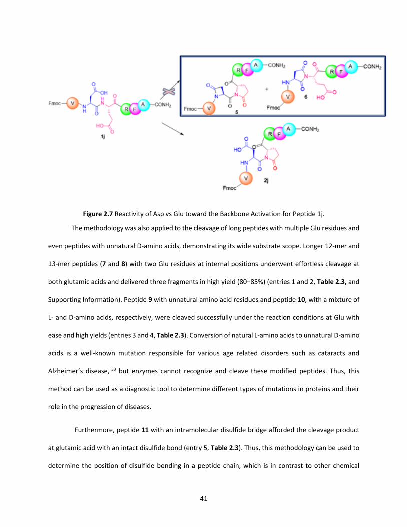
Peptide 1j, along with Glu, contains Asp, which also has free carboxylic group. This peptide under
the reaction conditions could give the four membered -lactam ring (5) or the five membered imide ring
(6) by cyclization of Asp with Glu (2j). However, product (5) was not observed due to strain involved in the
formation of the four membered ring. If product 6 had been formed, under the hydrolysis conditions, ring
opening would occur rather than cleavage of the peptide bond. Instead, hydrolysis led to the fission of
the peptide bond at the N-terminus of Glu generating two fragments as previously reported and
confirmed by HPLC and MS. Based on these results, the reactions conditions were determined to be
selective for Glu (Figure 2.7).
41
Figure 2.7 Reactivity of Asp vs Glu toward the Backbone Activation for Peptide 1j.
The methodology was also applied to the cleavage of long peptides with multiple Glu residues and
even peptides with unnatural D-amino acids, demonstrating its wide substrate scope. Longer 12-mer and
13-mer peptides (7 and 8) with two Glu residues at internal positions underwent effortless cleavage at
both glutamic acids and delivered three fragments in high yield (80−85%) (entries 1 and 2, Table 2.3, and
Supporting Information). Peptide 9 with unnatural amino acid residues and peptide 10, with a mixture of
L- and D-amino acids, respectively, were cleaved successfully under the reaction conditions at Glu with
ease and high yields (entries 3 and 4, Table 2.3). Conversion of natural L-amino acids to unnatural D-amino
acids is a well-known mutation responsible for various age related disorders such as cataracts and
Alzheimer’s disease, 33 but enzymes cannot recognize and cleave these modified peptides. Thus, this
method can be used as a diagnostic tool to determine different types of mutations in proteins and their
role in the progression of diseases.
Furthermore, peptide 11 with an intramolecular disulfide bridge afforded the cleavage product
at glutamic acid with an intact disulfide bond (entry 5, Table 2.3). Thus, this methodology can be used to
determine the position of disulfide bonding in a peptide chain, which is in contrast to other chemical
42
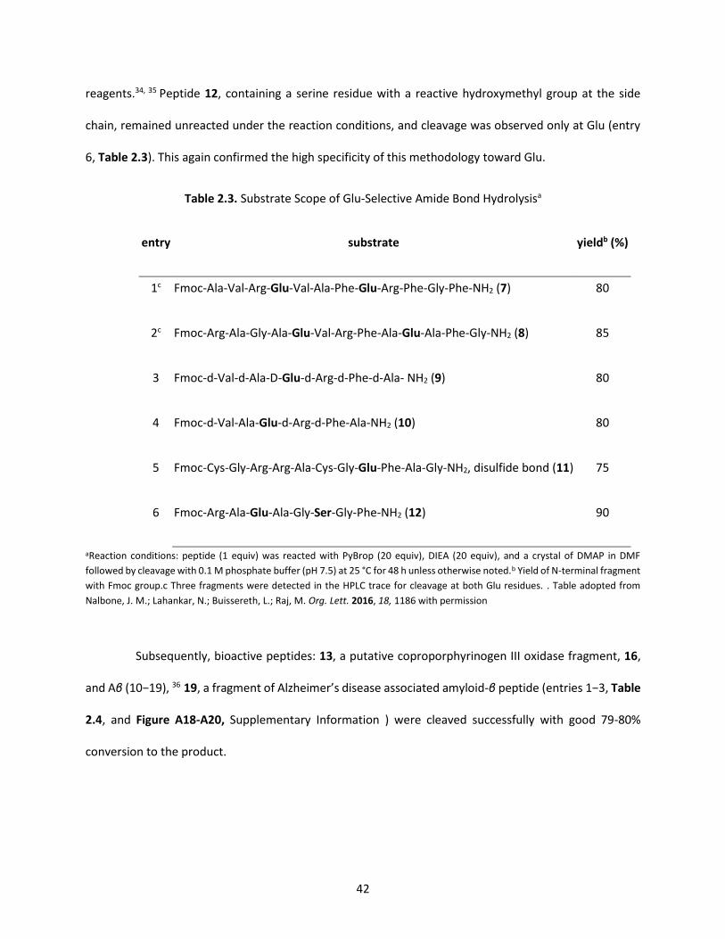
reagents.34, 35 Peptide 12, containing a serine residue with a reactive hydroxymethyl group at the side
chain, remained unreacted under the reaction conditions, and cleavage was observed only at Glu (entry
6, Table 2.3). This again confirmed the high specificity of this methodology toward Glu.
Table 2.3. Substrate Scope of Glu-Selective Amide Bond Hydrolysisa
entry substrate yieldb (%)
1c Fmoc-Ala-Val-Arg-Glu-Val-Ala-Phe-Glu-Arg-Phe-Gly-Phe-NH2 (7) 80
2c Fmoc-Arg-Ala-Gly-Ala-Glu-Val-Arg-Phe-Ala-Glu-Ala-Phe-Gly-NH2 (8) 85
3 Fmoc-d-Val-d-Ala-D-Glu-d-Arg-d-Phe-d-Ala- NH2 (9) 80
4 Fmoc-d-Val-Ala-Glu-d-Arg-d-Phe-Ala-NH2 (10) 80
5 Fmoc-Cys-Gly-Arg-Arg-Ala-Cys-Gly-Glu-Phe-Ala-Gly-NH2, disulfide bond (11) 75
6 Fmoc-Arg-Ala-Glu-Ala-Gly-Ser-Gly-Phe-NH2 (12) 90
aReaction conditions: peptide (1 equiv) was reacted with PyBrop (20 equiv), DIEA (20 equiv), and a crystal of DMAP in DMF
followed by cleavage with 0.1 M phosphate buffer (pH 7.5) at 25 °C for 48 h unless otherwise noted.b Yield of N-terminal fragment
with Fmoc group.c Three fragments were detected in the HPLC trace for cleavage at both Glu residues. . Table adopted from
Nalbone, J. M.; Lahankar, N.; Buissereth, L.; Raj, M. Org. Lett. 2016, 18, 1186 with permission
Subsequently, bioactive peptides: 13, a putative coproporphyrinogen III oxidase fragment, 16,
and Aβ (10−19), 36 19, a fragment of Alzheimer’s disease associated amyloid-β peptide (entries 1−3, Table
2.4, and Figure A18-A20, Supplementary Information ) were cleaved successfully with good 79-80%
conversion to the product.

43
Table 2.4. Glu-Selective Cleavage of Bioactive Peptides
entry substrate yielda (%)
1 Fmoc-Met-Gly-His-Gln-Glu-His-Leu-Pro-Tyr- NH2 (13) 79
2 Fmoc-Leu-Pro-Arg-Leu-Gln-Glu-Ala-Trp-Gln- NH2 (16) 75
3 Fmoc-Tyr-Glu-Val-His-His-Gln-Lys-Leu-Val-Phe-NH2 (19) 80
4 Fmoc-Ala-Gly-Leu-Pro-Glu-Lys-Tyr-NH2 (22) 82
a Yield of N-terminal fragment with Fmoc group. Table adopted from Nalbone, J. M.; Lahankar, N.; Buissereth, L.; Raj, M. Org.
Lett. 2016, 18, 1186 with permission.
Finally, this methodology was evaluated on a bioactive peptide, amyloid A protein fragment
(Homo sapiens) 22, with a proline residue next to Glu (entry 4, Table 4). The location of proline at a
neighboring position nearly blocks the cleavage by proteases completely independent of the amino acid
residue, 27, 28 but remarkably this method cleaved the Pro-Glu bond with 82% conversions to the peptide
fragments. Interestingly, even though peptides 19 and 22 contained lysine residues with a free side-chain,
cyclization was observed only at Glu to generate the kinetically favorable five-membered pGlu moiety
(Figure A20, A21 Supporting Information) further confirming high specificity and broad substrate scope
of the cleavage strategy.
2.7 CONCLUSIONS
In this study, a site-selective approach for the cleavage of peptides at the N-terminus of Glu under
mild and metal-free reaction conditions with high specificity was developed. The method was tolerant
to wide range of unprotected side chains functionalities within the tested peptides unlike some other
chemical cleavage methods. In addition, as disulfide bonds are stable toward reaction conditions, this
methodology can be useful for determining the position of disulfide pairing in peptides. Moreover, the

44
method exhibited broad substrate scope including the cleavage of peptides at proteolytically resistant
Pro-Glu sites. Similarly, hydrolysis of mutated peptides with unnatural amino acid residues such as D-
amino acids, unsuitable substrates for enzymes, proceeded with ease under the reaction conditions. The
strategy presents a potential valuable complementary tool for peptide and protein sequencing with a
broad substrate scope, including cleavage of bioactive peptides and mutated peptides with unnatural
amino acid residues such as D-amino acids implicated in various diseases. The results of this study have
provided a foundation for further studies aimed at developing artificial chemical proteases for the
cleavage of target proteins responsible for various diseases
2.8 EXPERIMENTAL SECTION
General.
All commercial materials (Aldrich, Fluka, Nova) were used without further purification. All solvents
were reagent grade or HPLC grade (Fisher). Anhydrous THF, diethyl ether, CH2Cl2, and DMF were obtained
from a dry solvent system (passed through column of alumina) and were used without further drying. All
reactions were performed under air in round bottom flask. Yields refer to chromatographically pure
compounds; % yields were obtained by comparison of HPLC peak areas of products and starting material.
HPLC was used to monitor the reaction progress.
Materials.
Fmoc-amino acids were obtained from Nova Biochem under (EMD Millipore Corporation) (Billerica,
Massachusetts) and CreoSalus (Louisville, Kentucky). Rink amide resin was obtained from ChemPep Inc
(Wellington, Florida). N,N,N′,N′-Tetramethyl-O-(1Hbenzotriazol-1-yl)uronium hexafluorophosphate
(HBTU) was obtained from CreoSalus (Louisville, Kentucky). N, N′-Disuccinimidyl carbonate (DSC) was
obtained from Nova Biochem, under (EMD Millipore Corporation) (Billerica, Massachusetts).
Bromotris(pyrrolidino)phosphonium hexafluoro-phosphate (PyBrop) was obtained from Sigma Aldrich (St.
Louis, Missouri). 4-Dimethylaminopyridine (DMAP): Merck KGaA (Darmstadt, Germany). N, N-
45
Dimethylformamide (DMF): Macron Fine Chemicals (Center Valley, Pennsylvania). Dichloroethane (DCE),
acetonitrile, N, N- Diisopropylethylamine (DIEA), N, N'diisopropylcarbodiimide (DIC), were purchased
from (EMD Millipore Corporation) (Billerica, Massachusetts). Piperidine was purchased from Alfa Aesar
(Ward Hill, Massachusetts).
Trifluoroacetic acid (TFA) was purchased from VWR (100 Matsonford Road Radnor, PA). Diethyl Ether:
Sigma Aldrich (St. Louis, Missouri). Water was purified using a Millipore MilliQ water purification system.
NMR:
Proton NMR spectra were recorded on a 600 MHz spectrometer and carbon NMR spectra on a 151
MHz spectrometer at ambient temperature. All NMR chemical shifts (δ) are referenced in ppm relative
to residual solvent or internal tetramethylsilane. 1H NMR chemical shifts are referenced to residual
DMSO-d5 at 2.50 ppm, and 13C NMR chemical shifts are referenced to DMSO-d6 at 39.52 ppm. Carbon
NMR spectra are proton decoupled. NMR spectral data are reported as chemical shift (multiplicity,
coupling constants (J), integration). Multiplicity is reported as follows: singlet (s), broad singlet (bs),
doublet (d), doublet of doubles (dd), doublet of triplet (td), triplet (t) and multiplet (m). Coupling constant
(J) in Hertz (Hz).
HPLC
Semi-Preparative HPLC:
Semi-preparative HPLC chromatography (HPLC) was performed on Beckman Coulter equipped with
System Gold 168 detector and 125P solvent module HPLC with a 10 mm C-18 reversed-phase column. All
separations involved a mobile phase of 0.1% FA (v/v) in water (solvent A) and 0.1% FA (v/v) in acetonitrile
(solvent B). Semi-preparative HPLC method using a linear gradient of 0–80% acetonitrile in 0.1% aqueous
FA over 30 min at room temperature with a flow rate of 3.0 mL min-1. The eluent was monitored by
absorbance at 220 nm and 254 nm unless otherwise noted.
46
Analytical HPLC:
Analytical HPLC chromatography (HPLC) was performed on an Agilent 1100 series HPLC equipped with
a 4.6 x150 mm (5µm) C-18 reversed-phase column. All separations involved a mobile phase of 0.1% FA
(v/v) in water (solvent A) and 0.1% FA (v/v) in acetonitrile (solvent B). Peptide compositions were
evaluated by analytical reverse phase HPLC using a gradient of 0.1% FA in acetonitrile versus 0.1% FA in
water. Analytical HPLC method using a linear gradient of 0–80% 0.1% FA (v/v) acetonitrile in 0.1% aqueous
FA over 30 min at room temperature with a flow rate of 1.0 mL min-1. The eluent was monitored by
absorbance at 254 nm unless otherwise noted.
LCMS:
Mass spectrometry was performed using ultra high-performance liquid chromatography-mass
spectrometry using the Agilent 1100 Series LCMSD VL MS Spectrometer using positive polarity
electrospray ionization (+ESI).
Fmoc Solid-Phase Peptide Synthesis.37
Peptides were synthesized manually on a 0.25 mmol scale using a Rink amide resin. The Fmoc–group
was deprotected using 20% piperidine–DMF for 20 min to obtain a deprotected peptide-resin. Fmoc-
protected amino acids (1.25 mm) were sequentially coupled on the resin using a HBTU (1.25 mmol) and
DIEA (1.25 mmol) for 2 h at room temperature. Peptides were synthesized using standard protocols.37 The
peptide was cleaved from the resin using a cocktail of 95:2.5:2.5, trifluoroacetic acid:triisopropyl
silane:water for 2 h. The resin was removed by filtration and the resulting solution was concentrated. The
oily residue was triturated with diethyl ether to obtain a white suspension. The resulting solid was purified
by HPLC.
47
General Procedure for cyclization of a peptide:
To a 5-mL round-bottom flask containing peptide (I) (20 mg, 1 equiv.) in 1 mL dimethylformamide
(DMF) was added a solution of PyBrop (20 equiv.), DIEA (20 equiv.) and crystal of DMAP in DMF (0.5 mL).
The mixture was stirred at room temperature for 24 h. The reaction was concentrated under vacuum and
resulting peptide was dissolved in 1:1 mixture of water and acetonitrile and purified by HPLC. The purified
fractions were lyophilized to afford cyclized peptide (II) as a white powder; yield: (80 %).
General procedure for the cleavage of peptides:
To a cyclized peptide (2a-j), 1 mL phosphate buffer (pH 7.5) was added. The reaction was stirred at
25 °C and monitored by analytical HPLC at regular intervals. The reaction mixture was lyophilized, purified
by HPLC and isolated. HPLC: 0.1% FA (v/v) in water (solvent A): 0.1% FA (v/v) acetonitrile (solvent B);
gradient 0-80 %, 0.1% FA (v/v) acetonitrile in 25 min, flow rate = 1.0 mL/min, detection wavelength 254
nm.
2.9 REFERENCES
1. Wallace, C. J. A. Protein Engineering by Semisynthesis; CRC Press: Boca Raton, FL, 2000.
2. Thorner, J.; Emr, S. D.; Abelson, J. N. Methods Enzymol. 2000, 326, 200.
3. Radzicka, A.; Wolfenden, R. J. Am. Chem. Soc. 1996, 118, 6105.
4. Lee, T. Y.; Suh, J. Chem. Soc. Rev. 2009, 38, 1949.
5. Back, J. W.; David, O.; Kramer, G.; Masson, G.; Kasper, P. T.; de Koning, L. J.; de Jong, L.; van
Maarseveen, J. H.; de Koster C. G.; Angew. Chem. Int. Ed. Engl., 2005, 44, 7946.
6. Thorner, J.; Emr, S. D.; Abelson, J. N. Methods Enzymol. 2000, 326, 200.
7. Arnau, J.; Lauritzen, C.; Petersen, G. E.; Pedersen, J. Protein Expression Purif. 2006, 48, 1.
8. Gross, E. Methods Enzymol. 1967, 11, 238.

48
9. B. J. Smith in The Protein Protocols Handbook, 2nd ed. Walker, J. M. Ed.; Humana Press, Totowa, NJ,
USA, 2002, p. 485.
10. Nalbone, J. M.; Lahankar, N.; Buissereth, L.; Raj, M. Org. Lett. 2016, 18, 1186.
11. Koida, M.; R Walter, R. J. Biol. Chem. 1976, 251, 7593.
12. Lee, T. Y.; Suh, J. Chem. Soc. Rev. 2009, 38, 1949.
13. Stepankova, V.; Bidmanova, S.; Koudelakova, T.; Prokop, Z.; Chaloupkova, R.; Damborsky, J. ACS Catal.
2013, 3, 2823.
14. Sanger, F. Biochem. J. 1945, 39, 507
15. Berg, J. M.; Tymoczko, J. L.; Stryer, L. Protein Structure and Function, In Biochemistry, 5th edition. W
H Freeman: New York, 2002.
16. Edman, P.; Högfeldt, E.; Sillén, L. G.; Kinell, P. Acta Chem. Scand. 1950, 4, 283.
17. Gross, E. Methods Enzymol. 1967, 11, 238.
18. Kaiser, R.; Metzka, L. Anal. Biochem. 1999, 266, 1.
19. Mahoney, W. C.; Smith, P. K.; Hermodson, M. A. Biochemistry 1981, 20, 443.
20. Seki, Y; Tanabe, K.; Sasaki, D.; Sohma, Y.; Oisaki, K., Kanai, M. Angew. Chem. Int. Ed. Engl. 2014, 16,
6501.
21. Kaminskaia, N. V.; Johnson, T. W.; Kostic, N. M. ́J. Am. Chem. Soc. 1999, 121, 8663.
22. Parac, T. N.; Ullmann, G. M.; Kostic, N. M. ́J. Am. Chem. Soc. 1999, 121, 3127.
23. Zhu, L.; Qin, L.; Parac, T. N.; Kostic, N. M. J. Am. Chem. Soc. 1994, 116, 5218.
24. Hoyer, D.; Cho, H.; Schultz, P. G. J. Am. Chem. Soc. 1990, 112, 3249.
25. Schepartz, A.; Cuenoud, B. J. Am. Chem. Soc. 1990, 112, 3247.
26. Kita, Y.; Nishii, Y.; Higuchi, T.; Mashima, K. Angew. Chem., Int. Ed. 2012, 51, 5723.
27. Milovic, N. M.; Kostic ́, N. M. ́J. Am. Chem. Soc. 2003, 125, 781.

49
28. Tofteng, A. P.; Sørensen, K. K.; Conde-Frieboes, K. W.; HoegJensen, T.; Jensen, K. J. Angew. Chem., Int.
Ed. 2009, 48, 7411.
29. Montalbetti, C. A. G. N.; Falque, V. Tetrahedron 2005, 61, 10827.
30. Alsina, J.; Barany, G.; Albericio, F.; Kates, S. Lett. Pept. Sci. 1999, 6, 243.
31. Coste, J.; Frerot, E.; Jouin, P. J. Org. Chem. 1994, 59, 2437.
32. Blinov K. A.; Smurnyy Y. D.; Churanova T. S.; Elyashberg M. E.; Williams, A. J. Chemometrics and
Intelligent Laboratory Systems. 2009, 97, 91.
33. Maeda, H.; Takata, T.; Fujii, N.; Sakaue, H.; Nirasawa, S.; Takahashi, S.; Sasaki, H.; Fujii, N. Anal. Chem.
2015, 87, 561.
34. Edman, P.; Begg, G. Eur. J. Biochem. 1967, 1, 80.
35. Gorman, J. J.; Wallis, T. P.; Pitt, J. J. Mass Spectrom. Rev. 2002, 21, 183.
36. Tanabe, K.; Taniguchi, A.; Matsumoto, T.; Oisaki, K.; Sohma, Y.; Kanai, M. Chem. Sci. 2014, 5, 2747.
37. Chan, W. C.; White, P. D. Fmoc solid phase peptide synthesis: a practical approach Oxford University
Press: New York, 2000.

50
CHAPTER 3. PEPTIDE STAPLING VIA UREA BRIDGING - CHEMICAL INSIGHTS INTO CYCLIC PEPTIDE SYNTHESIS
3.1. ABSTRACT
An efficient synthesis strategy has been developed for peptide stapling onto activated lysine
residues. In this approach, a two-step one pot reaction featuring activation of the side chain amino group
of lysine with carbonyl diimidazole followed by urea bond formation with an adjacent side chain amino
group of a second lysine residue was accomplished without the need of any metal catalyst. This
methodology was used to synthesize a stapled Axin mimetic analogue, which demonstrated increased
alpha helicity and improved proteolytic stability upon peptide stapling. Moreover, a bicyclic peptide was
prepared with this strategy, to further extend the scope of reaction. Finally, this methodology can be
potentially applied for the production of urea stapled peptides as mild and efficient synergistic catalysts
for carrying out a range of organic asymmetric transformations like Michael Addition, and C-H
functionalization reactions with high reactivity and selectivity.
3.2 GRAPHICAL ABSTRACT
Figure 3.1. General method for the synthesis of urea-bridged cyclic peptides
3.3. INTRODUCTION
Peptide drugs have gained a wide range of applications in treating human malignancies such as
cancer (Goserelin, a synthetic gonadotropin-releasing hormone analog), multiple sclerosis (Glatiramer

51
acetate, a synthetic peptide with four amino acids), and type 2 diabetes (Exenatide, a synthetic glucagon-
like peptide-1 analog).1 Peptides can provide target specificity, potency and longer resident times for
more effective duration of action which improves biological efficacy and minimizes side effects.2, 3
However, short synthetic peptides exhibit poor pharmacokinetic and pharmacodynamics properties
which limits their translation into the clinic.4 In addition, peptides are susceptible to proteolytic
degradation1 and they are poorly cell permeable which further restricts their drug-like characteristics.5
3.3.1 BIOACTIVE CYCLIC PEPTIDES
As stapling linear peptides constrains them into cyclic form, the likelihood of the formation of
intramolecular hydrogen bonds increases, which can decrease interactions with water, thus, increasing
the potential for membrane permeability by passive diffusion.6 In addition, constrained cyclic peptides
contain amide bonds which are more difficult to access by proteases, thereby improving their proteolytic
resistance. Moreover, the structural stability resulting from cyclization favors biologically active
conformations of cyclized peptides like Vasopressin and Desmopressin (antidiuretic hormones
Eptifibatide (an antithrombolytic agent), Oxytocin (hormone related to childbirth and social bonding)
(Figure 3.2 )7 with target molecules; thus decreasing the entropic cost upon binding to their target.8 For
the aforementioned reasons, stapling short peptides can greatly improve their pharmacological
performance.

52
Figure 3.2 Bioactive cyclic peptides
3.3.2 SYNTHESIS OF CYCLIC PEPTIDES
Since the development of stapling techniques by Grubbs9 and Verdine using Ring-Closing
Metathesis (RCM),10 various stapling methods have been explored in order to off-set some of their
limitations, including the use of transition metal catalysts, un-natural amino acid residues and harsh
reaction conditions that may denature peptide structure. In spite of their utility, these methods, including
the use of cysteine side chains to form disulfide bridges11 and thioether formation (crosslinking with ’-
dibromo-m-xylene,12 aromatic nucleophilic substitutions with perfluoroaromatic reactants),13 the use of
functionalized non-natural amino acids and biaryl linkages involving the borylated phenylalanine
derivatives,14- 17 or azide–alkyne cycloadditions (click chemistry)18 still required metal catalysts, usually
expensive, long stepwise syntheses and the use of unnatural amino acids.
Such limitations can be overcome by new practical and general stapling methods involving
modifications of naturally occurring residues. These stapling strategies are useful in modifying readily
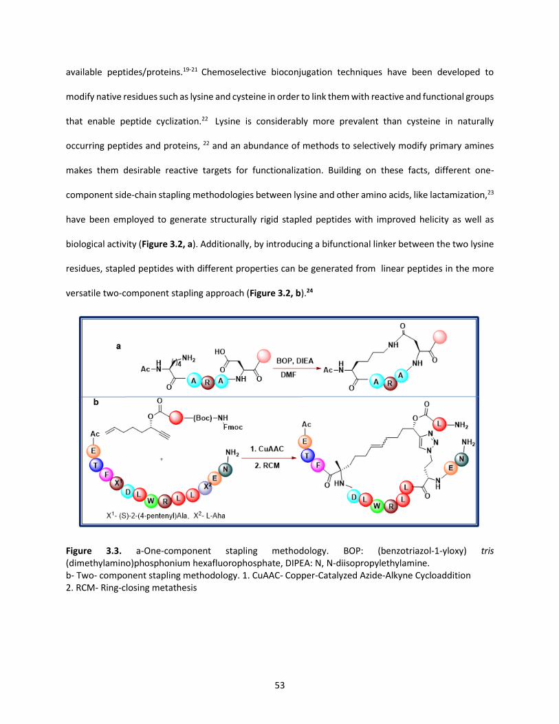
53
available peptides/proteins.19-21 Chemoselective bioconjugation techniques have been developed to
modify native residues such as lysine and cysteine in order to link them with reactive and functional groups
that enable peptide cyclization.22 Lysine is considerably more prevalent than cysteine in naturally
occurring peptides and proteins, 22 and an abundance of methods to selectively modify primary amines
makes them desirable reactive targets for functionalization. Building on these facts, different one-
component side-chain stapling methodologies between lysine and other amino acids, like lactamization,23
have been employed to generate structurally rigid stapled peptides with improved helicity as well as
biological activity (Figure 3.2, a). Additionally, by introducing a bifunctional linker between the two lysine
residues, stapled peptides with different properties can be generated from linear peptides in the more
versatile two‐component stapling approach (Figure 3.2, b).24
Figure 3.3. a-One-component stapling methodology. BOP: (benzotriazol-1-yloxy) tris (dimethylamino)phosphonium hexafluorophosphate, DIPEA: N, N-diisopropylethylamine. b- Two- component stapling methodology. 1. CuAAC- Copper-Catalyzed Azide-Alkyne Cycloaddition 2. RCM- Ring-closing metathesis

54
Thus, new synthetic chemical approaches for the selective and straightforward modification of lysine
are of great importance for stapling methodology.
3.4. CHAPTER OBJECTIVES
In this thesis chapter, a biocompatible, two‐component stapling methodology to modify the ε-
amino group of lysine 51 (pKa values of 10.4 in model compounds)52 with a reactive carbonyl donor leading
to the formation of a urea functionality with an adjacent deprotonated lysine is reported. We
hypothesized that in presence of a base and the electrophilic bifunctional linker carbonylating agent, the
lysine residue will be activated in an intermolecular SN2 reaction to form intermediate A, which will
subsequently generate the urea-stapled peptide B by an intramolecular nucleophilic attack by another
lysine residue (Scheme 3.1). This method will be optimized by exploring the influence of the base and
carbonylating reagent on the cyclization reaction in model peptides. With optimized conditions in hand,
a series of bio-active peptides will be subjected to the proposed urea stapling strategy to investigate the
influence of structure on peptide biological activity.
Scheme 3.1: Proposed mechanism for the urea stapling reaction
3.5 RESULTS AND DISCUSSION
3.5.1 PRELIMINARY RESULTS
To investigate this one-step, two component synthetic strategy, different carbonylating agents,
namely, N, N'-disuccinimidyl carbonate (DSC), 1,1'-carbonyldiimidazole (CDI), and 4-nitrophenyl
chloroformate (NPCF) were screened for lysine reactivity within a model N-terminus acetyl-protected
hexapeptide Ac-Phe-Arg-Lys-Gly-Lys-Ala-CONH2 in dimethylformamide (DMF) as reaction solvent and

55
N,N-diisopropylethylamine (DIEA) as base (Table 3.1). The desired urea-stapled peptide was observed and
confirmed by LCMS, HRMS, MS/MS, and NMR spectroscopy which validated H/D exchange of the product
(Figure 3.4).
H/D Exchange Data: # of exchangeable protons = 788 – 773 – 1 = 14

56
Figure 3.4: H/D exchange, MS, MS/MS, HRMS, NMR of stapled Ac-Phe-Arg-Lys-Gly-Lys-Ala-CONH2

57
Good conversions (69%) were detected by HPLC conversion with CDI as the active carbonylating
reagent at 250C, (Table 3.1, entry 3), and was subsequently selected as the reagent of choice for the
stapling reaction.
Table 3.1. Reagent screening for the urea stapling reaction.a
a Reaction conditions: peptide (1 equiv) was reacted with CDI (3 equiv), DIEA (3 equiv) in DMF at 25°C for 24h. b Conversion to product was calculated with HPLC by measuring absorbance at 220 nm.
The stapling reactions were conducted for 24 h since the reaction of the model peptide Ac-
FKGAKF with CDI, gave optimum conversion (64%) to the product following a one-day reaction (Table 3.2,
entry 3) (Figure 3.5)
Table 3.2. Optimization of conditions for the urea stapling reactiona : Time
entry time convb (%)
1 5 13.83
2 10 26.73
3 24 63.43
Reaction conditions: a peptide (1 equiv) was reacted with CDI (3 equiv), DIEA (3 equiv) in DMF at 25°C for
24h. b Conversion to product was calculated with HPLC by measuring absorbance at 220 nm.
entry reagent convb(%)
1 4-nitrophenyl chloroformate 40
2 DSC 50
3 CDI 69

58
Figure 3.5: HPLC trace and MS for stapled Ac-Phe-Lys-Gly-Ala-Lys-Phe- NH2 at 250C after 24 h
Next, in order to optimize the distance between two lysine residues and reaction temperature,
a series of peptides with two lysine residues separated by varying amino acids were synthesized, and
subsequently subjected to the optimized CDI conditions at 250C and 650C.
Table 3.3 Optimization of reaction conditions for urea stapling a: Distance between lysine residues and
temperature.
entry peptide position of
lysine residues
equiv of
CDI
convb to
product (%)
250C
equiv
of CDI
convb to
product (%)
650C
1 Ac-Phe-Lys-Lys-Phe-NH2c (3) i, i+1 3 0 - -
2 Ac-Phe-Arg-Lys-Gly-Lys-Ala-NH2 (1) I, i+2 3 69 - -
3 Ac-Phe-Lys-Gly-Ala-Lys-Phe- NH2 (2) i, i+3 3 63 - -
4 Ac-Arg-Lys-Leu-Gly-Ala-Lys-Tyr-NH2 (4) i, i+4 3 86 - -
5 Ac-Arg-Lys-Ala-Leu-Gly-Ala-Lys-Phe-NH2 (5) i, i+5 3 40 -

59
6 Ac-Phe-Lys-Val-Ala-Leu-Gly-Ala-Lys-
Phe-NH2 (6)
i, i+6 3 0
- -
7 Ac-Phe-Lys-Gly-Lys-Leu-NH2 (7) I, i+2 3 32 3 63
8 Ac-Phe-Lys-Phe-Lys-Leu-NH2 (8) I, i+2 3 9 3 74
9 Ac-Phe-Arg-Lys-Gly-Lys-Ala-NH2 (1) I, i+2 - - 9.2 94
10 Ac-Arg-Lys-Leu-Gly-Ala-Lys-Tyr-NH2 (4) i, i+4 - - 7 98
11
Ac-Glu-Asn-Pro-Glu-Lys-Ile-Leu-Asp-Lys-His-
Val-Gln-Arg-Val-Met-NH2 (9)
i, i+4 - - 6 89
12
Ac-Thr-Ser-Phe-Lys-Glu-Tyr-Trp-His-Leu-Leu
-Ser-NH2 (10)
i, i+4 - - 8 93
13 Ac- Ala-Lys-Leu-Gly-Phe-Lys-Leu-NH2 (11) i, i+4 - - 7 44
14
Ac- Ile-Lys-Leu-Gly-Phe-Lys-Leu-NH2 (12) i, i+4 - 7 67
15 Phe- Lys-Leu-Gly-Ala-Lys-Phe-Glu (13) i, i+4 - - 9 99
a Reaction conditions: peptide (1 equiv) was reacted with CDI, DIEA in DMF at 25°C for 24 h. bConversion to product was calculated with HPLC by measuring absorbance at 220 nm. c Reaction continued for 48 h, 23.587% intermediate.
At 250C, high conversion to the urea stapled peptide was obtained when two lysine residues were
at i and i+2, i and i+3 and i and i+4 positions with good HPLC conversions ranging from 69% to 86% (Table
3.3, entries 2-4) (Figure 3.4, 3.5, 3.6).

60
Figure 3.6: HPLC trace and MS for stapled Ac-Arg-Lys-Leu-Gly-Ala-Lys-Tyr-NH2 (4) at 250C after 24 h
Though stapled product was obtained with lysine residues at positions i and i+5, modest HPLC conversion
to product was observed (40%, Table 3.3, entry 5) (Figure 3.7), while no stapled product was detected
with lysine residues at i and i+ 6 positions (Table 3.3, entry 6).
Figure 3.7: HPLC trace and MS Ac-Arg-Lys-Ala-Leu-Gly-Ala-Lys-Phe-NH2 (5a) at 250C after 24 h

61
When the stapling reaction was carried out with 3 equiv of CDI at 650C, it gave better conversion
to product than for the reaction at 250C (Table 3.3, entries 7 and 8), though, the amount of CDI for
maximum conversion to product varied from peptide to peptide. (Table 3.3, entries 9 and 10) (Figure A23,
A31, Supplementary Information).
We then proceeded to investigate the selectivity of the stapling reaction toward other residues
in the peptide chain. Two model peptides, Ac-Glu-Asn-Pro-Glu-Lys-Ile-Leu-Asp-Lys-His-Val-Gln-Arg-Val-
Met-CONH2 (9) and Ac-Thr-Ser-Phe-Lys-Glu-Tyr-Trp-His-Leu-Leu-Ser-CONH2 (10) that contained all
nucleophilic residues except for cysteine, it being more nucleophilic than lysine (pKa around 8.5), 27 were
designed (Table 3.3, entries 11 and 12 respectively) (Figure 3.8).
Both the peptides reacted cleanly without any by-products even with unprotected side chains of
other amino acids in peptides. Urea stapled product was obtained for both peptides (9a and 10a) with
high HPLC conversion ranging from 89% to 93%. Thus, the urea stapling methodology is chemoselective
to the lysine side-chain amino groups in N- terminus Ac- protected peptides in the presence of reactive
functional groups, namely, alcohols and carboxylic acids in the peptides (Table 3.3, entries 11 and 12).
(Table 3.3, entries 11 and 12 respectively) (Figure3.8).
Figure 3.8: Selectivity of peptide stapling: Stapled peptides Ac-Glu-Asn-Pro-Glu-Lys-Ile-Leu-Asp-Lys-His-
Val-Gln-Arg-Val-Met-NH2 (9a) and Ac-Thr-Ser-Phe-Lys-Glu-Tyr-Trp-His-Leu-Leu-Ser-NH2) (10a)

62
We then aimed to explore the steric effect of side-chain functionality of the amino acids adjacent
to lysine residues on the efficiency of the stapling reaction. There was only 9% HPLC conversion to the
product for the peptide Ac-Phe-Lys-Phe-Lys-Leu-NH2 (8) with the bulky phenylalanine in between the
reactive lysine residues, whereas, 32% conversion was observed for the peptide Ac-Phe-Lys-Gly-Lys-Leu-
NH2 (7) at 250C which involved substitution of Phe3 for Gly3 (Table 3.3, entries 8 and 7, respectively) (Figure
3.9). Thus, the bulky Phe3 residue had an adverse effect on peptide stapling efficiency when compared to
the sterically unencumbered Gly3 residue.
Figure 3.9: HPLC traces of stapled Ac-Phe-Lys-Gly-Lys-Leu-NH2 (7a) and Ac-Phe-Lys-Phe-Lys-Leu-NH2 (8a)
At 650C, HPLC conversion to product for peptide Ac-Ile-Lys-Leu-Gly-Phe-Lys-Leu-NH2 (12) with bulky
isoleucine residue placed outside the stapling site was 67% compared to 44% for peptide Ac-Ala-Lys-Leu-
Gly-Phe-Lys-Leu-NH2 (11) which substituted Ile1 for Ala1 (Table 3.3, entries 14 and 13 respectively), while
peptide Ac-Phe-Lys-Phe-Lys-Leu-NH2 (8) with bulky Phe1 inside the stapling site gave 74% HPLC
conversion to product, as against 63% for peptide Ac-Phe-Lys-Gly-Lys-Leu-NH2 (7) (Table 3.3, entries 8
and 7 respectively), indicating that steric bulk did not affect efficiency of stapling reaction at elevated
temperature, whether outside or inside the staple to be formed.
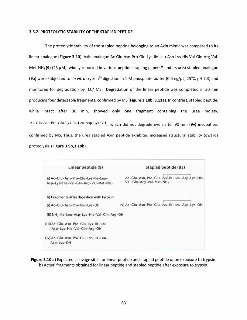
63
3.5.2. PROTEOLYTIC STABILITY OF THE STAPLED PEPTIDE
The proteolytic stability of the stapled peptide belonging to an Axin mimic was compared to its
linear analogue (Figure 3.10). Axin analogue Ac-Glu-Asn-Pro-Glu-Lys-Ile-Leu-Asp-Lys-His-Val-Gln-Arg-Val-
Met-NH2 (9) (25 μM) widely reported in various peptide stapling papers28 and its urea stapled analogue
(9a) were subjected to in vitro trypsin29 digestion in 1 M phosphate buffer (0.5 ng/μL, 250C, pH 7.3) and
monitored for degradation by LC/ MS. Degradation of the linear peptide was completed in 30 min
producing four detectable fragments, confirmed by MS (Figure 3.10b, 3.11a). In contrast, stapled peptide,
while intact after 30 min, showed only one fragment containing the urea moiety,
, which did not degrade even after 90 min (9v) incubation,
confirmed by MS. Thus, the urea stapled Axin peptide exhibited increased structural stability towards
proteolysis. (Figure 3.9b,3.10b).
Figure 3.10 a) Expected cleavage sites for linear peptide and stapled peptide upon exposure to trypsin. b) Actual fragments obtained for linear peptide and stapled peptide after exposure to trypsin.
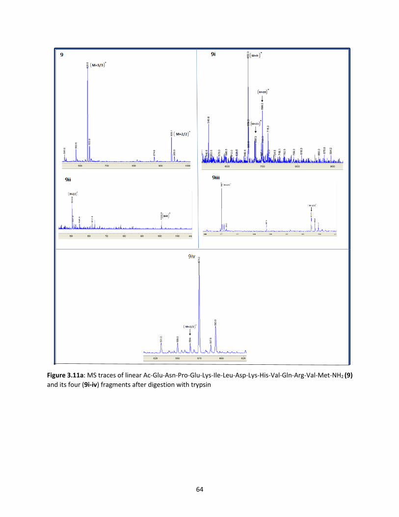
64
Figure 3.11a: MS traces of linear Ac-Glu-Asn-Pro-Glu-Lys-Ile-Leu-Asp-Lys-His-Val-Gln-Arg-Val-Met-NH2 (9)
and its four (9i-iv) fragments after digestion with trypsin

65
9a
9V
Figure 3.11b: MS trace of stapled Ac-Glu-Asn-Pro-Glu-Lys-Ile-Leu-Asp-Lys-His-Val-Gln-Arg-Val-Met-NH2 (a)
and fragment (9v) after digestion with trypsin.

66
3.5.3 SECONDARY STRUCTURE ANALYSIS OF THE STAPLED PEPTIDE
The Axin mimetic Ac-Glu-Asn-Pro-Glu-Lys-Ile-Leu-Asp-Lys-His-Val-Gln-Arg-Val-Met-NH2 (9) and its
urea stapled analogue (9a) were next explored to detect their secondary structures (if any) using Circular
Dichroism (CD) spectroscopy30 (Figure 3.12). When dissolved in TFE/water, the urea stapled peptide
clearly showed the α-helical signature with an observed minima at 208 nm and 222 nm and a maxima at
190 nm (Figure 31.2a). The comparison of the helical propensity indicated that the urea stapled peptide
had a higher tendency (50%) to adopt α-helical character in 1:9 v/v TFE: water while the unstapled linear
peptide exhibited a weaker (21%) helical structure (Figure 3.12b).
Figure 3.12: Helicity of linear and urea stapled peptide: CD spectra recorded for the peptide (5 μM) with different amounts of TFE in phosphate buffer (pH 7.3). Urea stapled peptide indicated a high tendency to adopt α-helical character.
3.5.4 SYNTHESIS OF A BICYCLIC PEPTIDE
In order to illustrate the application of the urea-stapling methodology, a bicyclic peptide was
synthesized as a new class of bioactive bicyclic compounds.31 In this synthesis strategy, the first ring was
constructed by protecting the acid side chain of glutamic acid. The free N-terminus amino group of the
peptide was used for on-resin head-to-tail cyclization with the C-terminus carboxylic acid group of the
model peptide NH2-Phe-Lys-Leu-Gly-Ala-Lys-Phe-Glu-COOH (13) (Scheme 3.2). Overnight reaction in the

67
presence of HOAT/DIC (Scheme 3.2) generated the first ring (13a), confirmed by HRMS and NMR (Figure
3.13). The second cyclization reaction was carried out by the urea stapling methodology of two internal
lysine residues (Scheme 3.2, 13b) and the product was confirmed by MS and NMR (Figure 3.14). From the
ion mobility mass spectrometry (IMS) data (Figure 3.15), the bicyclic peptide was shown to exist as two
conformational diastereomers, 13b-1 (retention time tR 11.2min) and 13b-2 (retention time tR 12.4min).
However, the conformer 13b-1 (92%) was found to dominate conformer 13b-2 (6%) (Figure 3.16) Thus, an
appropriate choice of the protection scheme allowed for the synthesis of structurally diverse bicyclic
peptides by our urea stapling strategy.
Scheme3.2: Synthesis of a bicyclic compound: stapling strategy
.

68
HRMS data- Monocycle Phe-Lys-Leu-Gly-Ala-Lys-Phe-Glu-CONH2: [C46H69N11O9+2H]2+: m/z [M+2/2]+
460.768 (calc.M+2/2]+ 460.77), [ M+H]+m/z 920.52 (calcd [M+H]+ =920.53)
Figure 3.13: HRMS and 1H NMR for Monocyclic peptide Phe-Lys-Leu-Gly-Ala-Lys-Phe-Glu-CONH2 (13a)

69
MS Bicycle Phe-Lys-Leu-Gly-Ala-Lys-Phe-Glu-NH2 (13b).
Figure 3.14: MS and 1H NMR of Bicycle Phe-Lys-Leu-Gly-Ala-Lys-Phe-Glu-NH2 (13b).

70
Extracted Mass Spectra: Bicyclic Peptide Phe-Lys-Leu-Gly-Ala-Lys-Phe-Glu-NH2 (13b).
Figure 3.15 Ion Mobility Resolved MS/MS (Overlay) (A) 328 Å2 and (B) 339 Å2 Conformations for
bicyclic Peptide Phe-Lys-Leu-Gly-Ala-Lys-Phe-Glu-NH2 (13b).

71
Figure 3.16: HPLC trace of two conformational diastereomers, 13b-1 and 13b-2 of the bicyclic Peptide
Phe-Lys-Leu-Gly-Ala-Lys-Phe-Glu-NH2 (13b).
3.6 CONCLUSIONS
In conclusion, a urea-based strategy for stapling and cyclizing peptides has been developed in this
thesis chapter. The methodology is chemoselective to lysine residues and utilizes an electrophilic
carbonylating reagent, CDI, to generate the urea stapled peptide. This mild, one-step, two-component
method works without any metal catalyst. A structural study of the urea stapled peptide displayed
increased α-helicity, and also improved proteolytic stability. Moreover, this stapling strategy was
successfully applied to generate bicyclic peptides as a new type of scaffold for peptide-based drug design.
Finally, the unique feature of this methodology is the generation of a urea moiety that is capable of
forming H-bonds and thus can play an important role in catalysis. Future studies will be used to address
the application of urea stapled peptides as efficient catalysts for carrying out a range of organic
asymmetric transformations such as the Michael Addition, and C-H functionalization reactions.

72
3.7. EXPERIMENTAL SECTION
General:
All commercial materials (Aldrich, Fluka, Nova) were used without further purification. All
solvents were reagent grade or HPLC grade (Fisher). CH2Cl2 and DMF were obtained from a dry solvent
system (passed through column of alumina) and were used without further drying. All reactions were
performed under air in a round bottom flask. Yields refer to chromatographically pure compounds;
% conversions were obtained by comparison of HPLC peak areas of products and starting material. HPLC
was used to monitor reaction progress.
Materials:
Fmoc-amino acids were obtained from Nova Biochem under (EMD Millipore Corporation) (Billerica,
Massachusetts) and CreoSalus (Louisville, Kentucky). Rink amide resin was obtained from ChemPep Inc
(Wellington, Florida). N,N,N′,N′-Tetramethyl-O(1H-benzotriazol-uronium hexafluorophosphate (HBTU)
was obtained from CreoSalus (Louisville, Kentucky). N, N′-Disuccinimidyl carbonate (DSC) was obtained
from Nova Biochem, under (EMD Millipore Corporation) (Billerica, Massachusetts). 4-
Dimethylaminopyridine (DMAP): Merck KGaA (Darmstadt, Germany). N, N-Dimethylformamide (DMF):
Macron Fine Chemicals (Center Valley, Pennsylvania). Acetonitrile, N, N-Diisopropylethylamine (DIEA), N,
N'-diisopropylcarbodiimide (DIC), were purchased from (EMD Millipore Corporation)(Billerica,
Massachusetts). Piperidine was purchased from Alfa Aesar (Ward Hill, Massachusetts). 1,1'-
Carbonyldiimidazole (CDI) was purchased from Nova Biochem (Calbiochem-Novabiochem corporation, La
Jolla, California) Trifluoroacetic acid (TFA) was purchased from VWR 100 Matsonford Road Radnor, PA.
Water was purified using a Millipore MilliQ water purification system.
Analytical HPLC:

73
Analytical HPLC chromatography (HPLC) was performed on an Agilent 1100 series HPLC equipped
with a 4.6 x150 mm (5µm) C-18 reversed-phase column. All separations involved mobile phase of 0.1% FA
(v/v) in water (solvent A) and 0.1% FA (v/v) in acetonitrile (solvent B). Peptide compositions were
evaluated by analytical reverse phase HPLC using a gradient of 0.1% FA in acetonitrile versus 0.1% FA in
water. Analytical HPLC method using a linear gradient of 0–80% 0.1% FA (v/v) acetonitrile in 0.1% aqueous
FA, unless otherwise noted, over 30 min at room temperature with a flow rate of 1.0 mL min-1. The peptide
absorbance was monitored at 220 nm (peptide bond) unless otherwise noted.
LC-MS:
Mass spectrometry was performed on an Agilent 1100 Series HPLC with MSD VL mass
spectrometer using positive polarity electrospray ionization (+ESI).
LC-HRMS:
Analysis was done at Merck facilities at Rahway, NJ by Ryan Cohen. High resolution MS data were
acquired using an Agilent 1290 UHPLC with a 6520 Q-ToF mass spectrometer under positive polarity
electrospray ionization (+ESI) with capillary and fragmentor voltages set to 3.5 kV and 175 V, respectively.
The instrument was calibrated prior to data acquisition using Agilent’s reference standard solution, which
provided accurate masses within 5 ppm. A reversed phase, linear gradient separation was performed at a
flow rate of 1.0 mL min-1 on a C18 column (Waters Acquity UPHLC peptide BEH C18, 1.7 µm particle size,
50 mm x 2.1 mm I.D.) thermostatted at 45°C and gradient from 95% solvent A (0.1% formic acid in water)
to 99% solvent B (0.1% formic acid in acetonitrile) in 4 minutes, followed by 1 minute re-equilibration.
MS/MS:
Analysis was done was done at Merck facilities at Rahway, NJ by Ryan Cohen. Tandem MS
analyses were performed either on an Agilent 1260 HPLC coupled with a 6530 Q-ToF or on an Agilent 1290
74
HPLC coupled with a 6520 Q-ToF mass spectrometer. Both mass spectrometers were operated under
electrospray ionization in positive polarity (+ESI). Reverse phase C18 gradient separation conditions were
used (mobile phase A = 0.1% formic acid in water and mobile phase B = 0.1% formic acid in acetonitrile).
Spectra were acquired in profile mode with collision energies ranging from 25-90 V and nitrogen (N2) as
collision gas.
NMR:
NMR was done by Ryan Cohen at Merck facilities at Rayway, NJ. NMR spectra were recorded on a
Bruker AVANCE III HD 600 MHz spectrometer equipped with a 5-mm HCN cryoprobe at 600 and 151 MHz
for proton and carbon, respectively, spectrometer at ambient temperature. An approximately 1 mg
sample was dissolved in DMSO-d6 solvent containing 0.05% v/v TMS for chemical shift referencing. 1H,
13C, gCOSY, TOCSY (80 ms mixing time), multiplicity-edited gHSQC, gHMBC, and ROESY (200 ms spin lock)
experiments were performed. NMR spectra were processed using MNova (ver. 11.0.4).
All NMR chemical shifts are referenced in ppm relative to residual solvent or internal
tetramethylsilane. 1H NMR chemical shifts referenced to residual DMSO-d5 at 2.50 ppm, and 13C NMR
chemical shifts referenced to DMSO-d6 at 39.52ppm. Carbon NMR spectra are proton decoupled. NMR
spectral data are reported as chemical shift (multiplicity, coupling constants (J), integration). Multiplicity
is reported as follows: singlet (s), broad singlet (bs), doublet (d), doublet of doubles (dd), doublet of triplet
(td), triplet (t) and multiplet (m). Coupling constant (J) in Hertz (Hz).
General Procedure for Fmoc Solid-Phase Peptide Synthesis32:
Peptides were synthesized manually on a 0.25 mmol scale using Rink amide resin. Fmoc–group
was deprotected using 20% piperidine–DMF to obtain a deprotected peptide-resin (2x 20 min). Fmoc-

75
protected amino acids (1.25 mmol) were sequentially coupled on the resin using a HBTU (1.25 mmol) and
DIEA (1.25 mmol) for 2 h at room temperature. Peptides were synthesized using standard protocols. The
peptide was cleaved from the resin using a cocktail of 95:2.5:2.5, trifluoroacetic acid (TFA):
triisopropylsilane (TIPS): water (H2O) for 2 h. The resin was removed by filtration and the resulting solution
was concentrated under vacuum. The resulting solid was dissolved in ACN-water solvent system, analyzed
and purified by RP HPLC.
General Procedure for Stapling Reaction:
To a 5-mL round-bottom flask containing peptide (1 equiv.) in 200 μL dimethylformamide (DMF)
was added a solution of CDI (3-9.2 equiv.) and DIEA (3-9.2 equiv.) in DMF. The mixture was stirred at
250C/650C for 24 h. The reaction mixture was concentrated under vacuum and resulting peptide was
dissolved in mixture of water and acetonitrile and purified by RP HPLC. The purified fractions were
lyophilized to afford stapled peptide as a white powder. % conversion was obtained by comparison of
HPLC peak areas of products and starting material. HPLC was used to monitor reaction progress.
Macrocyclization of Linear Peptide, Phe-Lys-Leu-Gly-Ala-Lys-Phe-Glu, on Solid Support:
To peptide, Phe-Lys-Leu-Gly-Ala-Lys-Phe-Glu, containing O-allyl protected glutamic acid on solid
support (50 mg, 0.69 mmol/g) was added a 3 mL solution of 20 mg Pd (PPh3)4 and 72 µL phenylsilane in
DCM. This solution was incubated on a shaker at room temperature for 40 minutes. The resin was washed
with DCM (3 X 2 min). The above reaction was repeated, and then the resin was washed with DCM (3 X 2
min), MeOH (3 X 2 min), and DMF (3 X 2 min). The palladium catalyst was removed from the resin by
washing with DIEA in DMF (2% v/v) (2 X 2 min) and then with DMF (2 X 2 min). Next, the peptide was
treated with 20% piperdine in DMF to remove the Fmoc protection. Macrocyclization was achieved by
exposing the resin to DIC, HOAt, and a catalytic amount of DMAP in DMF on a shaker for 15 h. The solution
was drained, and the resin was washed with DMF. To confirm complete macrocyclization, the Kaiser test32

76
was performed, which showed yellow bead coloration indicating absence of free amino groups. The
peptide was then cleaved from the resin by 2 hours incubation with a solution of TFA:TIPS:water
(95/2.5/2.5, v/v/v). Resin was removed by filtration, and the resulting solution was evaporated. The
peptide was purified by reversed-phase semi-preparative chromatography.

77
3.8 REFERENCES
1. Henninot A.; Collins, J. C.; Nuss, J. M. J Med Chem. 2018,61, 1382.
2. Craik, D. J.; Fairlie, D. P.; Liras, S.; Price, D. Chem. Biol. Drug Des. 2013, 81, 136.
3. Vlieghe, P.; Lisowski, V.; Martinez, J.; Khrestchatisky, M. Drug Discov. Today 2010, 15, 40.
4. Hill T. A.; Nicholas E.; Shepherd N. E.; Diness, F.; Fairlie D. P. Angew.Int.ed. 2014, 53, 13020.
5. Yang N. J.; Hinner, M. J. Methods Mol Biol. 2015, 1266, 29.
6. Rezai, T.; Yu, B.; Millhauser, G. L.; Jacobson, M. P.; Lokey, R. S. J. Am. Chem. Soc. 2006, 128, 2510.
7. Hamley, I. W. Chem. Rev., 2017, 117, 14015.
8. Khan, A. R.; Parrish, J.C.; Fraser, M.E.; Smith, W.W.; Bartlett, P.A.; James, M.N. Biochemistry, 1998, 37,
16839.
9. Blackwell, H. E.; Grubbs, R. H. Angew. Chem. Int. Ed. 1998, 37, 3281.
10. Schafmeister, C. E.; Po, J.; Verdine, G. L. J. Am. Chem. Soc. 2000, 122, 5891.
11. Benıtez, M. G.; Puche, J. T.; Albericio, F. Chem. Rev. 2014, 114, 901.
12. Jo H.; Meinhardt N.; Wu Y.; Kulkarni S.; Hu X.; Low K. E.; Davies P. L.; DeGrado W. F.; Greenbaum D. C.
J. Am. Chem. Soc. 2012, 134, 17704.
13. Spokoyny, A. M.; Zou, Y.; Ling, J. J.; Yu, H.; Lin, Y.S,; Pentelute, B. L. J. Am. Chem. Soc., 2013, 135,
5946.
14. Bois-Choussy, M.; Cristau, P.; Zhu, J. Angew. Chem. Int. Ed. 2003, 42, 4238.
15. Carbonnelle, A. C.; Zhu, J. Org. Lett. 2000, 2, 3477.
16. Afonso, A.; Feliu, L.; Planas, M. Tetrahedron 2011, 67, 2238.
17. Meyer, F. M.; Collins, J. C.; Borin B.; Bradow, J.; Liras, S.; Limberakis, C.; Mathiowetz, A. M.; Philippe,
L.; Price, D.; Song, K.; James, K. J. Org. Chem. 2012, 77, 3099.
18. Dharanipragada, R. Future Med. Chem. 2013, 5, 831.
78
19. Angelini, A.; Heinis, C. Curr. Opin. Chem. Biol., 2011, 15, 355.
20. Basle, E.; Joubert, N.; Pucheault, M.; Chem. Biol. 2010, 17, 213.
21. Smith, J. M.; Frost, J. R.; Fasan, R. J. Org. Chem. 2013, 78, 3525.
22. Sletten E. M.; Bertozzi, C. R. Angew. Chem. Int. Ed. 2009, 48, 6974.
23. Shepherd, N. E.; Hoang, H. N.; Abbenante, G.; Fairlie, D. P. J. Am. Chem. Soc. 2005, 127, 2974.
24. Serrano J.C.; Sipthorp J.; Xu W.; Itzhaki L. S.; Ley S. V. Chembiochem. 2017, 18,1066.
25. Hacker, S. M.; Backus, K. M.; Lazear, M. R.; Forli, S.; Correia, B. E.; Cravatt, B. F. Nat. Chem., 2017, 9,
1181.
26. Nozaki, Y.; Tanford, C. Methods Enzymol. 1967, 11, 715.
27. Kyte J. Structure in Protein Chemistry. New York: Garland Publishing, Inc; 1995.
28. Wang, Y; Chou, D. H.; Angew. Chem., Int. Ed. 2015, 54, 10931
29. Greenfield, N. J. Nat. Protoc. 2006, 1, 2876.
30. Kelly, S.M.; Jess, T.J.; Price, N.C. Biochim Biophys Acta. 2005,1751, 119.
31. Baeriswyl, V.; Rapley, H.; Pollaro, L.; Stace, C.; Teufel, D.; Walker, E.; Chen, S.; Winter, G.; Tite, J.;
Heinis, C. Chem. Med. Chem. 2012, 7, 1173.
32. Chan, W. C.; White, P. D. Fmoc solid phase peptide synthesis: a practical approach Oxford University
Press: New York, 2000.

79
CHAPTER 4: CONTRIBUTIONS TO KNOWLEDGE AND FUTURE DIRECTIONS
4.1 CONTRIBUTIONS TO KNOWLEDGE MADE IN THIS THESIS
In this thesis, side chain modified polypeptides have been developed to affect both conformational
as well as functional properties. The effect of cyclic conformational constraint on the peptide backbone
and the side chain geometry were explored as chemical tools for improving peptide structure–activity
relationships.
4.1.1 PYROGLUTAMATE - A CYCLIC BACKBONE INTERMEDIATE FOR SELECTIVE CHEMICAL CLEAVAGE
OF PEPTIDE BONDS
The second chapter of the thesis describes a site-selective cleavage at glutamic acid of unreactive
peptide bonds in native peptides as well as in peptidomimetics (Figure 4.1).
Figure 4.1: Graphical abstract for Glutamic Acid Selective Chemical Cleavage of Peptide Bond.
To develop this method, we optimized the reaction conditions for activation of the peptide
backbone amide bond at glutamic acid by the formation of a reactive pyroglutamyl (pGlu) imide
intermediate. Next, the reactive pGlu imide moiety was subjected to hydrolysis, resulting in cleavage of
the scissile peptide bond at the N-terminus of the modified glutamic acid residue. The method exhibited
broad substrate scope. When we explored different substrates to study the effect of steric bulk of
neighboring amino acid residues on the efficiency of chemical cleavage, there was no significant effect.

80
However, substrates with bulky residues such as isoleucine required longer incubation times for the
cleavage reaction to proceed to completion. Moreover, the cleavage reaction proceeded without any by-
products, demonstrating selectivity of the reaction. Cleavage of mutated peptides with unnatural amino
acid residues such as D-amino acids, unsuitable substrates for enzymes, proceeded with ease under the
reaction conditions. Moreover, disulfide bonds were stable toward the reaction conditions in contrast to
other chemical reagents such as the use of cyanogen bromide. Importantly, this methodology was
successful in cleaving peptide bonds next to proline, a site inaccessible to proteases. Finally, the scope of
the methodology was successfully extended to different bioactive peptides such as a fragment of putative
coproporphyrinogen III oxidase, amyloid-β peptide; and amyloid A protein.
The significance of this chemical cleavage strategy lies in the fact that it presents a potential
valuable complementary tool for peptide and protein sequencing (Figure 4.2).
Primary Structure determination
Steps Technique
I. Determination of amino acid sequence in a protein
Complete hydrolysis of a protein Pronase enzyme
Separation and estimation of amino acids Chromatography
II. Protein degradation in smaller fragments
Liberation and determination of number of polypeptide chains
Urea, Dansyl chloride
Cleavage of polypeptides into fragments Enzymatic, Glutamic acid selective

81
Figure 4.2: Peptide sequencing with glutamic acid selective chemical cleavage
The results of this study have provided a foundation for further studies aimed at developing artificial
chemical proteases for the cleavage of target proteins responsible for various diseases (Figure 4.2B). The
importance of this study was acknowledged by a publication in Organic Letters.
4.1.2 PEPTIDE STAPLING VIA UREA BRIDGING-CHEMICAL INSIGHTS INTO CYCLIC PEPTIDE SYNTHESIS
In the third chapter of this thesis, a urea-based methodology between two lysine residues for
stapling and cyclizing peptides has been reported. First, we explored a suitable base and a carbonylating
reagent to deprotonate and activate the lysine side chain. In our optimized conditions, 1,1'-
carbonyldiimidazole (CDI) and N, N-diisopropylethylamine (DIEA) were selected as the reagents of
choice. This activation step led to formation of a urea functionality with an adjacent lysine residue
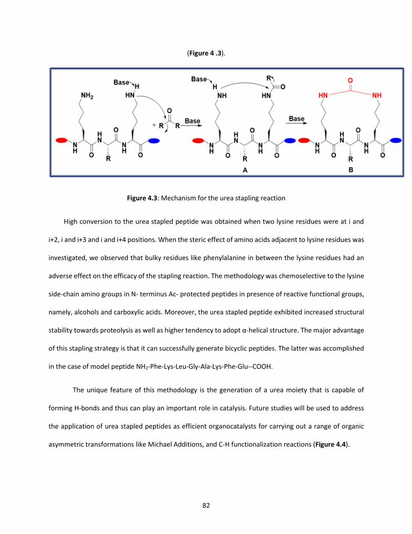
82
(Figure 4 .3).
Figure 4.3: Mechanism for the urea stapling reaction
High conversion to the urea stapled peptide was obtained when two lysine residues were at i and
i+2, i and i+3 and i and i+4 positions. When the steric effect of amino acids adjacent to lysine residues was
investigated, we observed that bulky residues like phenylalanine in between the lysine residues had an
adverse effect on the efficacy of the stapling reaction. The methodology was chemoselective to the lysine
side-chain amino groups in N- terminus Ac- protected peptides in presence of reactive functional groups,
namely, alcohols and carboxylic acids. Moreover, the urea stapled peptide exhibited increased structural
stability towards proteolysis as well as higher tendency to adopt α-helical structure. The major advantage
of this stapling strategy is that it can successfully generate bicyclic peptides. The latter was accomplished
in the case of model peptide NH2-Phe-Lys-Leu-Gly-Ala-Lys-Phe-Glu--COOH.
The unique feature of this methodology is the generation of a urea moiety that is capable of
forming H-bonds and thus can play an important role in catalysis. Future studies will be used to address
the application of urea stapled peptides as efficient organocatalysts for carrying out a range of organic
asymmetric transformations like Michael Additions, and C-H functionalization reactions (Figure 4.4).

83
Figure 4.4: Urea stapled peptides as efficient organocatalysts: a) Michael Addition b) Design of
Organic Catalyst c) Mechanistic Pathway of Peptide Catalysis
4.1.3 REFERENCES:
1. Nalbone, J.M.; Lahankar, N.; Buissereth, L.; Raj, M. Org. Lett. 2016, 18, 1186.
4.2 PUBLICATIONS, CONFERENCE PRESENTATIONS, AND AWARDS
PUBLICATIONS
• Glutamic Acid Selective Chemical Cleavage of Peptide Bonds – Nalbone, J. M.; Lahankar, N.;
Buissereth, L.; Raj, M. Org Lett. 2016, 18, 1186.
• Amino-Acid-Catalyzed Direct Aldol Bioconjugation – Howard T. S.; Cohen R. D.; Nwajiobi O.;
Muneeswaran Z. P.; Sim, Y. E.; Lahankar N. N.; Johannes T.-H. Y.; Raj, M. Org. Lett. 2018, 20, 5344.

84
MANUSCRIPTS IN PREPARATION
• Synthesis of Urea Stapled Helical Peptides with Lysine Carbonylation– Lahankar, N.; Pereira, Z.; Raj M.
• Proline Selective Bioconjugation – Sim, Y.; Lahankar, N.; Pereira, Z.; Buissereth, L.; Raj, M.
POSTER PRESENTATIONS
• Poster: Rapid Bio conjugation of Peptides using Aldehyde as an Anchor on the Solid Support – 24th
American Peptide Symposium, Florida, 6/2015.
• Poster: Rapid Bio conjugation of Peptides using Aldehyde as an Anchor on the Solid Support –
Petersheim Academic Exposition at Seton Hall University, 4/2015.
• Poster: Glutamic Acid Selective Chemical Cleavage of Peptide Bonds – Petersheim Academic
Exposition at Seton Hall University, 4/2016.
• Poster: A novel method for peptide stapling using two naturally occurring lysine residues – Petersheim
Academic Exposition at Seton Hall University, 4/2017.
• Poster: Urea stapled helical peptides as catalysts – Gordon Research Conference, 2/2018.
AWARDS AND HONORS
• Robert De Simone Graduate Fellowship– Department of Chemistry and Biochemistry Seton Hall
University 9/2017 – 6/2018.
• Outstanding Teaching Assistant, Department of Chemistry and Biochemistry, Seton Hall University
9/2017 – 6/2018.

85
BIBLIOGRAPHY
1. Afonso, A.; Feliu, L.; Planas, M. Tetrahedron 2011, 67, 2238.
2. Albericio, F. - Peptide Sci., 2000,55, 123.
3. Albericio, F. Curr. Opin. Chem. Biol. 2004, 8, 211.
4. Alsina, J.; Barany, G.; Albericio, F.; Kates, S. Lett. Pept. Sci. 1999, 6, 243.
5. Andersson, L.; Blomberg, L.; FlegeL, M.; Lepsa, L.; Nilsson, B.; Verlander, M. Biopolymers, 2000,
55, 227.
6. Angelini, A.; Heinis, C. Curr. Opin. Chem. Biol., 2011, 15, 355.
7. Anthony, L.; Freda, P. U. Curr. Med. Res. Opin. 2009, 25, 2989.
8. Arnau, J.; Lauritzen, C.; Petersen, G. E.; Pedersen, J. Protein Expression Purif. 2006, 48, 1.
9. B. J. Smith in The Protein Protocols Handbook, 2nd ed. Walker, J. M. Ed.; Humana Press, Totowa,
NJ, USA, 2002, p. 485.
10. Back, J. W.; David, O.; Kramer, G.; Masson, G.; Kasper, P. T.; de Koning, L. J.; de Jong, L.; van
Maarseveen, J. H.; de Koster C. G.; Angew. Chem. Int. Ed. Engl., 2005, 44, 7946.
11. Baeriswyl, V.; Rapley, H.; Pollaro, L.; Stace, C.; Teufel, D.; Walker, E.; Chen, S.; Winter, G.;
Tite, J.; Heinis, C. Chem. Med. Chem. 2012, 7, 1173.
12. Barlos, K.; Chatzi, O.; Gatos, D.; Stavropoulos, G. Int. J. Pept. Protein Res. 1991, 37, 513.
13. Basle, E.; Joubert, N.; Pucheault, M.; Chem. Biol. 2010, 17, 213.
14. Benıtez, M. G.; Puche, J. T.; Albericio, F. Chem. Rev. 2014, 114, 901.
15. Berg, J. M.; Tymoczko, J. L.; Stryer, L. Protein Structure and Function, In Biochemistry, 5th edition.
W H Freeman: New York, 2002.
16. Blackwell, H. E.; Grubbs, R. H. Angew. Chem. Int. Ed. 1998, 37, 3281.
17. Bois-Choussy, M.; Cristau, P.; Zhu, J. Angew. Chem. Int. Ed. 2003, 42, 4238.

86
18. Canne, L. E.; Botti, P.; Simon, R. J.; Chen, Y.; Dennis, E. A.; Kent, S. B. J. Am. Chem. Soc. 1999, 121,
8720.
19. Carbonnelle, A. C.; Zhu, J. Org. Lett. 2000, 2, 3477.
20. Carpino, L. A.; Beyermann, M.; Wenschuh, H.; Bienert, M. Acc. Chem. Res. 1996, 2, 268.
21. Carpino, L. A.; Han, G.Y. J. Am. Chem. Soc. 1970, 92, 5748.
22. Chan, W. C.; White, P. D. Fmoc solid phase peptide synthesis: a practical approach. Oxford
University Press: Oxford, 2000; p. 341.
23. Chartrel, N.; Conlon, J. M.; Danger, J. M.; Fournier, A.; Tonon, M. C.; Vaudry, H. Proc. Natl.
Acad. Sci. U.S.A. 1991, 88, 3862.
24. Blinov K. A.; Smurnyy Y. D.; Churanova T. S.; Elyashberg M. E.; Williams, A. J. Chemometrics and
Intelligent Laboratory Systems. 2009, 97, 91.
25. Chen, S.; Janda, K. D., Tetrahedron Lett. 1998, 39, 3943.
26. Choudhary, C.; Weinert, B. T.; Nishida, Y.; Verdin, E.; Mann, M. Nat. Rev. Mol. Cell Biol. 2014, 15,
536.
27. Coin, I.; Beyermann, M.; Bienert, M.; Nat Protoc. 2007, 2, 3247.
28. Coste, J.; Frerot, E.; Jouin, P. J. Org. Chem. 1994, 59, 2437.
29. Craik, D. J.; Fairlie, D. P.; Liras, S.; Price, D. Chem. Biol. Drug Des. 2013, 81, 136.
30. Dawson, P. E.; Muir, T. W.; Clark-Lewis, I.; Kent, S. B. Science 1994, 266, 776.
31. Deming, T. J. Chem. Rev. 2016, 116, 786.
32. Dharanipragada, R. Future Med. Chem. 2013, 5, 831.
33. du Vigneau, V.; Ressler, B.; Swan, J. M.; Roberts, J. M.; Katsoyannis, C. W. J. Am. Chem. Soc. 1954,
76, 3115.
34. Du, A. W.; Stenzel, M. H. Biomacromolecules 2014, 15, 1097.

87
35. Eaton L.; Rogers K. The Building Blocks of Life: Examining Basic Chemical Molecules; 1st ed.;
Britannica Digital Learning: New York, 2018; p. 5.
36. Edman, P.; Begg, G. Eur. J. Biochem. 1967, 1, 80.
37. Edman, P.; Högfeldt, E.; Sillén, L. G.; Kinell, P. Acta Chem. Scand. 1950, 4, 283.
38. Fischer, E.; Fourneau, E. Ber. dtsch. Chem. Ges. 1901, 34, 2868.
39. Garcia-Martin, F.; Quintanar-Audelo, M.; Garcia-Ramos, Y.; Cruz, L.J.; Gravel, C.; Furic, R.; Cote,
S.; Tulla-Puche, J.; Albericio, F. J Comb Chem. 2006, 8, 213.
40. García-Ramos Y.; Paradís-Bas M.; Tulla-Puche J.; Albericio F. J Pept Sci. 2010, 16, 675.
41. Gentilucci, L.; De Marco, R.; Cerisoli, L. Curr. Pharm. Des. 2010, 16, 3185.
42. Gilon, C.; Halle, D.; Chorev, M.; Selinger, Z.; Byk, G. Biopolymers 1991, 31, 745.
43. Gorman, J. J.; Wallis, T. P.; Pitt, J. J. Mass Spectrom. Rev. 2002, 21, 183.
44. Greenfield, N. J. Nat. Protoc. 2006, 1, 2876.
45. Grimsley G. R.; Scholtz J. M.; Pace C.N. Protein Sci. 2009, 18, 247.
46. Gross, E. Methods Enzymol. 1967, 11, 238.
47. Guzmán, F.; Barberis, S.; Illanes, A. Electronic J Biotechnol. 2007, 10, 279.
48. Hacker, S. M.; Backus, K. M.; Lazear, M. R.; Forli, S.; Correia, B. E.; Cravatt, B. F. Nat. Chem.,
2017, 9, 1181.
49. Hacker, S. M.; Backus, K. M.; Lazear, M. R.; Forli, S.; Correia, B. E.; Cravatt, B. F. Nat. Chem.
2017, 9,1181.
50. Hamley, I. W. Chem. Rev., 2017, 117, 14015.
51. Hancock, R. E.; Sahl H. G. Nat. Biotechnol. 2006, 24, 1551.
52. Henninot A.; Collins, J. C.; Nuss, J. M. J Med Chem. 2018,61, 1382.
53. Hill T. A.; Nicholas E.; Shepherd N. E.; Diness, F.; Fairlie D. P. Angew.Int.ed. 2014, 53, 13020.

88
54. Houk, K. N.; Leach, A. G.; Kim, S. P.; Zhang, X. Y. Angew. Chem. Int. Ed. 2003, 42, 4872.
55. Hoyer, D.; Cho, H.; Schultz, P. G. J. Am. Chem. Soc. 1990, 112, 3249.
56. Isidro-Llobet, A. Alvarez, M.; Albericio, F. Chem. Rev. 2009, 109, 2455.
57. Jo H.; Meinhardt N.; Wu Y.; Kulkarni S.; Hu X.; Low K. E.; Davies P. L.; DeGrado W. F.; Greenbaum
D. C. J. Am. Chem. Soc. 2012, 134, 17704.
58. Kaiser, R.; Metzka, L. Anal. Biochem. 1999, 266, 1.
59. Kaminskaia, N. V.; Johnson, T. W.; Kostic, N. M. ́ J. Am. Chem. Soc. 1999, 121, 8663.
60. Kamiński, Z. J. Biopolymers 2000, 55, 140.
61. Kelly, S.M.; Jess, T.J.; Price, N.C. Biochim Biophys Acta. 2005,1751, 119.
62. Khan, A. R.; Parrish, J.C.; Fraser, M.E.; Smith, W.W.; Bartlett, P.A.; James, M.N. Biochemistry,
1998, 37, 16839.
63. Khorana, H. G. Chem. Ind. (London) 1955, 33, 1087.
64. Kimmerlin, T.; Seebach, D. J. Peptide Res. 2005, 65, 229.
65. Kita, Y.; Nishii, Y.; Higuchi, T.; Mashima, K. Angew. Chem., Int. Ed. 2012, 51, 5723.
66. Kitts, D. D.; Weiler, K. Curr. Pharm. Des. 2003, 9, 1309.
67. Koida, M.; R Walter, R. J. Biol. Chem. 1976, 251, 7593.
68. Kyte J. Structure in Protein Chemistry. New York: Garland Publishing, Inc; 1995.
69. Lee, T. Y.; Suh, J. Chem. Soc. Rev. 2009, 38, 1949.
70. Maeda, H.; Takata, T.; Fujii, N.; Sakaue, H.; Nirasawa, S.; Takahashi, S.; Sasaki, H.; Fujii, N. Anal.
Chem. 2015, 87, 561.
71. Mahoney, W. C.; Smith, P. K.; Hermodson, M. A. Biochemistry 1981, 20, 443.

89
72. Mant, C.T.; Chen. Y.; Yan, Z.; Popa, T.V.; Kovacs, J. M.; Mills, J. B.; Tripet, B. P.; Hodges, R.S. Peptide
Characterization and Application Protocols; Fields, G. B., Ed.; Humana Press: Totawa, NJ, 2007;
p. 33.
73. Meisel, H.; FitzGerald, R. J. Curr. Pharm. Des. 2003, 9, 1289.
74. Meldal, M. Methods Enzymol. 1997, 289, 83.
75. Merrifield, R. B. J. Am. Chem. Soc. 1963, 85, 2149.
76. Merrifield, R. B. Recent Prog. Horm. Res. 1967, 23, 451.
77. Meyer, F. M.; Collins, J. C.; Borin B.; Bradow, J.; Liras, S.; Limberakis, C.; Mathiowetz, A. M.;
Philippe, L.; Price, D.; Song, K.; James, K. J. Org. Chem. 2012, 77, 3099.
78. Milovic, N. M.; Kostic ́, N. M. ́J. Am. Chem. Soc. 2003, 125, 781.
79. Montalbetti, C. A. G. N.; Falque, V. Tetrahedron 2005, 61, 10827.
80. Nalbone, J. M.; Lahankar, N.; Buissereth, L.; Raj, M. Org. Lett. 2016, 18, 1186.
81. Nozaki, Y.; Tanford, C. Methods Enzymol. 1967, 11, 715.
82. Ondetti, M. A.; Rubin, B.; Cushman, D.W. Science, 1977, 196, 441.
83. Ovadia, O.; Greenberg, S.; Laufer, B.; Gilon, C.; Hoffman, A.; Kessler, H. Expert Opin. Drug Discov.
2010, 5, 655.
84. Parac, T. N.; Ullmann, G. M.; Kostic, N. M. ́J. Am. Chem. Soc. 1999, 121, 3127.
85. Radzicka, A.; Wolfenden, R. J. Am. Chem. Soc. 1996, 118, 6105.
86. Rezai, T.; Yu, B.; Millhauser, G. L.; Jacobson, M. P.; Lokey, R. S. J. Am. Chem. Soc. 2006, 128, 2510.
87. Russell, P. J. iGenetics: A Molecular Approach, 3rd ed.; Pearson Education, 2010.
88. Sakakibara, S.; Shimonishi, Y.; Kishida, Y.; Okada, M.; Sugihara, H. Bull Chem. Soc. Jpn. 1967, 40,
2164.
89. Sanger, F. Biochem. J. 1945, 39, 507

90
90. Schafmeister, C. E.; Po, J.; Verdine, G. L. J. Am. Chem. Soc. 2000, 122, 5891.
91. Schepartz, A.; Cuenoud, B. J. Am. Chem. Soc. 1990, 112, 3247.
92. Seki, Y; Tanabe, K.; Sasaki, D.; Sohma, Y.; Oisaki, K., Kanai, M. Angew. Chem. Int. Ed. Engl. 2014,
16, 6501.
93. Serrano J.C.; Sipthorp J.; Xu W.; Itzhaki L. S.; Ley S. V. Chembiochem. 2017, 18, 1066.
94. Shaw, B. F.; Schneider, G. F.; Bilgiçer, B.; Kaufman, G. K.; Neveu, J. M.; Lane, W. S.; Whitelegge,
J. P.; Whitesides, G. M. Protein Sci. 2008, 17, 1446.
95. Sheehan, J. C.; Hess, G. P. J. Am. Chem. Soc. 1955, 77, 1067.
96. Shemyakin, M. M. Pure Appl. Chem. 1968, 17, 313.
97. Shen, S.; Casaccia-Bonnefil, P.; J Mol Neurosci. 2008, 35, 13.
98. Shepherd, N. E.; Hoang, H. N.; Abbenante, G.; Fairlie, D. P. J. Am. Chem. Soc. 2005, 127, 2974.
99. Sletten E. M.; Bertozzi, C. R. Angew. Chem. Int. Ed. 2009, 48, 6974.
100. Smith, J. M.; Frost, J. R.; Fasan, R. J. Org. Chem. 2013, 78, 3525.
101. Spokoyny, A. M.; Zou, Y.; Ling, J. J.; Yu, H.; Lin, Y.S,; Pentelute, B. L. J. Am. Chem. Soc.
2 2013, 135, 5946.
102. Stepankova, V.; Bidmanova, S.; Koudelakova, T.; Prokop, Z.; Chaloupkova, R.; Damborsky,
J. J. ACS Catal. 2013, 3, 2823.
103. Synthesis of peptides and peptidomimetics, In Houben-Weyl Methods of Organic
Chemistry; Goodman, M.; Felix, A.; Luis Moroder, L.; Toniolo, C., Eds.; Thieme Stuttgart:
New York, Vol E22a.
104. Tan, N. C.; Yu, P.; Kwon, Y. U.; Kodadek, T. Bioorg Med Chem. 2008, 16, 5853.
105. Tanabe, K.; Taniguchi, A.; Matsumoto, T.; Oisaki, K.; Sohma, Y.; Kanai, M. Chem. Sci. 2014,
5, 2747

91
106. Thorner, J.; Emr, S. D.; Abelson, J. N. Methods Enzymol. 2000, 326, 200.
107. Tofteng, A. P.; Sørensen, K. K.; Conde-Frieboes, K. W.; HoegJensen, T.; Jensen, K. J. Angew.
Chem., Int. Ed. 2009, 48, 7411.
108. Vagner, J.; Qu, H.; Hruby, V. J. Curr. Opin. Chem. Biol. 2008, 12, 292.
109. Vlieghe, P.; Lisowski, V.; Martinez, J.; Khrestchatisky, M. Drug Discov. Today 2010, 15, 40.
110. Wallace, C. J. A. Protein Engineering by Semisynthesis; CRC Press: Boca Raton, FL, 2000.
111. Wang, Y; Chou, D. H.; Angew. Chem., Int. Ed. 2015, 54, 10931
112. Yan, B. Comb. Chem. Anal High Throughput Screen. 1998, 1, 215.
113. Yang N. J.; Hinner, M. J. Methods Mol Biol. 2015, 1266, 29.

92
APPENDIX
TABLE OF CONTENTS
Figure A1 HPLC trace, 1H /13C HSQC, 1H /13C HMBC and 1H /1H ROSEY NMR of
l linear and HPLC trace of cyclized Fmoc-Gly-Glu 95
Figure A2 HPLC traces of Fmoc-Val-Ala-Glu-Arg-Phe-Ala-NH2 reaction 97
Figure A3 HPLC traces of Fmoc-Val-Gly-pGlu-Arg-Phe-Ala-NH2 reaction 98
Figure A4 HPLC traces of Fmoc- Val-Arg-pGlu-Arg-Phe-Ala -NH2 reaction 100
Figure A5 HPLC traces of Fmoc- Val-Met-pGlu-Arg-Phe-Ala- -NH2 reaction 101
Figure A6 HPLC traces of Fmoc- Val-Asn-pGlu-Arg-Phe-Ala -NH2 reaction 102
Figure A7 HPLC traces of Fmoc- Val-His-pGlu-Arg-Phe-Ala -NH2 reaction 104
Figure A8 HPLC traces of Fmoc--Val-Phe-pGlu-Arg-Phe-Ala- NH2 reaction 105
Figure A9 HPLC traces of Fmoc- Val-Tyr-pGlu-Arg-Phe-Ala -NH2 reaction 106
Figure A10 HPLC traces of Fmoc- Val-Ile-pGlu-Arg-Phe-Ala -NH2 reaction 108
Figure A11 HPLC traces of Fmoc- Val-Asp-pGlu-Arg-Phe-Ala -NH2 reaction 109
Figure A12 HPLC traces of Ala-Val-Arg-pGlu-Val-Ala-Phe-pGlu-Arg-Phe-Gly-Phe-
NH2 reaction 111
Figure A13 HPLC s traces of Fmoc-Arg-Ala-Gly-Ala-pGlu-Val-Arg-Phe-Ala-pGlu-Ala-
Phe-Gly -NH2 reaction 113
Figure A14 HPLC traces of Fmoc- D-Val-D-Ala-D-pGlu-D-Arg-D-Phe-D-Ala -NH2
reaction 115
Figure A15 HPLC traces of Fmoc-D-Val-Ala-pGlu-D-Arg-D-Phe-Ala-NH2 reaction 116
Figure A16 HPLC traces of Fmoc-Cys-Gly-Arg-Arg-Ala-Cys-Gly-pGlu-Phe-Ala-Gly-
-NH2 reaction 118
Figure A17 HPLC traces of Fmoc-Arg-Ala-pGlu-Ala-Gly-Ser-Gly-Phe-NH2
reaction 120
Figure A18 HPLC traces of Fmoc -Met-Gly-His-Gln-pGlu-His-Leu-Pro-Tyr -NH2
reaction 123
Figure A19 HPLC traces of Fmoc- Leu-Pro-Arg-Leu-Gln-pGlu-Ala-Trp-Gln -NH2
reaction 124
Figure A20 HPLC traces of Fmoc- Tyr-pGlu-Val-His-His-Gln-Lys-Leu-Val-Phe-NH2
reaction 126

93
Figure A21 HPLC traces of Fmoc- Ala-Gly-Leu-Pro-pGlu-Lys-Tyr -NH2 reaction 128
Figure A22 HPLC traces and MS of Ac-Phe-Arg-Lys-Gly-Lys-Ala-NH2 linear peptide 130
Figure A23 HPLC traces of Ac-Phe-Arg-Lys-Gly-Lys-Ala-NH2 stapled at 250C and
650C 131
Figure A24 13C NMR, 1H/1H COSY NMR, 1H /1H TOCSY NMR, 1H /13C HSQC NMR,
1H /13C HMBC NMR,1H /1H ROSEY for
stapled Ac-Phe-Arg-Lys- Gly-Lys-Ala-NH2 133
Figure A25 HPLC trace and MS of Ac- Phe-Lys-Gly-Ala-Lys-Phe-NH2 linear 135
Figure A26 HPLC trace of Ac-Phe-Lys-Gly-Ala-Lys-Phe-NH2 stapled after 5h, 10h 136
Figure A27 13C NMR, 1H/1H COSY NMR, 1H /1H TOCSY NMR, 1H /13C HSQC NMR,
1H /13C HMBC NMR,1H /1H ROSEY for
stapled Ac-Phe-Arg-Lys-Gly-Lys-Ala-NH2 141
Figure A28 Mass spectrum of Ac-Phe-Lys-Lys-Phe-NH2 linear peptide 142
Figure A29 HPLC and MS spectra of Ac-Phe-Lys-Lys-Phe-NH2 intermediate 143
Figure A30 HPLC trace and MS of Ac-Arg-Lys-Leu-Gly-Ala-Lys-Tyr-NH2 linear 144
Figure A31 HPLC trace (at 650C), MS, HRMS, H/D Exchange experiment of Ac-Arg-
Lys-Leu-Gly-Ala-Lys-Tyr-NH2 stapled peptide 147
Figure A32 1H NMR, 13C NMR, 1H/1H COSY NMR, 1H /1H TOCSY NMR, 1H/13C HSQC
NMR, 1H /13C HMBC NMR,1H /1H ROSEY for
Ac-Phe-Arg- Lys-Gly-Lys-Ala-NH2 stapled peptide 151
Figure A33 HPLC trace and MS of Ac-Arg-Lys-Ala-Leu-Gly-Ala-Lys-Phe-NH2 linear 152
Figure A34 MS of Ac-Phe-Lys-Gly-Lys-Leu-NH2 linear peptide 153
Figure A35 HPLC trace and MS Ac-Phe-Lys-Gly-Lys-Leu-NH2 stapled
peptide at 650C 154
Figure A36 HPLC trace of Ac-Phe-Lys-Phe-Lys-Leu-NH2 linear peptide
MS of Ac-Phe-Lys-Phe-Lys-Leu-NH2 linear peptide 155
Figure A37 HPLC trace of Ac-Phe-Lys-Phe-Lys-Leu-NH2 stapled peptide
MS Ac-Phe-Lys-Phe-Lys-Leu-NH2 stapled peptide 155
Figure A38 HPLC trace and MS Ac-Glu-Asn-Pro-Glu-Lys-Ile-Leu-Asp-Lys-His-Val-Gln-
Arg-Val-Met-NH2 linear peptide 158
Figure A39 HPLC trace and MS of Ac-Glu-Asn-Pro-Glu-Lys-Ile-Leu-Asp-Lys-His-Val-
Gln-Arg-Val-Met-NH2 stapled peptide 159

94
Figure A40 HPLC trace and MS of Ac-Thr-Ser-Phe-Lys-Glu-Tyr-Trp-His-Leu-Leu-Ser-
NH2 linear peptide 160
Figure A41 HPLC trace and MS of Ac-Thr-Ser-Phe-Lys-Glu-Tyr-Trp-His-Leu-Leu-Ser-
NH2 stapled peptide 161
Figure A42 HPLC trace of Ac- Ala-Lys-Leu-Gly-Phe-Lys-Leu-NH2 linear peptide
MS spectrum of Ac- Ala-Lys-Leu-Gly-Phe-Lys-Leu-NH2 linear peptide 162
Figure A43 HPLC trace of Ac- Ala-Lys-Leu-Gly-Phe-Lys-Leu-NH2 stapled peptide
MS of Ac- Ala-Lys-Leu-Gly-Phe-Lys-Leu-NH2 stapled peptide 163
Figure A44 HPLC trace of Ac- Ile-Lys-Leu-Gly-Phe-Lys-Leu-NH2 linear peptide
MS spectrum of Ac- Ile-Lys-Leu-Gly-Phe-Lys-Leu-NH2 linear peptide 164
Figure A45 HPLC trace and MS of Ac- Ile-Lys-Leu-Gly-Phe-Lys-Leu-NH2 stapled
peptide 165
Figure A46 Stability Study: Ac-Glu-Asn-Pro-Glu-Lys-Ile-Leu-Asp-Lys-His-Val-Gln-
Arg-Val-Met-NH2 stapled peptidecompared to its linear analogue 166
Figure A47 MS of Fmoc-Phe-Lys-Leu-Gly-Ala-Lys-Phe-Glu-(O-allyl)-CONH2
MS of Fmoc-Phe-Lys-Leu-Gly-Ala-Lys-Phe-Glu-CONH2 167
Figure A48 HPLC trace of Phe-Lys-Leu-Gly-Ala-Lys-Phe-Glu-NH2 monocyclic
MS spectrum of Phe-Lys-Leu-Gly-Ala-Lys-Phe-Glu-NH2 Monocyclic 173
Figure A49 13C NMR, 1H/1H COSY NMR, 1H /1H TOCSY NMR, 1H /13C HSQC NMR,
1H /13C HMBC NMR,1H /1H ROSEY for
Phe-Lys-Leu-Gly-Ala-Lys-Phe-Glu-NH2 monocyclic 176
Figure A50 Ion Mobility Resolved MS/MS for two Conformations and HPLC trace
of Phe-Lys-Leu-Gly-Ala-Lys-Phe-Glu-CONH2 bicyclic 178
Figure A51 13C NMR, 1H/1H COSY NMR, 1H /1H TOCSY NMR, 1H /13C HSQC NMR,
1H /13C HMBC NMR, 1H /1H ROSEY for Phe-Lys-Leu-Gly-Ala-Lys-
Phe-Glu-NH2 bicyclic 181

95
CHAPTER 2
The figures from A1 to A21 are reprinted with permission from Nalbone, J.M.; Lahankar, N.; Buissereth,
L.; Raj, M. Glutamic Acid Selective Cleavage of Peptide Bonds. Org. Lett. 2016, 18 (5), 1186–1189.
Copyright 2016, American Chemical Society.

96
Figure A1: HPLC trace, 1H /13C HSQC NMR, 1H /13C HMBC NMR,1H /1H ROSEY NMR for linear and
HPLC trace for cyclized Fmoc-Gly-Glu. Product peak is labeled.
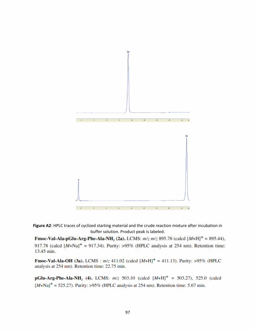
97
Figure A2: HPLC traces of cyclized starting material and the crude reaction mixture after incubation in
buffer solution. Product peak is labeled.

98
Figure A3: HPLC traces of cyclized starting material and the crude reaction mixture after incubation in
buffer solution. Product peak is labeled.

99
.

100
Figure A4: HPLC traces of cyclized starting material and the crude reaction mixture after incubation in
buffer solution. Product peak is labeled.

101
Figure A5: HPLC traces of cyclized starting material and the crude reaction mixture after incubation in
buffer solution. Product peak is labeled.

102
Figure A6: HPLC traces of cyclized starting material and the crude reaction mixture after incubation in
buffer solution. Product peak is labeled.

103

104
Figure A7: HPLC traces of cyclized starting material and the crude reaction mixture after incubation in
buffer solution. Product peak is labeled.
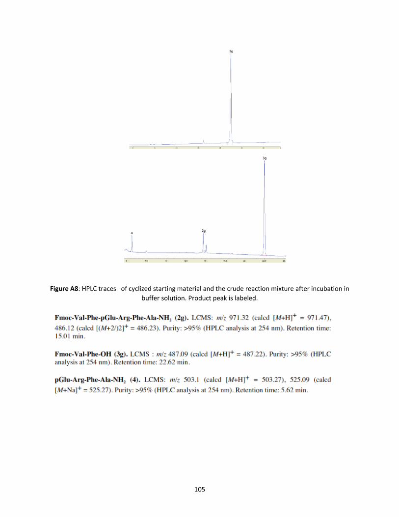
105
Figure A8: HPLC traces of cyclized starting material and the crude reaction mixture after incubation in
buffer solution. Product peak is labeled.

106
Figure A9: HPLC traces of cyclized starting material and the crude reaction mixture after incubation in
buffer solution. Product peak is labeled.

107
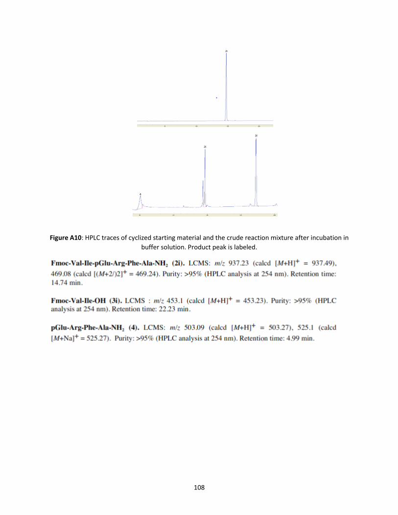
108
Figure A10: HPLC traces of cyclized starting material and the crude reaction mixture after incubation in
buffer solution. Product peak is labeled.

109
Figure A11: HPLC traces of cyclized starting material and the crude reaction mixture after incubation in
buffer solution. Product peak is labeled.

110

111
Figure A12: HPLC traces of cyclized starting material and the crude reaction mixture after incubation in
buffer solution. Product peak is labeled.

112
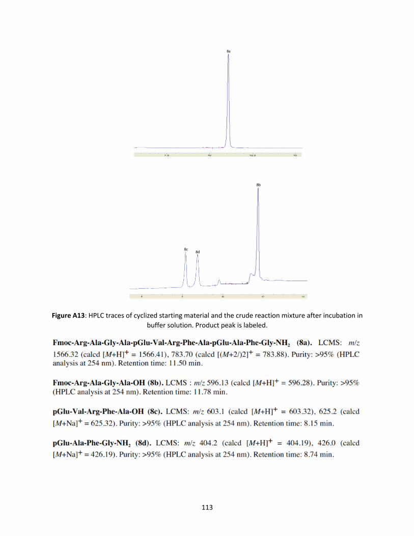
113
Figure A13: HPLC traces of cyclized starting material and the crude reaction mixture after incubation in
buffer solution. Product peak is labeled.

114
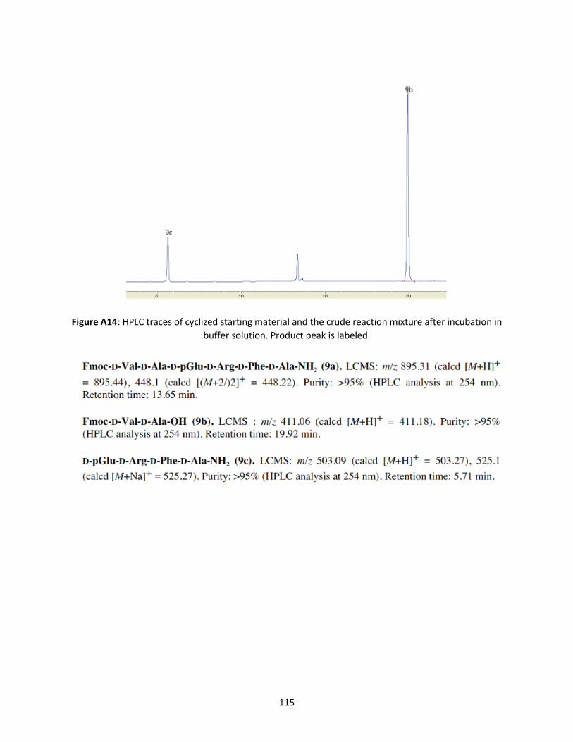
115
Figure A14: HPLC traces of cyclized starting material and the crude reaction mixture after incubation in
buffer solution. Product peak is labeled.
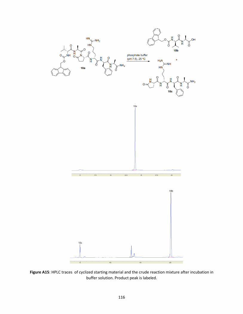
116
Figure A15: HPLC traces of cyclized starting material and the crude reaction mixture after incubation in
buffer solution. Product peak is labeled.
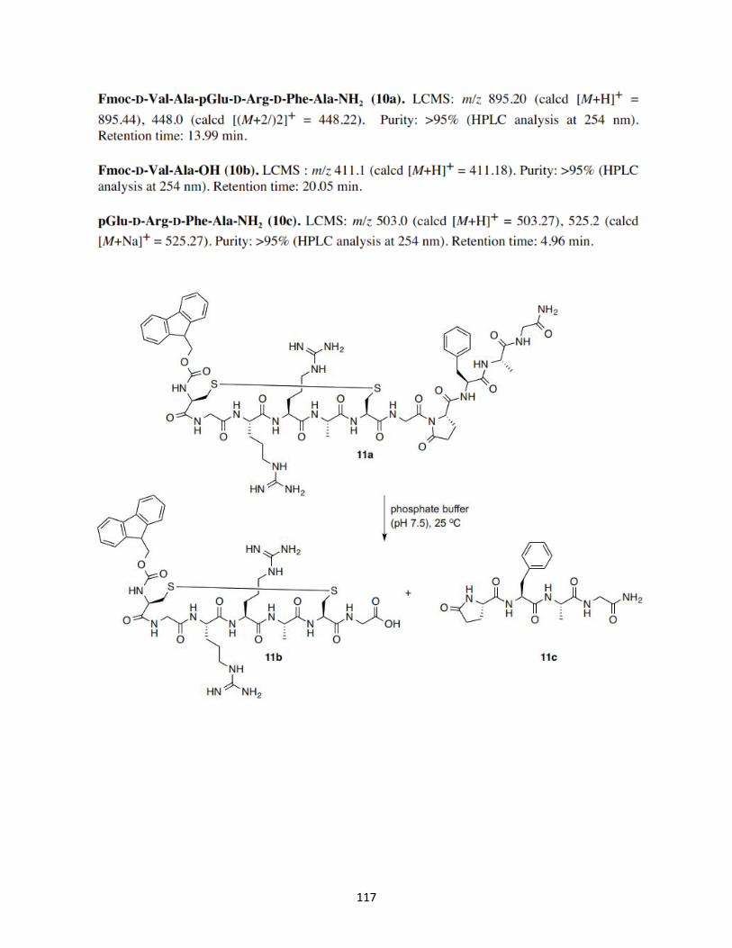
117

118
Figure A16: HPLC traces of cyclized starting material and the crude reaction mixture after incubation in
buffer solution. Product peak is labeled.

119

120
Figure A17: HPLC traces of cyclized starting material and the crude reaction mixture after incubation in
buffer solution. Product peak is labeled.

121

122
Figure A18: HPLC traces of cyclized starting material and the crude reaction mixture after incubation in
buffer solution. Product peak is labeled.

123
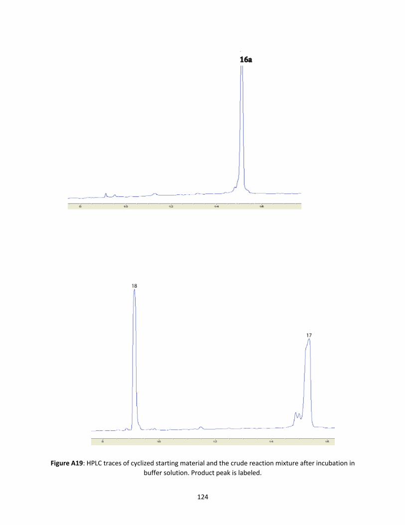
124
Figure A19: HPLC traces of cyclized starting material and the crude reaction mixture after incubation in
buffer solution. Product peak is labeled.

125

126
Figure A20: HPLC traces of cyclized starting material and the crude reaction mixture after incubation in
buffer solution. Product peak is labeled.

127

128
Figure A21: HPLC traces of cyclized starting material and the crude reaction mixture after incubation in
buffer solution. Product peak is labeled.

129

130
CHAPTER 3
Ac-Phe-Arg-Lys-Gly-Lys-Ala-NH2 Linear Peptide (1) - LCMS: m/z 747.6 (calcd [M+H]+ = 747.893), Purity:
>95% (HPLC analysis at 220 nm). Retention time: 6.1 min. (HPLC method of 0–80% 0.1% FA)
Figure A22 : HPLC trace and MS of Ac-Phe-Arg-Lys-Gly-Lys-Ala-NH2 Linear Peptide
Ac-Phe-Arg-Lys-Gly-Lys-Ala-NH2 Stapled peptide (1a) at 250C- LCMS: m/z 773.5 (calcd [M+H]+ = 773.88 ),
Purity: >95% (HPLC analysis at 220 nm). Retention time: Starting material 4.902min, Product 7.848 min
(HPLC method of 0–80% 0.1% FA).Reactant 31.30%, Product 68.70%.

131
Ac-Phe-Arg-Lys-Gly-Lys-Ala-NH2 Stapled peptide (1a) at 650C (9.2 equiv CDI) - LCMS: m/z 773.5 (calcd
[M+H]+ = 773.88 ), Purity: >95% (HPLC analysis at 220 nm). Retention time: Starting material 5.6i5 min.
Product 9.422 min (HPLC method of 0–80% 0.1% FA). Reactant 5.788%, Product 94.212%.
Figure A23: HPLC trace of Ac-Phe-Arg-Lys-Gly-Lys-Ala-NH2 Stapled Peptide at 250C and 650C. The product
peak is labeled.
13C NMR for Lys-Lys stapled Ac-Phe-Arg-Lys-Gly-Lys-Ala-NH2 (1a).
13C NMR (151 MHz, DMSO-d6) (ppm) 174, 171.6, 171.3, 171, 170.7, 169.3, 168.6, 157.9, 157, 137.9, 129,
127.9, 126.153.9, 52.2, 51.9, 51.8, 47.9, 42.3, 40.1, 38.8, 38.7, 37.3, 32.2, 31.7, 29, 28.8, 28.7, 24.8, 22.4,
22.3, 21.6, 18.2.
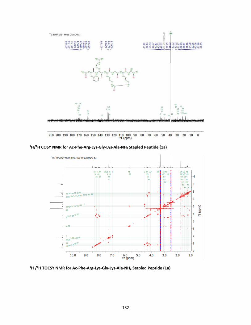
132
1H/1H COSY NMR for Ac-Phe-Arg-Lys-Gly-Lys-Ala-NH2 Stapled Peptide (1a)
1H /1H TOCSY NMR for Ac-Phe-Arg-Lys-Gly-Lys-Ala-NH2 Stapled Peptide (1a)

133
1H /13C HSQC NMR for Ac-Phe-Arg-Lys-Gly-Lys-Ala-NH2 Stapled Peptide (1a)
1H /13C HMBC NMR for Ac-Phe-Arg-Lys-Gly-Lys-Ala-NH2 Stapled Peptide (1a)
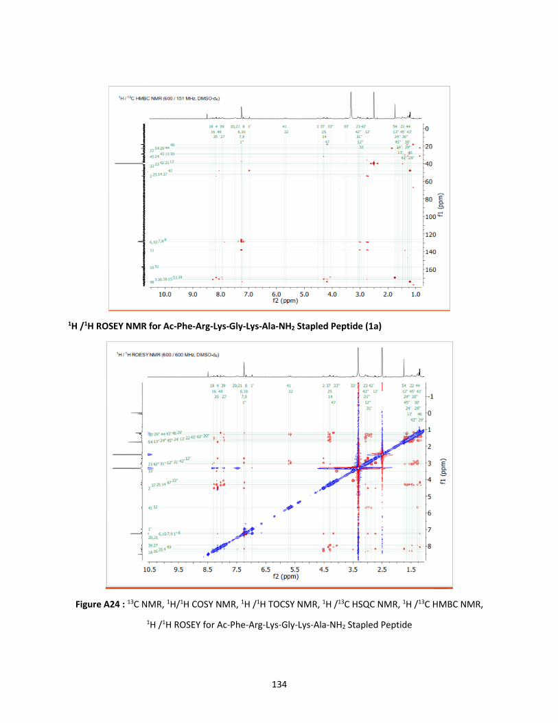
134
1H /1H ROSEY NMR for Ac-Phe-Arg-Lys-Gly-Lys-Ala-NH2 Stapled Peptide (1a)
Figure A24 : 13C NMR, 1H/1H COSY NMR, 1H /1H TOCSY NMR, 1H /13C HSQC NMR, 1H /13C HMBC NMR,
1H /1H ROSEY for Ac-Phe-Arg-Lys-Gly-Lys-Ala-NH2 Stapled Peptide

135
Ac- Phe-Lys-Gly-Ala-Lys-Phe-NH2 (2) - LCMS: m/z 738.9 (calcd [M+H]+ = 738.88), Purity: >95% (HPLC
analysis at 220 nm). Retention time: 9.9 min. (HPLC method of 0–80% 0.1% FA)
Figure A25 : HPLC trace and MS of Ac- Phe-Lys-Gly-Ala-Lys-Phe NH2 Linear Peptide

136
Ac- Phe-Lys-Gly-Ala-Lys-Phe-NH2 Stapled peptide (2a) after 5 h: LCM-: m/z 764.88 (calcd [M+H]+ =
764.87), Purity: >95% (HPLC analysis at 220 nm). Retention time: reactant 8.662 min., Product 11.273 min.
(HPLC method of 0–80% 0.1% FA).Reactant 86.167%. Product 13.833%.
Ac-Phe-Lys-Gly-Ala-Lys-Phe-NH2 peptide Stapled (2a) after 10 h: LCM-: m/z 764.88 (calcd [M+H]+ =
764.87), Purity: >95% (HPLC analysis at 220 nm). Retention time: reactant 8.642 min., Product 11.087 min.
(HPLC method of 0–80% 0.1% FA). Reactant 73.270%. Product 26.730%.
Figure A26: HPLC trace of Ac-Phe-Lys-Gly-Ala-Lys-Phe-NH2 Stapled after 5h and 10 h. The product peak is
labeled.

137
1H NMR for Ac- Phe-Lys-Gly-Ala-Lys-Phe- NH2 Stapled Peptide (2a).
1H NMR chemical shifts referenced to residual DMSO-d6 at 2.50 ppm and 13C NMR chemical shifts
referenced to DMSO-d6 at 39.52 ppm.
1H NMR for Ac- Phe-Lys-Gly-Ala-Lys-Phe- NH2 Stapled Peptide (2a
1H NMR (600 MHz, DMSO-d6) (ppm) 8.30 (m, 1H), 8.30 (m, 1H), 7.98 (d, J = 8.4Hz, 1H), 7.97(d, J = 8.5
Hz, 1H), 7.91 (d, J = 6.5 Hz, 1H), 7.65 (d, J = 8.3 Hz, 1H), 7.33(s, 1H), 7.17 (m, 2H), 7.17 (m, 2H), 7.17 (m,
2H), 7.13 (m, 2H), 7.11 (m, 1H), 7.11 (m, 1H), 7.02 (s, 1H), 5.76 (t, J = 5.2 Hz, 1H), 5.29 (t, J = 4.8 Hz, 1H),
4.45 (m, 1H), 4.34 (m, 1H), 4.19 (m, 1H), 4.18 (m, 1H), 4.12 (m, 1H), 3.93 ( dd, J = 16.4, 6.1 Hz, 1H), 3.39
(dd, J=16.1, 5.8 Hz, 1H), 3.02 (m, 1H), 2.99 (m, 1H), 2.94 (dd, J = 14.2, 5.4 Hz, 1H), 2.90 (m, 1H), 2.88 (m,
1H), 2.87 (m, 1H), 2.74 (dd, J=14.1, 8.0 Hz, 1H), 2.65 (m, 1H), 1.67 (s, 1H), 1.55 (m, 1H), 1.55 (m, 1H),
1.39 (m, 1H), 1.38 (m, 1H), 1.32 (m, 1H), 1.30 (m, 1H), 1.28 (m, 1H), 1.23 (m, 1H), 1.17 (m, 2H), 1.18 (m,
1H), 1.13 (d, J=7.2 Hz, 3H), 1.11 (m, 1H).
13C NMR for Ac- Phe-Lys-Gly-Ala-Lys-Phe- NH2 Stapled Peptide (2a).

138
13C NMR (151 MHz, DMSO-d6) (ppm) 172.2, 171.3, 171.2, 170.7, 169.4, 169, 157.8, 137.9, 129, 127.8,
126, 53.6, 51.9, 51.9, 48.1, 41.6, 38.2, 37.8, 37.3, 31.1, 30.5, 28.5, 27.8, 22.3, 22, 21, 17.5.
1H/1H COSY for Ac- Phe-Lys-Gly-Ala-Lys-Phe- NH2 Stapled Peptide (2a)
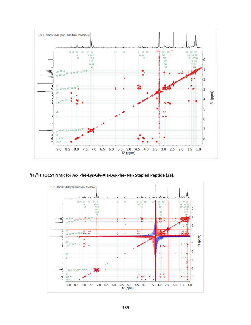
139
1H /1H TOCSY NMR for Ac- Phe-Lys-Gly-Ala-Lys-Phe- NH2 Stapled Peptide (2a).
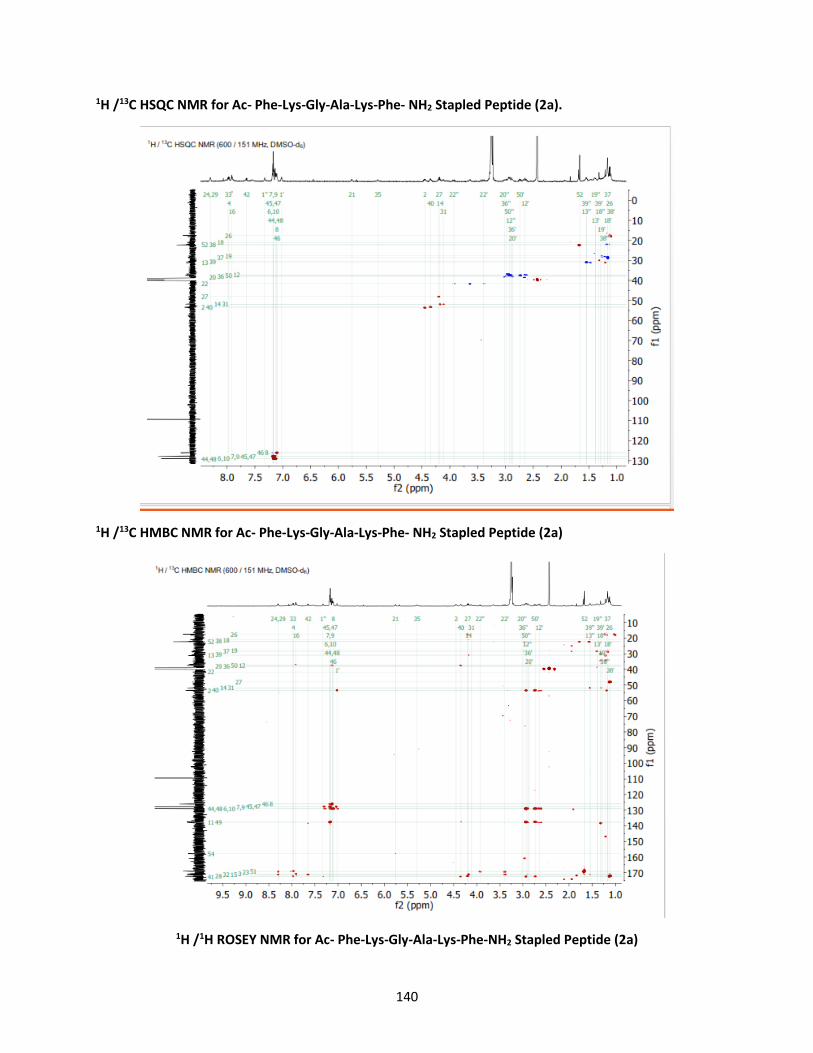
140
1H /13C HSQC NMR for Ac- Phe-Lys-Gly-Ala-Lys-Phe- NH2 Stapled Peptide (2a).
1H /13C HMBC NMR for Ac- Phe-Lys-Gly-Ala-Lys-Phe- NH2 Stapled Peptide (2a)
1H /1H ROSEY NMR for Ac- Phe-Lys-Gly-Ala-Lys-Phe-NH2 Stapled Peptide (2a)

141
Figure A27 : 1H NMR, 13C NMR, 1H/1H COSY NMR, 1H /1H TOCSY NMR, 1H /13C HSQC NMR, 1H /13C HMBC
NMR, 1H /1H ROSEY NMR for Stapled Peptide Ac-Phe-Arg-Lys-Gly-Lys-Ala-NH2
Ac-Phe-Lys-Lys-Phe-NH2 Linear Peptide (3): LCMS- m/z 609.9 (calcd [M+H]+ = 609.752), Purity: >95% (HPLC
analysis
at 220 nm).

142
Figure A28- MS of Ac-Phe-Lys-Lys-Phe-NH2 Linear Peptide
Expected Ac-Phe-Lys-Lys-Phe-NH2 Stapled Peptide (3a): (calcd [M+H]+ = 635.646) was not obtained, but
intermediate (3b) was obtained.
Intermediate (3b)-LCMS: m/z 703.1 (calcd [M+H]+ =703.823), Purity: >95% (HPLC analysis at 220 nm).
Retention time: Starting material 8.303 min intermediate - 12.582 min (HPLC method of 0–80% 0.1% FA)

143
Intermedia23.587%
Figure A29: HPLC trace and MS of Ac-Phe-Lys-Lys-Phe-NH2 Intermediate. The product peak is labeled.

144
Ac-Arg-Lys-Leu-Gly-Ala-Lys-Tyr-NH2 Linear Peptide (4): LCMS- m/z 876.5 (calcd [M+H]+ =877.05), Purity:
>95% (HPLC analysis at 220 nm). Retention time: 10.7 min. (HPLC method of 0–80% 0.1% FA)
Figure A30: HPLC trace and MS of Ac-Arg-Lys-Leu-Gly-Ala-Lys-Tyr-NH2 Linear Peptide

145
Ac-Arg-Lys-Leu-Gly-Ala-Lys-Tyr-NH2 Stapled peptide (4a) (CDI 7 equiv) at 650C -LCMS: m/z 903.04 (calcd
[M+H]+ = 902.6), Purity: >95% (HPLC analysis at 220 nm). Retention time: Staring material 10.141 min.
Product 13.697 min. (HPLC method of 0–80% 0.1% FA). Reactant 2.042%, Product 97.958%
HRMS data: Ac-Arg-Lys-Leu-Gly-Ala-Lys-Tyr-NH2 Stapled Peptide (4a) [C41H67N13O10+H]+: calc. m/z
902.5207, obs. m/z 902.5208

146
MS of Ac-Arg-Lys-Leu-Gly-Ala-Lys-Tyr-NH2 Stapled Peptide (4a)
H/D Exchange Experiment: Fmoc--Arg-Lys-Leu-Gly-Ala-Lys-Tyr-NH2 Stapled Peptide (4a)
Observed # of exchangeable protons = 1099 – 1082 – 1 = 16. Expected # of exchangeable protons = 16
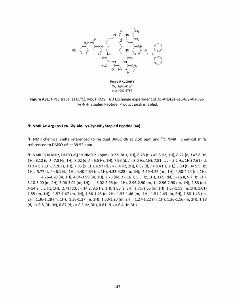
147
Figure A31: HPLC trace (at 650C), MS, HRMS, H/D Exchange experiment of Ac-Arg-Lys-Leu-Gly-Ala-Lys-Tyr-NH2 Stapled Peptide. Product peak is labled.
1H NMR Ac-Arg-Lys-Leu-Gly-Ala-Lys-Tyr-NH2 Stapled Peptide (4a)
1H NMR chemical shifts referenced to residual DMSO-d6 at 2.50 ppm and 13C NMR chemical shifts
referenced to DMSO-d6 at 39.52 ppm.
1H NMR (600 MHz, DMSO-d6) 1H NMR (ppm) 9.22( br s, 1H), 8.28 (t, J =5.8 Hz, 1H), 8.22 (d, J =7.8 Hz,
1H), 8.12 (d, J =7.8 Hz, 1H), 8.02 (d, J = 6.5 Hz, 1H), 7.99 (d, J = 8.0 Hz, 1H), 7.81( t, J = 5.2 Hz, 1H,) 7.61 ( d,
J Hz = 8.1,1H), 7.26 (s, 1H), 7.05 (s, 1H), 6.97 (d, J = 8.4 Hz, 2H), 6.62 (d, J = 8.4 Hz, 2H,) 5.80 (t, J= 5.9 Hz,
1H), 5.77 (t, J = 6.1 Hz, 1H), 4.40-4.34 (m, 1H), 4.33-4.28 (m, 1H), 4.30-4.26 ( m, 1H), 4.30-4.24 (m, 1H),
4.26-4.20 (m, 1H), 4.04-3.99 (m, 1H), 3.73 (dd, J = 16.7, 5.5 Hz, 1H), 3.69 (dd, J =16.8, 5.7 Hz, 1H),
3.10-3.00 (m, 2H), 3.06-3.02 (m, 1H), 3.02-2.96 (m, 1H), 2.96-2.90 (m, 1), 2.96-2.90 (m, 1H), 2.88 (dd,
J=14.2, 5.1 Hz, 1H), 2.71 (dd, J = 14.2, 8.5 Hz, 1H), 1.85 (s, 3H), 1.71-1.65 (m, 1H), 1.67-1.59 (m, 1H), 1.61-
1.55 (m, 1H), 1.57-1.47 (m, 1H), 1.56-1.45 (m,2H), 1.53-1.46 (m, 1H), 1.51-1.43 (m, 2H), 1.50-1.43 (m,
2H), 1.36-1.28 (m, 1H), 1.36-1.27 (m, 2H), 1.30-1.20 (m, 2H), 1.27-1.22 (m, 1H), 1.26-1.16 (m, 2H), 1.18
(d, J = 6.8, 3H Hz), 0.87 (d, J = 6.5 Hz, 3H), 0.81 (d, J = 6.4 Hz, 3H).

148
13C NMR Ac-Arg-Lys-Leu-Gly-Ala-Lys-Tyr-NH2 Stapled Peptide (4a).
13C NMR (151 MHz, DMSO-d6) (ppm) 172.7, 172.3, 172, 171.4, 171.3, 171.2, 169.2, 168, 158.5, 157,
155.7, 130, 127.6, 114.7, 53.7, 53.2, 52.2, 51.7, 51.1, 47.8, 41.9, 40.4, 40.1, 38.9, 38, 36.6, 31.5, 30.7,
29.5, 29.4, 29.1, 24.7, 24, 22.9, 22.4, 22.2, 21.4, 18.
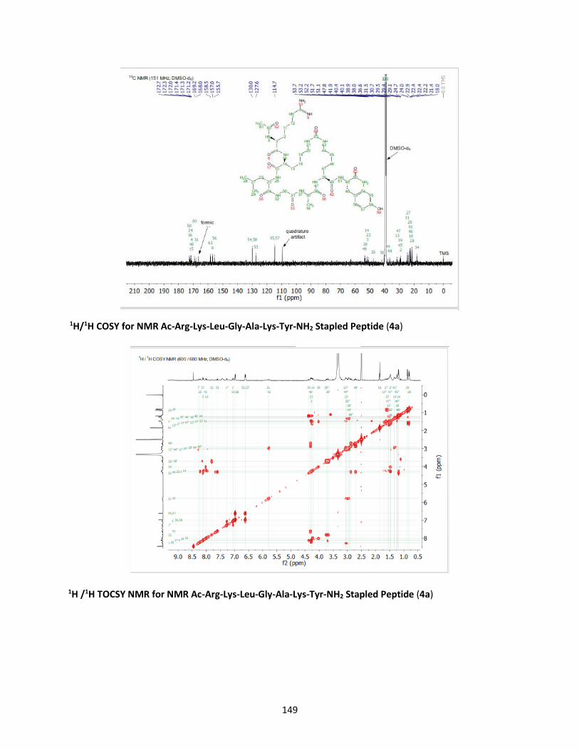
149
1H/1H COSY for NMR Ac-Arg-Lys-Leu-Gly-Ala-Lys-Tyr-NH2 Stapled Peptide (4a)
1H /1H TOCSY NMR for NMR Ac-Arg-Lys-Leu-Gly-Ala-Lys-Tyr-NH2 Stapled Peptide (4a)

150
1H /13C HSQC NMR for Ac-Arg-Lys-Leu-Gly-Ala-Lys-Tyr-NH2 Stapled Peptide (4a)
1H /13C HMBC NMR for Ac-Arg-Lys-Leu-Gly-Ala-Lys-Tyr-NH2 Stapled Peptide (4a)

151
1H /1H ROSEY NMR for Ac-Arg-Lys-Leu-Gly-Ala-Lys-Tyr-NH2 Stapled Peptide (4a)
Figure A32 : 1H NMR, 13C NMR, 1H/1H COSY NMR, 1H /1H TOCSY NMR, 1H /13C HSQC NMR, 1H /13C HMBC
NMR, 1H /1H ROSEY for Stapled Peptide Ac-Phe-Arg-Lys-Gly-Lys-Ala-NH2

152
Ac-Arg-Lys-Ala-Leu-Gly-Ala-Lys-Phe-NH2 Linear Peptide (5): LCMS- m/z 930.5 (calcd [M+H]+ =931.1),
[(M+2/)2] += 465.8 (calcd [(M+2/)2]+= 466.5) Purity: >95% (HPLC analysis at 220 nm). Retention time: 5.4
min. (HPLC method of 0–80% 0.1% FA).
Figure A33: HPLC and MS of linear peptide Ac-Arg-Lys-Ala-Leu-Gly-Ala-Lys-Phe-NH2 Linear Peptide.
Product peak is labled.
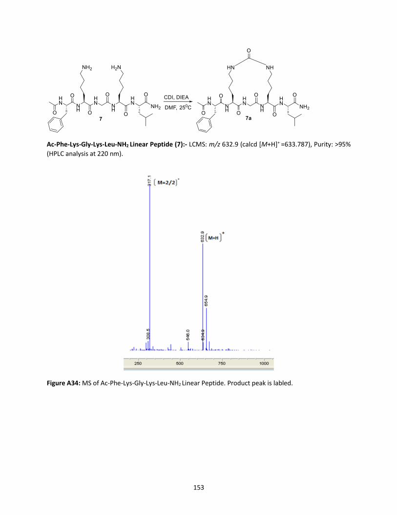
153
Ac-Phe-Lys-Gly-Lys-Leu-NH2 Linear Peptide (7):- LCMS: m/z 632.9 (calcd [M+H]+ =633.787), Purity: >95%
(HPLC analysis at 220 nm).
Figure A34: MS of Ac-Phe-Lys-Gly-Lys-Leu-NH2 Linear Peptide. Product peak is labled.
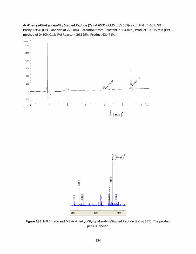
154
Ac-Phe-Lys-Gly-Lys-Leu-NH2 Stapled Peptide (7a) at 650C -LCMS: m/z 659(calcd [M+H]+ =659.781),
Purity: >95% (HPLC analysis at 220 nm). Retention time: Reactant 7.984 min., Product 10.915 min (HPLC
method of 0–80% 0.1% FA) Reactant 36.529%, Product 63.471%.
Figure A35: HPLC trace and MS Ac-Phe-Lys-Gly-Lys-Leu-NH2 Stapled Peptide (8a) at 650C. The product
peak is labeled.
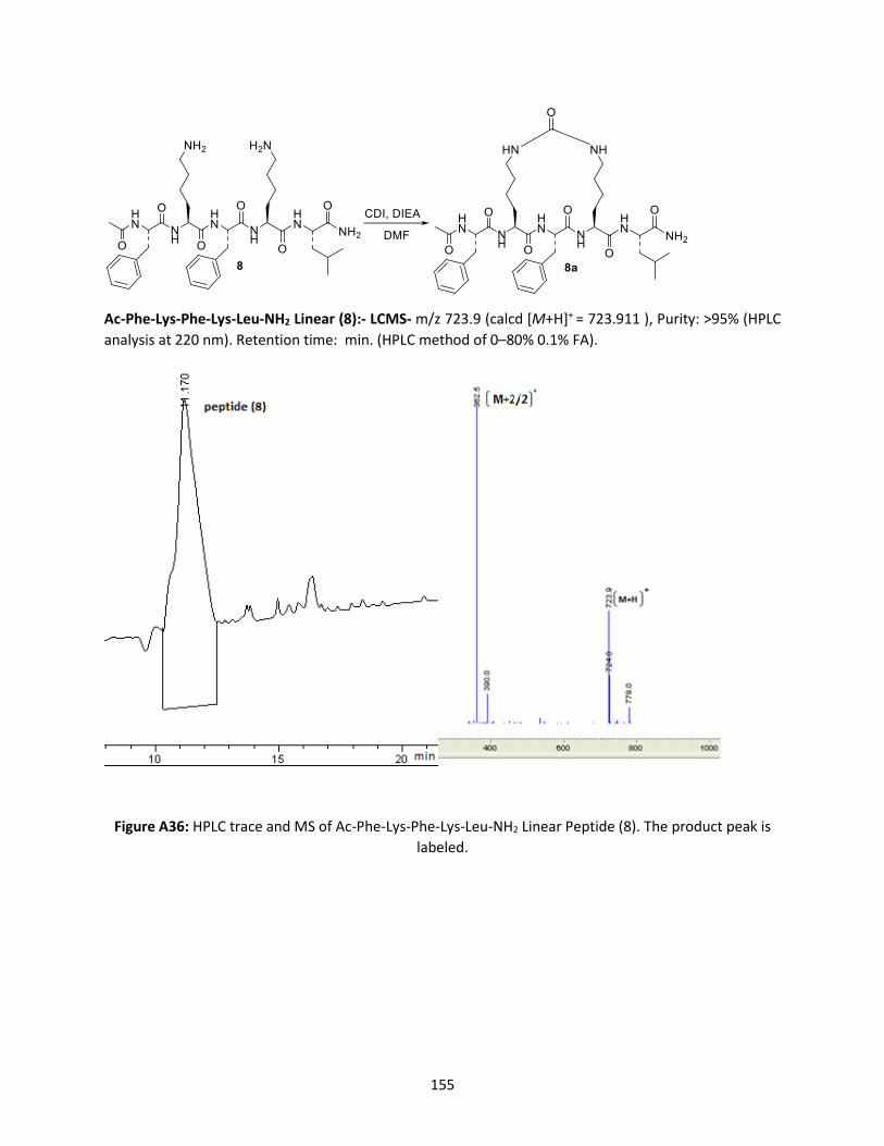
155
Ac-Phe-Lys-Phe-Lys-Leu-NH2 Linear (8):- LCMS- m/z 723.9 (calcd [M+H]+ = 723.911 ), Purity: >95% (HPLC
analysis at 220 nm). Retention time: min. (HPLC method of 0–80% 0.1% FA).
Figure A36: HPLC trace and MS of Ac-Phe-Lys-Phe-Lys-Leu-NH2 Linear Peptide (8). The product peak is
labeled.

156
Ac-Phe-Lys-Phe-Lys-Leu-NH2 Stapled Peptide (8a) at 650C: LCMS- m/z 749.8 (calcd [M+H]+ = 749.805),
Purity: >95% (HPLC analysis at 220 nm). Retention time: Reactant 10.796 min. Product 13.604 min.
Reactant 26.033%, Product 73.967% (HPLC method of 0–80% 0.1% FA).
Figure A37: HPLC trace and MS of Ac-Phe-Lys-Phe-Lys-Leu-NH2 Stapled Peptide (8a) at 650C. Product
peak is labled.

157
Ac-Glu-Asn-Pro-Glu-Lys-Ile-Leu-Asp-Lys-His-Val-Gln-Arg-Val-Met-NH2 Linear Peptide (9): LCMS: [(M+2/)2] += 939.1 (calcd [(M+2/)2]+= 939.58), [(M+3/)3] += 626.67 (calcd [(M+3/)3] += 626.5),Purity: > 95%. (HPLC analysis at 220 nm). Retention time: Reactant 11.305 min. (HPLC method of 0–60% 0.1% FA).

158
Figure A38: HPLC trace and MS of Ac-Glu-Asn-Pro-Glu-Lys-Ile-Leu-Asp-Lys-His-Val-Gln-Arg-Val-Met-NH2
Linear Peptide (9)
Ac-Glu-Asn-Pro-Glu-Lys-Ile-Leu-Asp-Lys-His-Val-Gln-Arg-Val-Met-NH2 Stapled Peptide (9a)-LCMS:
[(M+2/)2] += 952.58 (calcd [(M+2/)2]+ = 952.58), Purity: > 95% (HPLC analysis at 220 nm). Retention time:
Reactant 11.647 min, Product 14.189 min. (HPLC method of 0–60% 0.1% FA). Reactant 10.667%, Product
89.333%.
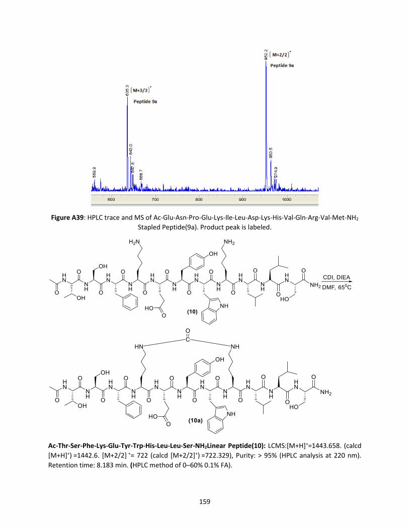
159
Figure A39: HPLC trace and MS of Ac-Glu-Asn-Pro-Glu-Lys-Ile-Leu-Asp-Lys-His-Val-Gln-Arg-Val-Met-NH2
Stapled Peptide(9a). Product peak is labeled.
Ac-Thr-Ser-Phe-Lys-Glu-Tyr-Trp-His-Leu-Leu-Ser-NH2Linear Peptide(10): LCMS:[M+H]+=1443.658. (calcd
[M+H]+) =1442.6. [M+2/2] += 722 (calcd [M+2/2]+) =722.329), Purity: > 95% (HPLC analysis at 220 nm).
Retention time: 8.183 min. (HPLC method of 0–60% 0.1% FA).

160
Figure A40: HPLC trace and MS of Ac-Thr-Ser-Phe-Lys-Glu-Tyr-Trp-His-Leu-Leu-Ser-NH2 (10) Linear
Peptide
Stapled peptide (10a):-LCMS: m/z 1470.9 (calcd [M+H]+ =1469.652), Purity: >95% (HPLC analysis at 220
nm). Retention time: Starting material 8.253 min. Product 18.861, 19.761 min (HPLC method of 0–60%
0.1% FA). Reactant 6.629%, Product 93.371%.

161
Figure A41: HPLC and MS of Ac-Thr-Ser-Phe-Lys-Glu-Tyr-Trp-His-Leu-Leu-Ser-NH2 Stapled Peptide (10) .
Product peak is labaled.
Ac- Ala-Lys-Leu-Gly-Phe-Lys-Leu-NH2 (11): Linear-LCMS: m/z 816.9 (calcd [M+H]+ =817.025), Purity: >95%
(HPLC analysis at 220 nm). Retention time: 7.826 min. (HPLC method of 0–80% 0.1% FA)

162
Figure A42: HPLC trace and MS of Ac- Ala-Lys-Leu-Gly-Phe-Lys-Leu-NH2 Linear Peptide (11)
Stapled peptide (11a)-LCMS: m/z 844.8 (calcd [M+H]+ =844.01), Purity: >95% (HPLC analysis at 220 nm).
Retention time: Reactant 7.891 min. Product 9.364 min, 9.692 min. (HPLC method of 0–80% 0.1% FA).
Reactant 56.632%, Product 43.639%.

163
Figure A43: HPLC trace and MS of Ac- Ala-Lys-Leu-Gly-Phe-Lys-Leu-NH2 (11a) Stapled Peptide. Product
peak is labeled
Ac- Ile-Lys-Leu-Gly-Phe-Lys-Leu-NH2 Linear Peptide (12): -LCMS: m/z 859.06 (calcd [M+H]+ =860.10),
Purity: >95% (HPLC analysis at 220 nm). Retention time: 11.170 min. (HPLC method of 0–80% 0.1% FA)

164
Figure A44: HPLC trace and MS of Ac- Ile-Lys-Leu-Gly-Phe-Lys-Leu-NH2 Linear Peptide (12). Product peak
is labeled.
Stapled Peptide (12a)-LCMS: m/z 885.8 (calcd [M+H]+ = 886.09), Purity: >95% (HPLC analysis at 220 nm).
Retention time- Reactant 12.650 min: Product 14.022 min, 14.215 min. (HPLC method of 0–80% 0.1% FA).
Reactant 32.916%, Product 67.084%.

165
Figure A45 : HPLC trace and MS of Ac- Ile-Lys-Leu-Gly-Phe-Lys-Leu-NH2 (12) Stapled Peptide. Product
peak is labled.

166
Stability study of Ac-Glu-Asn-Pro-Glu-Lys-Ile-Leu-Asp-Lys-His-Val-Gln-Arg-Val-Met-NH2 Linear
Peptide(9): -LCMS: [(M+2/)2] += 939.1 (calcd [(M+2/)2]+= 939.58), [(M+3/)3] += 626.67 (calcd [(M+3/)3] +=
626.5),Purity:> 95%. (HPLC analysis at 220 nm). Retention time: Reactant 11.305 min. (HPLC method of 0–
60% 0.1% FA).

167
Figure A46A -1: Ac-Glu-Asn-Pro-Glu-Lys-Ile-Leu-Asp-Lys-His-Val-Gln-Arg-Val-Met-NH2 Linear Peptide (9): 0
min
Figure A46 A-2: Ac-Glu-Asn-Pro-Glu-Lys-Ile-Leu-Asp-Lys-His-Val-Gln-Arg-Val-Met-NH2 Linear Peptide(9):
Fragmentation at 30 min: Fragments (9i), (9ii), (9iii), (9iv).

168
Figure A46 A-3: Ac-Glu-Asn-Pro-Glu-Lys-Ile-Leu-Asp-Lys-His-Val-Gln-Arg-Val-Met-NH2 Linear Peptide(9):
Fragmentation after 90 min: Fragments (9i), (9ii), (9iii), (9iv).
Figure A46: Stability Study: Ac-Glu-Asn-Pro-Glu-Lys-Ile-Leu-Asp-Lys-His-Val-Gln-Arg-Val-Met-NH2 Linear
Peptide (9):
Fragmentation after 90 min: Fragments (9i), (9ii), (9iii), (9iv).
Fragment Ac-Glu-Asn-Pro-Glu-Lys-OH (9i) - LCMS: m/z 658.3 (calcd [M+H]+ = 657.676), Purity: >95%
(HPLC analysis at 220 nm). Retention time- 5.703 min (HPLC method of 0–60% 0.1% FA).
Fragment Ile-Leu-Asp-Lys-His-Val-Gln-Arg-Val-Met-NH2 (9ii) - LCMS: m/z 1008.8 (calcd [M+H]+ =
1008.186), [M+2/2] += 504.0 (calcd [M+2/2]+) =504.593), Purity: >95% (HPLC analysis at 220 nm).
Retention time- 6.648 min (HPLC method of 0–60% 0.1% FA).
Fragment Ac-Glu-Asn-Pro-Glu-Lys-Ile-Leu-Asp-Lys-His-Val-Gln-Arg-OH (9iii ) - LCMS: [(M+2/)2] +=824.5
(calcd [(M+2/)2]+= 824.424), [(M+3/)3] += 549.7 (calcd [(M+3/)3] += 549.949),Purity:> 95%. (HPLC analysis
at 220 nm). Retention time: Reactant 9.831 min. (HPLC method of 0–60% 0.1% FA).
Fragment Ac-Glu-Asn-Pro-Glu-Lys-Ile-Leu-Asp-Lys-OH (9iv) - LCMS: calculated M+2/2= 564.129 LCMS:
[(M+2/) 2] +=564.1(calcd (M+2/) 2]+= 564.129. Purity:> 95%. (HPLC analysis at 220 nm). Retention time:
10.622 min. (HPLC method of 0–60% 0.1% FA).
Figure A46 A: The stability of the Ac-Glu-Asn-Pro-Glu-Lys-Ile-Leu-Asp-Lys-His-Val-Gln-Arg-Val-Met-NH2
linear (9).
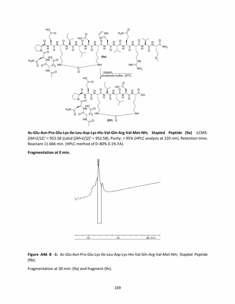
169
Ac-Glu-Asn-Pro-Glu-Lys-Ile-Leu-Asp-Lys-His-Val-Gln-Arg-Val-Met-NH2 Stapled Peptide (9a) -LCMS:
[(M+2/)2] += 952.58 (calcd [(M+2/)2]+ = 952.58), Purity: > 95% (HPLC analysis at 220 nm). Retention time:
Reactant 11.666 min. (HPLC method of 0–80% 0.1% FA).
Fragmentation at 0 min.
Figure A46 B -1: Ac-Glu-Asn-Pro-Glu-Lys-Ile-Leu-Asp-Lys-His-Val-Gln-Arg-Val-Met-NH2 Stapled Peptide (9b):
Fragmentation at 30 min: (9a) and fragment (9v).

170
Figure A46 B -2: Fragmentation at 30 min: (9a) and fragment (9v).
Figure A46 B -3: Fragmentation at 90 min: fragment ((9v).
Fragment stapled Ac-Glu-Asn-Pro-Glu-Lys-Ile-Leu-Asp-Lys-OH (9v).- LCMS: [(M+2/)2] +=577.3 (calcd
[(M+2/)2]+= 577. 126), [(M+3/)3] += 385.6 (calcd [(M+3/)3] += 385.083),Purity:> 95%. (HPLC analysis at 220
nm). Retention time: 10.548 min. (HPLC method of 0–60% 0.1% FA).
Figure A46 B : The stability of the stapled peptide.
Figure A46 : The stability study of Ac-Glu-Asn-Pro-Glu-Lys-Ile-Leu-Asp-Lys-His-Val-Gln-Arg-Val-Met-NH2
Stapled Peptide compared to its Linear Analogue.

171
Figure A47 A: MS of Fmoc-Phe-Lys-Leu-Gly-Ala-Lys-Phe-Glu-(O-allyl)-CONH2
LCMS: m/z 1201.4 (calcd[M+H]+=1201.43), Purity:>95%
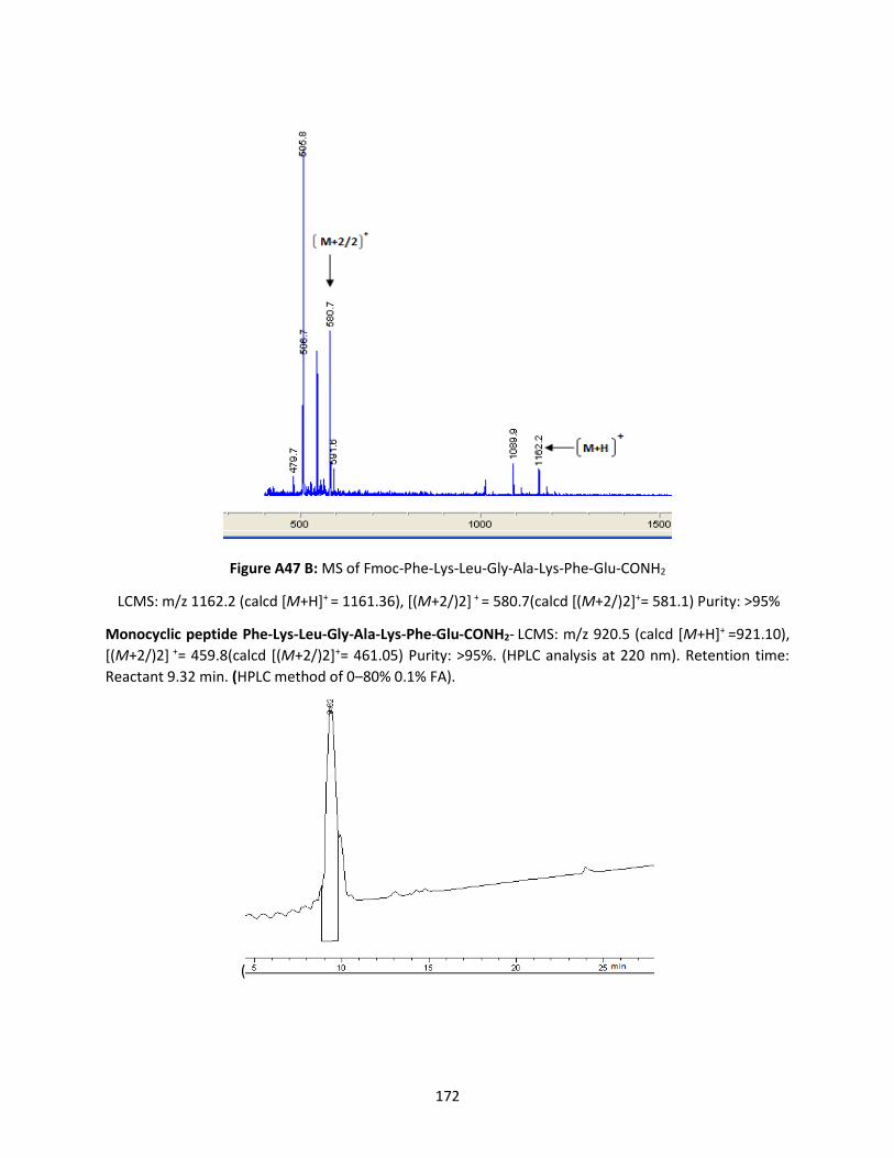
172
Figure A47 B: MS of Fmoc-Phe-Lys-Leu-Gly-Ala-Lys-Phe-Glu-CONH2
LCMS: m/z 1162.2 (calcd [M+H]+ = 1161.36), [(M+2/)2] + = 580.7(calcd [(M+2/)2]+= 581.1) Purity: >95%
Monocyclic peptide Phe-Lys-Leu-Gly-Ala-Lys-Phe-Glu-CONH2- LCMS: m/z 920.5 (calcd [M+H]+ =921.10),
[(M+2/)2] += 459.8(calcd [(M+2/)2]+= 461.05) Purity: >95%. (HPLC analysis at 220 nm). Retention time:
Reactant 9.32 min. (HPLC method of 0–80% 0.1% FA).
(

173
Figure A48 –HPLC trace and MS of Monocyclic Phe-Lys-Leu-Gly-Ala-Lys-Phe-Glu-NH2 (13a).
13C NMR Monocyclic Phe-Lys-Leu-Gly-Ala-Lys-Phe-Glu-NH2 (13a).
13C NMR (151 MHz, DMSO-d6) (ppm) 172.9, 172.6, 172.4, 172.2, 172.1, 171.6, 171, 170.6, 169.1, 137.9,
137.4, 129.2, 129, 128, 127.9, 126.2, 126.2, 54.8, 54.1, 53.7, 52.5, 52.5, 52, 49.1, 42.1, 39.4, 38.5, 38.5,
37.3, 36.7, 32.6, 31.6, 30.3, 29, 27, 26.9, 24, 22.6, 22.2, 21.8, 21.7, 17.

174
1H/1H COSY NMR Monocyclic Phe-Lys-Leu-Gly-Ala-Lys-Phe-Glu-NH2 (13a).
1H /1H TOCSY NMR Monocyclic Phe-Lys-Leu-Gly-Ala-Lys-Phe-Glu-NH2 (13a).

175
1H /13C HSQC NMR Monocyclic Phe-Lys-Leu-Gly-Ala-Lys-Phe-Glu-NH2 (13a).
1H /13C HMBC NMR Monocyclic Phe-Lys-Leu-Gly-Ala-Lys-Phe-Glu-NH2 (13a).

176
1H /1H ROSEY NMR Monocyclic Phe-Lys-Leu-Gly-Ala-Lys-Phe-Glu-NH2 (13a).
Figure A49 : 13C NMR, 1H/1H COSY NMR, 1H /1H TOCSY NMR, 1H /13C HSQC NMR, 1H /13C HMBC NMR,
1H /1H ROSEY for Monocyclic Phe-Lys-Leu-Gly-Ala-Lys-Phe-Glu-NH2 (13a).

177
Ion Mobility Resolved MS/MS
Ion Mobility Resolved MS/MS(A) 328 Å2 and (B) 339 Å2 Conformations
LCMS: m/z 946.7 (calcd [M+H]+ = 947), (M+23) += 968.8 (calcd [(M+23]+= 968.98), (M+39) += 984(calcd
[(M+39]+)= 985,Purity: >95%. (HPLC analysis at 220 nm). Retention time: Reactant 8.822 min, Product
11.177min, 12.374min (HPLC method of 0–80% 0.1% FA). Reactant 0.946%, Product 13b-1 92.491%, 13b-
2- 6.563%
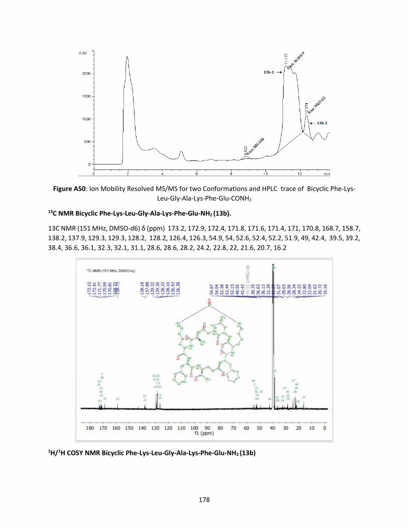
178
Figure A50: Ion Mobility Resolved MS/MS for two Conformations and HPLC trace of Bicyclic Phe-Lys-
Leu-Gly-Ala-Lys-Phe-Glu-CONH2
13C NMR Bicyclic Phe-Lys-Leu-Gly-Ala-Lys-Phe-Glu-NH2 (13b).
13C NMR (151 MHz, DMSO-d6) δ (ppm) 173.2, 172.9, 172.4, 171.8, 171.6, 171.4, 171, 170.8, 168.7, 158.7,
138.2, 137.9, 129.3, 129.3, 128.2, 128.2, 126.4, 126.3, 54.9, 54, 52.6, 52.4, 52.2, 51.9, 49, 42.4, 39.5, 39.2,
38.4, 36.6, 36.1, 32.3, 32.1, 31.1, 28.6, 28.6, 28.2, 24.2, 22.8, 22, 21.6, 20.7, 16.2
1H/1H COSY NMR Bicyclic Phe-Lys-Leu-Gly-Ala-Lys-Phe-Glu-NH2 (13b)

179
1H /1H TOCSY NMR Bicyclic Phe-Lys-Leu-Gly-Ala-Lys-Phe-Glu-NH2 (13b).
1H /13C HSQC NMR Bicyclic Phe-Lys-Leu-Gly-Ala-Lys-Phe-Glu-NH2 (13b).

180
1H /13C HMBC NMR Bicyclic Phe-Lys-Leu-Gly-Ala-Lys-Phe-Glu-NH2 (13b).
1H /1H ROSEY NMR Bicyclic Phe-Lys-Leu-Gly-Ala-Lys-Phe-Glu-NH2 (13b).

181
Figure A51 : 13C NMR, 1H/1H COSY NMR, 1H /1H TOCSY NMR, 1H /13C HSQC NMR, 1H /13C HMBC NMR,
1H /1H ROSEY for Bicyclic Phe-Lys-Leu-Gly-Ala-Lys-Phe-Glu-NH2 (13a).


