Download - Special Senses - Equilibrium and Hearing

Equilibrium
▪ vestibular apparatus - equilibriumreceptors in the semicircular canals andvestibule– send signals to the brain that initiate reflexes
needed to make the simplest changes in position as well as more complex moves

Equilibrium
Two Functional Parts
▪ Static Equilibrium (Vestibule) - monitor linear acceleration and the position of the head with respect to gravity (constant)
▪ Dynamic Equilibrium (Semicircular Canals) -monitor changes in head rotation
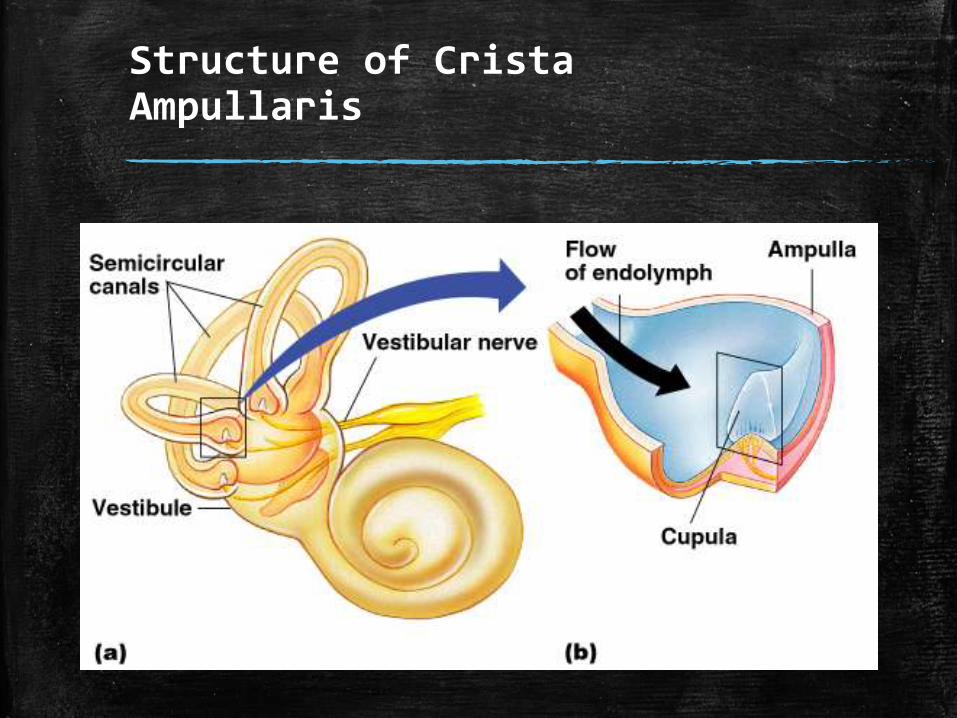
Structure of CristaAmpullaris

Static Equilibrium
Maculae
– receptors in the vestibule
Report on the position of the head
Send information via the vestibular nerve

Static Equilibrium
Anatomy of the Maculae
Hair cells are embedded in the otolithicmembrane
Otoliths (tiny stones made of calcium salts) float in a gel around the hair cells
respond to static equilibrium cues relative to the position of the head in space
Movements cause otoliths to bend the hair cells

Function of Maculae
Diagrammatic view of part of a macula

Function of Maculae
When the head is tipped, the maculae are stimulated by movement of the otoliths in the gelatinous otolithic membrane in the direction of gravitational pull, which creates a pull on the hair cells.

Dynamic Equilibrium
Crista ampullaris
– receptors in the semicircular canals
Tuft of hair cells
Cupula (gelatinous cap) covers the hair cells

Dynamic Equilibrium
Action of angular head movements
The cupula stimulates the hair cells
An impulse is sent via the vestibular nerve to the cerebellum

Crista Ampullaris


Recap
▪ What sense do the vestibule and semicircular canals serve?– Balance or equilibrium.
▪ Benji is enjoying a boat ride until a storm suddenly descends on the bay. So he is nauseated and can barely stand up. Which equilibrium receptors – static or dynamic – are operating furiously during such a rough voyage?– Dynamic receptors located in the semicircular canals (crista
ampullaris).

Recap
▪ What are otoliths, and what is their role in equilibrium?– Otoliths are tiny stones made of calcium
salts which are located in the maculae of the vestibule. They respond to static equilibrium cues relative to the position of the head in space.

Hearing
Spiral Organ of Corti
Located within the cochlear duct
Hearing receptors = hair cells on the basilar membrane
Gel-like tectorial membrane is capable of bending hair cells
Cochlear nerve attached to hair cells transmits nerve impulses to auditory cortex on temporal lobe – interpretation of sound or hearing occurs

Anatomy of the cochlea. (a) Lateral view of part of the internal ear with a wedge-shaped section removed from the cochlea.

Anatomy of the cochlea. (b) Magnified cross section of one turn of the cochlea, showing the relationship of the three scalae. This cross section has been rotated from its position in (a). (c) Detailed structure of the spiral organ.

Mechanisms of Hearing
Vibrations from sound waves move tectorial membrane
(pass through the endolymph fluid filling the membranous labyrinth in the cochlear duct)
Mechanisms of Hearing
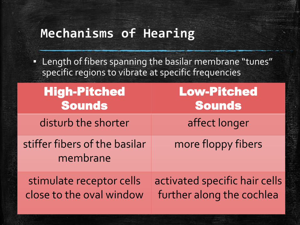
▪ Length of fibers spanning the basilar membrane “tunes” specific regions to vibrate at specific frequencies
High-Pitched
Sounds
Low-Pitched
Sounds
disturb the shorter affect longer
stiffer fibers of the basilar membrane
more floppy fibers
stimulate receptor cells close to the oval window
activated specific hair cells further along the cochlea
Mechanisms of Hearing
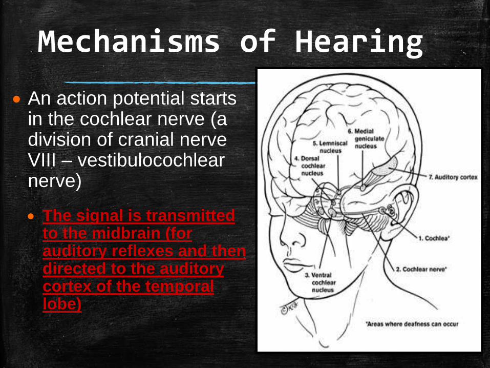
An action potential starts in the cochlear nerve (a division of cranial nerve VIII – vestibulocochlearnerve)
The signal is transmitted to the midbrain (for auditory reflexes and then directed to the auditory cortex of the temporal lobe)

Mechanisms of Hearing
▪ Continued stimulation can lead to adaptation–(over stimulation to the brain
makes it stop interpreting the sounds)

Hearing and Equilibrium Deficits
▪ Tuning fork or audiometry testing – try to diagnose ear problems or hearing deficits
▪ Deafness – hearing loss of any degree-from a slight loss to a total inability to hear sound
1. Conduction Deafness2. Sensorineural Deafness

Hearing Deficits
▪ Temporary or Permanent Conduction Deafness– something interferes with the conduction of sound
vibrations to the fluids of the inner ear
– Causes▪ build up of earwax
▪ fussion of ossicles (otosclerosis)
▪ ruptured eardrum
▪ otitis media (middle ear inflammation)
– still be able to hear by bone conduction (hearing aids)

Hearing Deficits
▪ Sensorineural Deafness– degeneration or damage to the receptor
cells in the spiral organ of Corti, to the cochlear nerve, or neurons of the auditory cortex
– Causes▪ Extended listening to excessively loud sounds
▪ Problem with nervous system structures
– Can’t hear better by either conduction route

Equilibrium Deficits
▪ Nausea, dizziness, and problems in maintaing balance– Impulses from the vestibular apparatus
“disagree” with what we see (visual input)
–Strange eye movements (jerky or rolling)
▪ Meniere’s Syndrome

Equilibrium Deficits
▪ Meniere’s Syndrome– Suspected causes▪ Arteriosclerosis, degeneration of cranial nerve VIII,
and increased pressure of the inner ear fluids
– Progressive deafness occurs
– Nauseted and howling or ringing sounds in the ears and vertigo (a sensation of spinning)
– So severe = cannot stand up without discomfort
– Anti-motion sickness drugs = decrease discomfort


