©2018 MFMER | slide-1
Prosthetic Valve DysfunctionAdvanced Echocardiography
Heidi M. Connolly
©2017 MFMER | 3690389-2
Prosthetic Valves - Outline
•Prosthesis types•Types of dysfunction
•High AVR gradient
•Differential and clues to diagnosis
•Others
•PV thrombosis
•Infection
•Calcification
•Pannus
•Structural deterioration
©2017 MFMER | 3690389-3
What year was the first prosthetic valve placed?
1. 1952
2. 1960
3. 1962
4. 1965
5. 1970
©2017 MFMER | 3690389-4
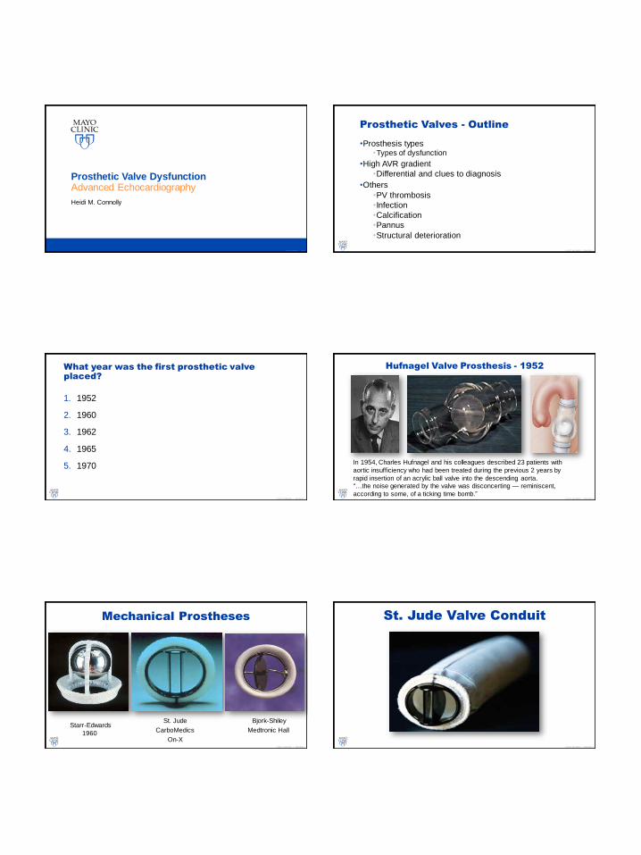
Hufnagel Valve Prosthesis - 1952
In 1954, Charles Hufnagel and his colleagues described 23 patients with
aortic insufficiency who had been treated during the previous 2 years by
rapid insertion of an acrylic ball valve into the descending aorta.
“…the noise generated by the valve was disconcerting — reminiscent,
according to some, of a ticking time bomb.”
©2017 MFMER | 3690389-5
Mechanical Prostheses
Bjork-Shiley
Medtronic Hall
St. Jude
CarboMedics
On-X
Starr-Edwards
1960
©2017 MFMER | 3690389-6
St. Jude Valve Conduit

©2017 MFMER | 3690389-7
Mechanical Valve Dysfunction
PannusThrombosisEndocarditis
©2017 MFMER | 3690389-8
Elevated AVR Velocity and Gradient
What is the Cause?
©2017 MFMER | 3690389-9
Increased PV Gradients
•Valve geometry/pressure recovery
•Small bi-leaflet prosthesis
•Prosthesis-patient mismatch
•Hemodynamically significant regurgitation
•High flow states
•Obstruction – thrombosis, infection
•Structural degeneration
©2017 MFMER | 3690389-10
Hemodynamic Profiles Differ by Prosthesis Type and Size
AutograftHomograft
Caged Ball
21 mm
MechanicalBileaflet
StentlessPorcine
31 mm
TranscatheterAortic prostheses
29 mm
StentedPericardial
27 mm
MechanicalTilting Disc
25 mm
Stented Porcine
23 mm
33 mm
Prosthesis type Prosthesis size
Most favorable
Least favorable 19 mm
©2017 MFMER | 3690389-11
Doppler Parameters of Prosthetic Aortic Valve Function in Mechanical and Stented Biologic Valves
NormalPossible
stenosis
Suggests
significant
stenosis
Peak velocity (m/s)* <3 3-4 >4
MG (mmHg)* <20 20-35 >35
DI / DVI ≥0.30 0.29-0.25 <0.25
EOA (cm2) >1.2 1.2-0.8 <0.8
Contour jet velocityTriangular,
early peaking
Triangular
to IntermediateRounded
AT (ms) <80 80-100 >100
Zoghbi WA: J Am Soc Echocardiogr, 2009©2017 MFMER | 3690389-12
19-mm St. Jude Mechanical AVP
AT 133 msec AT 89 msec
Rounded
contourPointed
contour
Pathologic Stenosis Functional Stenosis
Peak velocity 4.0 m/s
MG 40 mm Hg
DVI 0.30
EOA 0.85 cm2
iEOA 0.48 cm2/m2
Peak velocity 4.1 m/s
MG 40 mm Hg
DVI 0.32
EOA 1.25 cm2
iEOA 0.47 cm2/m2

©2017 MFMER | 3690389-13
Aortic Valve Prostheses With High Peak Velocity and/or High MG
Pathologic obstruction
•Pannus
•Thrombus
•Degenerative stenosis (bioprostheses)
Functional obstruction
•High flow state
•Pressure recovery (small mechanical bileaflet prostheses)
•Prosthesis-patient mismatch (PPM)
Pathologic regurgitation
©2017 MFMER | 3690389-14
75-Year-Old Man
• Early Postop 25-mm SJ Mechanical AVR
Echo-Doppler MG 50 mmHg, INR 3
What would you recommend?
1. CT2. TEE
3. Fluoroscopy
4. Observe
©2017 MFMER | 3690389-15
75-Year-Old ManEarly Postop 25-mm SJ Mechanical AVR Echo-Doppler MG 50 mmHg, INR 3
©2017 MFMER | 3690389-16
Evaluation of Abnormal Disc Motion
•Clinical assessment
•TTE to assess hemodynamic severity
•TOE to assess valve motion and echodensity
•Fluoroscopy or CT
Assess disc motion
Mechanism of dysfunction
©2017 MFMER | 3690389-17
Abnormal Disc Motion
Echodensity by TTE/TEE No Echodensity by TTE/TEE
CT/Fluoro
ThrombosisSuspect pannus
NO
Suspect
thrombus or IE
YESSystemic symptoms
endocarditis
Evaluation of Mechanical Valve Dysfunction
©2017 MFMER | 3690389-18
19-mm Bileaflet Mechanical AVR
MGH CV Images 2017

©2017 MFMER | 3690389-19
Other Examples
©2017 MFMER | 3690389-20
28-Year-Old Female CarboMedics MVR 30 weeks pregnant, recent dyspnea
Weight-based LMWH during pregnancy
MVR MG 16 mmHg
Baseline MG 4 mmHg
©2017 MFMER | 3690389-21 ©2017 MFMER | 3690389-22
28-Year-Old Female CM MVR
•30 weeks pregnant, recent dyspnea
•LMWH during pregnancy
What would you recommend?
1. Operation
2. Adjust anticoagulation3. Lytic therapy
4. Observation
©2017 MFMER | 3690389-23 ©2017 MFMER | 3690389-24
Prosthetic Valve Fibrinolysis
• Effective in 70-90% of patients
• Acute mortality 4-12%
• Complications
Thromboembolism 15%
Major bleed 5%
Non-disabling bleed 14%
Recurrent thrombosis 10-20%
ESC suggested regimens:
• Recombinant tPA 10 mg bolus + 90 mg in 90 min with UFH or
• Streptokinase 1.5 MU in 60 min
without UFH
©2017 MFMER | 3690389-25
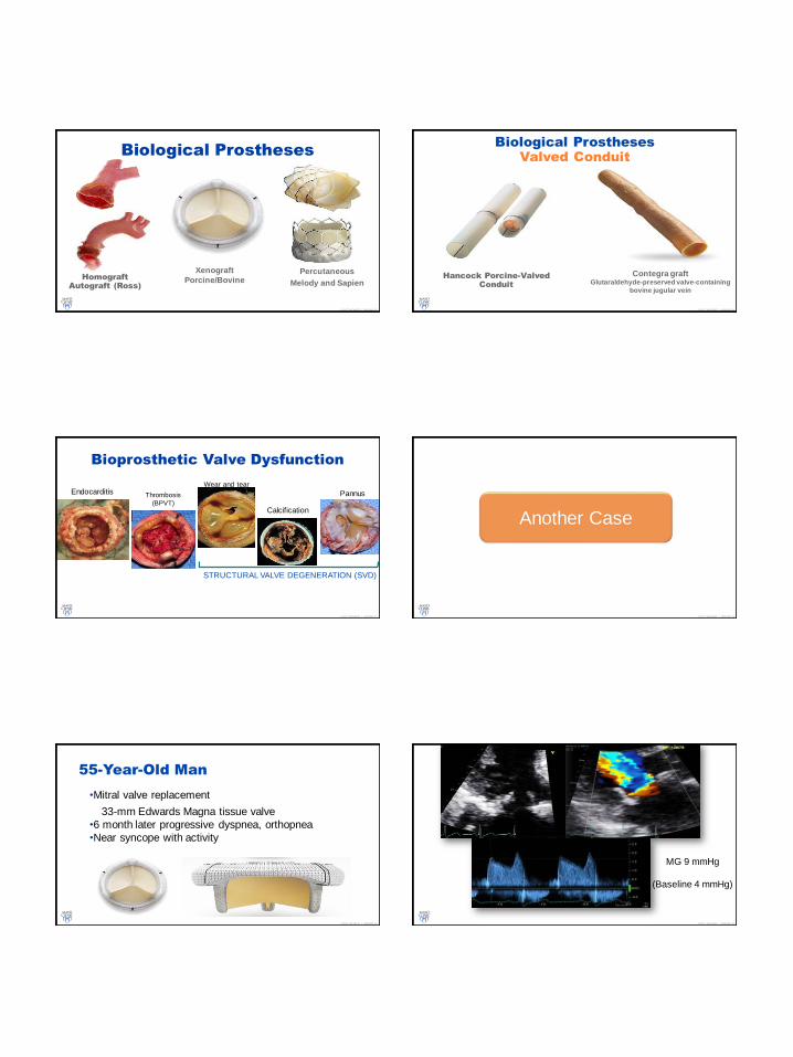
Biological Prostheses
Percutaneous
Melody and Sapien
Xenograft
Porcine/BovineHomograftAutograft (Ross)
©2017 MFMER | 3690389-26
Biological ProsthesesValved Conduit
Hancock Porcine-ValvedConduit
Contegra graftGlutaraldehyde-preserved valve-containing
bovine jugular vein
©2017 MFMER | 3690389-27
Bioprosthetic Valve Dysfunction
Wear and tear
PannusThrombosis
(BPVT)
Endocarditis
STRUCTURAL VALVE DEGENERATION (SVD)
Calcification
©2017 MFMER | 3690389-28
Another Case
©2017 MFMER | 3690389-29
55-Year-Old Man
•Mitral valve replacement
33-mm Edwards Magna tissue valve
•6 month later progressive dyspnea, orthopnea
•Near syncope with activity
©2017 MFMER | 3690389-30
MG 9 mmHg
(Baseline 4 mmHg)
©2017 MFMER | 3690389-31 ©2017 MFMER | 3690389-32
Increased Bioprosthetic GradientsDifferential Diagnosis
•Prosthetic valve degeneration
•Pannus ingrowth, calcification, wear and tear
•Infection
•Increased cardiac output
•Anemia, sepsis, thyrotoxicosis, A-V fistula
•Patient-prosthesis mismatch
•Pressure recovery – aortic prosthesis
•Bioprosthetic thrombosis
©2017 MFMER | 3690389-33
•Bioprosthetic MVR MG 9 mmHg
Baseline MG 4 mmHg, 6 months ago
What would you recommend?1. Anticoagulation
2. Surgical re-replacement 3. Percutaneous valve-in-valve
55-Year-Old Man with Dyspnea
©2017 MFMER | 3690389-34
55-Year-Old Man with Dyspnea
•Suspected bioprosthetic MV thrombosis
•Anticoagulation with VKA 2 months
•Follow up
•Clinically improved
©2017 MFMER | 3690389-35
MG 4 mmHgContinues on VKA
©2017 MFMER | 3690389-36
MG 9 mmHg MG 4 mmHgon VKA

©2017 MFMER | 3690389-37
Identify BPVT in all explanted BPV between 1997-2013 using Mayo Clinic
Pathology Database (397 cases)
J Am Coll Cardiol 2015;66: 2285-94
©2017 MFMER | 3690389-38
Study period 1997 - 2013
All v alv e
implanted
Egbe et al. JACC 2015
©2017 MFMER | 3690389-39
Echo Features of BPVT
• Gradient >50% baseline
• Restricted cusp mobility
• Thickened leaflets
• TOE when in doubt
©2017 MFMER | 3690389-40Egbe et al. JACC 2015
Clinical Predictors of BPVT
•Paroxysmal atrial fibrillation
•Sub-therapeutic or no warfarin therapy
©2017 MFMER | 3690389-41
N Engl J Med. 2015;373(21):2015-24
World Journal for Pediatric and Congenital Heart Surgery 2015 ©2017 MFMER | 3690389-42
Other Examples

©2017 MFMER | 3690389-43
63-Year-Old Male12 months after Freestyle AVR
©2017 MFMER | 3690389-44
63-Year-Old Male6 months after Freestyle AVR
12 Month postop echo
After 6 weeks of VKA
©2017 MFMER | 3690389-45
Tricuspid Prosthesis
©2017 MFMER | 3690389-46
58-Year Old Female with Carcinoid Heart Disease18 Months Postop – TVR MG 10 mmHg (Baseline 3 mmHg)
©2017 MFMER | 3690389-47
VKA Therapy 4 Months
TVR MG 3 mmHg
Still functioning normally >10 years postop
©2017 MFMER | 3690389-48
Baumgartner H. et al EHJ VHD Guidelines 2017

©2017 MFMER | 3690389-49
Bioprosthetic Valve ThrombosisThings to Remember
•May occur early or late after implantation
•When to suspect
•BPV gradient >50% over baseline
•Restricted cusp mobility, thickened leaflets
•Prosthesis regurgitation
•Findings may be subtle
•TEE or CT when in doubt
•Early screening may be beneficial
You have to think of it!!
©2017 MFMER | 3690389-50
Infection
©2017 MFMER | 3690389-51
31-Year-Old dTGA, RastelliStatus Post 22 mm Contegra Graft
Mean Gradient 34 mmHg
©2017 MFMER | 3690389-52
1. PPV IE – morbidity, mortality and high risk of reoperation
2. TTE and TEE – complementary diagnosotic techniques
3. PPV stenosis more common than regurg; consider IE in new
obstruction
©2017 MFMER | 3690389-53
Baumgartner H. et al EHJ VHD Guidelines 2017©2017 MFMER | 3690389-55
Bioprosthetic Valve Dysfunction
Wear and tear
PannusThrombosis
(BPVT)
Endocarditis
STRUCTURAL VALVE DEGENERATION (SVD)
Calcification

©2017 MFMER | 3690389-56
Calcification
©2017 MFMER | 3690389-57
Homograft CalcificationSternal Compression with Obstruction
©2017 MFMER | 3690389-58
Homograft Calcification and Obstruction
“Valve heavily calcified and severely obstructed – calcified valve cusps were excised and
debrided with a rongeur…”
©2017 MFMER | 3690389-59
Another Case
©2017 MFMER | 3690389-60
69-Year-Old-Female AVR for AS
• AVR 23-mm Mitroflow bovine pericardial
• DM, dyslipidemia, obesity, HTN
• 2 years postop →DOE, NYHA III
• New murmur on examination
©2017 MFMER | 3690389-61

©2017 MFMER | 3690389-62
Early Structural Valve Degeneration (SVD)
•Operative findings – severe SVD with calcification of
Mitroflow valve leaflets
•Redo AVR 21-mm CarboMedics TopHat valve
•Pathology – moderate commissural calcification without
cusp tear, thrombus, pannus or vegetation
©2017 MFMER | 3690389-63
Accelerated Structural Valve Degeneration
• Risk factorsDiabetes Young females Patient prosthesis mismatch Small prosthesis size Metabolic syndrome Mitral position CKD, abnormal Ca2+ metabolism
Lorusso et al. Circulation 2011
Meyer et al. J Card Surg 2012
• Wear and tear – accelerated calcification
• Host metabolic factors and specific valve design
©2017 MFMER | 3690389-64
One More Case
©2017 MFMER | 3690389-65
75-Year-Old Male
•Severe degenerative calcific AS
•AVR 25 mm Carbomedics Mitroflow prosthesis
• Increasing AV gradient noted postop
Year Mean gradient iEOA EF
2010 8 1.00 63%
2011 12 0.84 62%
2013 24 0.66 64%
2015 37 0.47 60%
©2017 MFMER | 3690389-66
What is the diagnosis?
1. Bioprosthetic AV thrombosis2. Accelerated degeneration
3. Pannus
©2017 MFMER | 3690389-67
Pannus Overgrowth

©2017 MFMER | 3690389-68
Pannus
•Difficult to differentiate from thrombus
• Failure of anticoagulation may be initial clue
•Host healing response to prosthesis
•Surgical injury → thrombus → inflammation → fibrosis/collagen deposition
•Pannus overgrowth → pathology
•Subvalvular thickening, stiffness → stenosis (majority)
•Can occur in isolated annuloplasty ring
©2017 MFMER | 3690389-69
Bioprosthetic Pannus - Histopathology
Thrombus and pannus often coexist
Cremer et al. JACC: CV Imaging, 2015
©2017 MFMER | 3690389-70
Prosthetic Valve Choice
•Every valve prosthesis introduces new disease process
•Randomized trials comparing mechanical vs bioprostheses:
•Similar survival
•No significant difference in rates of valve thrombosis
and thromboembolism
•Higher rates of bleeding with mechanical prostheses
•Higher rates of reintervention with bioprostheses
©2017 MFMER | 3690389-71
Patient with Prosthetic Heart Valve
•Lifelong follow-up
•Baseline TTE within 30 days •TTE at 1 year, and annually thereafter
•Repeat TTE for new symptoms or suspected complications
•TOE when TTE is poor quality and for all suspected prosthetic dysfunction or endocarditis
•Cine-fluoroscopy and MSCT for mechanical valves useful
if valve thrombus or pannus are suspected
©2017 MFMER | 3690389-72
Prosthetic Valve DysfunctionTake Home Points
Structural
Deterioration
Regurgitation
Multimodality
Assessment
Options
Normal
Appearance and
Hemodynamics
Know
Prosthesis
Type and
Size Infection
Calcification
Thrombus
Pannus
©2017 MFMER | 3690389-73
Questions & Discussion


