Download - Surgical pathology of the esophagus
By- Dr. Armaan SinghBy- Dr. Armaan Singh
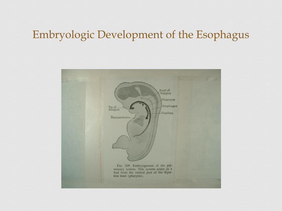
Embryologic Development of the Esophagus
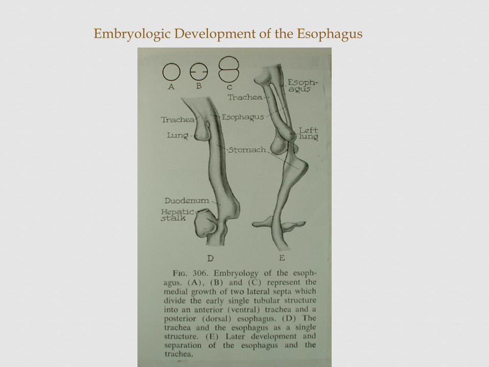
Embryologic Development of the Esophagus
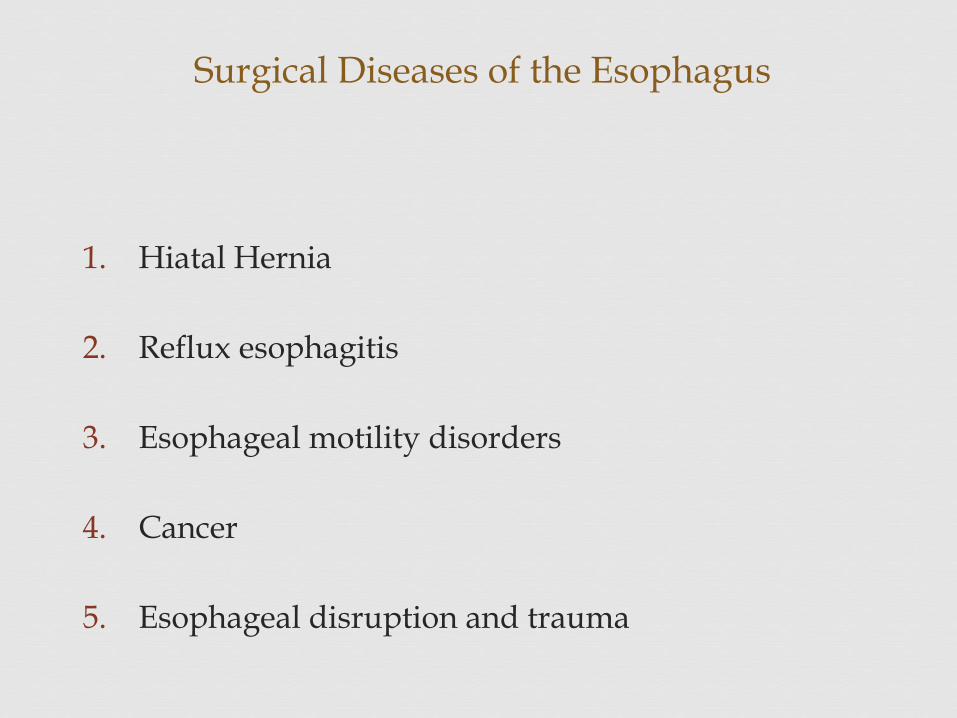
1. Hiatal Hernia
2. Reflux esophagitis
3. Esophageal motility disorders
4. Cancer
5. Esophageal disruption and trauma
Surgical Diseases of the Esophagus
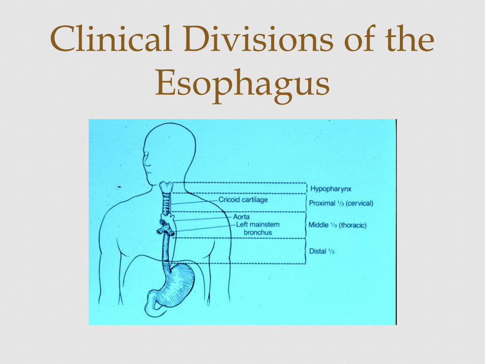
Clinical Divisions of the Esophagus
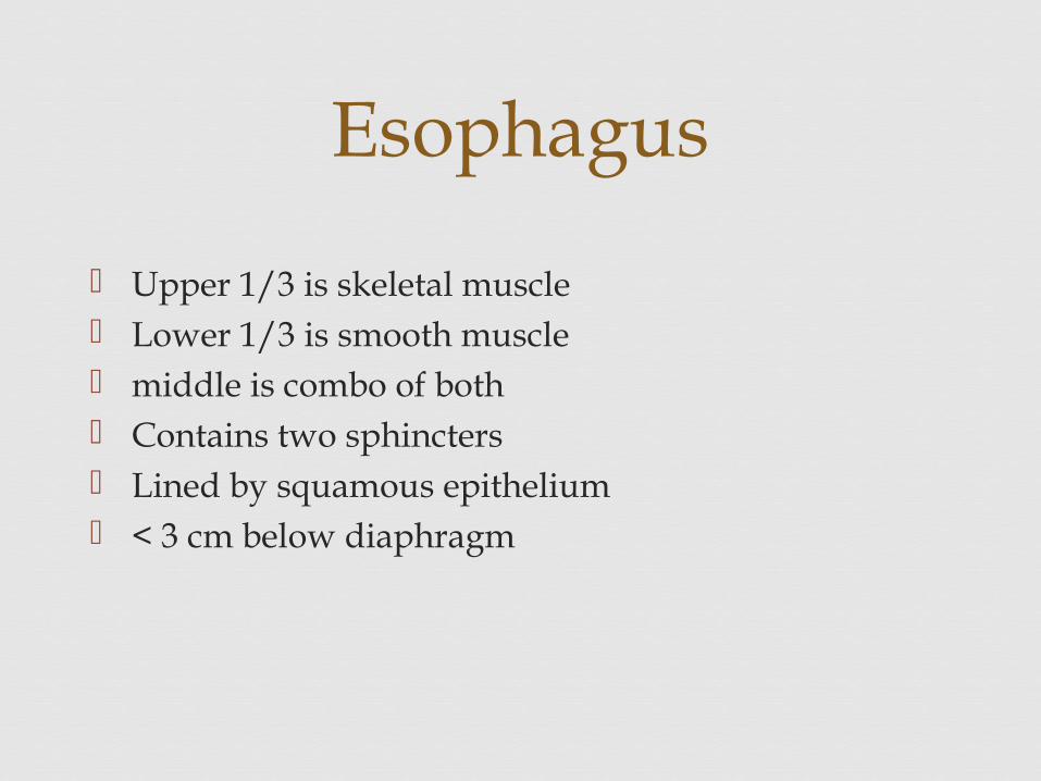
Upper 1/3 is skeletal muscle Lower 1/3 is smooth muscle middle is combo of both Contains two sphincters Lined by squamous epithelium < 3 cm below diaphragm
Esophagus
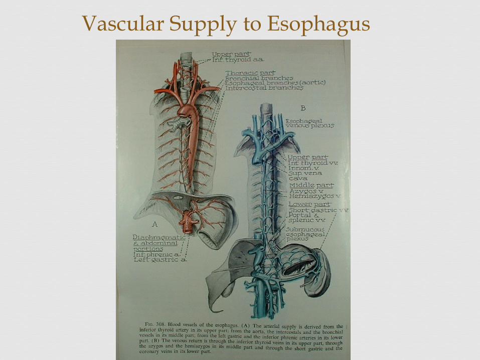
Vascular Supply to Esophagus
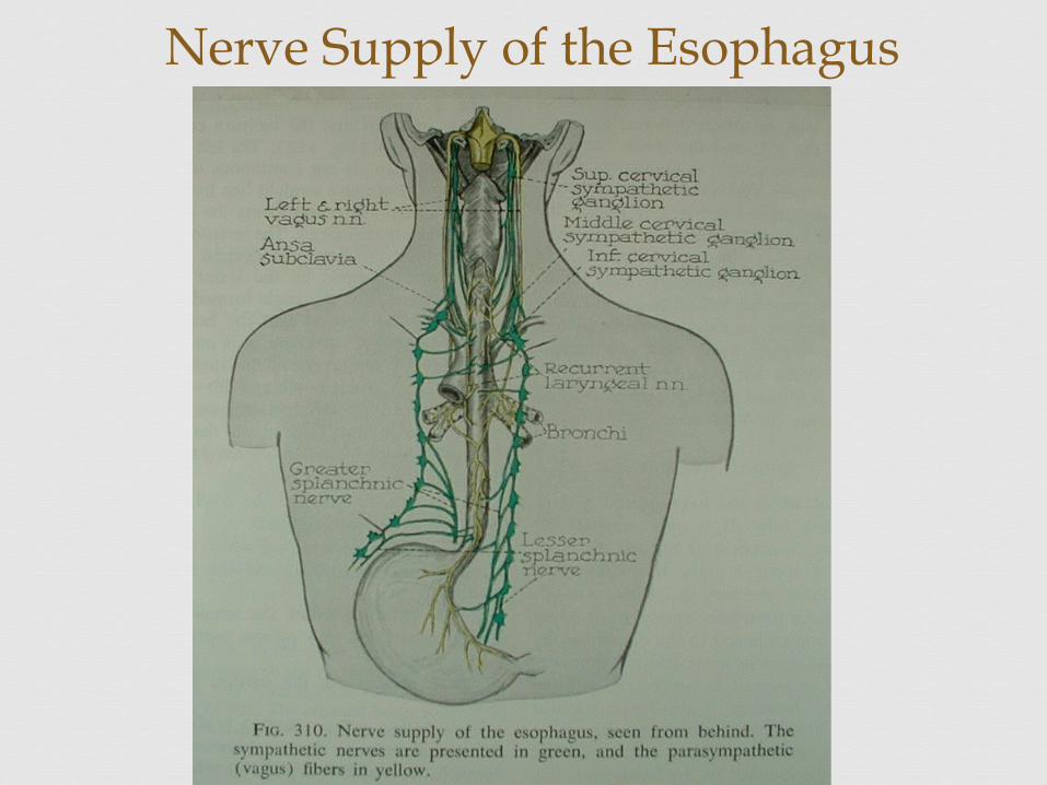
Nerve Supply of the Esophagus
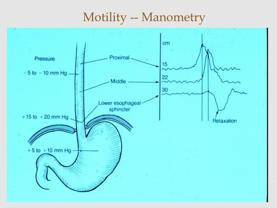
Motility -- Manometry
Esophageal Dysmotility
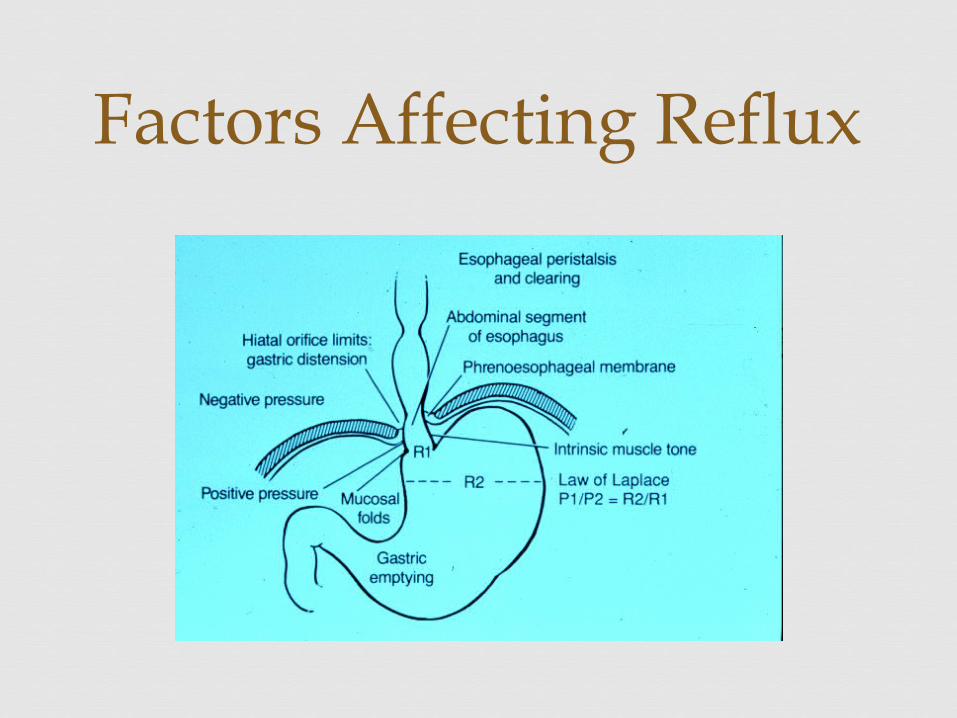
Factors Affecting Reflux
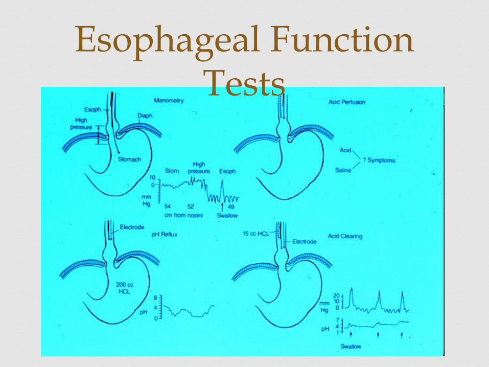
Esophageal Function Tests
Pathogenesis
two major types of hiatal hernia type I or "sliding" hiatal hernia type II paraesophageal hiatal hernia
Hiatal Hernia and Reflux Esophagitis
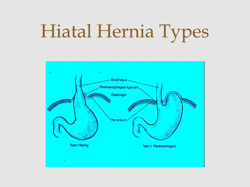
Hiatal Hernia Types
Sliding hiatal hernias are more common than paraesophageal hernias by 100:1
The lower esophageal sphincter mechanism becomes incompetent
Reflux of acid gastric juice produces a chemical burn Degree of mucosal injury is a function of the
duration of acid contact and not a disease of hyperacidity
Hiatus Hernia - Clinical Presentation
Continued inflammation of the distal esophagus may lead to mucosal erosion, ulceration, and eventually scarring and stricture
Predominantly in women who have been pregnant Men and women with increased intraabdominal
pressure
Hiatus Hernia - Clinical Presentation
Type I hiatal hernia with reflux is frequently found in patients who are overweight.
Many patients with type I hiatal hernia have no symptoms.
A burning epigastric or substernal pain or tightness Usually the pain does not radiate May be described as a tightness in the chest and can
be confused with the pain of myocardial ischemia
Clinical Presentation – Type I hernia
Clinical Presentation – Hiatus Hernia
Worse when the patient is supine or leaning over Antacid therapy frequently improves the symptoms. A lump or feeling that food is stuck beneath the
xyphoid Alcohol, aspirin, tobacco, and caffeine, may
exacerbate the symptoms Late symptoms of dysphagia and vomiting usually
suggest stricture formation
Hiatus Hernia - Clinical Presentation
Type II hernias Generally produce no symptoms until they
incarcerate and become ischemic
Dysphagia, bleeding, and occasionally respiratory distress are the presenting symptoms.
Hiatus Hernia - Clinical Presentation
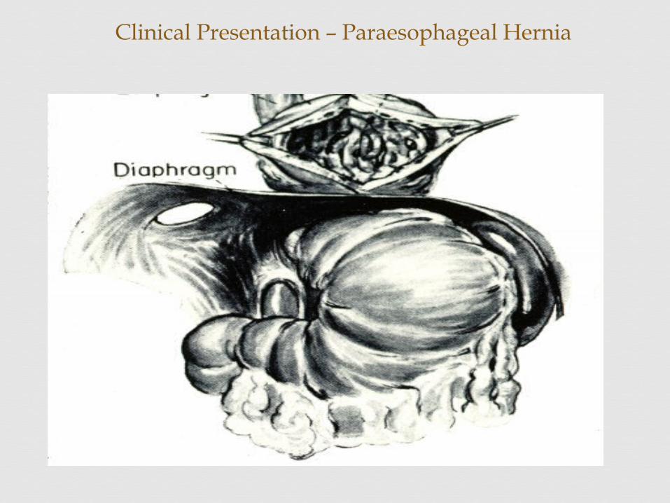
Clinical Presentation – Paraesophageal Hernia
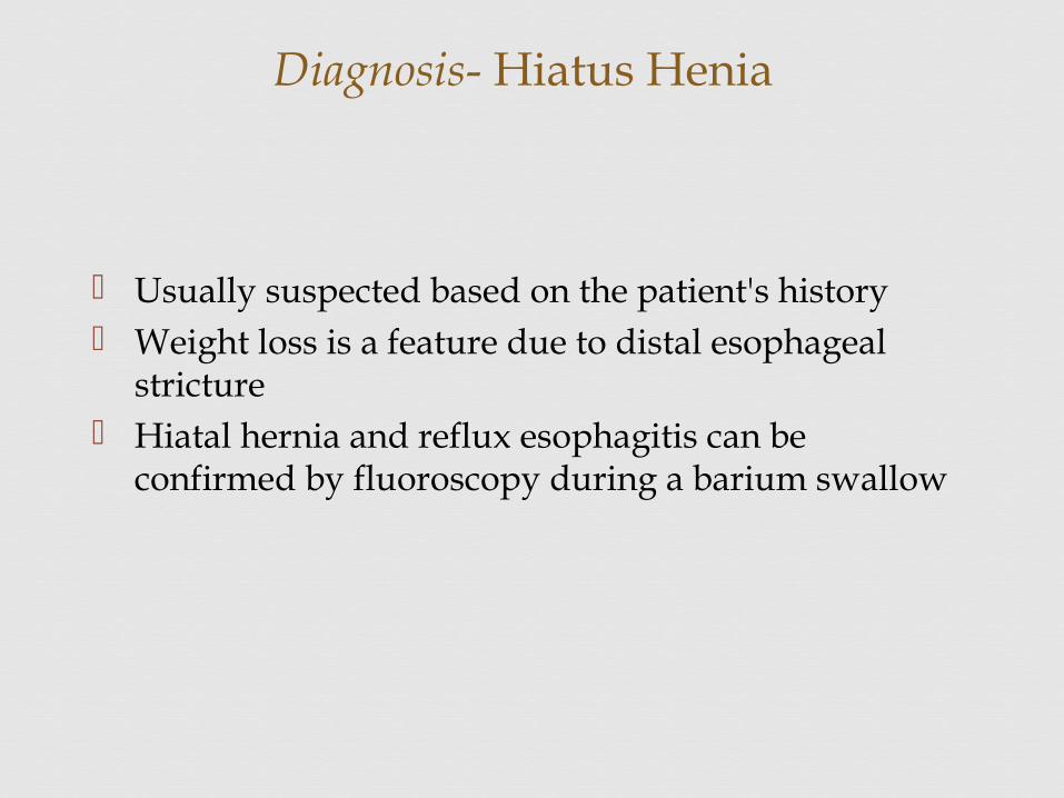
Usually suspected based on the patient's history Weight loss is a feature due to distal esophageal
stricture Hiatal hernia and reflux esophagitis can be
confirmed by fluoroscopy during a barium swallow
Diagnosis- Hiatus Henia
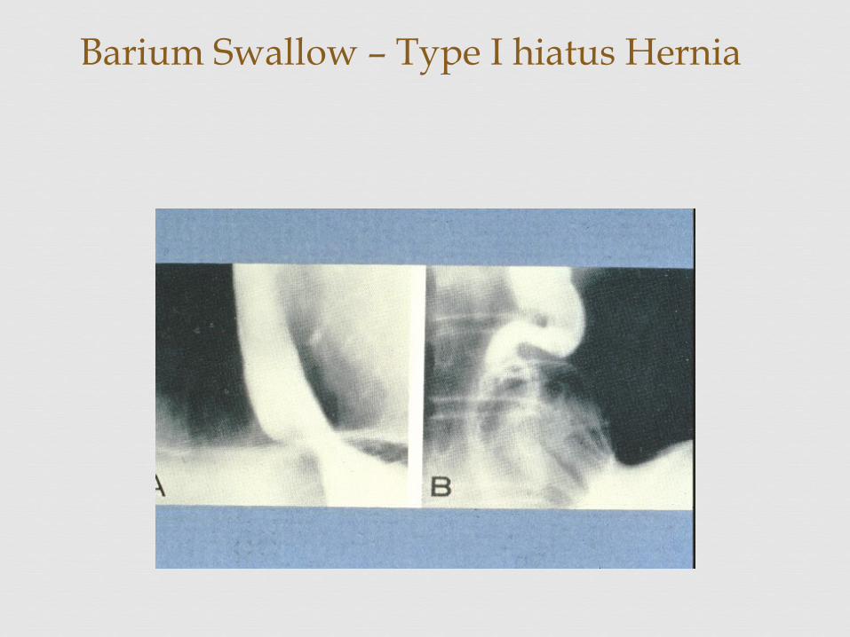
Barium Swallow – Type I hiatus Hernia
Esophagogastric endoscopy and biopsy of the inflamed esophagus
Manometry may show a loss of the lower esophageal high-pressure area
Diagnosis – Hiatus Hernia
Medical Therapy 1. Avoidance of gastric stimulants (coffee, tobacco, and
alcohol).
2. Elimination of tight garments that raise intraabdominal pressure, such as girdles or abdominal binders.
3. The regular use of antacids ( coat the esophagus), and antacid mints (Tums and Rolaids) to provide a steady stream of protection.
H2 blockers, to increase the pH of the refluxed gastric juice Metoclopramide (Reglan) to stimulate gastric emptying without
stimulating gastric, biliary, or pancreatic secretions
Treatment – Hiatus Hernia
4. Abstinence from drinking or eating within several hours of sleeping.
5. Sleeping with the head of the bed elevated to reduce nocturnal reflux.
6. Weight loss in obese patients.
About one third of patients fail to respond to initial medical treatment, and half of those who initially respond will ultimately relapse and require surgery.
Treatment – Hiatus Hernia
Correct the anatomic defect Prevent the reflux of gastric acid into the lower
esophagus by reconstruction of a valve mechanism
Treatment Hiatus Hernia -- Surgical
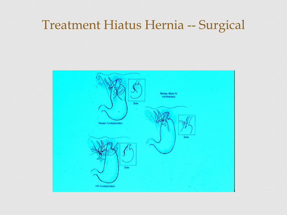
Treatment Hiatus Hernia -- Surgical
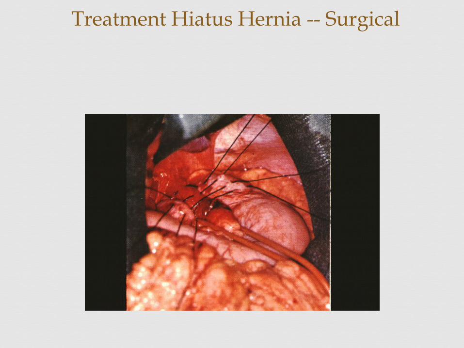
Treatment Hiatus Hernia -- Surgical
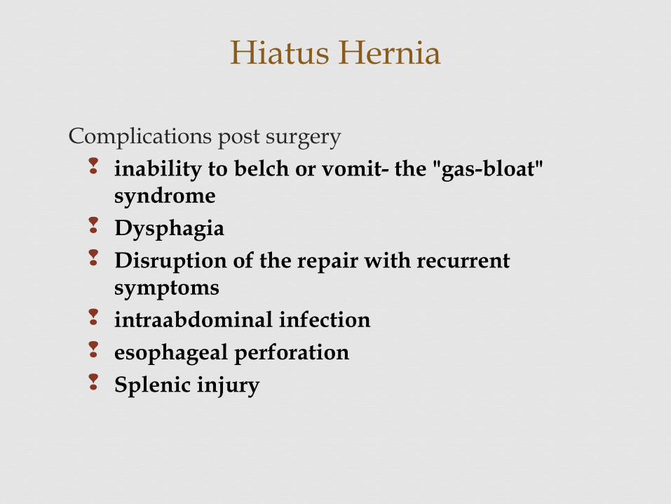
Complications post surgery inability to belch or vomit- the "gas-bloat"
syndrome Dysphagia Disruption of the repair with recurrent
symptoms intraabdominal infection esophageal perforation Splenic injury
Hiatus Hernia
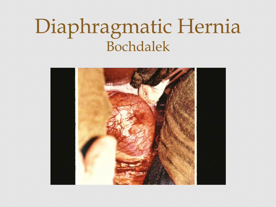
Congenital, left lateral area of diaphragm Through the pleuroperitoneal foramen of Bochdalek Symptoms of cyanosis, dyspnea, vomiting Treatment: surgery in first 48 hours of life
Also – retrosternal hernia through foramen of Morgagni in older children
Bochdalek Hernia
Diaphragmatic HerniaBochdalek
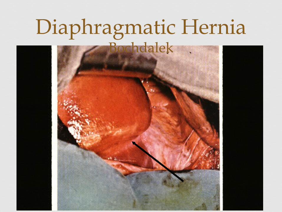
Diaphragmatic HerniaBochdalek
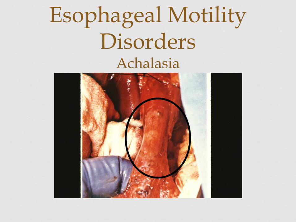
Failure to relax Not due to spasm Failure of the high-pressure zone sphincter to relax Painless dysphagia Progressive dilation of the proximal esophagus
Esophageal Motility Disorders
Achalasia
Dysphagia Regurgitation of undigested food Weight loss Pain in this condition is uncommon Aspiration pneumonia is common Complain of spitting up foul-smelling secretions
when simply leaning forward
Esophageal Motility Disorders
Achalasia -- Clinical Presentation
Generally first confirmed roentgenographically by contrast studies of the esophagus
Dilation of the proximal esophagus is classic
Esophageal diverticula may be present at any level
Endoscopy -- one needs to be particularly careful to avoid diverticular perforation
Esophageal manometry
Esophageal Motility Disorders
Achalasia -- Diagnosis
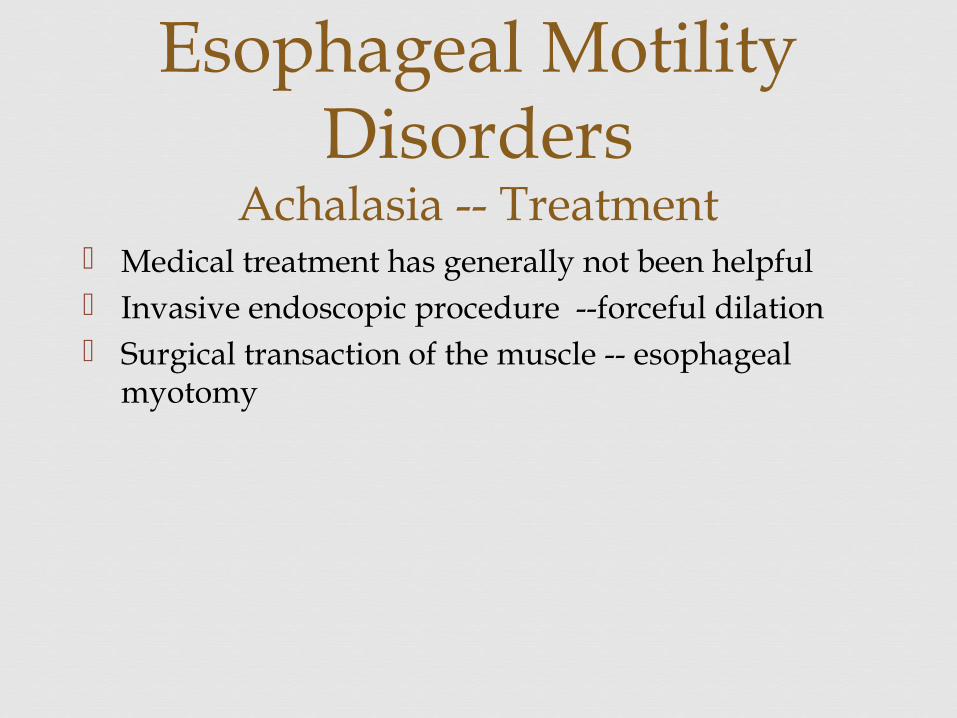
Medical treatment has generally not been helpful Invasive endoscopic procedure --forceful dilation Surgical transaction of the muscle -- esophageal
myotomy
Esophageal Motility Disorders
Achalasia -- Treatment
Esophageal Motility Disorders
AchalasiaSshove this down your own
throat
Esophageal Motility Disorders
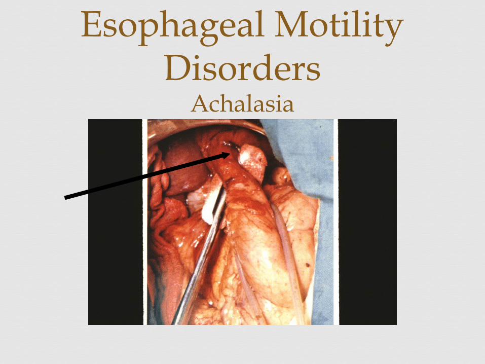
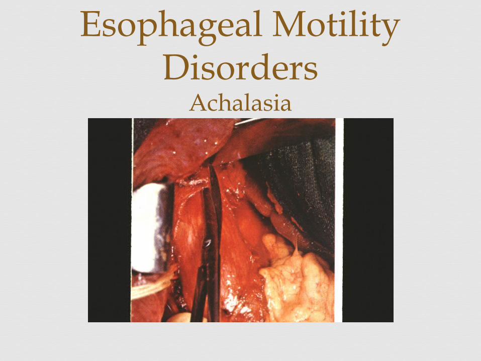
Achalasia
Esophageal Motility Disorders
Achalasia
Esophageal Motility Disorders
Achalasia
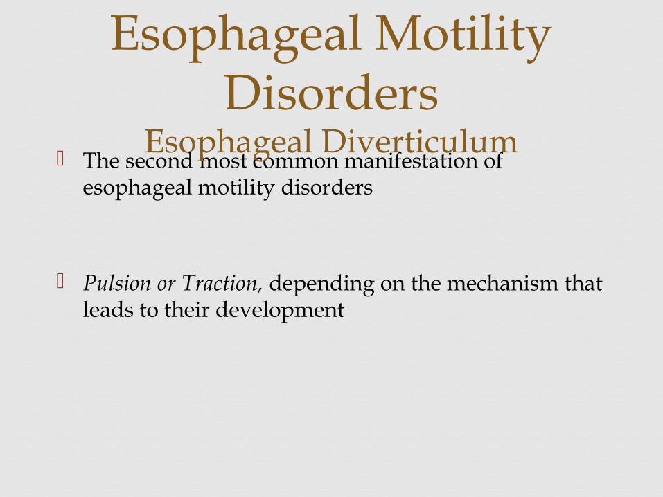
The second most common manifestation of esophageal motility disorders
Pulsion or Traction, depending on the mechanism that leads to their development
Esophageal Motility Disorders
Esophageal Diverticulum
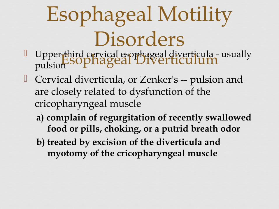
Upper third cervical esophageal diverticula - usually pulsion
Cervical diverticula, or Zenker's -- pulsion and are closely related to dysfunction of the cricopharyngeal musclea) complain of regurgitation of recently swallowed
food or pills, choking, or a putrid breath odorb) treated by excision of the diverticula and
myotomy of the cricopharyngeal muscle
Esophageal Motility Disorders
Esophageal Diverticulum
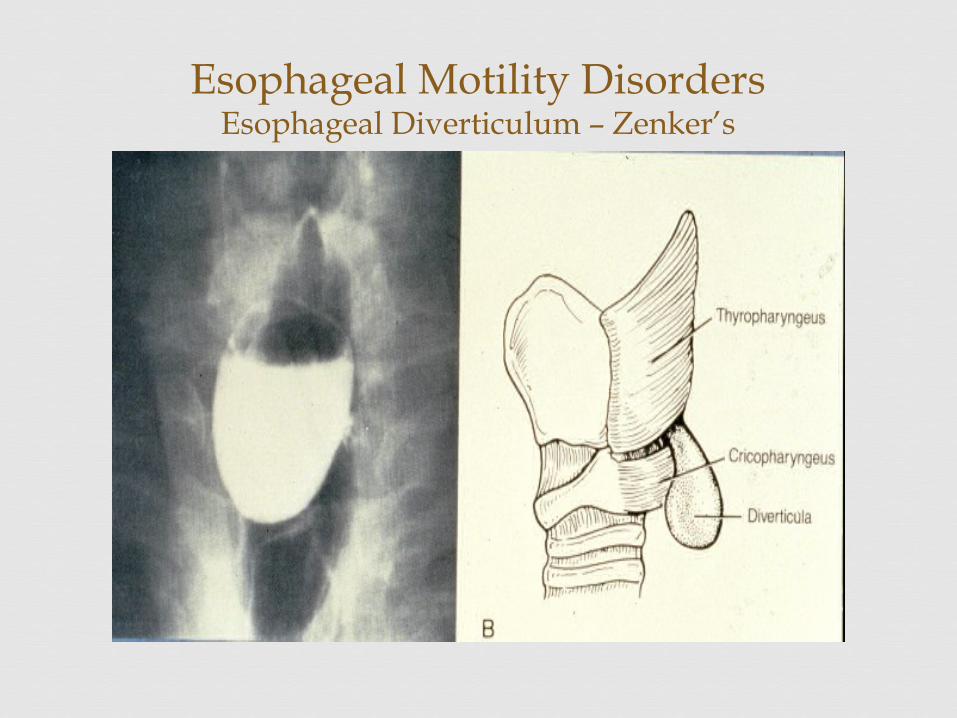
Esophageal Motility DisordersEsophageal Diverticulum – Zenker’s
Middle-third esophageal diverticula are almost always traction, not related to an intrinsic abnormality in esophageal motility
a) Result of mediastinal inflammation (usually inflammatory nodal disease from tuberculosis or
histoplasmosis, with formation and subsequent contracture that places "traction" on the
esophagus b) Usually asymptomatic and do not warrant
treatment.
Esophageal Motility Disorders
Esophageal Diverticulum
Diverticula of the distal third of the esophagus
a) associated with dysfunction of the esophagogastric junction due to chronic stricture from acid reflux, antireflux surgical procedures, achalasia
b) Excision of these diverticula should always be accompanied by correction of the underlying pathologic process
Esophageal Motility Disorders
Esophageal Diverticulum
Exceedingly rare – in middle and distal 1/3 Leiomyomas are the most common intramural
tumors1) potential for malignant degeneration appears to
be quite low2) indent the lumen of the esophagus on contrast
radiography3) tend to grow progressively and cause dysphagia3) Excised for possible dysphagia and malignancy
Esophageal NeoplasmsBenign
85% are squamous cell carcinomas
10% are adenocarcinomas
< 1% are malignant melanoma
Adenoid cystic tumors, sarcomas, APUDomas are rare
Esophageal NeoplasmsMalignant
Usually arises from squamous epithelium Commonly occurs in association with alcohol and/or
tobacco abuse Etiology has been related to diet, vitamin deficiency,
poor oral hygiene, surgical procedures, and a number of premalignant conditions, (caustic burns, Barrett's esophagus, radiation, Plummer-Vinson syndrome, and esophageal diverticula).
Esophageal NeoplasmsMalignant
Weight loss and pain may be present Difficulty in swallowing Acquired tracheoesophageal fistula due to erosion of
the tumor into the trachea or bronchus Frequent episodes of pneumonia due to recurrent
aspiration
Esophageal NeoplasmsMalignant
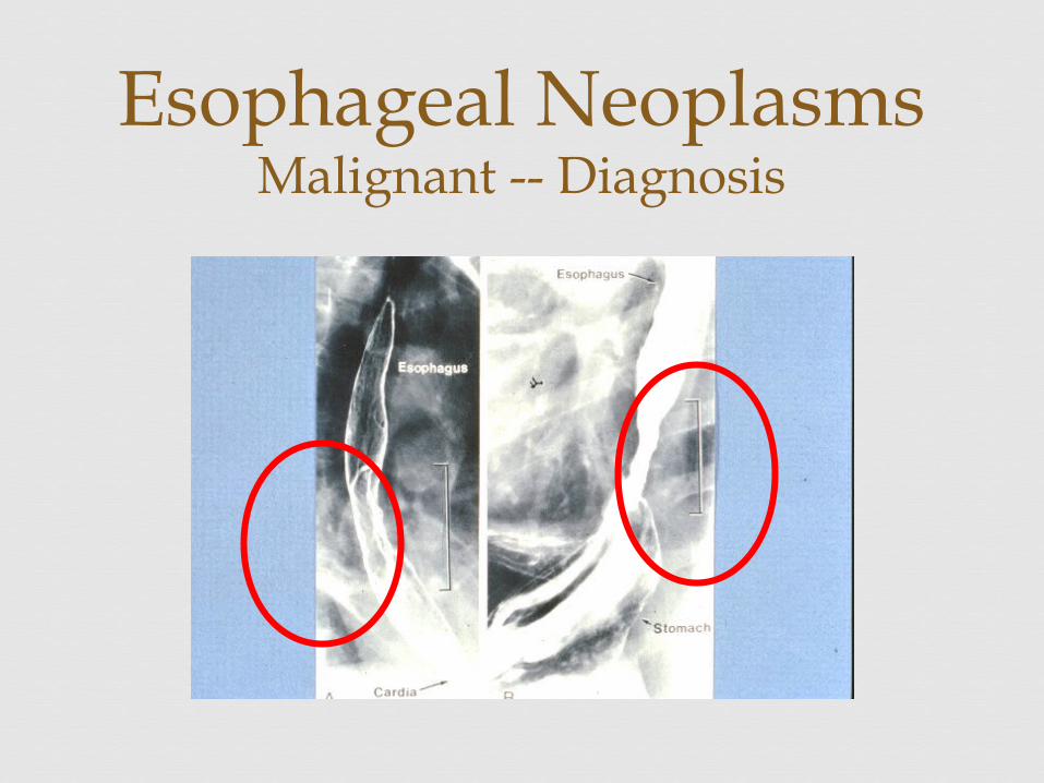
Barium contrast studies of the esophagus
Endoscopy and biopsy of the lesion
The extent of tumor involvement assessed by computed tomography (CT) of the chest and upper abdomen .
Esophageal NeoplasmsMalignant -- Diagnosis
Esophageal NeoplasmsMalignant -- Diagnosis
Approximately 10% of patients with Barrett's esophagus will develop adenocarcinoma
Symptoms produced by an esophageal malignancy frequently insidious at the onset, precluding early
diagnosis and thus the opportunity for effective treatment
As the tumor enlarges progressive dysphagia becomes the predominant symptom
Esophageal NeoplasmsMalignant
Tumors that involve the middle third of the esophagus are usually treated by a staged procedure with total thoracic esophagectomy and bypass
Cancer involving the lower third of the esophagus or proximal stomach is best treated by esophagogastric resection and an end-to-end anastomosis in the midchest.
Esophageal NeoplasmsMalignant -- Treatment
Squamous or adenocarcinomas of the esophagus - very poor prognosis
Palliation - restoration of effective swallowing Radiotherapy - primary mode of treatment for cancer
arising in the upper esophagus Surgical treatment of upper third usually
requires extirpation of the esophagus en bloc with the larynx, permanent tracheostomy, and restoration of swallowing by a free microsurgically constructed vascular pedicle of jejunum or colon into the neck.
Esophageal NeoplasmsMalignant -- Treatment
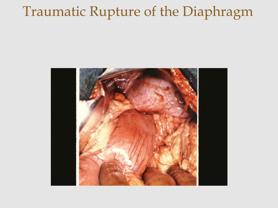
Traumatic Rupture of the Diaphragm
Instrumentation by endoscopic and/or biopsy Passage of blind nasogastric tubes Instruments designed for dilation of strictures Sengstaken-Blakemore tubes, balloon dilation for alchalasia Boerhaave’s syndrome -- spontaneous perforation secondary to
forceful vomiting (Plummer-Vinson) Treatment requires aggressive surgical intervention
Traumatic Esophageal DisordersPerforation
May be dramatic or occult Profound shock Mediastinal sepsis Severe chest or abdominal pain Hypotension Diaphoresis Nausea/Vomiting
Traumatic Esophageal DisordersPerforation -- Symptoms
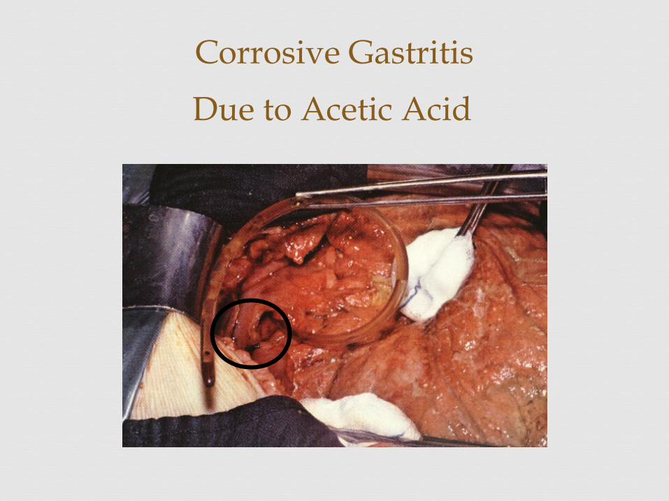
Corrosive Gastritis
Due to Acetic Acid
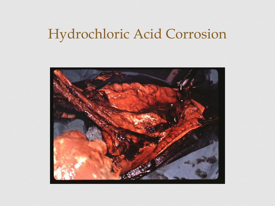
Hydrochloric Acid Corrosion
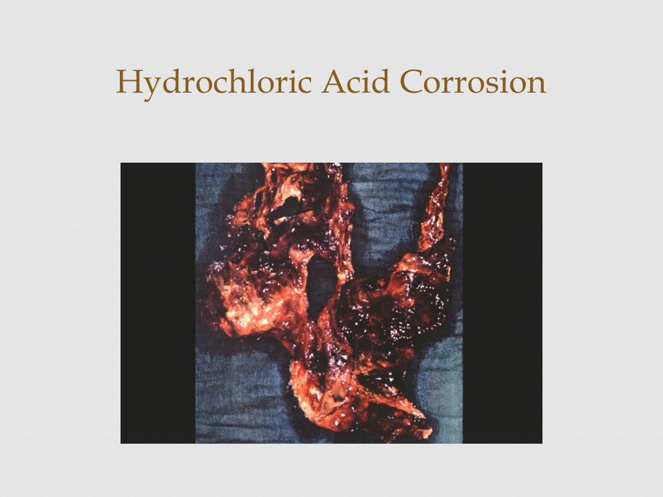
Hydrochloric Acid Corrosion
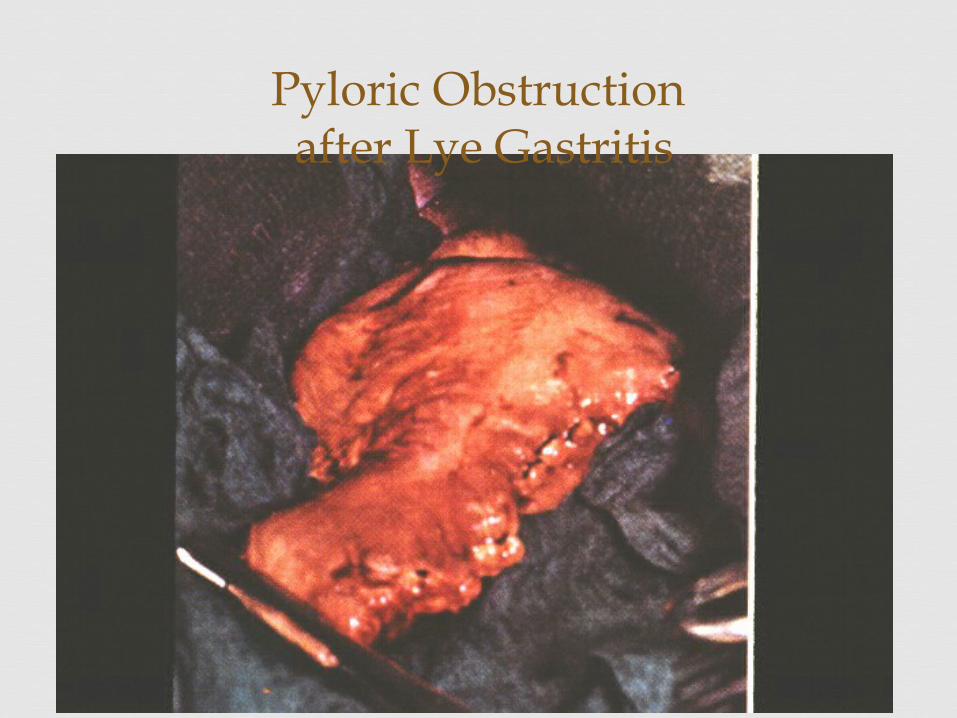
Pyloric Obstruction after Lye Gastritis
Medical Emergency Drano, Liquid Plumber -- alkaline containing
products Inspect mouth to assess injury Neutralization and induced emesis not usually
recommended Endoscopy, airway maintenance, patency of the
esophagus No steroids
Traumatic Esophageal DisordersIngestion of Caustic Materials


Diaphragmatic HerniaLarrey
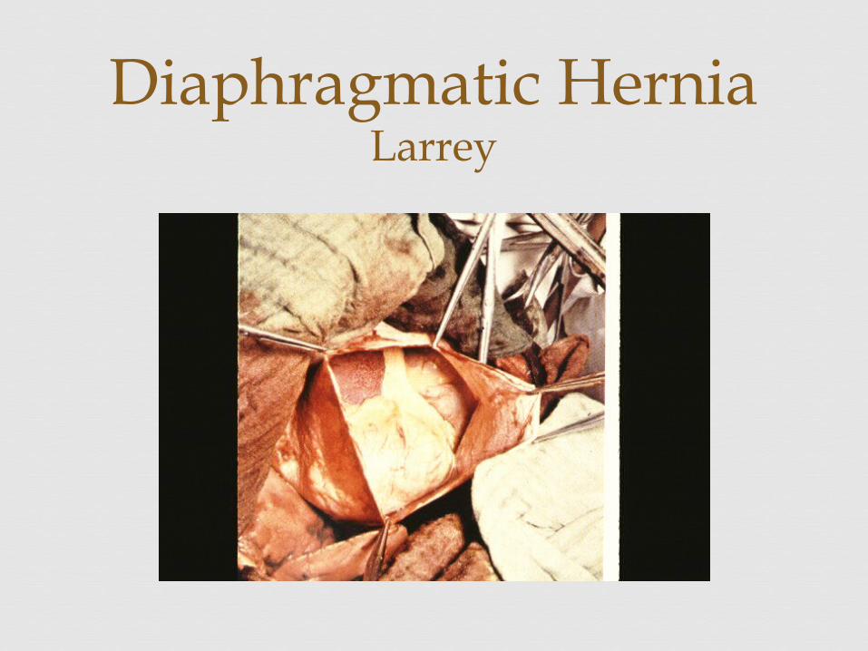
Diaphragmatic HerniaLarrey
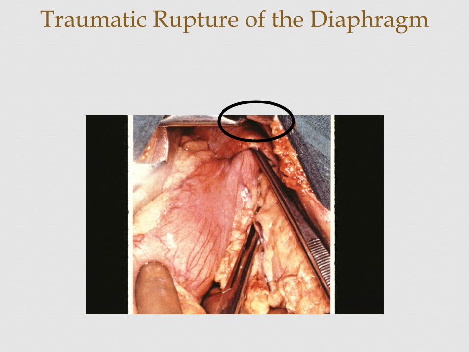
Traumatic Rupture of the Diaphragm
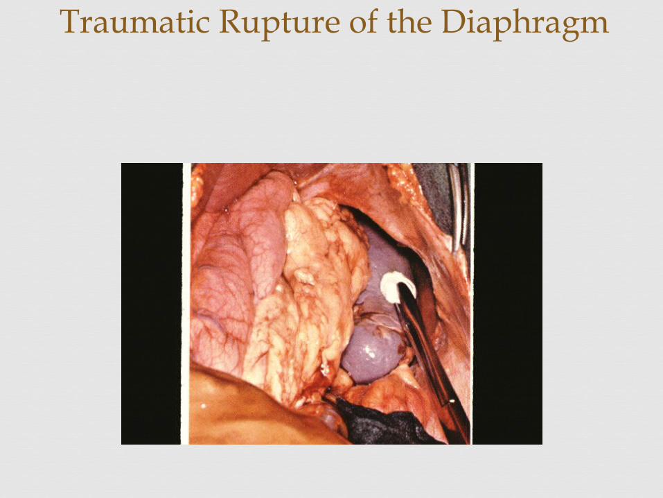
Traumatic Rupture of the Diaphragm
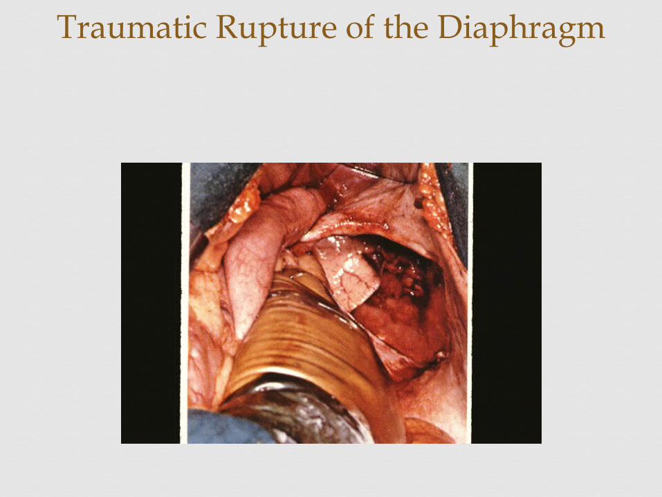
Traumatic Rupture of the Diaphragm
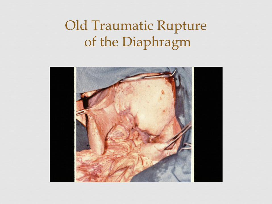
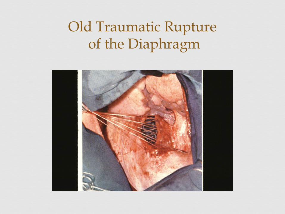
Old Traumatic Rupture of the Diaphragm
Old Traumatic Rupture of the Diaphragm


