
The caBIG Cancer Genome Atlas Radiology Project
Eliot Siegel, M.D.University of Maryland School of
Medicine Department of Diagnostic Radiology
Introduction
• One of the major original goals of caBIG
was to determine out how to create a
system that would enable extraction of
data for research or clinical decision
support that would:• Allow access to a variety of types and sources of data
including genomic, proteomic, clinical, lab,
demographic, and diagnostic imaging
• Take advantage of analytic potential of grid computing
to combine and cross-reference these for analysis for
research and clinical care
• The caBIG Imaging workspace has
worked to build basic tools toward this
goal and the TCGA imaging workspace
project represents an example of the
potential for caBIG to have a major
impact on the way in which data are
shared, research conducted, and patient
care is provided
Introduction to the caBIG in Vivo
Imaging Workspace
• caBIG in vivo Imaging workspace established
April 2005 a little more than a year after the
establishment of the other caBIG workspaces
• NCI funded effort by far the biggest and most
productive effort in imaging informatics today
• Subject matter experts from around country
with representation from major Universities,
informatics experts, industry, NCI

Review of Relevant Workspace
Projects XIP, AIM, Middleware, NBIA

Rapid application
development
environment for
diagnostic imaging
tasks that researchers
and others use to
create targeted
workflows customized
for specific projects
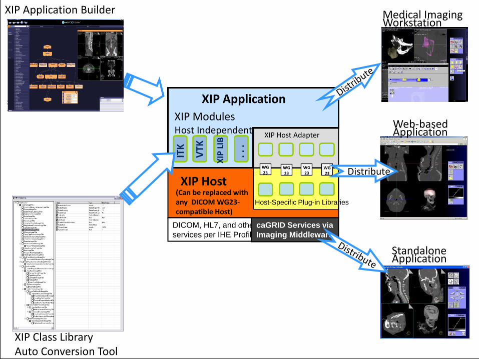
XIP Application
(Can be replaced with any DICOM WG23-compatible Host)
XIP Host Adapter
XIP
LIB
ITK
VTK
. . .
XIP ModulesHost Independent
WG23
XIP HostWG23
WG23
Web-based Application
Medical Imaging Workstation
Standalone Application
Distribute
DICOM, HL7, and other
services per IHE Profiles
caGRID Services via
Imaging Middleware
XIP Application Builder
XIP Class Library Auto Conversion Tool
Host-Specific Plug-in Libraries
WG23
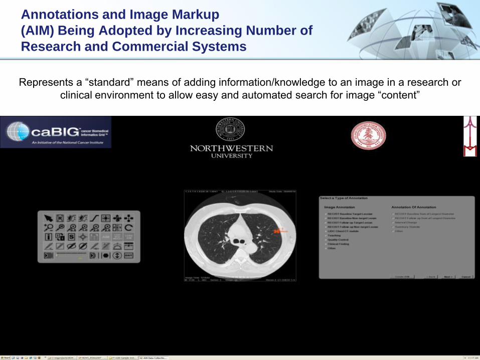
Annotations and Image Markup
(AIM) Being Adopted by Increasing Number of
Research and Commercial Systems
Represents a “standard” means of adding information/knowledge to an image in a research or
clinical environment to allow easy and automated search for image “content”
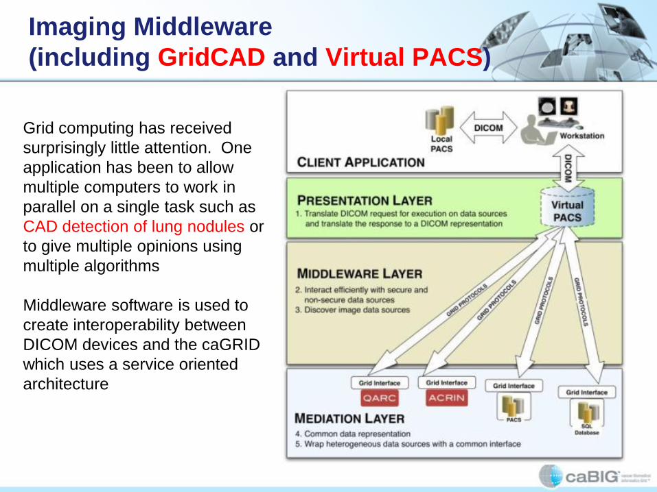
Imaging Middleware
(including GridCAD and Virtual PACS)
Grid computing has received
surprisingly little attention. One
application has been to allow
multiple computers to work in
parallel on a single task such as
CAD detection of lung nodules or
to give multiple opinions using
multiple algorithms
Middleware software is used to
create interoperability between
DICOM devices and the caGRID
which uses a service oriented
architecture

NBIA: National Cancer Imaging Archive
• Initially designed as repository for LIDC and
RIDER CT lung nodule studies
• Expanded to include multiple additional types
of image collections with role based security to
share with public or a selected group or to
support ongoing clinical trials or other reader
studies
• Open source and free
• Meant to be “federated” to create virtual
database across multiple instances of NCIA
software

NBIA Demo: Home Page

NBIA Demo: Using the
Search Criteria

NBIA Demo: Search Results/
Selecting Images for Download
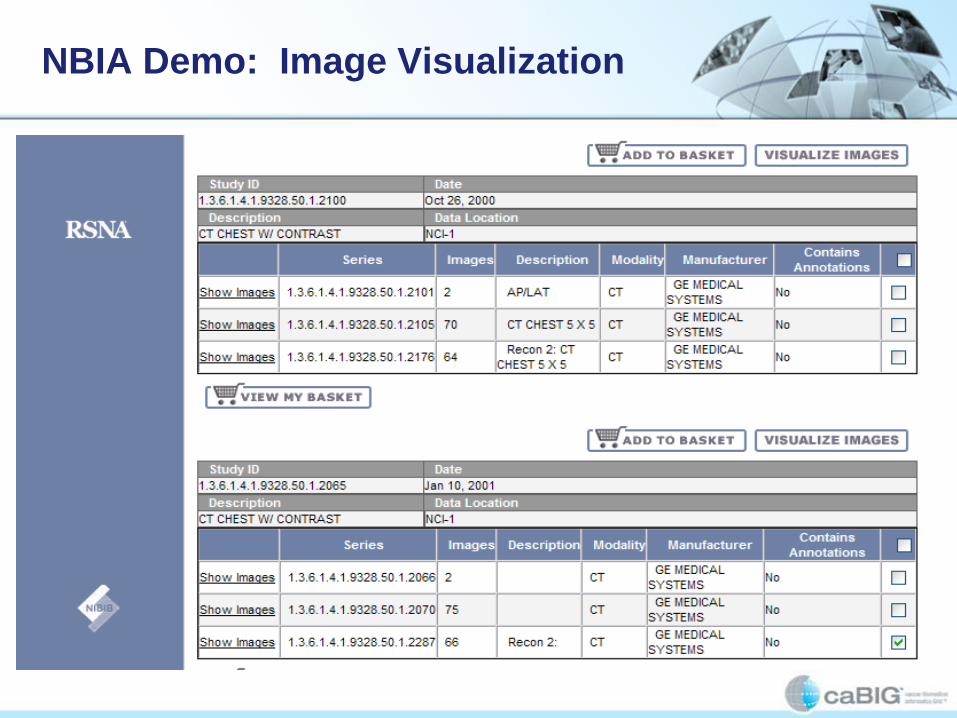
NBIA Demo: Image Visualization

NBIA PC DICOM Viewer: Cedara i-
Response

NBIA Mac DICOM Viewer: OSIRIX
Download or “Virtual PACS”

The Cancer Genome
Atlas (TCGA) In Vivo
Imaging Project
Initial Phase
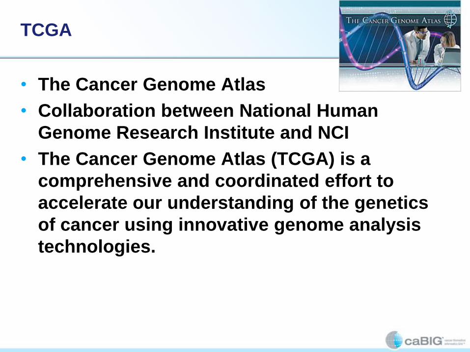
TCGA
• The Cancer Genome Atlas
• Collaboration between National Human
Genome Research Institute and NCI
• The Cancer Genome Atlas (TCGA) is a
comprehensive and coordinated effort to
accelerate our understanding of the genetics
of cancer using innovative genome analysis
technologies.

The Cancer Genome Atlas
• TCGA researchers have identified four
distinct molecular subtypes of
glioblastoma multiforme (GBM), and
demonstrated that response to
aggressive chemotherapy and radiation
differed by subtype
• These findings, reported in the January
19 issue of Cancer Cell, may result in
more personalized approaches to
treating groups of GBM patients based
on their genetic alterations

TCGA Second Study in Cancer Cell
• Another study published in April by The Cancer
Genome Atlas Research Network also in
Cancer Cell used epigenomic profiling
• Maps specific chemical changes or 'marks' to different
areas of the genome, to reveal a new subtype of
Glioblastoma Multiforme (GBM)
• Most patients with GBM survive only 12-15
months after their initial diagnosis
• However, patients with this specific subtype,
called Glioma CpG Island Methylator Phenotype
(G-CIMP), have a median survival of three years

Goals of TCGA Imaging Workspace
Project
• Investigate the added value of highly
structured interpretation and quantification of
MRI images of the TCGA dataset using AIM
• Determine the correlation between MRI
imaging and genotypic information and
response to therapy and prognosis
• Revise Cell article to include impact of MRI
data
• Determine the potential for these tools in
routine clinical practice

Feature Set – Controlled Vocabulary
• 20 features clustered by categories.
• Lesion Location
• Morphology of Lesion Substance
• Morphology of Lesion Margin
• Alterations in Vicinity of Lesion
• Extent of Resection
• Goal is to capture imaging features of
entire tumor and imaging features of
resection specimen.
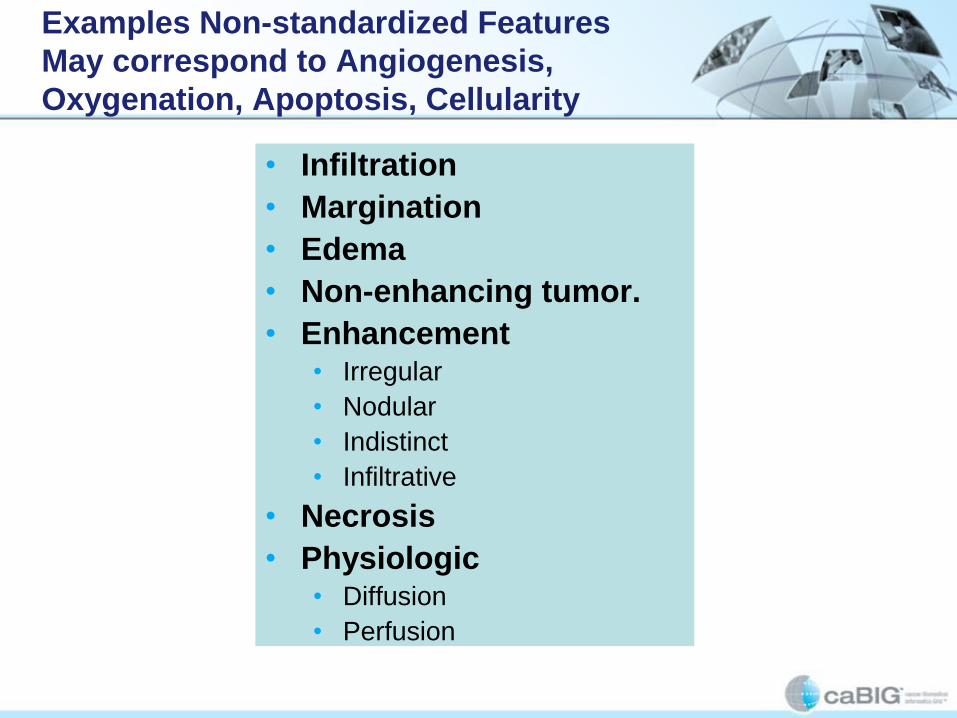
Examples Non-standardized Features
May correspond to Angiogenesis,
Oxygenation, Apoptosis, Cellularity
• Infiltration
• Margination
• Edema
• Non-enhancing tumor.
• Enhancement• Irregular
• Nodular
• Indistinct
• Infiltrative
• Necrosis
• Physiologic• Diffusion
• Perfusion

Well marginated Non-enhancing
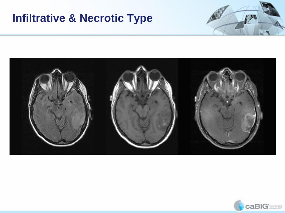
Infiltrative & Necrotic Type

Nodular Predominantly Non-enhancing
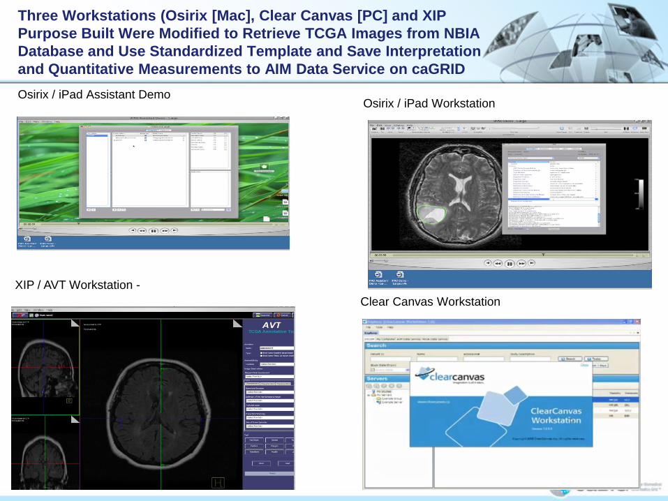
Osirix / iPad Assistant Demo
Three Workstations (Osirix [Mac], Clear Canvas [PC] and XIP
Purpose Built Were Modified to Retrieve TCGA Images from NBIA
Database and Use Standardized Template and Save Interpretation
and Quantitative Measurements to AIM Data Service on caGRID
Osirix / iPad Workstation
Clear Canvas Workstation
XIP / AVT Workstation -

Purpose of TCGA Radiology Phase II Project
Project Goals
Utilize multiple CBIIT/caBIG® technologies together to create
a practical system to capture diagnostic imaging “knowledge”
in a structured, standardized manner and to allow for the
integration with genomic and clinical data
Have at least two radiologists interpret the TCGA MRI brain
images associated with the Cancer Cell article
Utilize caBIG tools to create a repository of the qualitative
and quantitative information associated with the analysis of
the images
Utilize caBIG tools to perform cross database comparisons
for research purposes
Demonstrate potential of caBIG tools to assist in clinical
decision support

Achievements:
Radiology Reading
TCGA cases in NBIA have been read by at least two funded neuro-radiologists:
Images retrieved
from NBIA at CBIIT
New markups created
on Workstation and
saved to the AIME.
Existing markups and
annotation retrieved
from AIM Data Service
at Emory (AIME).
A radiologist fills out
AIM based reporting
template.
New annotation data
is saved on AIME.
Achievements:
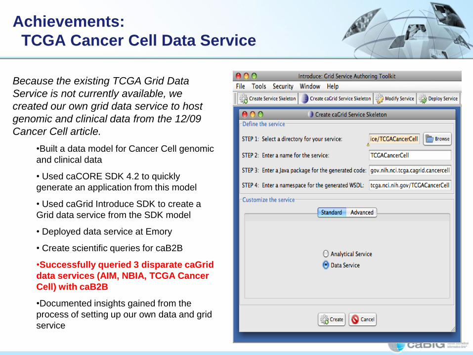
TCGA Cancer Cell Data Service
Because the existing TCGA Grid Data
Service is not currently available, we
created our own grid data service to host
genomic and clinical data from the 12/09
Cancer Cell article.
•Built a data model for Cancer Cell genomic
and clinical data
• Used caCORE SDK 4.2 to quickly
generate an application from this model
• Used caGrid Introduce SDK to create a
Grid data service from the SDK model
• Deployed data service at Emory
• Create scientific queries for caB2B
•Successfully queried 3 disparate caGrid
data services (AIM, NBIA, TCGA Cancer
Cell) with caB2B
•Documented insights gained from the
process of setting up our own data and grid
service

Achievements:
caB2B Query of NBIA, AIM and TCGA CC
Data Services
• Successfully
queried 3
disparate caGrid
data services
(AIM, NBIA,
TCGA Cancer
Cell) with caB2B
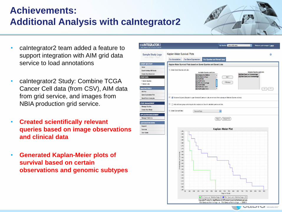
Achievements:
Additional Analysis with caIntegrator2
• caIntegrator2 team added a feature to
support integration with AIM grid data
service to load annotations
• caIntegrator2 Study: Combine TCGA
Cancer Cell data (from CSV), AIM data
from grid service, and images from
NBIA production grid service.
• Created scientifically relevant
queries based on image observations
and clinical data
• Generated Kaplan-Meier plots of
survival based on certain
observations and genomic subtypes

Achievements:
Preliminary Scientific Findings
• Survival of patients who had larger
thickness of enhancement tumors
with hemorrhage was significantly for
shorter than those who did not.
• Survival of patients who had
tumors that crossed midline was
significantly for shorter than those
who did not.
• Survival of patients with greater
thickness of enhancement (who appear
to have had tumors with a thicker “rim”)
was significantly for shorter than those
who had less.
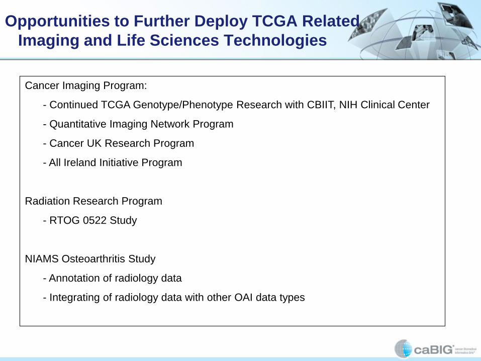
Opportunities to Further Deploy TCGA Related
Imaging and Life Sciences Technologies
Cancer Imaging Program:
- Continued TCGA Genotype/Phenotype Research with CBIIT, NIH Clinical Center
- Quantitative Imaging Network Program
- Cancer UK Research Program
- All Ireland Initiative Program
Radiation Research Program
- RTOG 0522 Study
NIAMS Osteoarthritis Study
- Annotation of radiology data
- Integrating of radiology data with other OAI data types

How the TCGA Radiology Project Fits Into
the caBIG® Imaging Program Roadmap
The Workstation provides a template for the type of visualization service
that we wish to make available as part of the suite of Imaging web-based
services.
The AIM Data Service is part of the proposed suite of web-based services
offered by CBIIT.
All of the TCGA technologies are part of the proposed software refactoring
for SAIF/ECCF compliance.

Proposed Next Steps for TCGA Radiology
1. Ongoing operation and maintenance of NBIA, Clear Canvas, AIM Data Service
and TCGA Cancer Cell Data Service.
2. Communication to community that radiologists can continue to read the cases
and add to the AIM TCGA data set
3. CIP recruited additional radiologists to read the cases since the AIM model
allows any number of readers to refer to one or more instances of the AIM data
service
4. CIP also says that they are working with TCGA sites to get additional TCGA
radiology cases to be loaded on CBIIT’s NBIA.

1. Plan to create a hosted instance of AIM Data Service,
and TCGA Cancer Cell Data Service at CBIIT and in the
cloud
2. Communication to community that researchers can
now query across the three data services. CIP is also
working with Carl Schaefer and Robert Clifford to
begin to do research correlations among the clinical,
genomic and image annotation data.
3. Solicit feedback from community regarding desired
features for the Workstation and AIM Data Service.

Future Plans
• Provide software to NCI clinical cancer centers for
their own clinical trials/research studies involving
diagnostic imaging
• Extend work from in-vivo Imaging to pathology

Future Plans for TCGA Imaging Project
• Include higher order analysis, such as quantitative
diffusion imaging and perfusion imaging metrics,
that could be more sensitive predictors of disease
severity, candidates for effective therapy, and
expected outcomes combining human with semi-
automated and automated analysis of images

Future Plans for TCGA Project
• Ultimately would like to develop a “service” that
has capability to provide immediate feedback for
radiologist or oncologist on patient survival,
patient treatment, etc.
• Incorporate genomic and other data display
during radiology interpretation at a workstation
General Access TCGA Data
• We plan to offer the study for public
consumption [on the production tier] by the
end of September.
Providing Radiology Observation Data for Genotypic/Phenotypic
Analysis in Support of TCGA
caIntegrator2 Demo

caIntegrator 2: Login

caIntegrator 2: Home
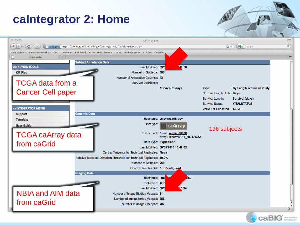
caIntegrator 2: Home
196 subjects
TCGA data from a
Cancer Cell paper
TCGA caArray data
from caGrid
NBIA and AIM data
from caGrid

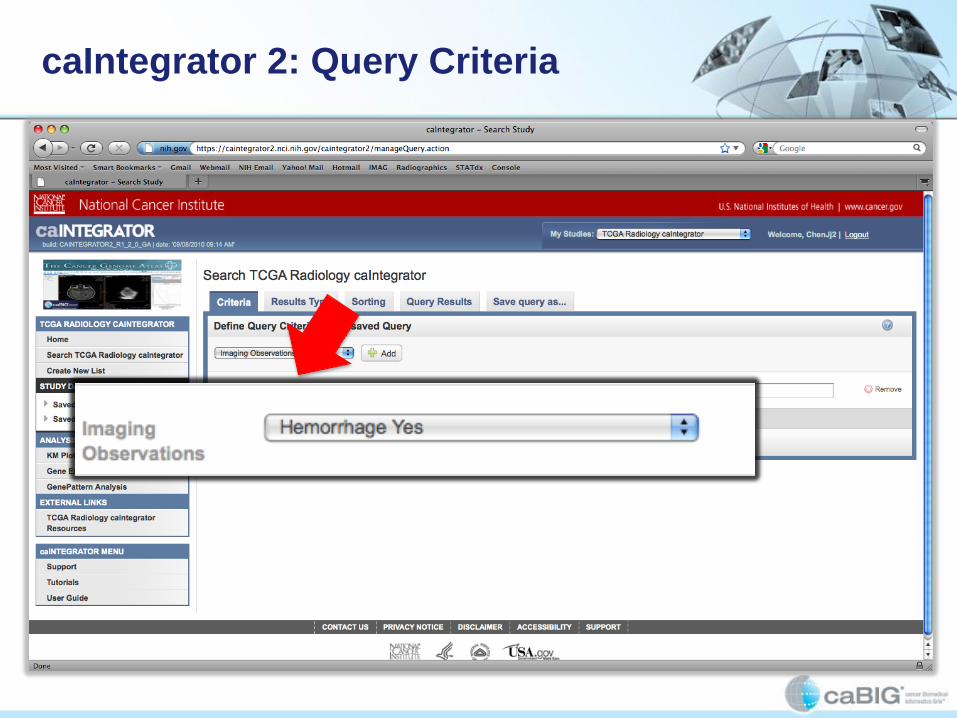
caIntegrator 2: Query Criteria
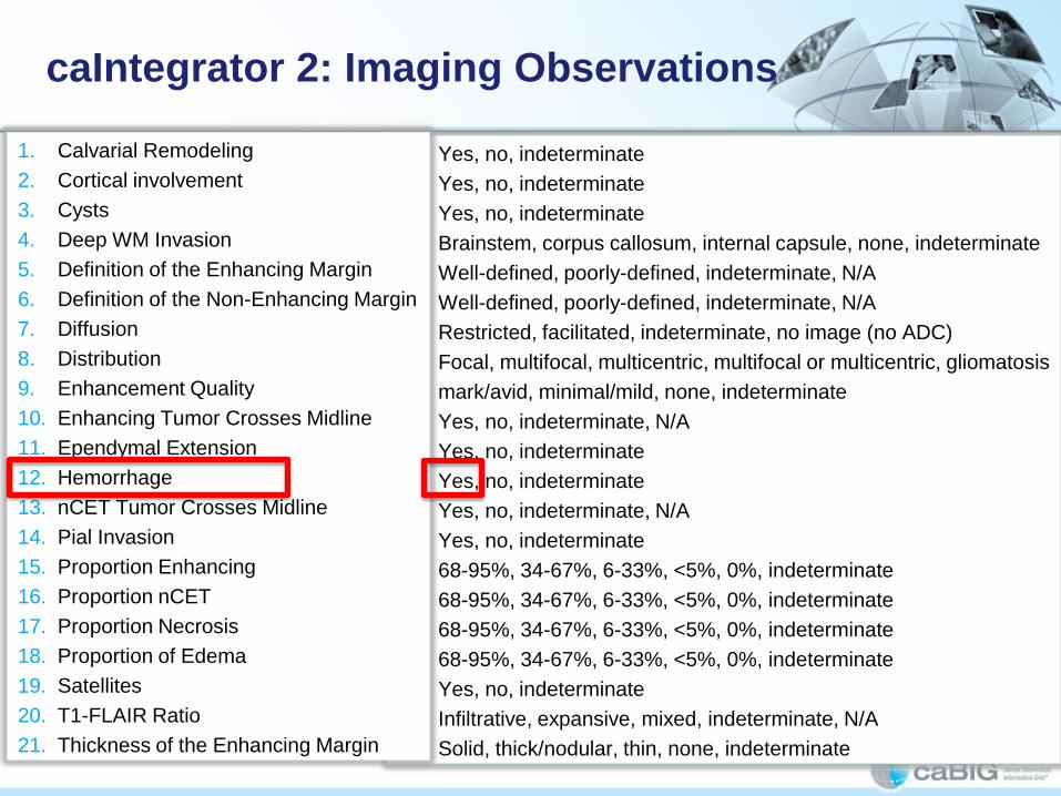
• Yes, no, indeterminate
• Yes, no, indeterminate
• Yes, no, indeterminate
• Brainstem, corpus callosum, internal capsule, none, indeterminate
• Well-defined, poorly-defined, indeterminate, N/A
• Well-defined, poorly-defined, indeterminate, N/A
• Restricted, facilitated, indeterminate, no image (no ADC)
• Focal, multifocal, multicentric, multifocal or multicentric, gliomatosis
• mark/avid, minimal/mild, none, indeterminate
• Yes, no, indeterminate, N/A
• Yes, no, indeterminate
• Yes, no, indeterminate
• Yes, no, indeterminate, N/A
• Yes, no, indeterminate
• 68-95%, 34-67%, 6-33%, <5%, 0%, indeterminate
• 68-95%, 34-67%, 6-33%, <5%, 0%, indeterminate
• 68-95%, 34-67%, 6-33%, <5%, 0%, indeterminate
• 68-95%, 34-67%, 6-33%, <5%, 0%, indeterminate
• Yes, no, indeterminate
• Infiltrative, expansive, mixed, indeterminate, N/A
• Solid, thick/nodular, thin, none, indeterminate
caIntegrator 2: Imaging Observations
1. Calvarial Remodeling
2. Cortical involvement
3. Cysts
4. Deep WM Invasion
5. Definition of the Enhancing Margin
6. Definition of the Non-Enhancing Margin
7. Diffusion
8. Distribution
9. Enhancement Quality
10. Enhancing Tumor Crosses Midline
11. Ependymal Extension
12. Hemorrhage
13. nCET Tumor Crosses Midline
14. Pial Invasion
15. Proportion Enhancing
16. Proportion nCET
17. Proportion Necrosis
18. Proportion of Edema
19. Satellites
20. T1-FLAIR Ratio
21. Thickness of the Enhancing Margin
caIntegrator 2: Query Criteria

caIntegrator 2: Results Type
1. Age At First
Diagnosis
2. Gender
3. Karnofsky Score
4. Survival (days)
5. Vital Status
6. Subtype
7. % Necrosis
8. % Tumor Nuclei
9. Etc.

caIntegrator 2: Query Results

caIntegrator 2: NBIA
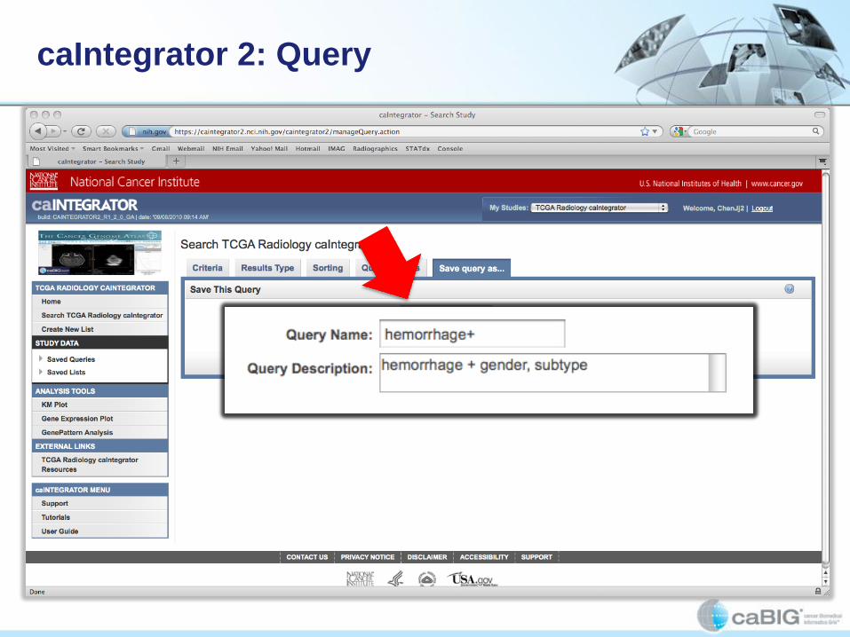
caIntegrator 2: Query

caIntegrator 2: KM Plot

caIntegrator 2: KM Plot

caIntegrator 2: KM Plot

caIntegrator 2: KM Plot

caIntegrator 2: Genomic Data

caIntegrator 2: Genomic Data

caIntegrator 2: Genomic Data

“Grandpa Jones”

“Grandpa Jones”

“Grandpa Jones”
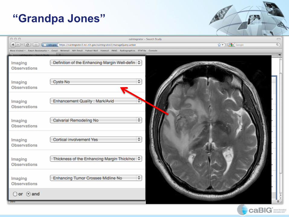
“Grandpa Jones”

“Grandpa Jones”

“Grandpa Jones”

“Grandpa Jones”
Conclusions
• Query
• Analysis
• Prognosis
• Clinical Decision Support
Thank you
Adam Flanders
CBITT Government Sponsors:
• Ed Helton
• Robert Shirley
• Mervi Heiskanen
• Juli Klemm
In collaboration with:
• NCI Cancer Imaging Program
• Carl Jaffe
• John Freyman
• Justin Kirby
Supported by:
• 5AM
• Booz Allen Hamilton
• Buckler Biomedical, LLC.
• Capability Plus Solutions
• ClearCanvas, Inc.
• Emory University
• Northwestern University
• SAIC
• Stanford University
• Thomas Jefferson University
• University of Maryland
• University of Virginia


