
The Cellular Level of Organization • Basic, living, structural and functional unit of the body
– compartmentalization of chemical reactions within specialized structures – regulate inflow & outflow of materials – use genetic material to direct cell activities
• Cytology = study of cellular structure
• Cell physiology = study of cellular function
Generalized Cell Structures • Plasma membrane = cell membrane • Nucleus = genetic material of cell
• Cytoplasm = everything between the membrane and the nucleus – cytosol = intracellular fluid – organelles = subcellular structures with specific functions
The Typical Cell
• Not all cells contain all of these organelles.
Plasma Membrane
• Flexible but sturdy barrier that surround cytoplasm of cell
• Fluid mosaic model describes its structure – “sea of lipids in which proteins float like icebergs” – membrane is 50 % lipid & 50 % protein

• held together by hydrogen bonds – lipid is barrier to entry or exit of polar substances – proteins are “gatekeepers” -- regulate traffic
• 50 lipid molecules for each protein molecule
Lipid Bilayer of the Cell Membrane
• Two back-to-back layers of 3 types of lipid molecules • Cholesterol and glycolipids scattered among a double row of phospholipid molecules
Phospholipids
• Comprises 75% of lipids • Phospholipid bilayer = 2 parallel layers of molecules
• Each molecule is amphipathic (has both a polar & nonpolar region) – polar parts (heads) are hydophilic and face on both surfaces a watery environment – nonpolar parts (tails) are hydrophobic and line up next to each other in the interior
Glycolipids within the Cell Membrane • Comprises 5% of the lipids of the cell membrane
• Carbohydrate groups form a polar head only on the side of the membrane facing the extracellular fluid
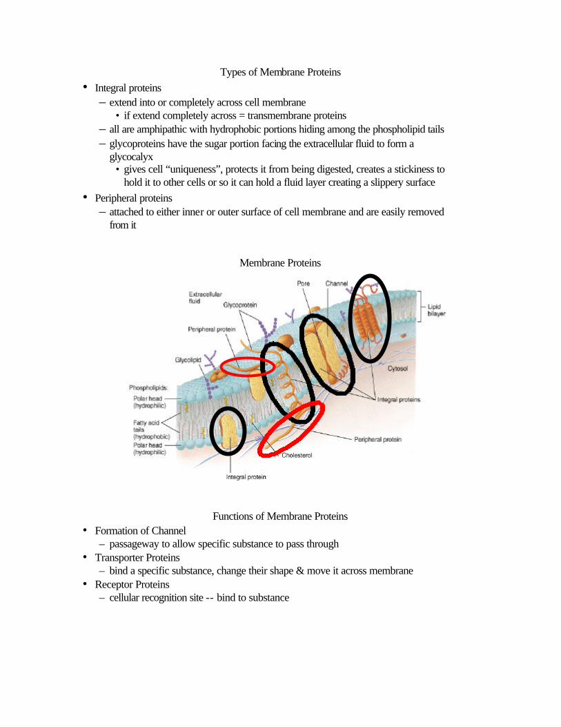
Types of Membrane Proteins • Integral proteins
– extend into or completely across cell membrane • if extend completely across = transmembrane proteins
– all are amphipathic with hydrophobic portions hiding among the phospholipid tails – glycoproteins have the sugar portion facing the extracellular fluid to form a
glycocalyx • gives cell “uniqueness”, protects it from being digested, creates a stickiness to
hold it to other cells or so it can hold a fluid layer creating a slippery surface • Peripheral proteins
– attached to either inner or outer surface of cell membrane and are easily removed from it
Membrane Proteins
Functions of Membrane Proteins • Formation of Channel
– passageway to allow specific substance to pass through • Transporter Proteins
– bind a specific substance, change their shape & move it across membrane • Receptor Proteins
– cellular recognition site -- bind to substance

Functions of Membrane Proteins • Cell Identity Marker
– allow cell to recognize other similar cells • Linker
– anchor proteins in cell membrane or to other cells – allow cell movement – cell shape & structure
• Act as Enzyme – speed up reactions
Membrane Fluidity • Membranes are fluid structures (oil layer)
– self-sealing if punctured with needle
• Explanation -- a compromise of forces – membrane molecules can rotate & move freely – need to stay in one half of lipid bilayer
• difficult for hydrophilic parts to pass through hydrophobic core of bilipid layer – fluidity is reduced by presence of cholesterol
• increases stiffness of membrane it forms hydrogen bonds with neighboring phospholipid heads

Selective Permeability of Membrane • Lipid bilayer
– permeable to nonpolar, uncharged molecules -- oxygen, CO2, steroids – permeable to water which flows through gaps that form in hydrophobic core of
membrane as phospholipids move about • Transmembrane proteins act as specific channels
– small and medium polar & charged particles
• Macromolecules unable to pass through the membrane – vesicular transport
Gradients Across the Plasma Membrane
• Membrane can maintain difference in concentration of a substance inside versus outside of the membrane (concentration gradient) – more O2 & Na+ outside of cell membrane – more CO2 and K+ inside of cell membrane
• Membrane can maintain a difference in charged ions between inside & outside of membrane (electrical gradient or membrane potential)
• Thus, substances move down their concentration gradient and towards the oppositely charged area – ions have electrochemical gradients
Gradients Across Membrane • Concentration gradient • Electrical gradient
Transport Across the Plasma Membrane • Substances cross membranes by a variety of processes:
– mediated transport moves materials with the help of a transporter protein
– nonmediated transport does not use a transporter protein

– active transport uses ATP to drive substances against their concentration gradients
– passive transport moves substances down their concentration gradient with only their kinetic energy – vesicular transport move materials across membranes in small vesicles -- either by exocytosis or endocytosis
Principles of Diffusion • Random mixing of particles in a solution as a result of the particle’s kinetic energy
– more molecules move away from an area of high concentration to an area of low concentration
• the greater the difference in concentration between the 2 sides of the membrane, the faster the rate of diffusion
• the higher the temperature, the faster the rate of diffusion • the larger the size of the diffusing substance, the slower the rate of diffusion • an increase in surface area, increases the rate of diffusion • increasing diffusion distance, slows rate of diffusion
• When the molecules are evenly distributed, equilibrium has been reached Diffusion
• Crystal of dye placed in a cylinder of water • Net diffusion from the higher dye concentration to the region of lower dye • Equilibrium has been reached in the far right cylinder
Osmosis • Net movement of water through a selectively permeable membrane from an area of
high water concentration to an area of lower water concentration – diffusion through lipid bilayer – aquaporins (transmembrane proteins) that function as water channels
• Only occurs if membrane is permeable to water but not to certain solutes

• Pure water on the left side & a membrane impermeable to the solute found on the right side
• Net movement of water is from left to right, until hydrostatic pressure (osmotic pressure ) starts to push water back to the left
Affects of Tonicity on RBCs in Lab
• Normally the osmotic pressure of the inside of the cell is equal to the fluid outside the cell – cell volume remains constant (solution is isotonic)
• Effects of fluids on RBCs in lab – water enters the cell faster than it leaves – water enters & leaves the cell in equal amounts – water leaves the cell
Effects of Tonicity on Cell Membranes • Isotonic solution
– water concentration the same inside & outside of cell results in no net movement of water across cell membrane
• Hypotonic solution – higher concentration of water outside of cell results in hemolysis
• Hypertonic solution – lower concentration of water outside of cell causes crenation
Diffusion Through the Lipid Bilayer • Important for absorption of nutrients -- excretion of wastes • Nonpolar, hydrophobic molecules
– oxygen, carbon dioxide, nitrogen, fatty acids, steroids, small alcohols, ammonia and fat-soluble vitamins (A, E, D and K)

Diffusion Through Membrane Channels • Each membrane channel specific for particular ion (K+, Cl-, Na+ or Ca+2) • Slower than diffusion through membrane but still 1million K+ through a channel in
one second • Channels may be open all the time or gated (closed randomly or as ordered)
Facilitated Diffusion
• Substance binds to specific transporter protein • Transporter protein conformational change moves substance across cell membrane • Facilitated diffusion occurs down concentration gradient only
– if no concentration difference exists, no net movement across membrane occurs
• Rate of movement depends upon – steepness of concentration gradient – number of transporter proteins (transport maximum)
Active Transport
• Movement of polar or charged substances against their concentration gradient – energy-requiring process
• energy from hydrolysis of ATP (primary active transport) • energy stored in an ionic concentration gradient (secondary active transport)
• Exhibits transport maximums and saturation • Na+, K+, H+, Ca+2, I- and Cl-, amino acids and monosaccharides
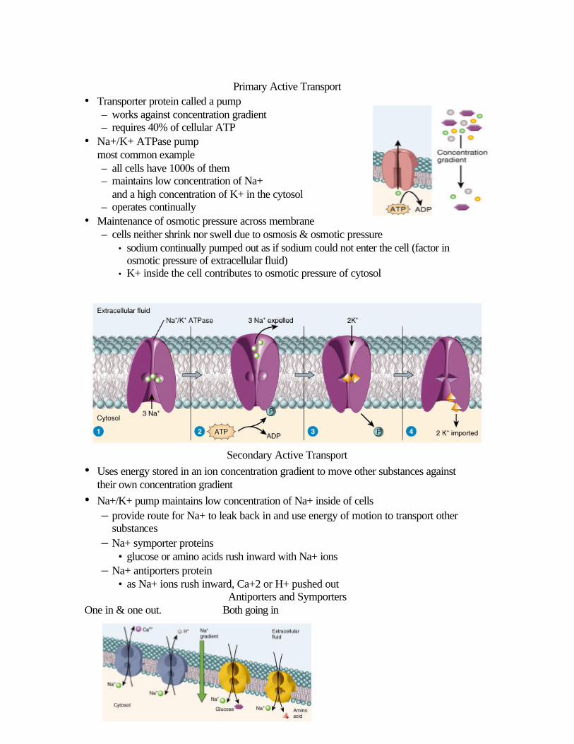
Primary Active Transport
• Transporter protein called a pump – works against concentration gradient – requires 40% of cellular ATP
• Na+/K+ ATPase pump most common example – all cells have 1000s of them – maintains low concentration of Na+
and a high concentration of K+ in the cytosol – operates continually
• Maintenance of osmotic pressure across membrane – cells neither shrink nor swell due to osmosis & osmotic pressure
• sodium continually pumped out as if sodium could not enter the cell (factor in osmotic pressure of extracellular fluid)
• K+ inside the cell contributes to osmotic pressure of cytosol
Secondary Active Transport • Uses energy stored in an ion concentration gradient to move other substances against
their own concentration gradient • Na+/K+ pump maintains low concentration of Na+ inside of cells
– provide route for Na+ to leak back in and use energy of motion to transport other substances
– Na+ symporter proteins • glucose or amino acids rush inward with Na+ ions
– Na+ antiporters protein • as Na+ ions rush inward, Ca+2 or H+ pushed out
Antiporters and Symporters One in & one out. Both going in

Vesicular Transport of Particles • Endocytosis = bringing something into cell
– phagocytosis = cell eating by macrophages & WBCs • particle binds to receptor protein • whole bacteria or viruses are engulfed & later digested
– pinocytosis = cell drinking • no receptor proteins
– receptor-mediated endocytosis = selective input • mechanism by which HIV virus enters cells
• Exocytosis = release something from cell • Vesicles form inside cell, fuse to cell membrane • Release their contents
– digestive enzymes, hormones, neurotransmitters or waste products
• replace cell membrane lost by endocytosis
Receptor-Mediated Endocytosis • Mechanism for uptake of specific substances -- ligands • Desired substance binds to receptor protein in clathrin-coated pit region of cell membrane causing membrane to fold inward • Vesicles become uncoated & combine with endosome • Receptor proteins separate from ligands and return to surface • Ligands are digested by lysosomal enzymes or transported across cell -- epithelial cell crossing accomplished Pinocytosis and Phagocytosis No pseudopods form Nonselective drinking of Pseudopods extend to form phagosome extracellular fluid Lysosome joins it

Cytosol = Intracellular fluid • 55% of cell volume
• 75-90% water with other components – large organic molecules (proteins, carbos & lipids)
• suspended by electrical charges – small organic molecules (simple sugars) & ions
• dissolved – inclusions (large aggregates of one material)
• lipid droplets • glycogen granules
• Site of many important chemical reactions – production of ATP, synthesis of building blocks
Cell Organelles
• Nonmembranous organelles lack membranes & are indirect contact with cytoplasm • Membranous organelles surrounded by one or two lipid bilayer membranes
Cytoskeleton • Network of protein filaments throughout the cytosol • Functions
– cell support and shape – organization of chemical reactions – cell & organelle movement
• Continually reorganized

Centrosome • Found near nucleus • Pericentriolar area
– formation site for mitotic spindle and microtubules • Centrosome
– 2 centrioles(90 degrees to each other) – 9 clusters of 3 microtubules (9+0 array) – role in formation of cilia & flagella
Cilia and Flagella
• Structure – pairs of microtubules
(9+2 array) – covered by cell membrane – basal body is centriole
responsible for initiating its assembly
• Differences – cilia
• short and multiple – flagella
• longer and single
Movement of Cilia and Flagella • Cilia
– stiff during power stroke but flexible during recovery – many coordinated together – airways & uterine tube
• Flagella – single flagella wiggles in a wavelike pattern – propels sperm forward
Ribosomes
• Packages of Ribosomal RNA & protein • Free ribosomes are loose in cytosol
– synthesize proteins found inside the cell
• Membrane-bound ribosomes – attached to endoplasmic reticulum or nuclear membrane – synthesize proteins needed for plasma membrane or for export – 10 to 20 together form a polyribosome
• Inside mitochondria, synthesize mitochondrial proteins
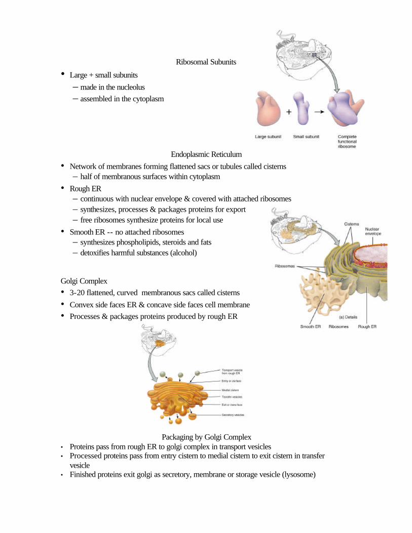
Ribosomal Subunits
• Large + small subunits
– made in the nucleolus – assembled in the cytoplasm
Endoplasmic Reticulum
• Network of membranes forming flattened sacs or tubules called cisterns – half of membranous surfaces within cytoplasm
• Rough ER – continuous with nuclear envelope & covered with attached ribosomes – synthesizes, processes & packages proteins for export – free ribosomes synthesize proteins for local use
• Smooth ER -- no attached ribosomes – synthesizes phospholipids, steroids and fats – detoxifies harmful substances (alcohol)
Golgi Complex • 3-20 flattened, curved membranous sacs called cisterns
• Convex side faces ER & concave side faces cell membrane • Processes & packages proteins produced by rough ER
Packaging by Golgi Complex • Proteins pass from rough ER to golgi complex in transport vesicles • Processed proteins pass from entry cistern to medial cistern to exit cistern in transfer
vesicle • Finished proteins exit golgi as secretory, membrane or storage vesicle (lysosome)

Lysosomes
• Membranous vesicles – formed in Golgi complex – filled with digestive enzymes – pumps in H+ ions until internal pH reaches 5.0
• Functions – digest foreign substances – autophagy(autophagosome forms)
• recycles own organelles – autolysis
• lysosomal damage after death
Peroxisomes • Membranous vesicles
– smaller than lysosomes – form by division of preexisting peroxisomes – contain enzymes that oxidize organic material
• Function – part of normal metabolic breakdown of amino acids and fatty acids – oxidizes toxic substances such as alcohol and formaldehyde – contains catalase which decomposes H2O2
Mitochondria • Double membrane organelle
– central cavity known as matrix – inner membrane folds known as crista
• surface area for chemical reactions of cellular respiration • Function
– generation of ATP – powerhouse of cell

• Mitochondria self-replicate – increases with need for ATP – circular DNA with 37 genes – only inherited from mother
Nucleus
• Large organelle with double membrane nuclear envelope – outer membrane continuous with rough ER – perforated by water-filled nuclear pores (10X channel pore size)
• Nucleolus – spherical, dark bodies within the nucleus (no membrane) – site of ribosome assembly
Function of Nucleus
• 46 human DNA molecules or chromosomes – genes found on chromosomes – gene is directions for a specific protein
• Non-dividing cells contain nuclear chromatin – loosely packed DNA
• Dividing cells contain chromosomes – tightly packed DNA – it doubled (copied itself) before condensing
Protein Synthesis • Instructions for making specific
proteins is found in the DNA (your genes) – transcribe that information onto a
messenger RNA molecule • each sequence of 3 nucleotides in DNA
is called base triplet • each base triplet is transcribed as 3 RNA
nucleotides (codon) – translate the “message” into a sequence of amino acids in order to build a protein

molecule • each codon must be matched by an anticodon found on the tRNA carrying a
specific amino acid
Transcription • DNA sense strand is template for the creation of
messenger RNA strand
Translation • Process where mRNA, rRNA & tRNA are used
to form a specific protein
Normal Cell Division • Mitosis (somatic cell division)
– one parent cell gives rise to 2 identical daughter cells • mitosis is nuclear division • cytokinesis is cytoplasmic division
– occurs in billions of cells each day – needed for tissue repair and growth
• Meiosis (reproductive cell division) – egg and sperm cell production – in testes and ovary only
The Cell Cycle in Somatic Cells • Process where cell duplicates its contents & divides in two
– 23 homologous pairs of chromosomes must be duplicated – genes must be passed on correctly to the next generation of cells
• Nuclear division = mitosis – continuous process divided into 4 stages – prophase, metaphase, anaphase & telophase
• Cytoplasmic division = cytokinesis
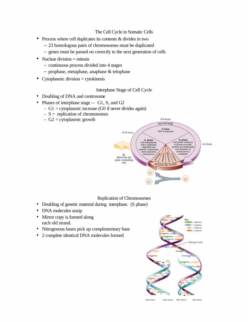
Interphase Stage of Cell Cycle • Doubling of DNA and centrosome • Phases of interphase stage -- G1, S, and G2
– G1 = cytoplasmic increase (G0 if never divides again) – S = replication of chromosomes – G2 = cytoplasmic growth
Replication of Chromosomes • Doubling of genetic material during interphase. (S phase) • DNA molecules unzip • Mirror copy is formed along
each old strand. • Nitrogenous bases pick up complementary base • 2 complete identical DNA molecules formed

Stages of Nuclear Division:Mitosis
• Prophase
• Metaphase
• Anaphase
• Telophase
Control of Cell Destiny
• Cell destiny is either to remain alive & functioning, to grow & divide or to die
• Homeostasis must maintain balance between cell multiplication & cell death • The protein cyclin builds up during interphase and triggers mitosis • Programmed cell death (apoptosis) occurs if a triggering agent turns on suicide
enzymes that kills the cell • Necrosis is cell death caused by injury or infection
Aging
• Age alters the body’s ability to adapt to changes in the environment
• Theories to explain aging – cells have a limited number of divisions – glucose bonds irreversibly with proteins – free radical theory---electrically charged molecules with an unpaired electron
cause cell damage – autoimmune responses due to changes in cell identity markers
• Evidence of aging – damaged skin, hardened arteries, stiff joints
Cellular Diversity
• 100 trillion cells in the body -- 200 different types • Vary in size and shape related to their function

Cancer = out of control cell division
• Hyperplasia = increased number of cell divisions – benign tumor does not metatasize or spread – malignant---spreads due to cells that detach from tumor and enter blood or lymph
• Causes -- carcinogens, x-rays, viruses – every cell has genes that regulate growth & development – mutation in those genes due to radiation or chemical agents causes excess
production of growth factors
• Carcinogenesis – multistep process that takes years and many different mutations that need to occur


