of March 25, 2018.This information is current as
Amyotrophic Lateral SclerosisSOD1-G93A Microglia Model of
Receptor in the7Dysregulated by the P2XThe NADPH Oxidase Pathway Is
Volonté and Nadia D'AmbrosiCinziaSimona Rossi, Maria Teresa Carrì, Mauro Cozzolino,
Savina Apolloni, Chiara Parisi, Maria Grazia Pesaresi,
http://www.jimmunol.org/content/190/10/5187doi: 10.4049/jimmunol.1203262April 2013;
2013; 190:5187-5195; Prepublished online 15J Immunol
Referenceshttp://www.jimmunol.org/content/190/10/5187.full#ref-list-1
, 10 of which you can access for free at: cites 43 articlesThis article
average*
4 weeks from acceptance to publicationFast Publication! •
Every submission reviewed by practicing scientistsNo Triage! •
from submission to initial decisionRapid Reviews! 30 days* •
Submit online. ?The JIWhy
Subscriptionhttp://jimmunol.org/subscription
is online at: The Journal of ImmunologyInformation about subscribing to
Permissionshttp://www.aai.org/About/Publications/JI/copyright.htmlSubmit copyright permission requests at:
Email Alertshttp://jimmunol.org/alertsReceive free email-alerts when new articles cite this article. Sign up at:
Print ISSN: 0022-1767 Online ISSN: 1550-6606. Immunologists, Inc. All rights reserved.Copyright © 2013 by The American Association of1451 Rockville Pike, Suite 650, Rockville, MD 20852The American Association of Immunologists, Inc.,
is published twice each month byThe Journal of Immunology
by guest on March 25, 2018
http://ww
w.jim
munol.org/
Dow
nloaded from
by guest on March 25, 2018
http://ww
w.jim
munol.org/
Dow
nloaded from
The Journal of Immunology
The NADPH Oxidase Pathway Is Dysregulated by the P2X7
Receptor in the SOD1-G93A Microglia Model ofAmyotrophic Lateral Sclerosis
Savina Apolloni,*,† Chiara Parisi,*,† Maria Grazia Pesaresi,† Simona Rossi,†,‡
Maria Teresa Carrı,†,‡ Mauro Cozzolino,†,x Cinzia Volonte,*,† and Nadia D’Ambrosi*,†
Inflammation and oxidative stress are thought to play determinant roles in the pathogenesis of amyotrophic lateral sclerosis (ALS).
Degenerating motor neurons produce signals that activate microglia to release reactive oxygen species (ROS) and proinflammatory
cytokines, resulting in a vicious cycle of neurodegeneration. The ALS-causing mutant protein Cu+/Zn+ superoxide dismutase
SOD1-G93A directly enhances the activity of the main ROS-producing enzyme in microglia, NADPH oxidase 2 (NOX2), a well-
known player in the pathogenesis of ALS. Considering that extracellular ATP through P2X7 receptor constitutes a neuron-to-
microglia alarm signal implicated in ALS pathology, we used primary microglial cells derived from transgenic SOD1-G93A mice
and SOD1-G93A mice lacking the P2X7 receptor to investigate the effects of both pharmacological induction and genetic ablation
of receptor activity on the NOX2 pathway. We observed that, in SOD1-G93A microglia, the stimulation of P2X7 receptor by 29-39-
O-(benzoyl-benzoyl) ATP enhanced NOX2 activity in terms of translocation of p67phox to the membrane and ROS production; this
effect was totally dependent on Rac1. We also found that, following P2X7 receptor stimulation, the phosphorylation of ERK1/2 was
augmented in ALS microglia, and there was a mutual dependency between the NOX2 and ERK1/2 pathways. All of these
microglia-mediated damaging mechanisms were prevented by knocking out P2X7 receptor and by the use of specific antagonists.
These findings suggest a noxious mechanism by which P2X7 receptor leads to enhanced oxidative stress in ALS microglia and
identify the P2X7 receptor as a promising target for the development of therapeutic strategies to slow down the progression of
ALS. The Journal of Immunology, 2013, 190: 5187–5195.
Inflammation and oxidative stress are thought to play deter-minant roles in the pathogenesis of amyotrophic lateralsclerosis (ALS) (1). Degenerating motor neurons, the main
pathological hallmark of ALS, produce signals that activatemicroglia, CNS-resident immune cells, to release free radicals andproinflammatory cytokines. This, in turn, causes further motor neu-ron stress and initiates a self-propagating cytotoxic cascade resultingin muscle weakness, atrophy, spasticity, and compromised speech,swallowing, and breathing (2, 3).NADPH oxidase 2 (NOX2 or phagocytic oxidase-gp91phox) is one
of the major sources of extracellular and intracellular reactive oxy-gen species (ROS) in microglial cells, where it operates by transfer-ring electrons across membranes from NADPH to molecular oxygen,generating O2
d2 (4). Activation of NOX2 in microglia increasesboth extracellular and intracellular ROS, causing neurotoxicity.
The former are directly toxic to neurons, and the latter function assignals to amplify the production of several proinflammatory andneurotoxic cytokines through the activation of downstream mol-ecules, including MAPK (i.e., p38 and ERK1/2) (5).A central activator of NOX2 is the Rho-GTPase family member
Rac1, acting as a molecular switch cycling between its inactiveGDP-bound state and active GTP-bound state (6). GTP hydrolysisby Rac1 is controlled through a redox-dependent interaction withthe ubiquitous enzyme Cu+/Zn+ superoxide dismutase (SOD1),which is found mutated in ALS patients. When SOD1 is boundto Rac1 under reducing conditions, GTP hydrolysis is inhibited;in oxidizing conditions, SOD1 dissociates from Rac1, no longerinhibiting GTP hydrolysis. The ALS-causing glycine-to-alaninesubstitution at position 93 (G93A) on SOD1 protein renders theenzyme less sensitive to redox uncoupling, leading to enhancedRac1/NOX2 activation (7) and resulting in overproduction ofdamaging ROS (8). Consistently, NOX2 is upregulated in acti-vated ALS microglia, and the deletion of NOX2 can improvedisease progression and survival of SOD1-G93A mice (9–11).Furthermore, uncontrolled ROS, together with inflammatorycytokines produced by microglia, mediate disease progression,causing direct neurotoxicity and increasing motor neuron sus-ceptibility to ALS-triggering factors (2, 12). Among these, ex-tracellular ATP was recently suggested as a possible candidate.The role of extracellular nucleotides is well recognized in prolifer-ation, chemotaxis, phagocytosis, and the release of proinflammatorymolecules, namely in all aspects of microglia functioning (13). Amajor player in the response of microglia to extracellular ATP is theionotropic purinergic P2X7 receptor, which is responsible for therelease of cytokines and induction of proinflammatory factors, suchas TNF-a, IL-6, IL-1b, plasminogen, and cyclooxygenase-2 (COX-2) (14). Indeed, the proinflammatory function of P2X7 receptor has
*Cellular Biology and Neurobiology Institute, National Research Council, Rome00143, Italy; †Santa Lucia Foundation, Rome 00143, Italy; ‡Department of Biology,University of “Tor Vergata”, Rome 00133, Italy; and xInstitute of TranslationalPharmacology, National Research Council, Rome, Italy
Received for publication November 27, 2012. Accepted for publication March 5,2013.
This work was supported by Agenzia Italiana per la Ricerca sulla Sclerosi LateraleAmiotrofica (Grant PRALS 2009, cofinanced with the support of “53 1000,” Health-care Research of the Ministry of Health).
Address correspondence and reprint requests to Dr. Nadia D’Ambrosi, NationalResearch Council/Santa Lucia Foundation, Via del Fosso di Fiorano 65, Rome00143, Italy. E-mail address: [email protected]
Abbreviations used in this article: ALS, amyotrophic lateral sclerosis; BBG, BrilliantBlue G; BzATP, 29-39-O-(benzoyl-benzoyl) ATP; COX-2, cyclooxygenase-2; MOI,multiplicity of infection; NOX2, NADPH oxidase 2; nt, nontransgenic; PAK, p21activated kinase 1 protein; ROS, reactive oxygen species; SOD1, Cu+/Zn+ superoxidedismutase.
Copyright� 2013 by The American Association of Immunologists, Inc. 0022-1767/13/$16.00
www.jimmunol.org/cgi/doi/10.4049/jimmunol.1203262
by guest on March 25, 2018
http://ww
w.jim
munol.org/
Dow
nloaded from

been involved in different neuropathologies with an inflammatorycomponent, such as Alzheimer’s and Huntington’s diseases, mul-tiple sclerosis, spinal cord injury, and neuropathic pain (15, 16).Recent articles reported that P2X7 receptor expression andmodulation are linked to neuroinflammation in human ALS andin different animal models for the disease (17, 18). In particular,human postmortem ALS spinal cord displays a greater density ofP2X7 receptor immunoreactivity in microglial cells, together withan increased production of COX-2 (19). A conspicuous P2X7 re-ceptor immunolabeling clearly delineating microglial cells was alsopresent at advanced stages of disease in spinal cord sections oftransgenic rats expressing mutant SOD1 (20). Thus, this distribu-tion of P2X7 receptor suggests its involvement in active micro-gliosis, which goes along with motor neuron degeneration in vivo.Consistently, in a previous study we showed that P2X7 receptormRNA and protein are upregulated in both primary and immor-talized microglial cells from SOD1-G93A mice. As a functionalconsequence, the exposure of SOD1-G93A microglia to the P2X7
receptor preferential agonist 29-39-O-(benzoyl-benzoyl) ATP(BzATP) amplified the morphological transition from resting/surveying to activated microglia and enhanced the content ofTNF-a and COX-2. Furthermore, only microglia expressingSOD1-G93A protein and preactivated by BzATP displayed tox-icity toward neuronal cells. All of these actions were prevented bythe P2X7 receptor–specific antagonist Brilliant Blue G (BBG),thus confirming the involvement of this receptor subtype inALS (21). In a similar way, endogenous ATP and BzATP werelater found to cause astrocyte-dependent neurotoxicity in cul-ture, by inducing motor neuron death. Moreover, SOD1-G93Aastrocytes displayed increased ATP-dependent proliferation anda basal increase in extracellular ATP degradation. Again, all ofthese events were prevented by BBG (22). Taken together, theseresults clearly suggest that the release/degradation of ATP and theactivation of the P2X7 receptor are involved in the microglia–astrocyte–motor neuron cross-talk that occurs during the pro-gression of ALS.The aim of this work is to define the molecular mechanisms
within P2X7 receptor activation that can account for an exacer-bated response in SOD1-G93A microglia. In particular, we ana-lyzed Rac1 and NOX2 activation and investigated upstream anddownstream signaling mechanisms. To ascertain unequivocally therole of P2X7 receptor in the microglial inflammatory pathway, wetook advantage of both pharmacological and molecular inhibitionof P2X7 receptor activity by using different antagonists and gen-erating SOD1-G93A mice lacking this receptor.
Materials and MethodsGeneration of P2X7
2/2/SOD1-G93A transgenic mice
Adult B6.Cg-Tg(SOD1-G93A)1Gur/J mice expressing a high copy numberof mutant human SOD1 with a Gly93Ala substitution (SOD1-G93A) andB6.129P2-P2rx7tm1Gab/J mice (P2X7
2/2) were originally obtained fromThe Jackson Laboratory (Bar Harbor, ME) and bred in the indoor animalfacility, where they were kept until experiments were carried out. SOD1-G93A transgenic animals were crossed with background-matched B6Jwild-type females, and selective breeding maintained each transgene in thehemizygous state. Nontransgenic (nt) littermate mice were used as con-trols. To generate SOD1-G93A mice lacking the P2X7 receptor (P2X7
2/2/SOD1-G93A), SOD1-G93A males were first cross-bred with P2X7
2/2
females to generate F1 progeny (P2X7+/2/SOD1-G93A), and the P2X7
+/2/SOD1-G93A males were cross-bred with P2X7
2/2 females to obtain theF2 progeny (P2X7
2/2/SOD1-G93A). All transgenic mice were identifiedby analyzing tissue extracts from tail tips. Animals were housed at constanttemperature (22 6 1˚C) and relative humidity (50%) with a regular 12-hlight cycle (light 7 AM–7 PM) throughout the experiments. Food and waterwere freely available. All animal procedures were performed according tothe European Guidelines for the use of animals in research (86/609/CEE)
and the requirements of Italian laws (D.L. 116/92). The ethical procedurewas approved by the Animal Welfare Office, Department of Public Healthand Veterinary, Nutrition and Food Safety, General Management of Ani-mal Care and Veterinary Drugs of the Italian Ministry of Health. All effortswere made to minimize animal suffering and to use only the number ofanimals necessary to produce reliable results.
Reagents
BzATP, BBG, and all other reagents, unless otherwise stated, were obtainedfrom Sigma-Aldrich (Milan, Italy). PD98059 and catalase (bovine liver)were purchased from Calbiochem (San Diego, CA). A-839977 andA-438079 were from Tocris Bioscience (Bristol, U.K.).
Abs
P2X7 rabbit polyclonal Ab (1:500) was purchased from Alomone Labs(Jerusalem, Israel); Rac1 mouse Ab (1:1000) was obtained from Millipore(Merck Millipore, Merck KGaA, Darmstadt, Germany); p67phox (1:200 forimmunofluorescence and 1:500 for Western blot) and gp91phox (1:1000–1:2000) mouse mAbs were from BD Transduction Laboratories (Lex-ington, KY); p44/42 MAPK (ERK1/2; L34F12; 1:2000) mouse Ab andphospho-p44/42 MAPK (ERK1/2; Thr202/Tyr204; 1:1000) and phospho-p38 MAPK (Thr180/Tyr182; 12F8; 1:700) rabbit Abs were from CellSignaling Technology (Beverly, MA). HRP-linked anti-rabbit and anti-mouse Abs were from Cell Signaling Technology.
Primary microglia cell cultures
Mixed glial cultures from brain cortex were prepared as we describedpreviously (21). Briefly, neonatal SOD1-G93A, P2X7
2/2/SOD1-G93A,and nt mice were sacrificed; after removing the meninges, cortices wereminced and digested with 0.01% trypsin and 10 mg/ml DNase I. Afterdissociation and passage through 70-mm filters, cells were resuspended inDMEM/F-12 media with GlutaMAX (Life Technologies, Invitrogen,Paisley, U.K.) plus 10% FBS, 100 U/ml gentamicin, and 100 mg/mlstreptomycin/penicillin at a density of 62,500 cells/cm2. After ∼15 d,a mild trypsinization (0.08% trypsin in DMEM/F-12 without FBS) wasperformed for 40 min at 37˚C to remove nonmicroglial cells (23). Theresultant adherent microglial cells (.99% pure) were washed twice withDMEM/F-12 and kept in 50% mixed glial cell–conditioned medium at37˚C in a 5% CO2 and 95% air atmosphere for 48 h until use.
Protein extraction, SDS-PAGE, and Western blotting
To isolate total-protein extracts, cells in serum-free medium were harvestedin ice-cold RIPA buffer (PBS, 1% Nonidet P-40, 0.5% sodium deoxy-cholate, 0.1% SDS) added to protease inhibitor mixture (Sigma-Aldrich).Lysates were kept on ice for 30 min and then centrifuged for 10 min at14,000 3 g at 4˚C. Supernatants were collected and assayed for proteinquantification with the BCA protein assay (Thermo Fischer Scientific,Rockford, IL). Analysis of protein components was performed using Mini-PROTEAN TGX Gels (Bio-Rad Laboratories, Milan, Italy) and transferonto nitrocellulose membranes (Amersham Biosciences, Cologno Monzese,Italy). After saturation with ECL Advance Blocking Agent (AmershamBiosciences), blots were probed overnight at 4˚C with the specified Ab,incubated for 1 h with HRP-conjugated secondary Abs, and detected onx-ray film (Aurogene, Rome, Italy), using an ECL Advance WesternBlotting Detection Kit (Amersham Biosciences). Quantifications wereperformed using a Kodak Image Station.
Membrane translocation of p67phox
After drug treatment, primary microglia were lysed in relaxation buffer(100 mM KCl, 3 mM NaCl, 3.5 mM MgCl2, 1.25 mM EGTA, and 10 mMPIPES [pH 7.3]) added to protease inhibitor mixture (Sigma-Aldrich) andsonicated (3 3 10 s, at 4˚C using an ultrasonic processor [Hielscher,Teltow, Germany]). Cell lysate was centrifuged at 500 3 g to removemitochondria and nuclei, generating a postnuclear supernatant that wassubsequently ultracentrifuged at 100,000 3 g for 1 h at 4˚C to spin downtotal membranes. The membrane fraction was resuspended in 100 ml re-laxation buffer added to 1% Triton X-100, protein concentration was esti-mated using BCA assay, and 5 mg protein was analyzed using Western blotwith anti-p67phox Ab. To normalize the total amount of cell membranes,Western blot with an Ab recognizing the mouse NOX2 membrane–residentgp91phox subunit was performed.
Rac1 constructs, transfection, and virus transduction
Human cDNA coding for Rac1 (GenBank accession number NM_006908)was cloned by RT-PCR from human SH-SY5Y neuroblastoma cells using
5188 NOX2 ACTIVATION BY P2X7 RECEPTOR IN ALS MICROGLIA
by guest on March 25, 2018
http://ww
w.jim
munol.org/
Dow
nloaded from
the forward primer 59-AAA GGA TCC ATG CAG GCC ATC AAG TGTG-39 and the reverse primer 59-AAA TCT AGA TTA CAA CAG CAGGCA TTT TCT C-39. The resulting PCR fragment was inserted intoBamHI/XbaI restriction sites of pRRLSIN.cPPT.PGK-eGFP lentiviralvector. Rac1-V12 and Rac1-N17 were produced by PCR site-directedmutagenesis using wild-type Rac1 plasmid as a template, followed bydigestion with Dpn1. All of the pRRLSIN.cPPT.PGK-eGFP constructswere verified by automated sequencing and then purified and cotransfectedtogether with packaging vectors into HEK-293FT cells. Supernatants werecollected after 48 and 72 h, and viral particles were concentrated by ul-tracentrifugation for 2 h at 26,000 rpm (Ultraclear Tubes, SW28 rotor, andOptima l-100 XP Ultracentrifuge; Beckman Coulter, Milan, Italy) andrecovered by suspension in HBSS (Sigma-Aldrich). Titers of viral particlesranged between 106 and 107 TU/ml. Viral particles and polybrene (8 mg/mlfinal concentration) were then added to isolated primary microglia. Len-tiviral particles, at a multiplicity of infection (MOI) of 30, and 8 mg/mlpolybrene (Sigma-Aldrich) were added to the culture. Supernatant wasremoved 5 h postinfection and replaced with DMEM-F12 medium con-taining 10% FBS. In all of the experiments, the efficiency of microgliatransduction was $90%, as determined by counting the number ofmicroglia expressing the GFP molecule and counterstaining nuclei withHoechst 33258 (1 mg/ml for 5 min) by means of a fluorescent microscope.Cell morphology was visualized by Cy3-conjugated phalloidin staining(5 mg/ml). GFP–adenovirus construction was described previously (24)and used at an MOI of 30–50. All experiments were performed at 72 hpostinfection.
Rac1 pull-down assay
The Rac-GTP pull-down assay was performed as previously described (24).Briefly, cells were lysed in a buffer containing 50 mM Tris [pH 7.2],100 mM NaCl, 5 mM MgCl2, 1 mM DTT, 10% glycerol, and 1% Nonidet-P40 plus protease inhibitors. One fiftieth of the cell lysate was subjectedto Western blotting. Cell lysates were mixed with 10 mg bacteriallyexpressed GST-p21 activated kinase 1 protein (PAK) (rat PAK aa 1–252)bound to glutathione-Sepharose and incubated at 4˚C with tumbling for30 min. Beads were collected by centrifugation and washed twice in lysisbuffer prior to the addition of Laemmli buffer and analysis with anti-Rac1Ab by Western blot.
Intracellular ROS measurement
Because of the many limitations of standard fluorescent probes whenmeasuring intracellular ROS by fluorescence microscope analysis (25),intracellular production of ROS was measured by monitoring the oxidationof the cell-permeable nonfluorescent reagent CellROX Deep Red Reagent(Molecular Probes, Carlsbad, CA), which, upon oxidation, exhibits astrong fluorogenic signal that is stable after fixation. Briefly, 2 3 105 cellswere starved with serum-free medium and then exposed to treatments.Subsequently, cells were incubated with 5 mM CellROX Deep Red Re-agent and 1 mg/ml Hoechst 33258 (for nuclei) for 30 min at 37˚C and, afterthree washes with PBS, were fixed for 10 min in 4% paraformaldehyde.Cells were mounted and cover-slipped with Gel/Mount antifading (Bio-Meda, Burlingame, CA). Fluorescence was analyzed using a confocal laserscanning microscope (LSM700; Zeiss, Oberkochen, Germany) equippedwith four laser lines: 405, 488, 561, and 639 nm. Images were analyzedwith ImageJ software.
H2O2-production assay
Microglial cells were grown in 96-well fluorescence tissue culture platesat a density of 104 cells/well. After treatment, an Amplex Red HydrogenPeroxide/Peroxidase Assay (Molecular Probes) was performed to detectH2O2 released from cells. In the presence of HRP, the Amplex Red reagent(10-acetyl-3,7-dihydroxyphenoxazine) reacts with H2O2 in a 1:1 stoichi-ometry to produce the red-fluorescent oxidation product resorufin. Fluo-rescence was recorded from the blank, control, and treated cells, togetherwith a positive control, in a multilabel counter (Victor3-V; PerkinElmerLife Sciences, Milan, Italy) by measuring (530–560Ex/590Em) fluorescenceevery 5 min over the experimental time period. To normalize the cellnumber, the CellTiter-Blue Cell Viability Assay (Promega Italia, Milan,Italy) was then performed. Briefly, after incubating cells with the CellTiter-Blue Reagent, fluorescence was recorded in the same multilabel counter atthe same experimental excitation and detection (530–560Ex/590Em).
Immunofluorescence microscopy
SOD1-G93A and P2X72/2/SOD1-G93A primary cells were fixed for 15 min
in 4% paraformaldehyde and permeabilized for 5 min in PBS containing0.1% Triton X-100. The cells were then incubated for 1.5 h at 37˚C with
mouse anti-p67phox (1:200; BD Biosciences) in 1% BSA in PBS. The cul-tures were stained for 1 h with Cy2-conjugated donkey anti-mouse IgG(1:200; Jackson ImmunoResearch, West Grove, PA). Nuclei were stainedwith 1 mg/ml Hoechst 33258 for 5 min, and the cells were mounted andcover-slipped with Gel/Mount antifading (BioMeda). Immunofluorescencewas analyzed using a confocal microscope, as described above.
Data analysis
Data are presented as mean 6 SEM. Statistical differences were verifiedby the Student t test using MedCalc (Medcalc Software, Mariakerke, Bel-gium), followed by individual post hoc comparisons (Fisher protected leastsignificant difference). The p values , 0.05 were considered significant.
ResultsModulation of P2X7 receptor affects p67
phox subunittranslocation
To investigate the modulation of the NOX2 pathway by P2X7
receptor activation in SOD1-G93A microglia, we first examinedthe translocation of the NOX2 activator p67phox to cell mem-branes. As shown in Fig. 1A, under basal conditions, SOD1-G93Amicroglia display a higher amount of membrane-associatedp67phox with respect to cells derived from nt animals (normal-ized to the membrane-resident gp91phox subunit). Activation of theP2X7 receptor by addition of BzATP increases the presence ofp67phox in membranes of both nt and SOD1-G93A cells. However,10 mM BzATP increases p67phox subunit translocation ∼3.5-fold(with respect to nt cells) only in SOD1-G93A microglia, whereasa higher BzATP concentration (100 mM) results in only a 2-foldincrease in the p67phox subunit in membranes of nt cells and a4-fold increase in SOD1-G93A cells. The translocation of p67phox
in SOD1-G93A membranes by BzATP was confirmed by immu-nofluorescence (Fig. 1B), because the generally diffused cytosolicstaining of anti-p67phox Ab becomes enriched on cell membranesupon 100 mM agonist stimulation. This effect is completely pre-vented by the addition of BBG, A-839977, or A-438079 (all at1 mM), as shown by immunofluorescence and Western blotting(Fig. 1B, 1C). Thus, the effectiveness of BzATP and the inhibitionby specific antagonists indicate that the P2X7 receptor mediatesthe translocation of p67phox. Indeed, in SOD1-G93A microglia inwhich the P2X7 receptor is knocked out (Fig. 1D, left panel),stimulation with 100 mM BzATP fails to increase p67phox incell membranes, as demonstrated by immunofluorescence andWestern blotting (Fig. 1D). These data reinforce the P2X7 receptoras a likely candidate to modulate the NOX2 pathway in ALSmicroglia (Fig. 8).
P2X7 receptor influences GTP-bound Rac1 level
The Rho-GTPase family member Rac1 is a central activator ofNOX2 via a multistep mechanism involving binding to GTP,translocation to cell membrane and direct interaction with gp91phox,followed by a subsequent interaction with p67phox (26). Thus, weanalyzed the effect of BzATP on Rac1 activation in nt and SOD1-G93A microglia by measuring GTP-bound active Rac1 witha GST-PAK1 pull-down assay (Fig. 2). Western blot analysis ofpulled-down Rac1 shows that, already under unstimulated con-ditions, SOD1-G93A microglia display a 3-fold higher amount ofGTP-Rac1 than do nt cells, as normalized to the expression oftotal Rac1. Despite this elevated Rac1 activity under basal con-ditions, the addition of BzATP at 10 mM and at 100 mM is able todramatically increase the content of active Rac1 in SOD1-G93Amicroglia compared with unstimulated nt cells (3- and 6-fold,respectively) and with stimulated nt cells (3-fold in each case).With regard to p67phox translocation, the addition of 1 mM BBG(Fig. 2A, left panel, 2B) or the molecular deletion of P2X7 re-ceptor (Fig. 2A, right panel) completely abolishes Rac-1 activa-
The Journal of Immunology 5189
by guest on March 25, 2018
http://ww
w.jim
munol.org/
Dow
nloaded from
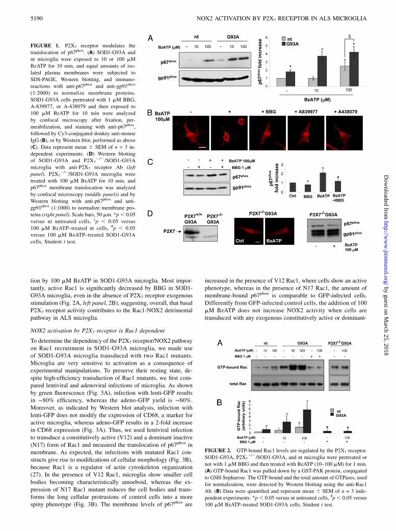
tion by 100 mM BzATP in SOD1-G93A microglia. Most impor-tantly, active Rac1 is significantly decreased by BBG in SOD1-G93A microglia, even in the absence of P2X7 receptor exogenousstimulation (Fig. 2A, left panel, 2B), suggesting, overall, that basalP2X7 receptor activity contributes to the Rac1-NOX2 detrimentalpathway in ALS microglia.
NOX2 activation by P2X7 receptor is Rac1 dependent
To determine the dependency of the P2X7 receptor/NOX2 pathwayon Rac1 recruitment in SOD1-G93A microglia, we made useof SOD1-G93A microglia transduced with two Rac1 mutants.Microglia are very sensitive to activation as a consequence ofexperimental manipulations. To preserve their resting state, de-spite high-efficiency transduction of Rac1 mutants, we first com-pared lentiviral and adenoviral infections of microglia. As shownby green fluorescence (Fig. 3A), infection with lenti-GFP resultsin ∼80% efficiency, whereas the adeno-GFP yield is ∼60%.Moreover, as indicated by Western blot analysis, infection withlenti-GFP does not modify the expression of CD68, a marker foractive microglia, whereas adeno-GFP results in a 2-fold increasein CD68 expression (Fig. 3A). Thus, we used lentiviral infectionto transduce a constitutively active (V12) and a dominant inactive(N17) form of Rac1 and measured the translocation of p67phox inmembrane. As expected, the infections with mutated Rac1 con-structs give rise to modifications of cellular morphology (Fig. 3B),because Rac1 is a regulator of actin cytoskeleton organization(27). In the presence of V12 Rac1, microglia show smaller cellbodies becoming characteristically amoeboid, whereas the ex-pression of N17 Rac1 mutant reduces the cell bodies and trans-forms the long cellular protrusions of control cells into a morespiny phenotype (Fig. 3B). The membrane levels of p67phox are
increased in the presence of V12 Rac1, where cells show an activephenotype, whereas in the presence of N17 Rac1, the amount ofmembrane-bound p67phox is comparable to GFP-infected cells.Differently from GFP-infected control cells, the addition of 100mM BzATP does not increase NOX2 activity when cells aretransduced with any exogenous constitutively active or dominant-
FIGURE 1. P2X7 receptor modulates the
translocation of p67phox. (A) SOD1-G93A and
nt microglia were exposed to 10 or 100 mM
BzATP for 10 min, and equal amounts of iso-
lated plasma membranes were subjected to
SDS-PAGE, Western blotting, and immuno-
reactions with anti-p67phox and anti-gp91phox
(1:2000) to normalize membrane proteins.
SOD1-G93A cells pretreated with 1 mM BBG,
A-839977, or A-438079 and then exposed to
100 mM BzATP for 10 min were analyzed
by confocal microscopy after fixation, per-
meabilization, and staining with anti-p67phox,
followed by Cy3-conjugated donkey anti-mouse
IgG (B), or by Western blot, performed as above
(C). Data represent mean 6 SEM of n = 3 in-
dependent experiments. (D) Western blotting
of SOD1-G93A and P2X72/2/SOD1-G93A
microglia with anti-P2X7 receptor Ab (left
panel). P2X72/2/SOD1-G93A microglia were
treated with 100 mM BzATP for 10 min, and
p67phox membrane translocation was analyzed
by confocal microscopy (middle panels) and by
Western blotting with anti-p67phox and anti-
gp91phox (1:1000) to normalize membrane pro-
teins (right panel). Scale bars, 50 mm. *p, 0.05
versus nt untreated cells, xp , 0.05 versus
100 mM BzATP–treated nt cells, #p , 0.05
versus 100 mM BzATP–treated SOD1-G93A
cells, Student t test.
FIGURE 2. GTP-bound Rac1 levels are regulated by the P2X7 receptor.
SOD1-G93A, P2X72/2/SOD1-G93A, and nt microglia were pretreated or
not with 1 mM BBG and then treated with BzATP (10–100 mM) for 1 min.
(A) GTP-bound Rac1 was pulled down by a GST-PAK protein, conjugated
to GSH-Sepharose. The GTP-bound and the total amount of GTPases, used
for normalization, were detected by Western blotting using the anti-Rac1
Ab. (B) Data were quantified and represent mean 6 SEM of n = 3 inde-
pendent experiments. *p, 0.05 versus nt untreated cells, #p , 0.05 versus
100 mM BzATP–treated SOD1-G93A cells, Student t test.
5190 NOX2 ACTIVATION BY P2X7 RECEPTOR IN ALS MICROGLIA
by guest on March 25, 2018
http://ww
w.jim
munol.org/
Dow
nloaded from
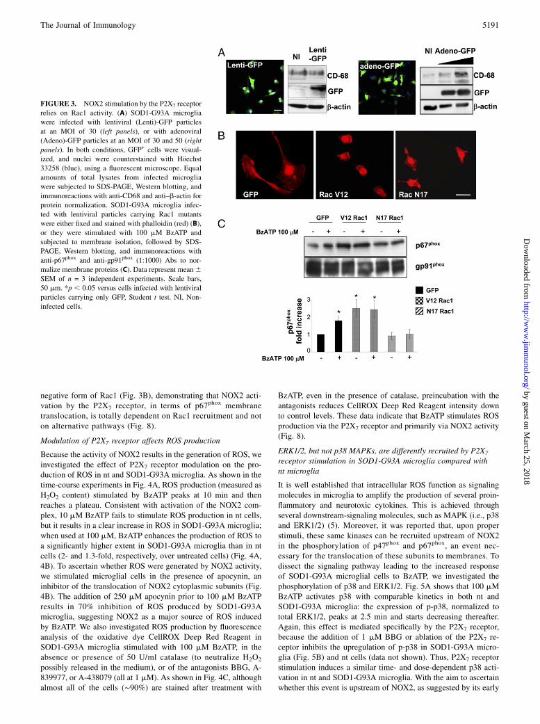
negative form of Rac1 (Fig. 3B), demonstrating that NOX2 acti-vation by the P2X7 receptor, in terms of p67phox membranetranslocation, is totally dependent on Rac1 recruitment and noton alternative pathways (Fig. 8).
Modulation of P2X7 receptor affects ROS production
Because the activity of NOX2 results in the generation of ROS, weinvestigated the effect of P2X7 receptor modulation on the pro-duction of ROS in nt and SOD1-G93A microglia. As shown in thetime-course experiments in Fig. 4A, ROS production (measured asH2O2 content) stimulated by BzATP peaks at 10 min and thenreaches a plateau. Consistent with activation of the NOX2 com-plex, 10 mM BzATP fails to stimulate ROS production in nt cells,but it results in a clear increase in ROS in SOD1-G93A microglia;when used at 100 mM, BzATP enhances the production of ROS toa significantly higher extent in SOD1-G93A microglia than in ntcells (2- and 1.3-fold, respectively, over untreated cells) (Fig. 4A,4B). To ascertain whether ROS were generated by NOX2 activity,we stimulated microglial cells in the presence of apocynin, aninhibitor of the translocation of NOX2 cytoplasmic subunits (Fig.4B). The addition of 250 mM apocynin prior to 100 mM BzATPresults in 70% inhibition of ROS produced by SOD1-G93Amicroglia, suggesting NOX2 as a major source of ROS inducedby BzATP. We also investigated ROS production by fluorescenceanalysis of the oxidative dye CellROX Deep Red Reagent inSOD1-G93A microglia stimulated with 100 mM BzATP, in theabsence or presence of 50 U/ml catalase (to neutralize H2O2
possibly released in the medium), or of the antagonists BBG, A-839977, or A-438079 (all at 1 mM). As shown in Fig. 4C, althoughalmost all of the cells (∼90%) are stained after treatment with
BzATP, even in the presence of catalase, preincubation with theantagonists reduces CellROX Deep Red Reagent intensity downto control levels. These data indicate that BzATP stimulates ROSproduction via the P2X7 receptor and primarily via NOX2 activity(Fig. 8).
ERK1/2, but not p38 MAPKs, are differently recruited by P2X7
receptor stimulation in SOD1-G93A microglia compared withnt microglia
It is well established that intracellular ROS function as signalingmolecules in microglia to amplify the production of several proin-flammatory and neurotoxic cytokines. This is achieved throughseveral downstream-signaling molecules, such as MAPK (i.e., p38and ERK1/2) (5). Moreover, it was reported that, upon properstimuli, these same kinases can be recruited upstream of NOX2in the phosphorylation of p47phox and p67phox, an event nec-essary for the translocation of these subunits to membranes. Todissect the signaling pathway leading to the increased responseof SOD1-G93A microglial cells to BzATP, we investigated thephosphorylation of p38 and ERK1/2. Fig. 5A shows that 100 mMBzATP activates p38 with comparable kinetics in both nt andSOD1-G93A microglia: the expression of p-p38, normalized tototal ERK1/2, peaks at 2.5 min and starts decreasing thereafter.Again, this effect is mediated specifically by the P2X7 receptor,because the addition of 1 mM BBG or ablation of the P2X7 re-ceptor inhibits the upregulation of p-p38 in SOD1-G93A micro-glia (Fig. 5B) and nt cells (data not shown). Thus, P2X7 receptorstimulation induces a similar time- and dose-dependent p38 acti-vation in nt and SOD1-G93A microglia. With the aim to ascertainwhether this event is upstream of NOX2, as suggested by its early
FIGURE 3. NOX2 stimulation by the P2X7 receptor
relies on Rac1 activity. (A) SOD1-G93A microglia
were infected with lentiviral (Lenti)-GFP particles
at an MOI of 30 (left panels), or with adenoviral
(Adeno)-GFP particles at an MOI of 30 and 50 (right
panels). In both conditions, GFP+ cells were visual-
ized, and nuclei were counterstained with Hoechst
33258 (blue), using a fluorescent microscope. Equal
amounts of total lysates from infected microglia
were subjected to SDS-PAGE, Western blotting, and
immunoreactions with anti-CD68 and anti–b-actin for
protein normalization. SOD1-G93A microglia infec-
ted with lentiviral particles carrying Rac1 mutants
were either fixed and stained with phalloidin (red) (B),
or they were stimulated with 100 mM BzATP and
subjected to membrane isolation, followed by SDS-
PAGE, Western blotting, and immunoreactions with
anti-p67phox and anti-gp91phox (1:1000) Abs to nor-
malize membrane proteins (C). Data represent mean6SEM of n = 3 independent experiments. Scale bars,
50 mm. *p , 0.05 versus cells infected with lentiviral
particles carrying only GFP, Student t test. NI, Non-
infected cells.
The Journal of Immunology 5191
by guest on March 25, 2018
http://ww
w.jim
munol.org/
Dow
nloaded from
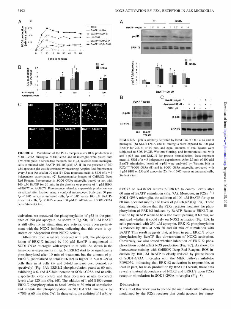
activation, we measured the phosphorylation of p38 in the pres-ence of 250 mM apocynin. As shown in Fig. 5B, 100 mM BzATPis still effective in stimulating p38 activity, even upon pretreat-ment with the NOX2 inhibitor, indicating that this event is up-stream or independent from NOX2 activity.Differently from what we observed with p38, the phosphory-
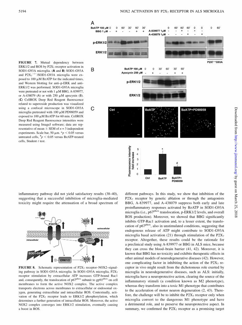
lation of ERK1/2 induced by 100 mM BzATP is augmented inSOD1-G93A microglia with respect to nt cells. As shown in thetime-course experiments in Fig. 6, ERK1/2 start to be significantlyphosphorylated after 10 min of treatment, but the amount of p-ERK1/2 (normalized to total ERK1/2) is higher in SOD1-G93Acells than in nt cells (5- and 3-fold increase over control, re-spectively) (Fig. 6A); ERK1/2 phosphorylation peaks at 60 min,exhibiting a 6- and 4.5-fold increase in SOD1-G93A and nt cells,respectively, over control and then decreases nearly to controllevels after 120 min (Fig. 6B). The addition of 1 mM BBG returnsERK1/2 phosphorylation to basal levels at 30 min of stimulationand inhibits the phosphorylation in SOD1-G93A microglia by∼70% at 60 min (Fig. 7A). In these cells, the addition of 1 mM A-
839977 or A-438079 returns p-ERK1/2 to control levels after60 min of BzATP stimulation (Fig. 7A). Moreover, in P2X7
2/2/SOD1-G93A microglia, the addition of 100 mM BzATP for up to60 min does not modify the levels of p-ERK1/2 (Fig. 7A). Thesedata strongly indicate that the P2X7 receptor mediates the phos-phorylation of ERK1/2 induced by BzATP. Because ERK1/2 ac-tivation by BzATP seems to be a late event, peaking at 60 min, weanalyzed whether it could rely on NOX2 activation (Fig. 7B). Incells pretreated with 250 mM apocynin, ERK1/2 phosphorylationis reduced by 30% at both 30 and 60 min of stimulation withBzATP. This result suggests that, at least in part, ERK1/2 phos-phorylation by BzATP lies downstream of NOX2 activation.Conversely, we also tested whether inhibition of ERK1/2 phos-phorylation could affect ROS production (Fig. 7C). As shown byfluorescence staining with CellROX Deep Red Reagent, ROS in-duction by 100 mM BzATP is clearly reduced by preincubationof SOD1-G93A microglia with the MEK pathway inhibitorPD98059, indicating that ERK1/2 activation is responsible, atleast in part, for ROS production by BzATP. Overall, these datareveal a mutual dependency of NOX2 and ERK1/2 upon P2X7
receptor stimulation in SOD1-G93A microglia (Fig. 8).
DiscussionThe aim of this work was to decode the main molecular pathwaysmodulated by the P2X7 receptor that could account for neuro-
FIGURE 4. Modulation of the P2X7 receptor alters ROS production in
SOD1-G93A microglia. SOD1-G93A and nt microglia were plated onto
a 96-well plate in serum-free medium, and H2O2 released from microglial
cells stimulated with BzATP (10–100 mM) (A, B) in the presence of 250
mM apocynin (B) was determined by measuring Amplex Red fluorescence
every 5 min (A) or after 10 min (B). Data represent mean 6 SEM of n = 3
independent experiments. (C) Representative images of CellROX Deep
Red Reagent fluorescence in SOD1-G93A microglia treated or not with
100 mM BzATP for 30 min, in the absence or presence of 1 mM BBG,
A839977, or A438079. Fluorescence related to superoxide production was
visualized after fixation using a confocal microscope. Scale bar, 50 mm.
*p , 0.05 versus nt untreated cells, xp , 0.05 versus 100 mM BzATP–
treated nt cells, #p , 0.05 versus 100 mM BzATP–treated SOD1-G93A
cells, Student t test.
FIGURE 5. p38 is similarly activated by BzATP in SOD1-G93A and nt
microglia. (A) SOD1-G93A and nt microglia were exposed to 100 mM
BzATP for 2.5, 5, or 10 min, and equal amounts of total lysates were
subjected to SDS-PAGE, Western blotting, and immunoreactions with
anti–p-p38 and anti-ERK1/2 for protein normalization. Data represent
mean 6 SEM of n = 3 independent experiments. After 2.5 min of 100 mM
BzATP stimulation, levels of p-p38 were analyzed by Western blot in
P2X72/2/SOD1-G93A (B) and in SOD1-G93A microglia pretreated with
1 mM BBG or 250 mM apocynin (C). *p , 0.05 versus nt untreated cells,
Student t test.
5192 NOX2 ACTIVATION BY P2X7 RECEPTOR IN ALS MICROGLIA
by guest on March 25, 2018
http://ww
w.jim
munol.org/
Dow
nloaded from

inflammation during ALS pathogenesis and to identify early sig-naling targets and effectors that might be useful in intercepting theprogression of the pathology. To this end, we made use of primarymicroglial cells derived from transgenic SOD1-G93A mice andSOD1-G93A mice lacking the P2X7 receptor as models to in-vestigate the effects of pharmacological induction or genetic ab-lation of the receptor activity. We found that P2X7 receptorstimulation in SOD1-G93A microglia increases NOX2 activity,ROS production, and GTP-Rac1 and p-ERK1/2 levels. Based onour results, we propose a novel mechanism by which the P2X7
receptor may lead to enhanced oxidative stress in ALS microglia.P2X7 receptor stimulated by extracellular ATP directly activatesERK1/2 and NOX2, and both of these pathways converge inROS generation; the induction of NOX2 relies on GTP-Rac1 anddetermines further ERK1/2 phosphorylation, with consequentROS overproduction. This mechanism is consistent with what isalready known about the P2X7 receptor in ALS and with thepathway of NOX2 activation by ALS-related genes. Indeed, ina previous work we demonstrated that the extracellular ATP poolis better preserved and the P2X7 receptor is upregulated in SOD1-G93A microglia, which translates into an increase in released
proinflammatory factors and neurotoxicity induced by microgliaupon P2X7 receptor stimulation (21). In addition, several reportsdescribe NOX2 as a source of damaging ROS in ALS (7, 9, 10,12). Consistent with this, in the present study we found a basalupregulation of the Rac1-NOX2 pathway in the model of SOD1-G93A primary microglia, in line with previous results obtained incell lines transiently infected with ALS-related genes (7, 12), andwe also demonstrated that activation of the P2X7 receptor furtherenhances this pathway, thus producing additional oxidative stress.Furthermore, by overexpression of a dominant-negative or con-stitutively active form of Rac1 in ALS microglia, we establishedthat p67phox subunit translocation on membranes is totally de-pendent on Rac1 recruitment and not on alternative pathways;thus, Rac1 activation is a prerequisite for NOX2 activation by theP2X7 receptor.Because NOX2-derived ROS may activate MAPKs, which are
known to be the core of the cell stress–response signaling networkand, conversely, that oxidative stress also can occur through ac-tivation of MAPKs (28), we investigated the role of these relevantkinases, in particular p38 and ERK1/2, which are known to beactivated by the P2X7 receptor in microglial cells (29) and to beimplicated in ALS pathology (30, 31). Although the role forERK1/2 in ALS is controversial, being overphosphorylated ordephosphorylated in different cells and conditions (32–34), per-sistent activation of p38 signaling was suggested to mediateneuronal apoptosis in ALS. Indeed, increased levels of p-p38MAPKs are present in the motor neurons and microglia of theventral spinal cord of mutant SOD1 mice, and p38 inhibitionlargely protects motor neurons and prevents proximal axon de-generation (35). In this study, we found that, following P2X7 re-ceptor stimulation, the phosphorylation of ERK1/2 is augmentedparticularly in ALS microglia, and this effect is inhibited by theROS inhibitor apocynin. We also demonstrated that inhibition ofERK1/2 phosphorylation decreases ROS production, suggestinga mutual dependency between ERK1/2 and NOX2 upon P2X7
receptor stimulation. In these same experimental conditions, ac-tivation of p38 by BzATP is comparable in SOD1-G93A andhealthy microglia. This may be explained by the fact that severalkinases, in addition to MAPKs, such as protein kinase C, PAK,Akt and PI3K, may be involved in NOX2 activation (28). Thiscomplexity in the phosphorylation of p47phox and p67phox suggeststhat the intracellular signaling pathways responsible for this eventmay be cell type and stimulus specific. Taken together, all of thesedata led us to hypothesize that, in SOD1-G93A mice, ATP re-leased in vivo in the surrounding extracellular space of damagedmotor neurons and slowly hydrolyzed in the proximity of micro-glia overactivates the P2X7 receptor, which is highly expressed inthese cells, leading to oxidative stress via the Rac1-NOX2 andERK1/2 pathways, as well as to the induction of proinflammatoryfactors. This effect adds to ROS production and activation ofproinflammatory signals exerted by SOD1-G93A protein, causingfurther detrimental effects on the surrounding motor neurons.Our findings contribute to improving the understanding of the
biological processes that cause neuroinflammation in ALS andidentify the P2X7 receptor as a promising target to halt the viciouscycle between uncontrolled neuroinflammation and neuron de-generation, because its inactivation affects the earliest steps of theproinflammatory action exerted by ALS microglia. The importantrole of microglia in the progression of ALS has been widelyrecognized (36); indeed, a substantial slowing of disease pro-gression was obtained by excision of mutant SOD1 in the myeloidlineage of SOD1-G93A mice (37). However, treatment with tra-ditional nonsteroidal anti-inflammatory drugs, with minocyclineand apocynin, or the knocking down of specific components of the
FIGURE 6. Phosphorylation of ERK1/2 by BzATP is significantly dif-
ferent in SOD1-G93A microglia compared with nt microglia. SOD1-G93A
and nt microglia were exposed to 100 mM BzATP for 2.5, 5, or 10 min (A)
and for 30, 60, or 120 min (B), and equal amounts of total lysates were
subjected to SDS-PAGE, Western blotting, and immunoreactions with
anti–p-ERK1/2 and anti–ERK1/2 for protein normalization. Data represent
mean 6 SEM of n = 3 independent experiments. *p , 0.05 versus nt
untreated cells, #p , 0.05 versus 100 mM BzATP–treated nt counterpart,
Student t test.
The Journal of Immunology 5193
by guest on March 25, 2018
http://ww
w.jim
munol.org/
Dow
nloaded from
inflammatory pathway did not yield satisfactory results (38–40),suggesting that a successful inhibition of microglia-mediatedtoxicity might require the attenuation of a broad spectrum of
different pathways. In this study, we show that inhibition of theP2X7 receptor by genetic ablation or through the antagonistsBBG, A-839977, and A-438079 suppress both early and lateproinflammatory responses activated by BzATP in SOD1-G93A
microglia (i.e., p67phox translocation, p-ERK1/2 levels, and overallROS production). Moreover, we showed that BBG significantly
inhibits GTP-Rac1 activation and, to a lesser extent, the translo-cation of p67phox, also in unstimulated conditions, suggesting thatendogenous release of ATP might contribute to SOD1-G93A
microglia basal activation (21) through stimulation of the P2X7
receptor. Altogether, these results could be the rationale for
a preclinical study using A-839977 or BBG in ALS mice, becausethey can cross the blood–brain barrier (41, 42). Moreover, it is
known that BBG has no toxicity and exhibits therapeutic effects inother animal models of neurodegenerative diseases (42). However,one complicating factor in inhibiting the action of the P2X7 re-
ceptor in vivo might result from the dichotomous role exerted bymicroglia in neurodegenerative diseases, such as ALS: initially,
microglia have a neuroprotective action, clearing the source of theinflammatory stimuli (a condition known as M2 phenotype),whereas they transform into a toxic M1 phenotype that contributes
to the acceleration of motor neuron degeneration (2, 43). There-fore, the challenge will be to inhibit the P2X7 receptor only when
microglia convert to the dangerous M1 phenotype and havea detrimental role, and to preserve the neuroprotective aspect. Insummary, we confirmed the P2X7 receptor as a promising target
FIGURE 7. Mutual dependency between
ERK1/2 and ROS by P2X7 receptor activation in
SOD1-G93A microglia. (A and B) SOD1-G93A
and P2X72/2/SOD1-G93A microglia were ex-
posed to 100 mM BzATP for the indicated times,
and Western blotting for anti–p-ERK and anti-
ERK1/2 was performed. SOD1-G93A microglia
were pretreated or not with 1 mMBBG, A-839977,
or A-438079 (A) or with 250 mM apocynin (B).
(C) CellROX Deep Red Reagent fluorescence
related to superoxide production was visualized
using a confocal microscope in SOD1-G93A
microglia pretreated with 100 mM PD98059 and
exposed to 100 mM BzATP for 60 min. CellROX
Deep Red Reagent fluorescence intensities were
measured using ImageJ software; data are rep-
resentative of mean6 SEM of n = 3 independent
experiments. Scale bar, 50 mm. *p , 0.05 versus
untreated cells, #p , 0.05 versus BzATP-treated
cells, Student t test.
FIGURE 8. Schematic representation of P2X7 receptor–NOX2–signal-
ing pathway in SOD1-G93A microglia. In SOD1-G93A microglia, P2X7
receptor stimulation by extracellular ATP increases GTP-bound Rac1
and, consequently, the translocation of p67phox subunit to gp91phox on cell
membranes to form the active NOX2 complex. The active complex
transports electrons across membranes to extracellular or endosomal ox-
ygen, generating extracellular and intracellular ROS. Contextually, acti-
vation of the P2X7 receptor leads to ERK1/2 phosphorylation, which
determines a further generation of intracellular ROS. Moreover, the active
NOX2 complex converges into ERK1/2 stimulation, eventually causing
a boost in ROS.
5194 NOX2 ACTIVATION BY P2X7 RECEPTOR IN ALS MICROGLIA
by guest on March 25, 2018
http://ww
w.jim
munol.org/
Dow
nloaded from
for the development of therapeutic strategies to slow down theprogression of ALS.
DisclosuresThe authors have no financial conflicts of interest.
References1. Cozzolino, M., M. G. Pesaresi, V. Gerbino, J. Grosskreutz, and M. T. Carrı. 2012.
Amyotrophic lateral sclerosis: new insights into underlying molecular mecha-nisms and opportunities for therapeutic intervention. Antioxid. Redox Signal. 17:1277–1330.
2. Henkel, J. S., D. R. Beers, W. Zhao, and S. H. Appel. 2009. Microglia in ALS:the good, the bad, and the resting. J. Neuroimmune Pharmacol. 4: 389–398.
3. Ilieva, H., M. Polymenidou, and D. W. Cleveland. 2009. Non-cell autonomoustoxicity in neurodegenerative disorders: ALS and beyond. J. Cell Biol. 187: 761–772.
4. Bedard, K., and K. H. Krause. 2007. The NOX family of ROS-generatingNADPH oxidases: physiology and pathophysiology. Physiol. Rev. 87: 245–313.
5. Block, M. L. 2008. NADPH oxidase as a therapeutic target in Alzheimer’sdisease. BMC Neurosci. 9(Suppl. 2): S8.
6. Hordijk, P. L. 2006. Regulation of NADPH oxidases: the role of Rac proteins.Circ. Res. 98: 453–462.
7. Harraz, M. M., J. J. Marden, W. Zhou, Y. Zhang, A. Williams, V. S. Sharov,K. Nelson, M. Luo, H. Paulson, C. Schoneich, and J. F. Engelhardt. 2008. SOD1mutations disrupt redox-sensitive Rac regulation of NADPH oxidase in a familialALS model. J. Clin. Invest. 118: 659–670.
8. Boillee, S., and D. W. Cleveland. 2008. Revisiting oxidative damage in ALS:microglia, Nox, and mutant SOD1. J. Clin. Invest. 118: 474–478.
9. Wu, D. C., D. B. Re, M. Nagai, H. Ischiropoulos, and S. Przedborski. 2006. Theinflammatory NADPH oxidase enzyme modulates motor neuron degeneration inamyotrophic lateral sclerosis mice. Proc. Natl. Acad. Sci. USA 103: 12132–12137.
10. Marden, J. J., M. M. Harraz, A. J. Williams, K. Nelson, M. Luo, H. Paulson, andJ. F. Engelhardt. 2007. Redox modifier genes in amyotrophic lateral sclerosis inmice. J. Clin. Invest. 117: 2913–2919.
11. Valdmanis, P. N., E. Kabashi, P. A. Dion, and G. A. Rouleau. 2008. ALS pre-disposition modifiers: knock NOX, who’s there? SOD1 mice still are. Eur. J.Hum. Genet. 16: 140–142.
12. Li, Q., N. Y. Spencer, N. J. Pantazis, and J. F. Engelhardt. 2011. Alsin and SOD1(G93A) proteins regulate endosomal reactive oxygen species production by glialcells and proinflammatory pathways responsible for neurotoxicity. J. Biol. Chem.286: 40151–40162.
13. Di Virgilio, F., S. Ceruti, P. Bramanti, and M. P. Abbracchio. 2009. Purinergicsignalling in inflammation of the central nervous system. Trends Neurosci. 32:79–87.
14. Skaper, S. D., P. Debetto, and P. Giusti. 2010. The P2X7 purinergic receptor:from physiology to neurological disorders. FASEB J. 24: 337–345.
15. Weisman, G. A., J. M. Camden, T. S. Peterson, D. Ajit, L. T. Woods, and L. Erb.2012. P2 receptors for extracellular nucleotides in the central nervous system:role of P2X7 and P2Y2 receptor interactions in neuroinflammation. Mol. Neu-robiol. 46: 96–113.
16. Volonte, C., S. Apolloni, S. D. Skaper, and G. Burnstock. 2012. P2X7 receptors:channels, pores and more. CNS Neurol. Disord. Drug Targets 11: 705–721.
17. Volonte, C., S. Apolloni, M. T. Carrı, and N. D’Ambrosi. 2011. ALS: focus onpurinergic signalling. Pharmacol. Ther. 132: 111–122.
18. Amadio, S., S. Apolloni, N. D’Ambrosi, and C. Volonte. 2011. Purinergic sig-nalling at the plasma membrane: a multipurpose and multidirectional mode todeal with amyotrophic lateral sclerosis and multiple sclerosis. J. Neurochem.116: 796–805.
19. Yiangou, Y., P. Facer, P. Durrenberger, I. P. Chessell, A. Naylor, C. Bountra,R. R. Banati, and P. Anand. 2006. COX-2, CB2 and P2X7-immunoreactivitiesare increased in activated microglial cells/macrophages of multiple sclerosis andamyotrophic lateral sclerosis spinal cord. BMC Neurol. 6: 12.
20. Casanovas, A., S. Hernandez, O. Tarabal, J. Rossello, and J. E. Esquerda. 2008.Strong P2X4 purinergic receptor-like immunoreactivity is selectively associatedwith degenerating neurons in transgenic rodent models of amyotrophic lateralsclerosis. J. Comp. Neurol. 506: 75–92.
21. D’Ambrosi, N., P. Finocchi, S. Apolloni, M. Cozzolino, A. Ferri, V. Padovano,G. Pietrini, M. T. Carrı, and C. Volonte. 2009. The proinflammatory action of
microglial P2 receptors is enhanced in SOD1 models for amyotrophic lateralsclerosis. J. Immunol. 183: 4648–4656.
22. Gandelman, M., H. Peluffo, J. S. Beckman, P. Cassina, and L. Barbeito. 2010.Extracellular ATP and the P2X7 receptor in astrocyte-mediated motor neurondeath: implications for amyotrophic lateral sclerosis. J. Neuroinflammation 7: 33.
23. Saura, J., J. M. Tusell, and J. Serratosa. 2003. High-yield isolation of murinemicroglia by mild trypsinization. Glia 44: 183–189.
24. Cozzolino, M., V. Stagni, L. Spinardi, N. Campioni, C. Fiorentini, E. Salvati,S. Alema, and A. M. Salvatore. 2003. p120 Catenin is required for growth factor-dependent cell motility and scattering in epithelial cells. Mol. Biol. Cell 14:1964–1977.
25. Kalyanaraman, B., V. Darley-Usmar, K. J. Davies, P. A. Dennery, H. J. Forman,M. B. Grisham, G. E. Mann, K. Moore, L. J. Roberts, II, and H. Ischiropoulos.2012. Measuring reactive oxygen and nitrogen species with fluorescent probes:challenges and limitations. Free Radic. Biol. Med. 52: 1–6.
26. Leto, T. L., S. Morand, D. Hurt, and T. Ueyama. 2009. Targeting and regulationof reactive oxygen species generation by Nox family NADPH oxidases. Anti-oxid. Redox Signal. 11: 2607–2619.
27. Allen, W. E., G. E. Jones, J. W. Pollard, and A. J. Ridley. 1997. Rho, Rac andCdc42 regulate actin organization and cell adhesion in macrophages. J. Cell Sci.110: 707–720.
28. Wilkinson, B. L., and G. E. Landreth. 2006. The microglial NADPH oxidasecomplex as a source of oxidative stress in Alzheimer’s disease. J. Neuro-inflammation 3: 30.
29. Friedle, S. A., V. M. Brautigam, M. Nikodemova, M. L. Wright, andJ. J. Watters. 2011. The P2X7-Egr pathway regulates nucleotide-dependent in-flammatory gene expression in microglia. Glia 59: 1–13.
30. Kim, E. K., and E. J. Choi. 2010. Pathological roles of MAPK signaling path-ways in human diseases. Biochim. Biophys. Acta 1802: 396–405.
31. Chung, Y. H., K. M. Joo, H. C. Lim, M. H. Cho, D. Kim, W. B. Lee, andC. I. Cha. 2005. Immunohistochemical study on the distribution of phosphory-lated extracellular signal-regulated kinase (ERK) in the central nervous systemof SOD1G93A transgenic mice. Brain Res. 1050: 203–209.
32. Ayala, V., A. B. Granado-Serrano, D. Cacabelos, A. Naudı, E. V. Ilieva, J. Boada,V. Caraballo-Miralles, J. Llado, I. Ferrer, R. Pamplona, and M. Portero-Otin.2011. Cell stress induces TDP-43 pathological changes associated with ERK1/2dysfunction: implications in ALS. Acta Neuropathol. 122: 259–270.
33. Perlson, E., G. B. Jeong, J. L. Ross, R. Dixit, K. E. Wallace, R. G. Kalb, andE. L. Holzbaur. 2009. A switch in retrograde signaling from survival to stress inrapid-onset neurodegeneration. J. Neurosci. 29: 9903–9917.
34. Yang, E. J., J. H. Jiang, S. M. Lee, S. C. Yang, H. S. Hwang, M. S. Lee, andS. M. Choi. 2010. Bee venom attenuates neuroinflammatory events and extendssurvival in amyotrophic lateral sclerosis models. J. Neuroinflammation 7: 69.
35. Dewil, M., V. F. dela Cruz, L. Van Den Bosch, and W. Robberecht. 2007. In-hibition of p38 mitogen activated protein kinase activation and mutant SOD1(G93A)-induced motor neuron death. Neurobiol. Dis. 26: 332–341.
36. Philips, T., and W. Robberecht. 2011. Neuroinflammation in amyotrophic lateralsclerosis: role of glial activation in motor neuron disease. Lancet Neurol. 10:253–263.
37. Boillee, S., K. Yamanaka, C. S. Lobsiger, N. G. Copeland, N. A. Jenkins,G. Kassiotis, G. Kollias, and D. W. Cleveland. 2006. Onset and progression ininherited ALS determined by motor neurons and microglia. Science 312: 1389–1392.
38. Turner, B. J., and K. Talbot. 2008. Transgenics, toxicity and therapeutics inrodent models of mutant SOD1-mediated familial ALS. Prog. Neurobiol. 85: 94–134.
39. Riboldi, G., M. Nizzardo, C. Simone, M. Falcone, N. Bresolin, G. P. Comi, andS. Corti. 2011. ALS genetic modifiers that increase survival of SOD1 mice andare suitable for therapeutic development. Prog. Neurobiol. 95: 133–148.
40. Phani, S., D. B. Re, and S. Przedborski. 2012. The Role of the Innate ImmuneSystem in ALS. Front. Pharmacol. 3: 150.
41. Honore, P., D. Donnelly-Roberts, M. Namovic, C. Zhong, C. Wade, P. Chandran,C. Zhu, W. Carroll, A. Perez-Medrano, Y. Iwakura, and M. F. Jarvis. 2009. Theantihyperalgesic activity of a selective P2X7 receptor antagonist, A-839977, islost in IL-1alphabeta knockout mice. Behav. Brain Res. 204: 77–81.
42. Takenouchi, T., K. Sekiyama, A. Sekigawa, M. Fujita, M. Waragai, S. Sugama,Y. Iwamaru, H. Kitani, and M. Hashimoto. 2010. P2X7 receptor signalingpathway as a therapeutic target for neurodegenerative diseases. Arch. Immunol.Ther. Exp. (Warsz.) 58: 91–96.
43. Liao, B., W. Zhao, D. R. Beers, J. S. Henkel, and S. H. Appel. 2012. Trans-formation from a neuroprotective to a neurotoxic microglial phenotype ina mouse model of ALS. Exp. Neurol. 237: 147–152.
The Journal of Immunology 5195
by guest on March 25, 2018
http://ww
w.jim
munol.org/
Dow
nloaded from


