The The Nervous Nervous SystemSystem

EQEQ
How does the nervous system work How does the nervous system work to control and maintain bodily to control and maintain bodily functions and activities?functions and activities?

GPSGPSSAP3. Students will assess the integration and SAP3. Students will assess the integration and coordination of body functions and their dependence coordination of body functions and their dependence on the endocrine and nervous systems to regulate on the endocrine and nervous systems to regulate physiological activities.physiological activities.a. Interpret interactions among hormones, senses, and a. Interpret interactions among hormones, senses, and nerves which make possible the coordination of functions of nerves which make possible the coordination of functions of the body.the body.b. Investigate the physiology of electrochemical impulses b. Investigate the physiology of electrochemical impulses and neural integration and trace the pathway of an impulse, and neural integration and trace the pathway of an impulse, relating biochemical changes involved in the conduction of relating biochemical changes involved in the conduction of the impulse.the impulse.c. Describe how the body perceives internal and external c. Describe how the body perceives internal and external stimuli and responds to maintain a stable internal stimuli and responds to maintain a stable internal environment, as it relates to biofeedback.environment, as it relates to biofeedback.

Functions of the Nervous Functions of the Nervous SystemSystem
1.Sensory input—gathering 1.Sensory input—gathering informationinformation– To monitor changes occurring inside & To monitor changes occurring inside &
outside the bodyoutside the body– Changes = stimuliChanges = stimuli
2.Integration2.Integration– To process and interpret sensory input & To process and interpret sensory input &
decide if action is neededdecide if action is needed

Functions of the Nervous Functions of the Nervous SystemSystem
3.Motor output3.Motor output– A response to integrated stimuliA response to integrated stimuli– The response activates muscles or The response activates muscles or
glandsglands

Functions of the Nervous Functions of the Nervous SystemSystem
Figure 7.1
Structural Classification Structural Classification of the Nervous Systemof the Nervous System
Central nervous system (CNS)Central nervous system (CNS)– BrainBrain– Spinal cordSpinal cord
Peripheral nervous system (PNS)Peripheral nervous system (PNS)– Nerves outside the brain and spinal cordNerves outside the brain and spinal cord
Spinal nervesSpinal nerves
Cranial nervesCranial nerves
Functional Classification of Functional Classification of the Peripheral Nervous Systemthe Peripheral Nervous SystemSensory (afferent) divisionSensory (afferent) division– Nerve fibers that carry information to the Nerve fibers that carry information to the
central nervous systemcentral nervous system– InputInput
Cutaneous (skin) sense organsCutaneous (skin) sense organsMechanoreceptors- detect somatic/ body stimulation Mechanoreceptors- detect somatic/ body stimulation
Proprioceptors—detect stretch or tensionProprioceptors—detect stretch or tensionNociceptorsNociceptors --detect pain & temp. --detect pain & temp.
changes.changes.
Motor (efferent) divisionMotor (efferent) division– Nerve fibers that carry impulses away from the Nerve fibers that carry impulses away from the
central nervous systemcentral nervous system– outputoutput

Organization of the Nervous Organization of the Nervous SystemSystem
Figure 7.2

Functional Classification ofFunctional Classification ofthe Peripheral Nervous Systemthe Peripheral Nervous System
Motor (efferent) division (continued)Motor (efferent) division (continued)– Two subdivisionsTwo subdivisions
Somatic nervous system = voluntarySomatic nervous system = voluntary
Autonomic nervous system = involuntaryAutonomic nervous system = involuntary

Support CellsSupport Cells
Support cells in the CNS are grouped Support cells in the CNS are grouped together as “neuroglia”together as “neuroglia”
Function: to support, insulate, and Function: to support, insulate, and protect neuronsprotect neurons
(9x more numerous than neurons)(9x more numerous than neurons)

Support CellsSupport CellsAstrocytesAstrocytes– Abundant, star-Abundant, star-
shaped cellsshaped cells– Brace neuronsBrace neurons– Form barrier Form barrier
between capillaries between capillaries and neuronsand neurons
– Control the Control the chemical chemical environment of environment of the brainthe brain

Support CellsSupport CellsMicrogliaMicroglia– Spiderlike Spiderlike
phagocytesphagocytesPhagocytes (white-Phagocytes (white-blood cells that fight blood cells that fight infections)infections)
– Dispose of debrisDispose of debris

Support CellsSupport CellsEpendymal cellsEpendymal cells– Line cavities of the brain and spinal cordLine cavities of the brain and spinal cord– Circulate cerebrospinal fluid (CSF)Circulate cerebrospinal fluid (CSF)
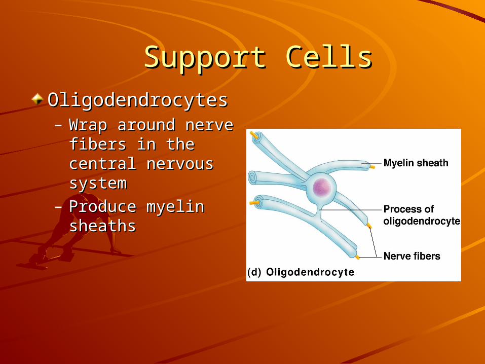
Support CellsSupport CellsOligodendrocytesOligodendrocytes– Wrap around nerve Wrap around nerve
fibers in the central fibers in the central nervous systemnervous system
– Produce myelin Produce myelin sheaths sheaths

Support CellsSupport CellsSatellite cellsSatellite cells– Protect neuron cell bodiesProtect neuron cell bodies
Schwann cellsSchwann cells– Form myelin sheath in jelly Form myelin sheath in jelly
roll–like fashionroll–like fashion– in the peripheral nervous in the peripheral nervous
systemsystem
Myelin sheathMyelin sheath——whitish, fatty material whitish, fatty material covering axonscovering axons
Nodes of RanvierNodes of Ranvier — —gaps in myelin sheath gaps in myelin sheath along the axon; allows along the axon; allows for quick transmission for quick transmission of electrical impulses.of electrical impulses.

NeuronsNeuronsNeurons = nerve cellsNeurons = nerve cells– large, complex cells made of cell body & large, complex cells made of cell body &
extensions or processes (Axons/ dendrites)extensions or processes (Axons/ dendrites)– Cells specialized to transmit messagesCells specialized to transmit messages– Mature neurons don’t replace themselves. Mature neurons don’t replace themselves. – Major regions of neuronsMajor regions of neurons
Cell body—nucleus & metabolic center of the cellCell body—nucleus & metabolic center of the cell
Processes—fibers that extend from the cell body Processes—fibers that extend from the cell body
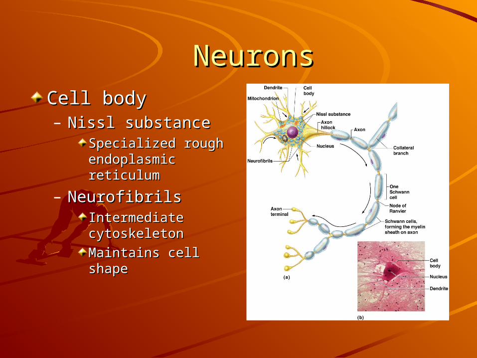
NeuronsNeuronsCell bodyCell body– Nissl substanceNissl substance
Specialized rough Specialized rough endoplasmic endoplasmic reticulumreticulum
– Neurofibrils Neurofibrils Intermediate Intermediate cytoskeleton cytoskeleton
Maintains cell shapeMaintains cell shape

NeuronsNeuronsCell bodyCell body– NucleusNucleus– Large nucleolusLarge nucleolus
Processes outside Processes outside the cell bodythe cell body– Dendrites—conduct Dendrites—conduct
impulses toward impulses toward the cell bodythe cell body
– Axons—conduct Axons—conduct impulses away from impulses away from the cell bodythe cell body
NeuronsNeurons
Axons end in axonal terminalsAxons end in axonal terminals
Axonal terminals contain vesicles w/ Axonal terminals contain vesicles w/ neurotransmitters (NT)neurotransmitters (NT)
Axonal terminals are separated from Axonal terminals are separated from the next neuron by a gapthe next neuron by a gap– Synaptic cleft—gap between adjacent Synaptic cleft—gap between adjacent
neuronsneurons– Synapse—junction btwn nervesSynapse—junction btwn nerves
Neuron Cell Body LocationNeuron Cell Body Location
Most neuron cell bodies are found in Most neuron cell bodies are found in the CNSthe CNS– Gray matter—cell bodies & Gray matter—cell bodies &
unmyelinated fibersunmyelinated fibers– Nuclei—clusters of cell bodies w/in the Nuclei—clusters of cell bodies w/in the
white matter of the CNSwhite matter of the CNS
Ganglia—collections of cell bodies Ganglia—collections of cell bodies outside the CNSoutside the CNS

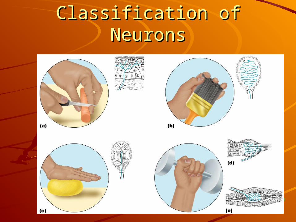
Functional Classification of Functional Classification of NeuronsNeurons
1. Sensory (afferent) neurons:1. Sensory (afferent) neurons:– Carry impulses from the sensory receptors to Carry impulses from the sensory receptors to
the CNSthe CNSCutaneous (skin) sense organsCutaneous (skin) sense organs
Mechanoreceptors- detect somatic/ body stimulation Mechanoreceptors- detect somatic/ body stimulation Proprioceptors—detect stretch or Proprioceptors—detect stretch or
tensiontensionNociceptorsNociceptors --detect pain & temp. --detect pain & temp.
changes.changes.
2. Motor (efferent) neurons:2. Motor (efferent) neurons:– Carry impulses from the CNS to viscera, Carry impulses from the CNS to viscera,
muscles, or glandsmuscles, or glands
Functional Classification of Functional Classification of NeuronsNeurons
Figure 7.7

Functional Classification of Functional Classification of NeuronsNeurons
3. Interneurons (association neurons)3. Interneurons (association neurons)– Found in neural pathways in the CNSFound in neural pathways in the CNS– Connect sensory and motor neuronsConnect sensory and motor neurons

Neuron ClassificationNeuron Classification
Figure 7.6

Figure 7.8a
Structural Classification of Structural Classification of NeuronsNeurons
1. Multipolar neurons—many 1. Multipolar neurons—many extensions from the cell bodyextensions from the cell body

Structural Classification of Structural Classification of NeuronsNeurons
2. Bipolar neurons—one axon & one 2. Bipolar neurons—one axon & one dendritedendrite
Figure 7.8b

Structural Classification of Structural Classification of NeuronsNeurons
3. Unipolar neurons—have a short 3. Unipolar neurons—have a short single process leaving the cell bodysingle process leaving the cell body
Figure 7.8c
Functional Properties of Functional Properties of NeuronsNeurons
1. Irritability1. Irritability– Ability to respond to stimuliAbility to respond to stimuli
2. Conductivity2. Conductivity– Ability to transmit an impulseAbility to transmit an impulse

Nerve ImpulsesNerve ImpulsesResting neuronResting neuron– The plasma membrane at rest is polarizedThe plasma membrane at rest is polarized– Fewer positive ions are inside the cell than Fewer positive ions are inside the cell than
outside the celloutside the cell
Depolarization Depolarization – A stimulus depolarizes the neuron’s membraneA stimulus depolarizes the neuron’s membrane– A depolarized membrane allows sodium (Na+) A depolarized membrane allows sodium (Na+)
to flow inside the membraneto flow inside the membrane
The exchange of ions initiates an The exchange of ions initiates an action potential (AP) in the neuronaction potential (AP) in the neuron

Nerve ImpulsesNerve Impulses
Figure 7.9a–b

Nerve ImpulsesNerve ImpulsesAction potentialAction potential– If the action potential (nerve impulse) starts, it If the action potential (nerve impulse) starts, it
is propagated (travels) over the entire axonis propagated (travels) over the entire axon– Impulses travel faster when fibers have a Impulses travel faster when fibers have a
myelin sheath (for insulation)myelin sheath (for insulation)

Nerve ImpulsesNerve ImpulsesRepolarizationRepolarization– Potassium ions rush out of the neuron after sodium ions Potassium ions rush out of the neuron after sodium ions
rush in, which repolarizes the membranerush in, which repolarizes the membrane– The sodium-potassium pump, using ATP, restores the The sodium-potassium pump, using ATP, restores the
original configurationoriginal configuration

Transmission of a Signal at SynapsesTransmission of a Signal at Synapses
Impulses are able to Impulses are able to cross the synapse to cross the synapse to another nerveanother nerve– NT (chemicals) is NT (chemicals) is
released from a nerve’s released from a nerve’s axon terminalaxon terminal
– The dendrite of the next The dendrite of the next neuron has receptors neuron has receptors that are stimulated by that are stimulated by the NTthe NT
– An AP is started in the An AP is started in the dendritedendrite
Axonterminal
Vesicles
Synapticcleft
Actionpotentialarrives
Synapse
Axon oftransmittingneuron
Receivingneuron
Neurotrans-mitter is re-leased intosynaptic cleft
Neurotrans-mitter bindsto receptoron receivingneuron’smembrane
Vesiclefuses withplasmamembrane
Synaptic cleftNeurotransmittermolecules
Ion channels Receiving neuron
Transmitting neuron
Receptor
Neurotransmitter
Na+
Na+
Neurotransmitterbroken downand released
Ion channel opens Ion channel closes


