Download - The permanent teeth
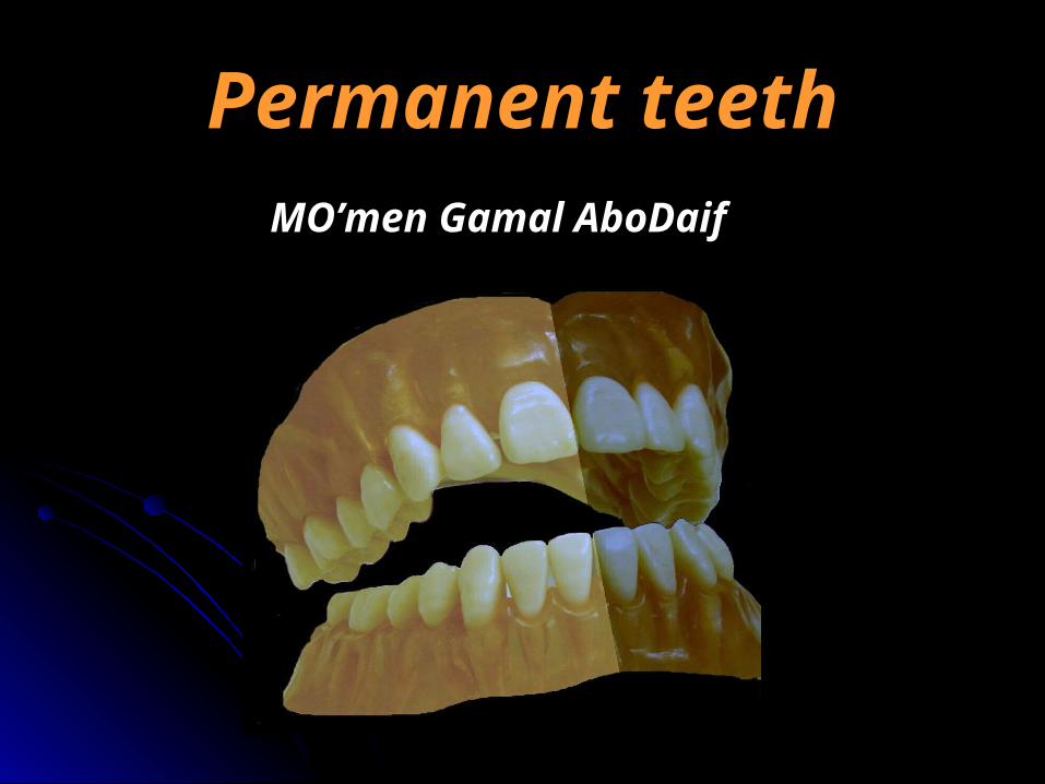
Permanent teethPermanent teethMO’men Gamal AboDaifMO’men Gamal AboDaif

The ideal way to describe a toothThe ideal way to describe a tooth
It is more easier to follow these stepsIt is more easier to follow these steps::- - ChronologyChronology (i.e. dates of events for each tooth as enamel (i.e. dates of events for each tooth as enamel
organ appearance, beginning of calcification, crown completed, organ appearance, beginning of calcification, crown completed, eruption and root completed).eruption and root completed).
- TypeType ( anterior or posterior)( anterior or posterior) and functionand function ( incising, holding ( incising, holding or grinding etc ….).or grinding etc ….).
- No. of lobes.No. of lobes.- RelationsRelations ( contact with what tooth mesially and distally). ( contact with what tooth mesially and distally).- No. of surfaces.No. of surfaces.- No. of rootNo. of root(s).(s).
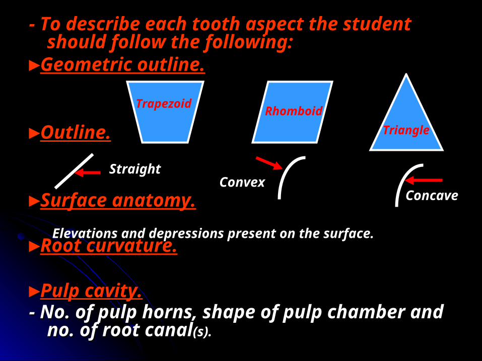
- To describe each tooth aspect the student - To describe each tooth aspect the student should follow the following:should follow the following:
►►Geometric outline.Geometric outline.
►►Outline.Outline.
►►Surface anatomy.Surface anatomy.
►►Root curvature.Root curvature.
►►Pulp cavity.Pulp cavity.- No. of pulp horns, shape of pulp chamber and - No. of pulp horns, shape of pulp chamber and
no. of root canalno. of root canal(s).(s).
Trapezoid RhomboidTriangle
StraightConvex
Concave
Elevations and depressions present on the surface.
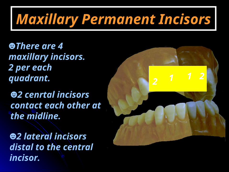
Maxillary Permanent IncisorsMaxillary Permanent Incisors
☻There are 4 maxillary incisors. 2 per each quadrant.
☻2 cenrtal incisors contact each other at the midline.
☻2 lateral incisors distal to the central incisor.
12 1 2

Maxillary Maxillary permanent permanent
central incisorcentral incisor
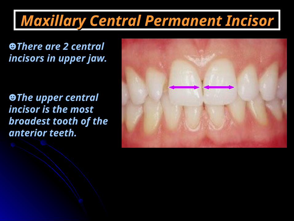
Maxillary Central Permanent IncisorMaxillary Central Permanent Incisor☻There are 2 central incisors in upper jaw.
☻The upper central incisor is the most broadest tooth of the anterior teeth.
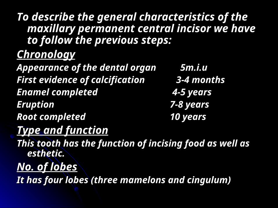
To describe the general characteristics of the To describe the general characteristics of the maxillary permanent central incisor we have maxillary permanent central incisor we have to follow the previous steps:to follow the previous steps:
ChronologyChronologyAppearance of the dental organ 5m.i.uAppearance of the dental organ 5m.i.uFirst evidence of calcification 3-4 monthsFirst evidence of calcification 3-4 monthsEnamel completed 4-5 yearsEnamel completed 4-5 yearsEruption 7-8 yearsEruption 7-8 yearsRoot completed 10 yearsRoot completed 10 yearsType and functionType and functionThis tooth has the function of incising food as well as This tooth has the function of incising food as well as
esthetic.esthetic.No. of lobesNo. of lobesIt has four lobes (three mamelons and cingulum) It has four lobes (three mamelons and cingulum)
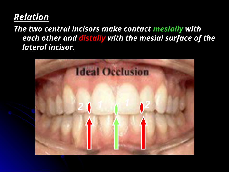
RelationRelationThe two central incisors make contact The two central incisors make contact mesiallymesially with with
each other and each other and distallydistally with the mesial surface of the with the mesial surface of the lateral incisor.lateral incisor.
1 1 22
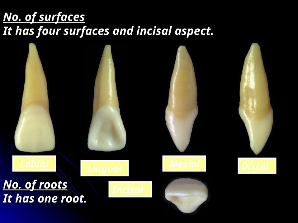
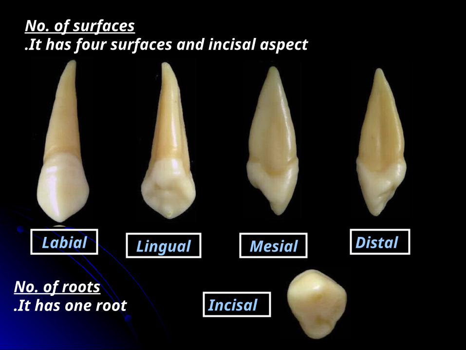
No. of surfacesNo. of surfacesIt has four surfaces and incisal aspect.It has four surfaces and incisal aspect.
No. of rootsNo. of rootsIt has one root.It has one root.
Labial Lingual Mesial Distal
Incisal

Surface AnatomySurface Anatomy Labial aspect
☻Geometrical outline of the crown: Trapezoid
-The short side cervically-The long side incisally
☻The outline: -Mesial outline is slightly convex.
-Distal outline is more convex. -Incisal outline is straight and
perpendicular to the long axis of the tooth.
-The cervical line is convex root-wards.
D M
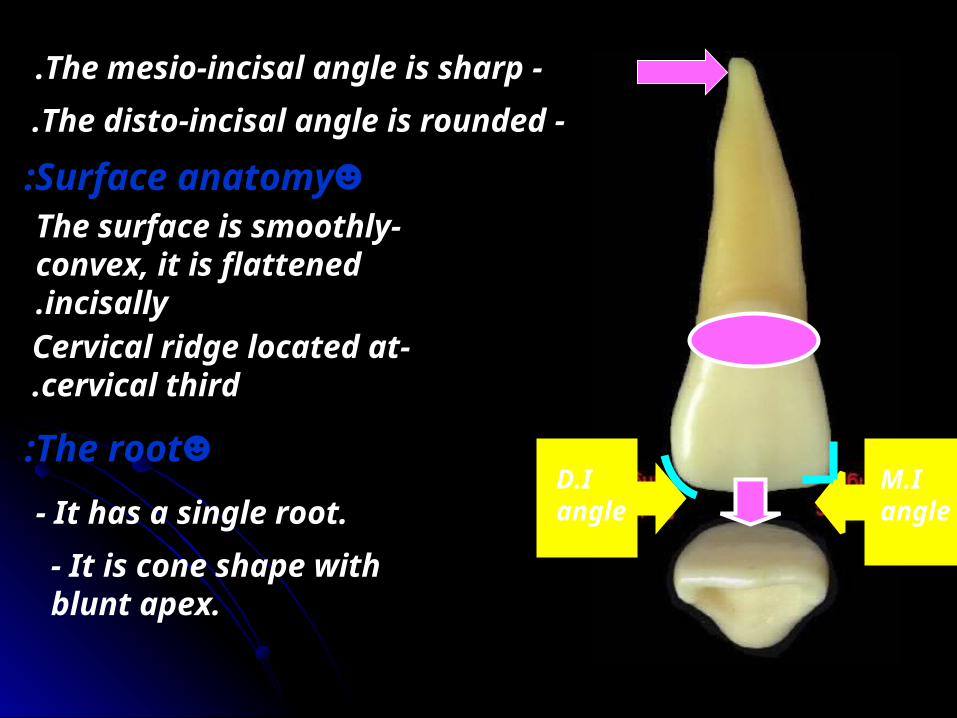
-The mesio-incisal angle is sharp. -The disto-incisal angle is rounded.
M.I angle
D.I angle
☻Surface anatomy:-The surface is smoothly
convex, it is flattened incisally.
-Cervical ridge located at cervical third.
☻The root:- It has a single root.- It is cone shape with blunt apex.

Lingual aspect -It has the same geometrical outline
and outline as the labial surface.
M D
- The mesial and distal sides of the crown and root converge ligually ( the lingual surface is narrower than the labial surface).
- This convergence to accommodate with the horse shoe shaped of the alveolar process.
The facial surface is larger than the lingual surface.

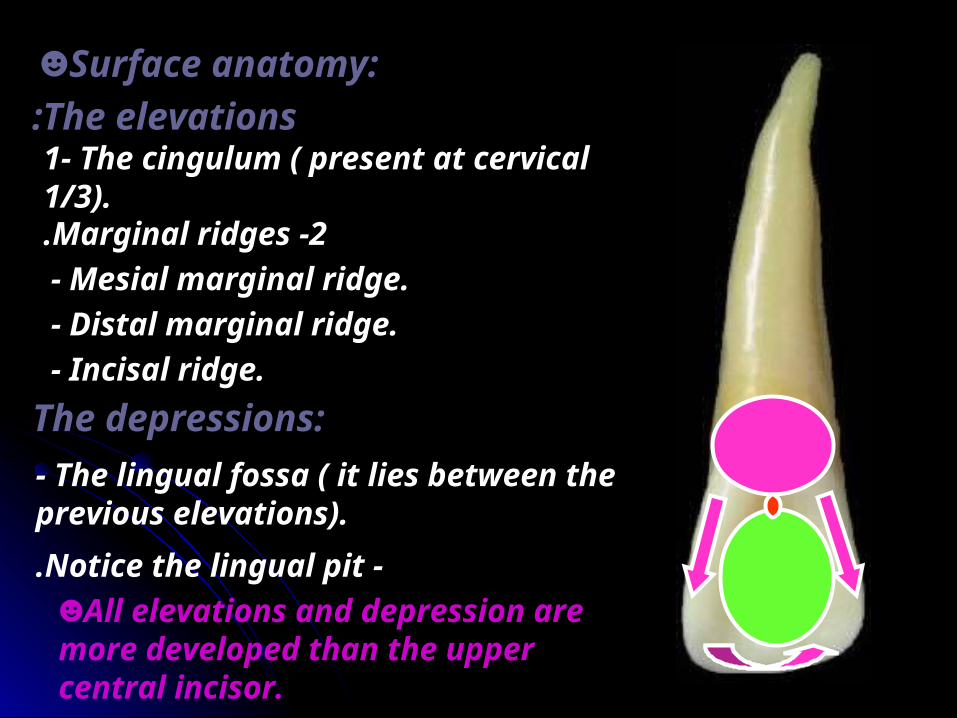
☻Surface anatomy:
The elevations:1 -The cingulum ( present
at cervical 1/3.2 -Marginal ridges.
- Mesial marginal ridge.- Distal marginal ridge.- Incisal ridge.
The depressions:- The lingual fossa ( it lies between the previous elevations).
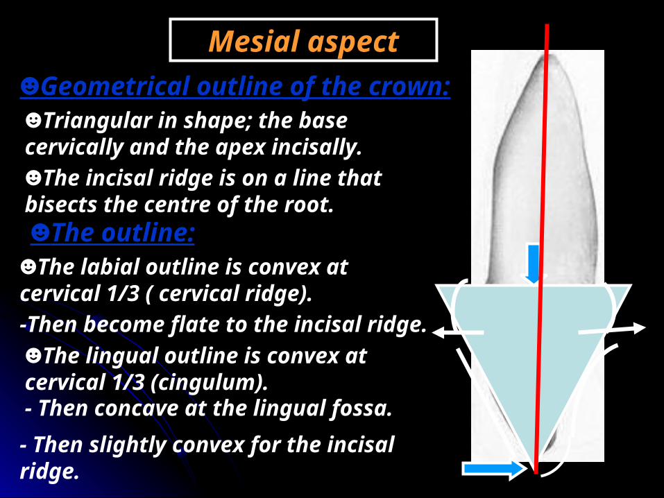
Mesial aspect☻Geometrical outline of the crown:☻Triangular in shape; the base cervically and the apex incisally.☻The incisal ridge is on a line that bisects the centre of the root.☻The outline:
☻The labial outline is convex at cervical 1/3 ( cervical ridge).-Then become flate to the incisal ridge.☻The lingual outline is convex at cervical 1/3 (cingulum).- Then concave at the lingual fossa.- Then slightly convex for the incisal ridge.
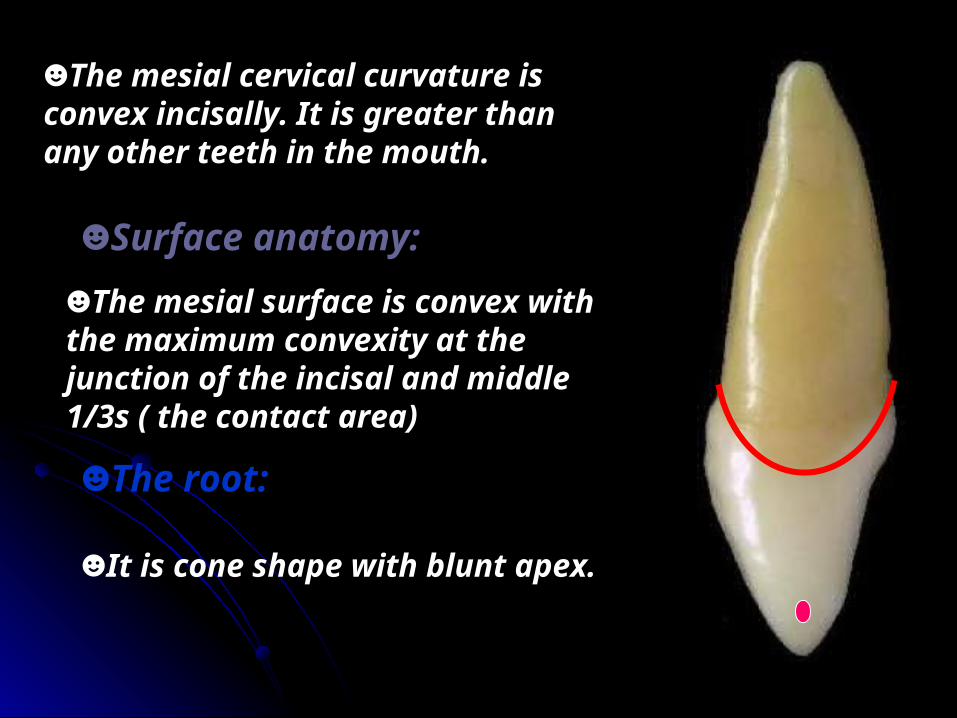
☻The mesial cervical curvature is convex incisally. It is greater than any other teeth in the mouth.
☻Surface anatomy:☻The mesial surface is convex with the maximum convexity at the junction of the incisal and middle 1/3s ( the contact area)
☻The root:
☻It is cone shape with blunt apex.

The distal aspect
☻Similar to the mesial aspect but differ in.
- The cervical line curvature is less than mesial ( by 1 mm).
-The contact area located at the middle 1/3.

Incisal aspect☻Triangular in shape.
-The base is placed labially and the apex lingually.
☻The labial surface is broad and flat. The cervical portion of the crown is convex ( cervical ridge).
☻The lingual outline tapers lingually to the cingulum (ligual
convergence) .
-The cingulum is shifted distally.
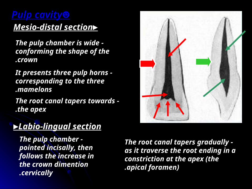
►Mesio-distal section
-The pulp chamber is wide conforming the shape of the crown.
-It presents three pulp horns corresponding to the three mamelons.
-The root canal tapers towards the apex.
►Labio-lingual section -The pulp chamber
pointed incisally, then follows the increase in the crown dimention cervically.
-The root canal tapers gradually as it traverse the root ending in a constriction at the apex (the apical foramen).
☻Pulp cavity
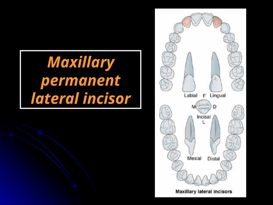
Maxillary permanent
lateral incisor
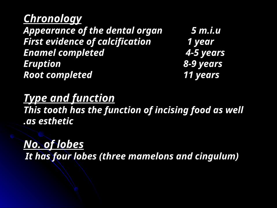
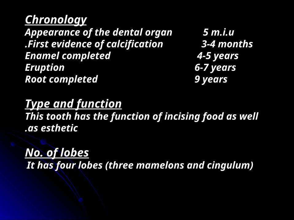
ChronologyAppearance of the dental organ 5 m.i.uFirst evidence of calcification 1 yearEnamel completed 4-5 yearsEruption 8-9 yearsRoot completed 11 years
Type and functionThis tooth has the function of incising food as well as esthetic.
No. of lobesIt has four lobes (three mamelons and cingulum)
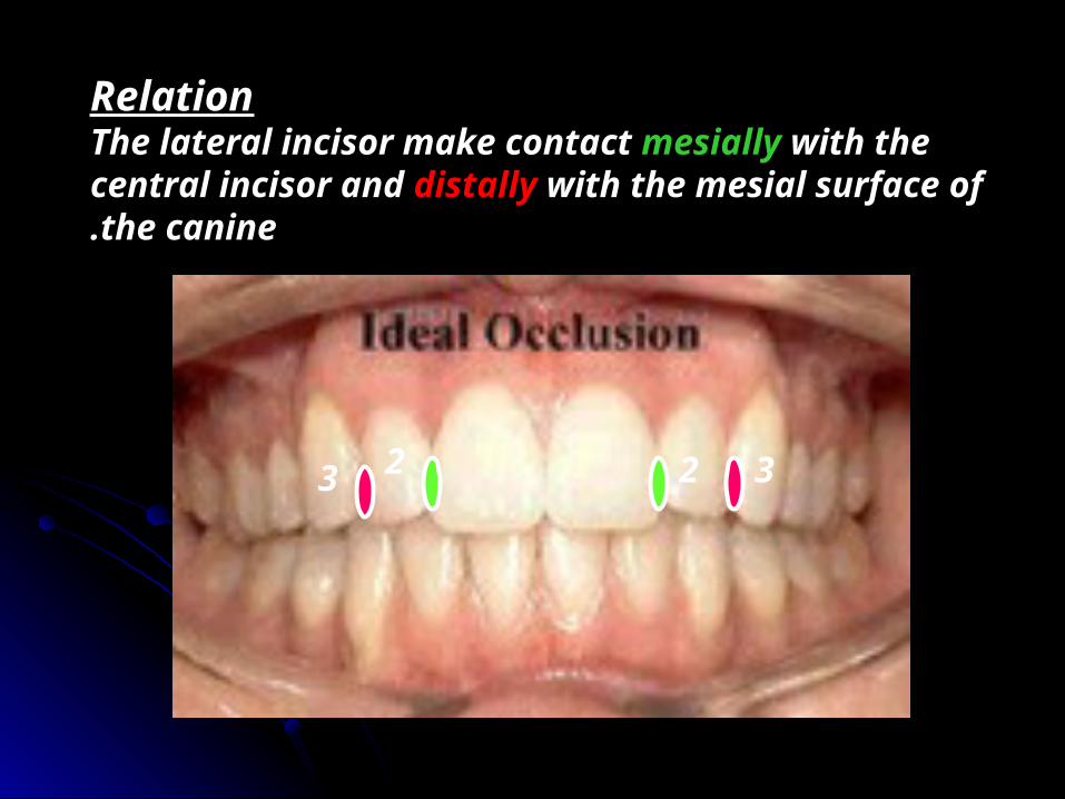
RelationThe lateral incisor make contact mesially with the central incisor and distally with the mesial surface of the canine.
2 23 3
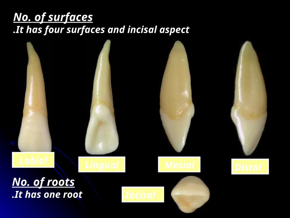
No. of surfacesIt has four surfaces and incisal aspect.
Labial Lingual Mesial Distal
IncisalNo. of rootsIt has one root.

Surface AnatomySurface Anatomy Labial aspect☻Geometrical outline of the
crown: Trapezoid-The short side cervically-The long side incisally
☻The outline: -Mesial outline is slightly convex The
crest at junction of incisal and middle thirds.
-Distal outline is more convex. -Incisal outline is more curved
than the central incisor.
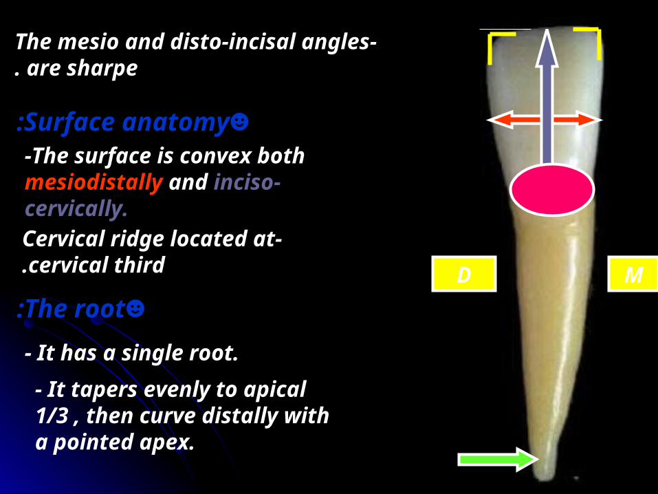
-The cervical line is convex root-wards.
D M

-The mesio-incisal angle is rounded. -The disto-incisal angle is more rounded.
☻Surface anatomy:-The surface is more convex than the central incisor.
-Cervical ridge located at cervical third.
☻The root:- It has a single root.- It tapers evenly to apical 1/3 , then curve distally with a pointed apex.
D M

Lingual aspect -It has the same geometrical outline
and outline as the labial surface.
M D
- The mesial and distal sides of the crown and root converge lingual ( the lingual surface is narrower than the labial surface).
- This convergence to accommodate with the horse shoe shaped of the alveolar process.
☻Surface anatomy:The elevations:1- The cingulum ( present at cervical 1/3).
2 -Marginal ridges.- Mesial marginal ridge.- Distal marginal ridge.- Incisal ridge.
The depressions:- The lingual fossa ( it lies between the previous elevations).
☻All elevations and depression are more developed than the upper central incisor.
-Notice the lingual pit.

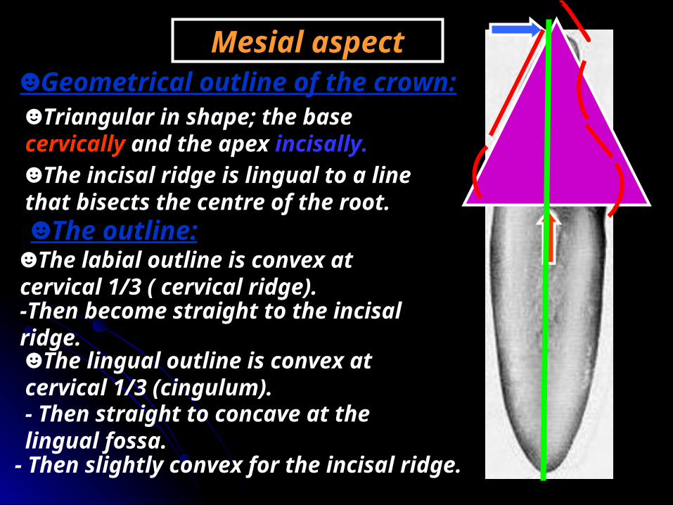
Mesial aspect☻Geometrical outline of the crown:☻Triangular in shape; the base cervically and the apex incisally.☻The incisal ridge is on a line that bisects the centre of the root.☻The outline:
☻The labial outline is convex at cervical 1/3 ( cervical ridge).-Then become slightly convex to the incisal ridge.☻The lingual outline is convex at cervical 1/3 (cingulum).- Then concave at the lingual fossa.
- Then slightly convex for the incisal ridge.
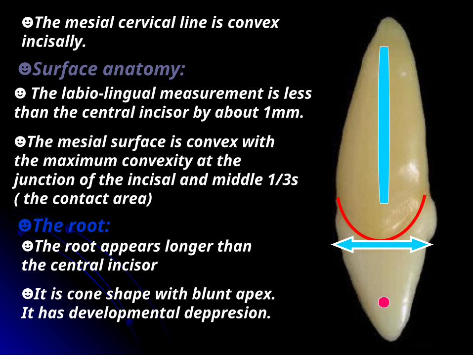
☻The mesial cervical line is convex incisally.
☻Surface anatomy:
☻The mesial surface is convex with the maximum convexity at the junction of the incisal and middle 1/3s ( the contact area)
☻The root:☻The root appears longer than the central incisor
☻It is cone shape with blunt apex. It has developmental deppresion.
☻ The labio-lingual measurement is less than the central incisor by about 1mm.
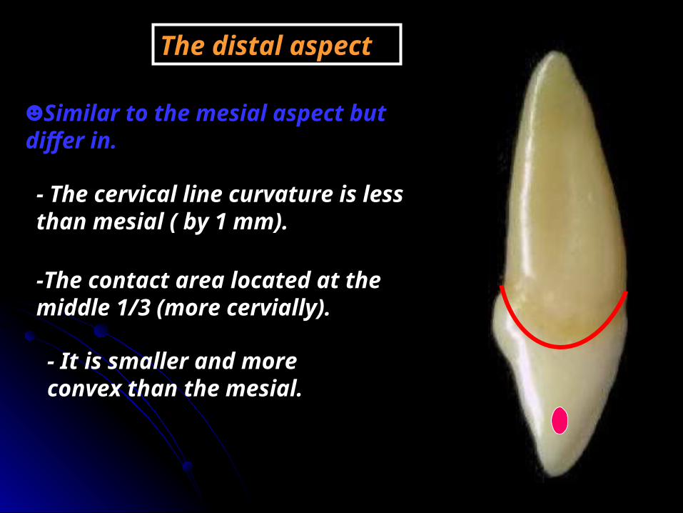
The distal aspect
☻Similar to the mesial aspect but differ in.
- The cervical line curvature is less than mesial ( by 1 mm).
-The contact area located at the middle 1/3 (more cervially).
- It is smaller and more convex than the mesial.
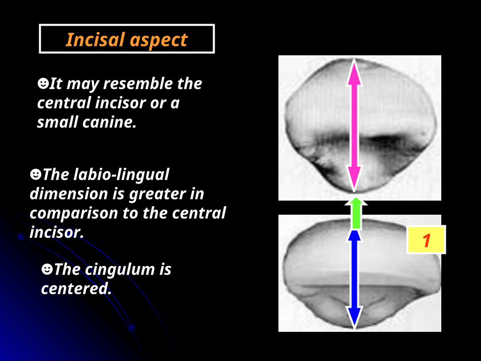
Incisal aspect
☻It may resemble the central incisor or a small canine.
☻The labio-lingual dimension is greater in comparison to the central incisor. 1
☻The cingulum is centered.
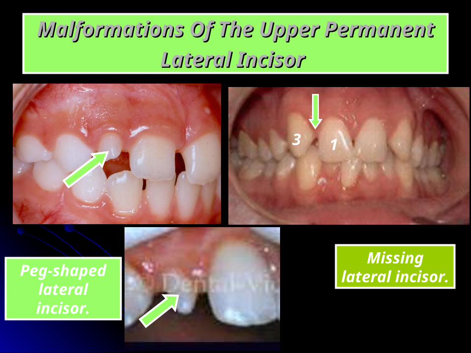
Malformations Of The Upper Permanent Malformations Of The Upper Permanent Lateral IncisorLateral Incisor
Peg-shaped lateral
incisor.
Missing lateral incisor.
13

►Mesio-distal section
-The pulp chamber is wide conforming the shape of the crown.
- It does not have three sharp pulp horns.
-The root canal tapers towards the apex.
►Labio-lingual section -The pulp chamber
pointed incisally, then follows the increase in the crown dimention cervically.
-The root canal tapers gradually as it traverse the root ending in a constriction at the apex (the apical foramen).
☻Pulp cavity


Mandibular Permanent Incisors
1 122
☻They are four in number.
☻They have smaller mesio-distal dimention than any of the other teeth.

Mandibular Central Incisor
ChronologyAppearance of the dental organ 5 m.i.uFirst evidence of calcification 3-4 months.
Enamel completed 4-5 yearsEruption 6-7 yearsRoot completed 9 years
Type and functionThis tooth has the function of incising food as well as esthetic.
No. of lobesIt has four lobes (three mamelons and cingulum)

RelationThe two central incisors make contact mesially with each other and distally with the mesial surface of the lateral incisor.
11 22

No. of surfacesNo. of surfaces It has four surfaces and incisal aspect.It has four surfaces and incisal aspect.
No. of rootsNo. of roots It has one root.It has one root.
Labial Lingual Mesial Distal
Incisal

Surface AnatomySurface Anatomy
Labial aspect☻Geometrical outline of the
crown: Trapezoid.-The short side cervically.-The long side incisally.
☻The outline: -Mesial and distal outlines are straight.
-Incisal outline is straight and perpendicular to the long axis of the tooth.
-The cervical line is convex root-wards.
MD
-The mesio and disto-incisal angles are sharpe.
☻Surface anatomy:-The surface is convex both mesiodistally and inciso-cervically.
-Cervical ridge located at cervical third.
☻The root:- It has a single root.- It tapers evenly to apical 1/3 , then curve distally with a pointed apex.
D M

Lingual aspect
-It has the same geometrical outline and outline as the labial surface.
M D
- The mesial and distal sides of the crown and root converge ligually ( the lingual surface is narrower than the labial surface).- This convergence to accommodate with the horse shoe shaped of the alveolar process.

☻Surface anatomy:The elevations:1- The cingulum ( present at cervical 1/3).
2 -Marginal ridges.- Mesial marginal ridge.- Distal marginal ridge.- Incisal ridge.
The depressions:
☻All elevations and depression are poorly developed than the upper incisor.
- The lingual fossa ( it lies between the previous elevations).
Mesial aspect☻Geometrical outline of the crown:☻Triangular in shape; the base cervically and the apex incisally.☻The incisal ridge is lingual to a line that bisects the centre of the root.☻The outline:
☻The labial outline is convex at cervical 1/3 ( cervical ridge).
☻The lingual outline is convex at cervical 1/3 (cingulum).
-Then become straight to the incisal ridge.
- Then straight to concave at the lingual fossa.
- Then slightly convex for the incisal ridge.

☻The mesial cervical line is convex incisally.
☻Surface anatomy:☻The mesial surface is convex with the maximum convexity near the incisal ridge ( the contact area).
☻The root:
☻The root outline is straight labially and lingually. It tapers in the middle third to a blunted or rounded apex.
-The root has a broad developmental depression.

The distal aspect
☻Similar to the mesial aspect but differ in.
- The cervical line curvature is less than mesial ( by 1 mm).
-The distal contact area at the same level as the mesial surface.
-The distal developmental depression on the root is deeper than mesial and may have developmental groove.

Incisal aspect
-The aspect is four sided or diamond shape.
-The incisal edge is straight.
-The mesial and distal halves are identical .
-The incisal edge is perpendicular to a line bisecting the crown labiolingually.

☻Pulp cavity
►Mesio-distal section
►Labio-lingual section
- The outline of the pulp cavity conform to the crown and root outline.
- The pulp chamber has 2 pulp horns. The root canal tapers to the apical foramen.
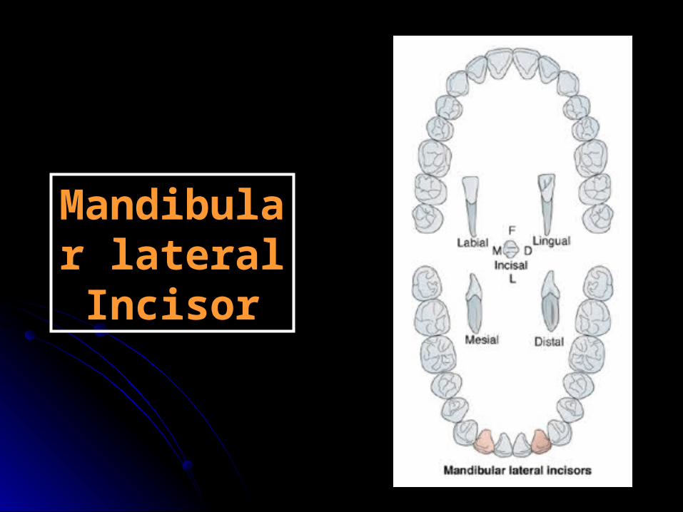
Mandibular lateral Incisor
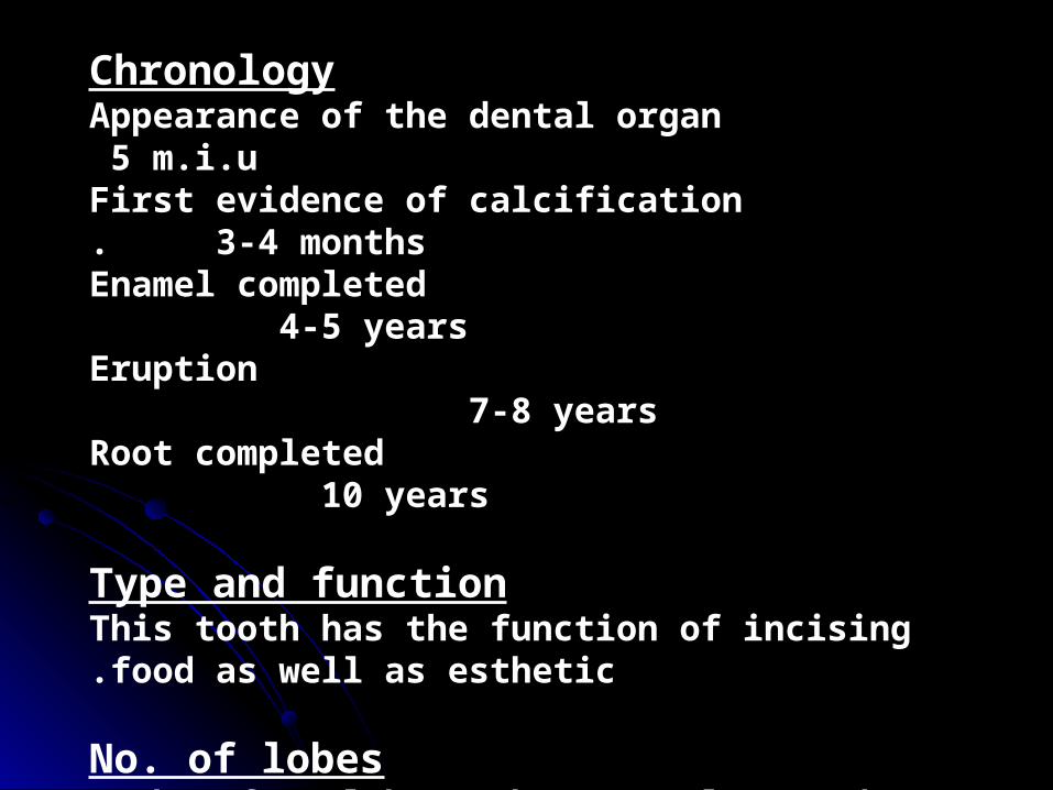
ChronologyAppearance of the dental organ 5 m.i.uFirst evidence of calcification 3-4 months.
Enamel completed 4-5 yearsEruption 7-8 yearsRoot completed 10 years
Type and functionThis tooth has the function of incising food as well as esthetic.
No. of lobesIt has four lobes (three mamelons and cingulum)

RelationThe lateral incisors make contact mesially with the distal surface of the central incisors and distally with the mesial surface of the canines.
11 223 3
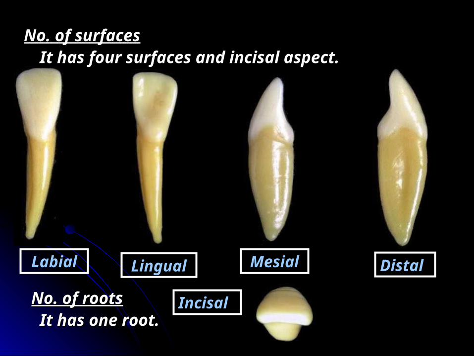
No. of surfacesNo. of surfaces It has four surfaces and incisal aspect.It has four surfaces and incisal aspect.
No. of rootsNo. of roots It has one root.It has one root.
Labial Lingual Mesial Distal
Incisal

Surface AnatomySurface Anatomy ☻It appear to have nearly the same
form as the mandibular central incisor, SO direct comparison will be discussed.
Labial aspect
-It is larger than the central by about 0.5 mm in all dimensions.
-The incisal edge is straight and slop downward in a distal direction.
MD
-The distal angle is rounded.
-The root is longer than the central incisor.

Lingual aspect
-The mesial outline is longer than the distal outline.
-The mesial marginal ridge is longer than the distal marginal ridge.
-The cingulum is deviated distal to the center of the lingual surface.
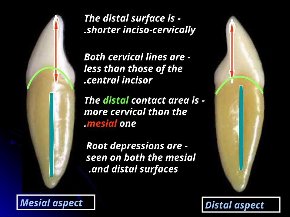
Mesial aspect Distal aspect
-The distal surface is shorter inciso-cervically.
-Both cervical lines are less than those of the central incisor.
-The distal contact area is more cervical than the mesial one.
-Root depressions are seen on both the mesial
and distal surfaces .
Incisal aspect
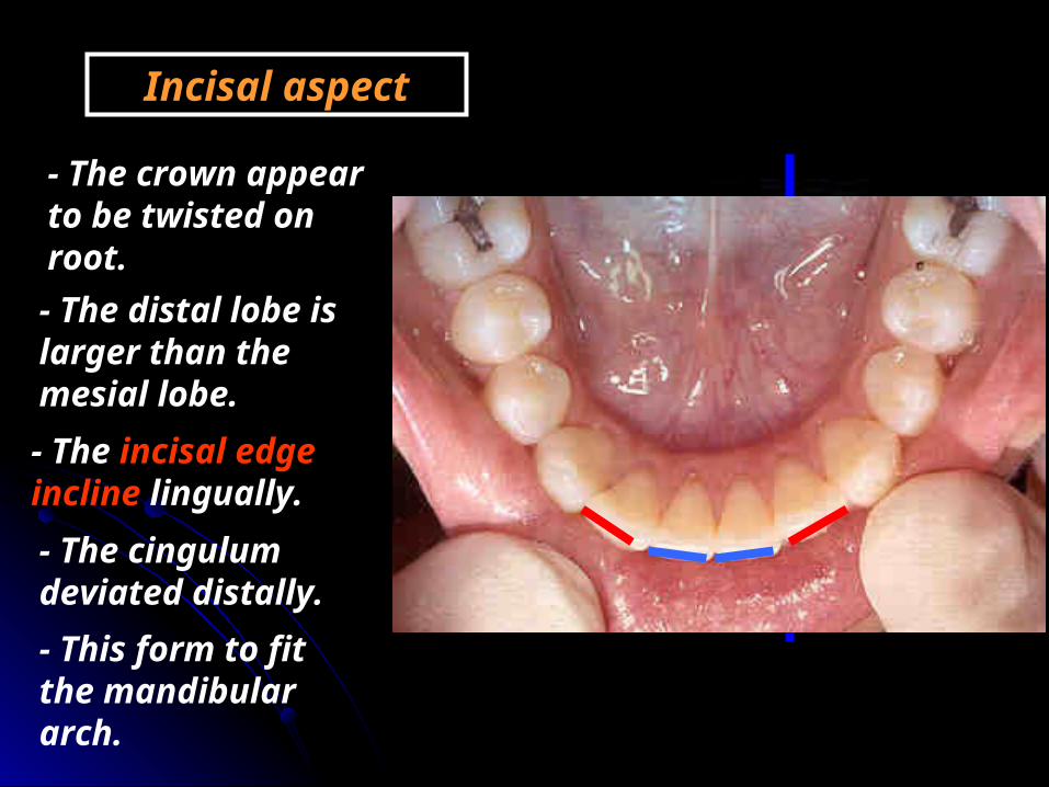
- The crown appear to be twisted on root. - The distal lobe is larger than the mesial lobe.
- This form to fit the mandibular arch.
- The incisal edge incline lingually.- The cingulum deviated distally.


Maxillary Maxillary permanent permanent
caninecanine
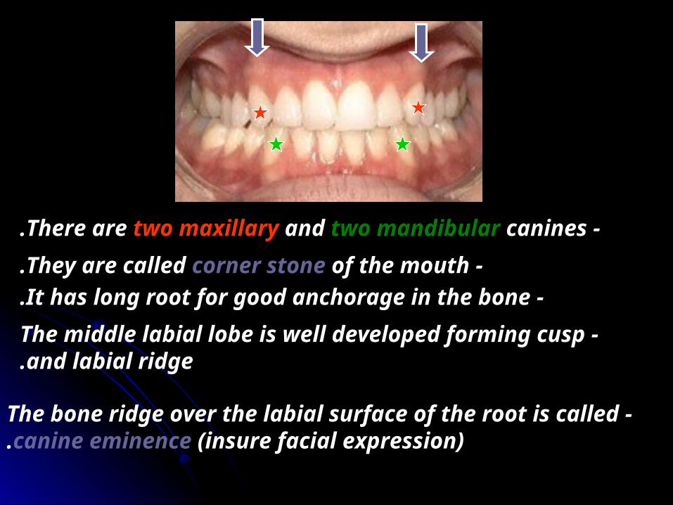
-There are two maxillary and two mandibular canines. -They are called corner stone of the mouth.
-It has long root for good anchorage in the bone. -The middle labial lobe is well developed forming cusp
and labial ridge.
-The bone ridge over the labial surface of the root is called canine eminence (insure facial expression).

ChronologyAppearance of the dental organ 6 m.i.uFirst evidence of calcification 4-5 months.
Enamel completed 6-7 yearsEruption 11-12 yearsRoot completed 14-15 years
Type and functionThis tooth has the function of incising, holding and tearing food as well as esthetic.
No. of lobesIt has four lobes (three labial-the middle is well developed- and cingulum)

33 2 24 4
RelationThe upper canine make contact mesially with the distal surface of the lateral incisors and distally with the mesial surface of the 1st premolar.
No. of surfaces It has four surfaces and incisal aspect.
Labial Lingual Mesial Distal
IncisalNo. of roots
It has one root.
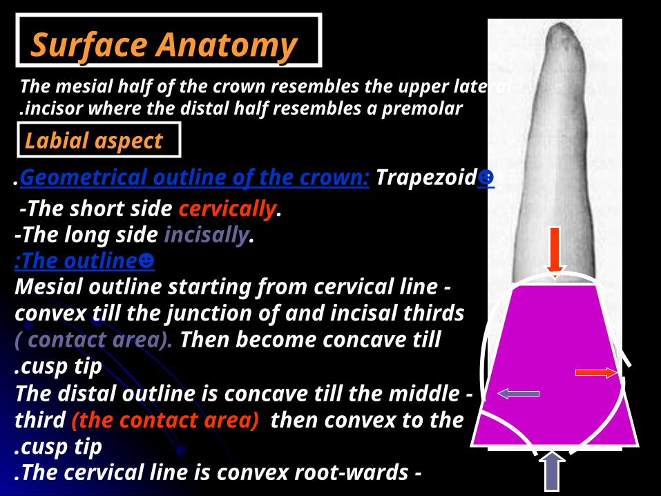
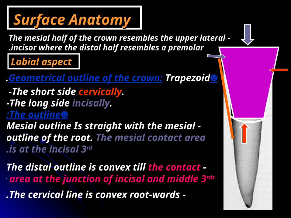
Surface AnatomySurface Anatomy -The mesial half of the crown resembles the upper lateral
incisor where the distal half resembles a premolar.
Labial aspect☻Geometrical outline of the crown: Trapezoid.
-The short side cervically.-The long side incisally.
☻The outline: -Mesial outline starting from cervical line
convex till the junction of and incisal thirds ( contact area). Then become concave till cusp tip.
-The cervical line is convex root-wards.
-The distal outline is concave till the middle third (the contact area) then convex to the cusp tip.

-The distal slope is longer than the mesial slope.
☻Surface anatomy:-The surface is convex both mesiodistally and inciso-cervically.Elevations
-Cervical ridge. -Labial ridge.
Depressions -Two developmental grooves.
☻The root: -It is long, slender, conical in
shape with distal curvature of the apical 3rd.
-Cusp.

Lingual aspect
-It has the same geometrical outline and outline as the labial surface.- The mesial and distal sides of the crown and root converge lingually.☻Surface anatomy:
The elevations: -The cingulum.
-The marginal ridges (mesial and distal). -The lingual ridge.
-Mesio and disto-incisal ridges.
The depressions -Two lingual fossae.

Cingulum.
Mesial marginal ridge.
Distal marginal ridge
M.L.FD.L.F
M.I.RidgeD.I.Ridge
Lingual ridge
Enumerate the names of the following elevations and
depressions?

Mesial aspect☻Geometrical outline of the crown:☻Triangular in shape; the base cervically and the apex incisally.☻The cusp tip may be on a line that bisects the centre of the root or slightly labial to it.☻The outline:☻The labial outline is convex with the greatest convexity at cervical 1/3 ( cervical ridge).☻The lingual outline is convex at cervical 1/3 (cingulum).- Then straight at the middle 3rd and convex at incisal 3rd.

☻The mesial cervical line is convex incisally.
☻Surface anatomy:☻The mesial surface is convex with the maximum convexity at the junction of the incisal and middle 1/3s ( the contact area).
☻The root: -The root is broad and taper to blunt
apex.
-It has a distal developmental depression.

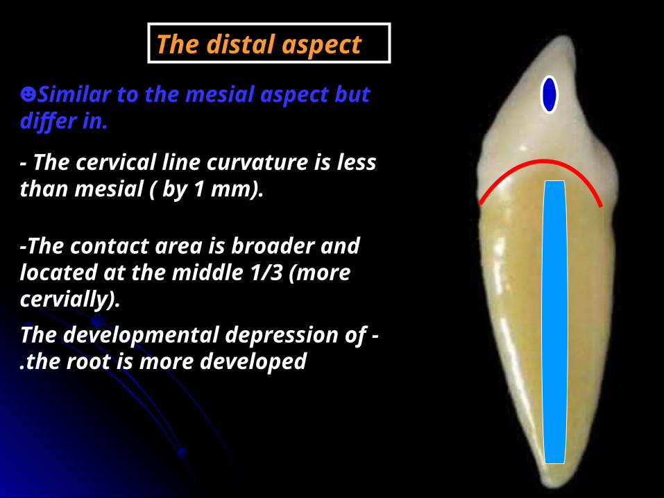
The distal aspect
☻Similar to the mesial aspect but differ in.
- The cervical line curvature is less than mesial ( by 1 mm).
-The contact area is broader and located at the middle 1/3 (more cervially).
-The developmental depression of the root is more developed.
Incisal aspect
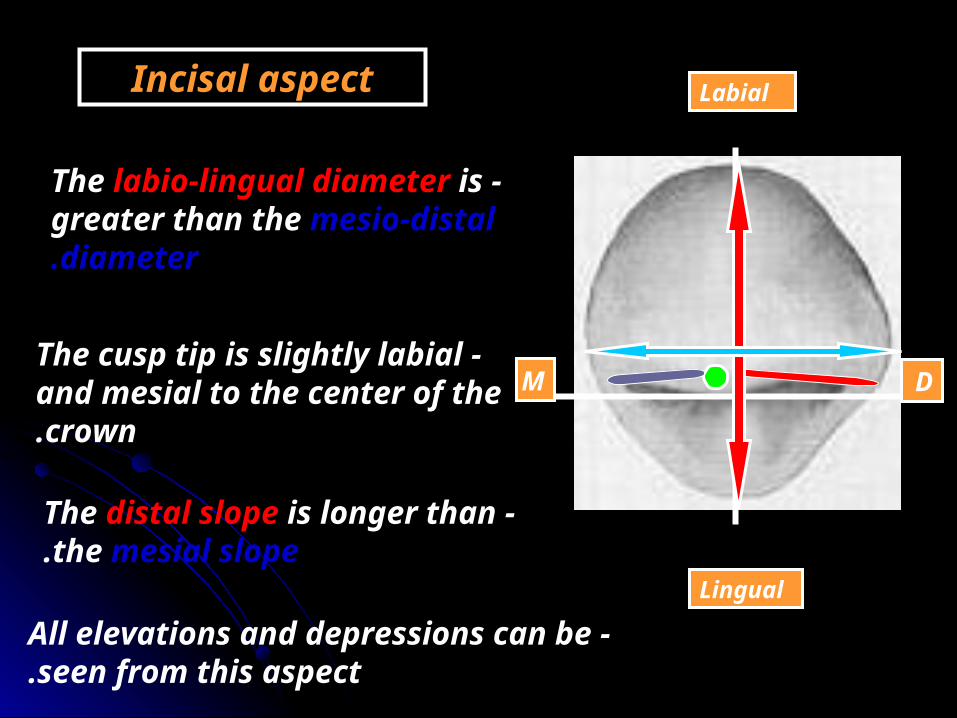
-The labio-lingual diameter is greater than the mesio-distal diameter.
-The cusp tip is slightly labial and mesial to the center of the crown.
Labial
Lingual
M D
-The distal slope is longer than the mesial slope.
-All elevations and depressions can be seen from this aspect.
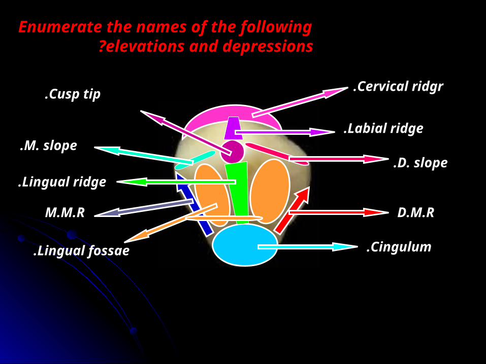
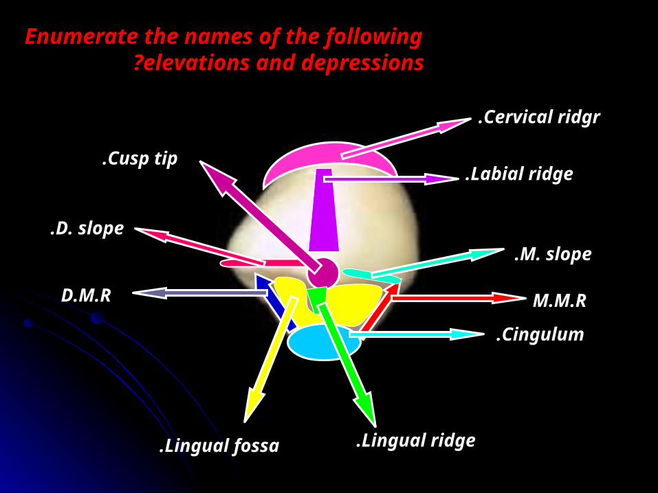
Enumerate the names of the following elevations and depressions?
Cervical ridgr.
Labial ridge.
D. slope.
D.M.R
Cusp tip.
M. slope.
Lingual ridge.
M.M.R
Lingual fossae. Cingulum.

☻Pulp cavity
►Mesio-distal section
►Labio-lingual section
-Has narrow pulp chamber.
-The root canal is long and tapering down to the apical foramen.
-The pulp chamber is pointed incisally.
-The root canal start cervically wide till about the middle then narrows to the apical foramen.
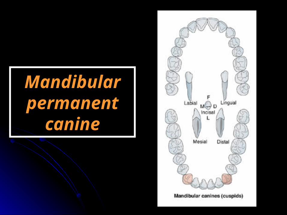
Mandibular Mandibular permanent permanent
caninecanine

ChronologyAppearance of the dental organ 6 m.i.uFirst evidence of calcification 4-5 months.
Enamel completed 6-7 yearsEruption 9-10 yearsRoot completed 12-13 years
Type and functionThis tooth has the function of incising, holding and tearing food as well as esthetic.
No. of lobesIt has four lobes (three labial-the middle is well developed- and cingulum)
332 2
4 4
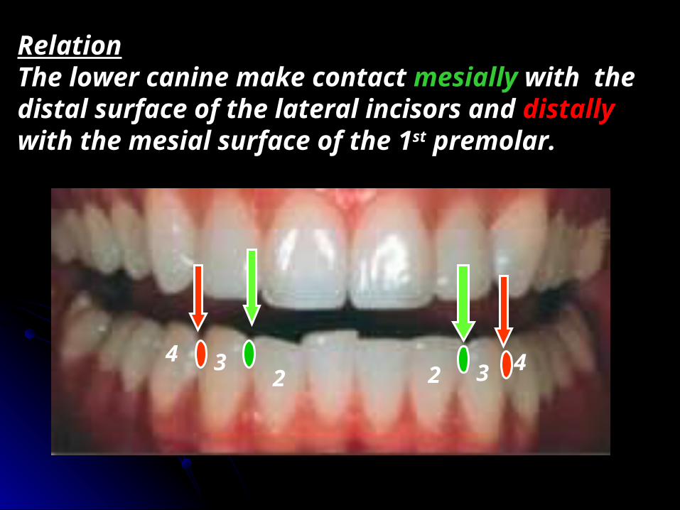
RelationThe lower canine make contact mesially with the distal surface of the lateral incisors and distally with the mesial surface of the 1st premolar.

No. of surfaces It has four surfaces and incisal aspect.
Labial Lingual Mesial Distal
IncisalNo. of roots
It has one root.
Surface AnatomySurface Anatomy -The mesial half of the crown resembles the upper lateral
incisor where the distal half resembles a premolar.
Labial aspect☻Geometrical outline of the crown: Trapezoid.
-The short side cervically.-The long side incisally.
☻The outline: -Mesial outline Is straight with the mesial
outline of the root. The mesial contact area is at the incisal 3rd.
-The cervical line is convex root-wards.
-The distal outline is convex till the contact area at the junction of incisal and middle 3rds .
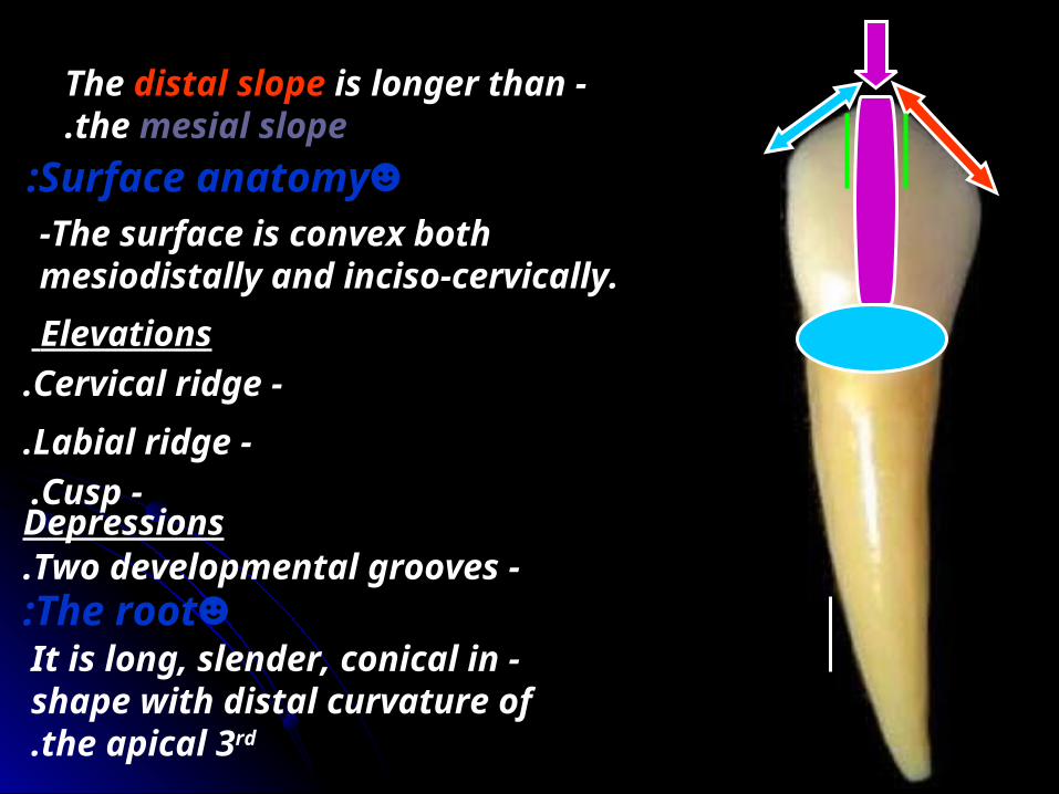
-The distal slope is longer than the mesial slope.
☻Surface anatomy:-The surface is convex both mesiodistally and inciso-cervically.Elevations
-Cervical ridge. -Labial ridge.
Depressions -Two developmental grooves.
☻The root: -It is long, slender, conical in
shape with distal curvature of the apical 3rd.
-Cusp.

Lingual aspect
-It has the same geometrical outline and outline as the labial surface.- The mesial and distal sides of the crown and root converge lingually.☻Surface anatomy:The elevations: (poorly developed)
-The cingulum. -The marginal ridges (mesial and distal).
-The lingual ridge. -Mesio and disto-incisal ridges.
The depressions -one lingual fossae.

Enumerate the names of the following elevations and
depressions? Mesio-incisal R.
Disto-incisal R. Lingual ridgeLingual fossa
Mesial marginal ridge
Distal marginal ridge
Cingulum

Mesial aspect☻Geometrical outline of the crown:☻Triangular in shape; the base cervically and the apex incisally.☻The cusp tip may be on a line that bisects the centre of the root or slightly lingual to it.☻The outline:☻The labial outline is slightlu convex with the greatest convexity at cervical 1/3 ( cervical ridge).☻The lingual outline is convex at cervical 1/3 (cingulum).- Then concave at the middle 3rd and convex at incisal 3rd.

☻The mesial cervical line is convex incisally.
☻Surface anatomy:☻The mesial surface is convex with the maximum convexity at the incisal third ( the contact area).
☻The root: -The root is broad and taper to blunt
apex.
-It has a distal developmental depression.
The distal aspect
☻Similar to the mesial aspect but differ in.
- The cervical line curvature is less than mesial ( by 1 mm).
-The contact area is broader and located at the middle 1/3 (more cervially).
-The developmental depression of the root is more developed.

Incisal aspect
-The labio-lingual diameter is greater than the mesio-distal diameter but in less proportion.
-The cusp tip is slightly lingual and mesial to the center of the crown.
Labial
Lingual
M D
-The distal slope is longer than the mesial slope.
-All elevations and depressions can be seen from this aspect.
-The mesial cusp ridge incline lingual.
Enumerate the names of the following elevations and depressions?
Cervical ridgr.
Labial ridge.
D. slope.
D.M.R
Cusp tip.
M. slope.
Lingual ridge.
M.M.R
Lingual fossa.
Cingulum.


Maxillary premolars (bicuspids)
-We have 4 maxillary premolars; two in right and two in left side.
-Mesial to them is the canine and distal the molars present.
-They have two cusps (one buccal and one lingual) {bicuspid}
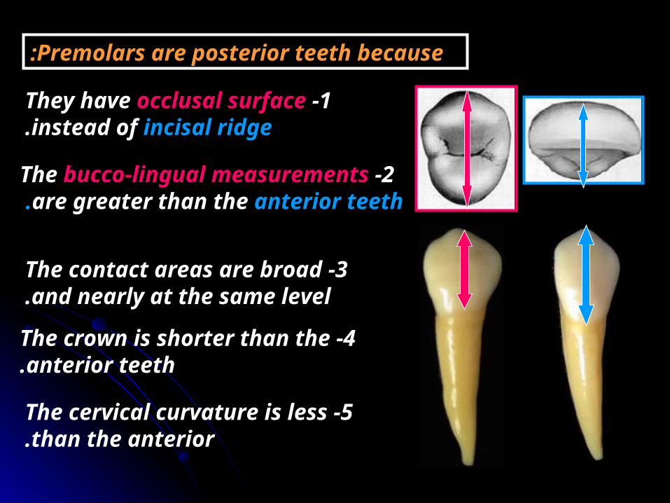
Premolars are posterior teeth because:
1 -They have occlusal surface instead of incisal ridge.
2 -The bucco-lingual measurements are greater than the anterior teeth.
4 -The crown is shorter than the anterior teeth.
5 -The cervical curvature is less than the anterior.
3 -The contact areas are broad and nearly at the same level.

Maxillary first Maxillary first premolarpremolar

ChronologyAppearance of the dental organ 7 m.i.uFirst evidence of calcification 11/2-13/4 years.
Enamel completed 5-6 yearsEruption 10-11 yearsRoot completed 12-13 years
Type and functionThis tooth has the function of tearing and grinding food.
No. of lobesIt has four lobes (three buccal -the middle is well developed (the buccal cusp)- and one lingual (the lingual cusp).
43 5
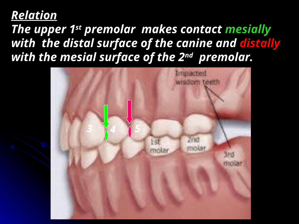
RelationThe upper 1st premolar makes contact mesially with the distal surface of the canine and distally with the mesial surface of the 2nd premolar.
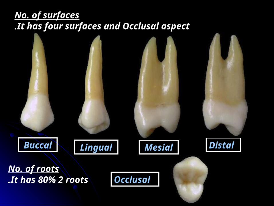
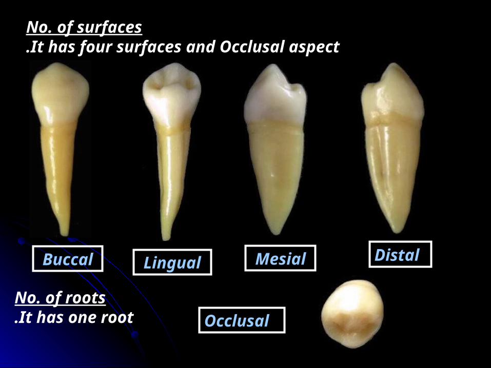
No. of surfaces It has four surfaces and Occlusal aspect.
Buccal Lingual Mesial Distal
OcclusalNo. of roots
It has 80% 2 roots.
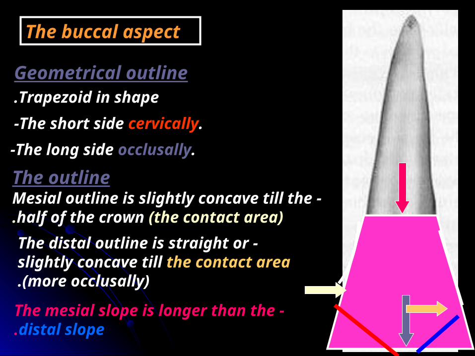
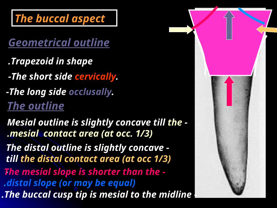
The buccal aspect
Geometrical outline
-The short side cervically.-The long side occlusally.
Trapezoid in shape.
The outline -Mesial outline is slightly concave till
the half of the crown (the contact area). -The distal outline is straight or
slightly concave till the contact area (more occlusally).
-The mesial slope is longer than the distal slope.

Surface anatomyThe elevations
-The cervical ridge
-The buccal ridge
The depressions
Two developmental grooves mesial and distal to the buccal ridge.
The root
The buccal root is similar to that of the canine but shorter.

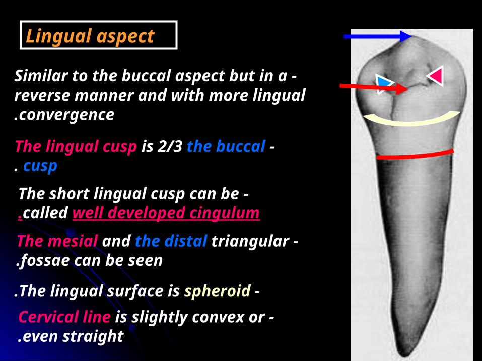
Lingual aspect
-Similar to the buccal aspect but in a reverse manner and with lingual convergence.
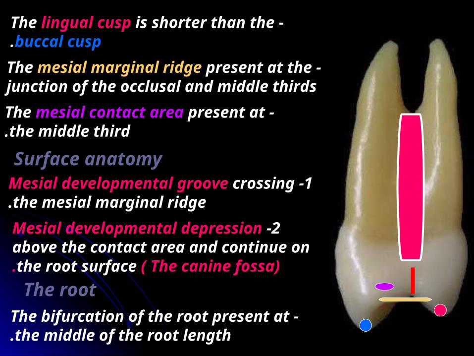
-The lingual cusp is shorter than the buccal cusp by 1mm.
-The mesial and the distal slopes of the lingual cusp have right angle.
-The lingual surface is spheroid and has a less developed lingual ridge Than the buccal ridge.
-Cervical line is slightly convex or even straight.

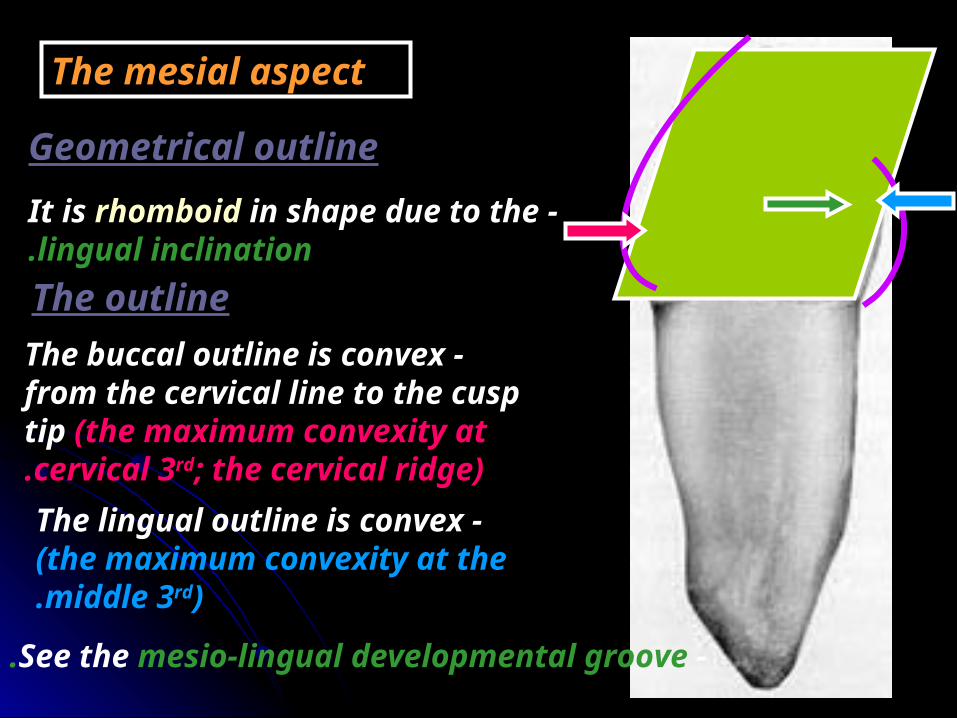
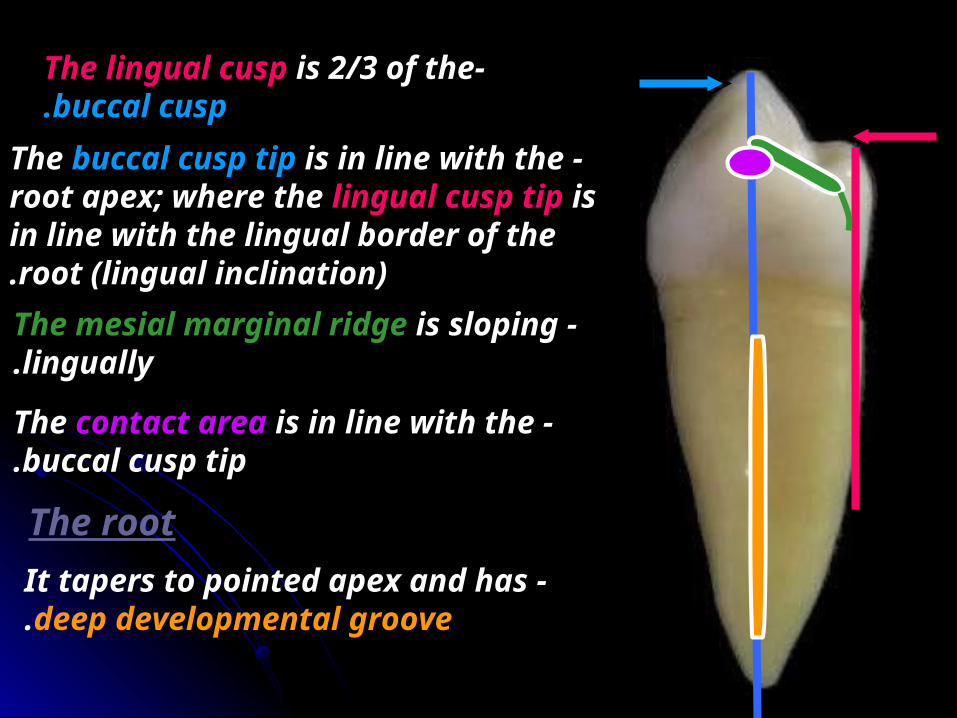
Mesial aspect
Geometrical outlineTrapezoid in shape.
-The short side occlusally.-The long side cervically.
The outline -The buccal outline is convex till the
buccal cusp tip with the maximum convexity at cervical 3rd.
-The lingual outline is convex with the maximum convexity at the center.
-The lingual cusp is shorter than the buccal cusp.
-The mesial marginal ridge present at the junction of the occlusal and middle thirds
-The mesial contact area present at the middle third.
Surface anatomy1 -Mesial developmental groove crossing
the mesial marginal ridge.2 -Mesial developmental depression
above the contact area and continue on the root surface ( The canine fossa).
-The bifurcation of the root present at the middle of the root length.
The root
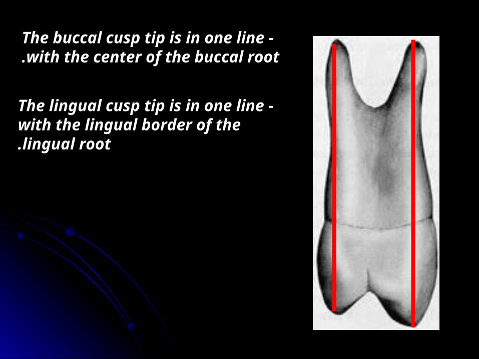
-The buccal cusp tip is in one line with the center of the buccal root.
-The lingual cusp tip is in one line with the lingual border of the lingual root.
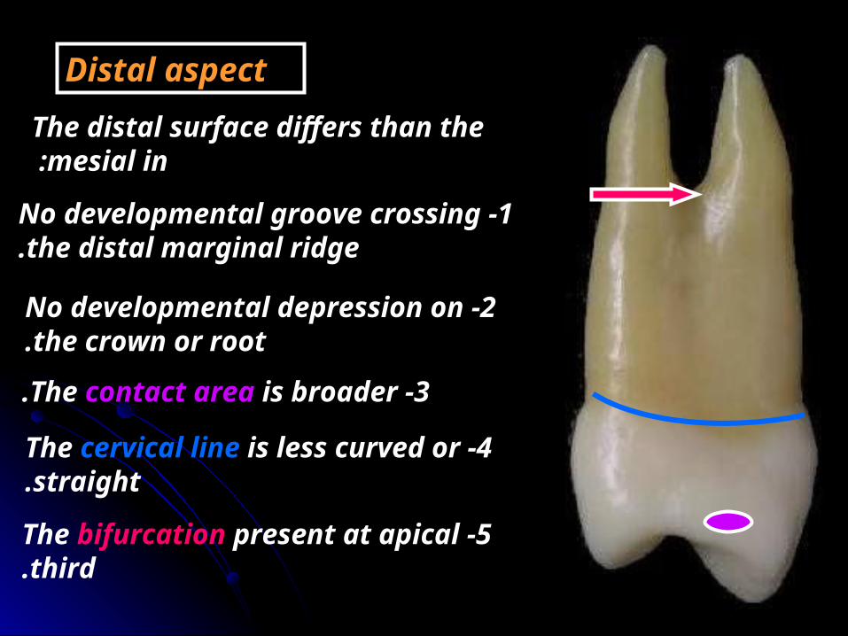
Distal aspectThe distal surface differs than the
mesial in :
1 -No developmental groove crossing the distal marginal ridge.
2 -No developmental depression on the crown or root.
4 -The cervical line is less curved or straight.
5 -The bifurcation present at apical third.
3 -The contact area is broader.

Occlusal aspect
It is hexagonal in shapeMesio-B. Disto-B.
Distal. Mesial
Disto-L. Mesio-L.
-Central developmental groove.
-Distal and mesial triangular fossa.
-Distal and mesial developmental pit.
-Distal and mesial marginal ridges.

-The buccal and lingual cusps have triangular ridge.
-The mesial marginal ridge is crossed by mesial developmental groove.
-The bucco-lingual measurement is larger than the mesio-distal measurement
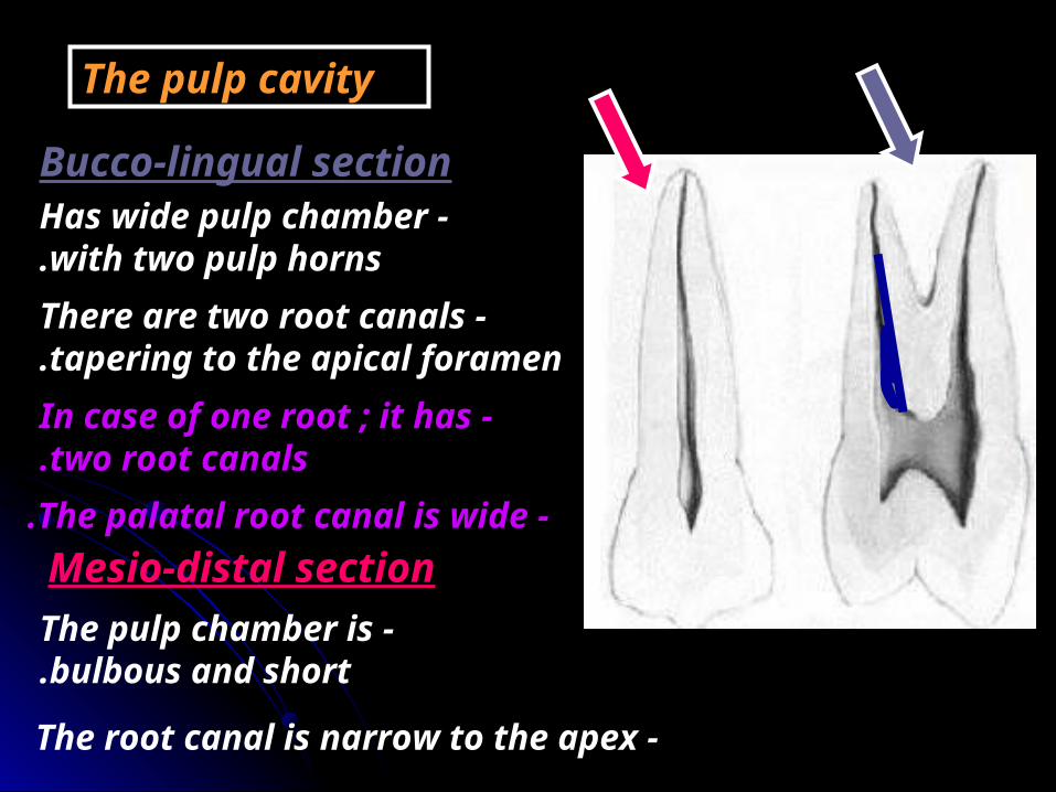
The pulp cavity
Bucco-lingual section -Has wide pulp chamber
with two pulp horns. -There are two root canals
tapering to the apical foramen. -In case of one root ; it has
two root canals.
Mesio-distal section -The pulp chamber is
bulbous and short.
-The root canal is narrow to the apex
-The palatal root canal is wide.
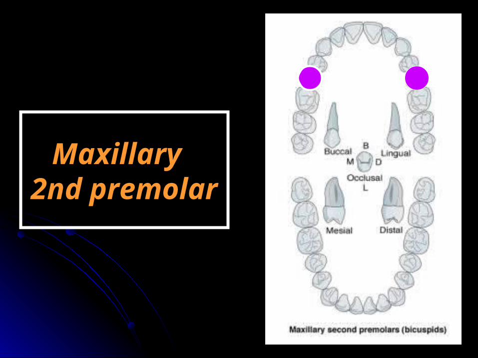
Maxillary Maxillary 2nd premolar2nd premolar

ChronologyAppearance of the dental organ 8 m.i.uFirst evidence of calcification 2-21/4 years.
Enamel completed 6-7 yearsEruption 10-12 yearsRoot completed 13-15 years
Type and functionThis tooth supplements the maxillary first premolar in function of tearing and grinding food.
No. of lobesIt has four lobes (three buccal -the middle is well developed (the buccal cusp)- and one lingual (the lingual cusp).

4 5
RelationThe upper 2ndt premolar makes contact mesially with the distal surface of the 1st premolar and distally with the mesial surface of the 1st permanent molar.
6
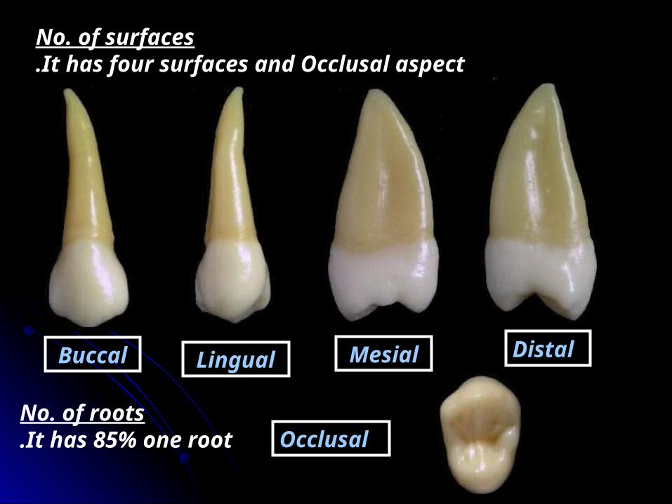
No. of surfaces It has four surfaces and Occlusal aspect.
Buccal Lingual Mesial Distal
OcclusalNo. of roots
It has 85% one root.

The buccal aspect
1 -The buccal cusp is less pointed and shorter.
2 -The mesial slope is shorter than the distal slope.
3 -The buccal ridge is less prominent.
-Similar to that of the 1st premolar and differs in:
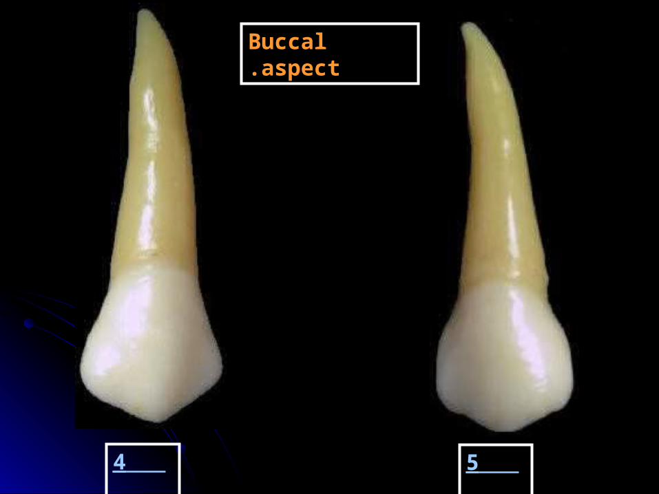
4 5
Buccal aspect.

The lingual aspect
-The lingual cusp is longer than that of upper 1st premolar.
-The buccal and lingual cusps are nearly equal.
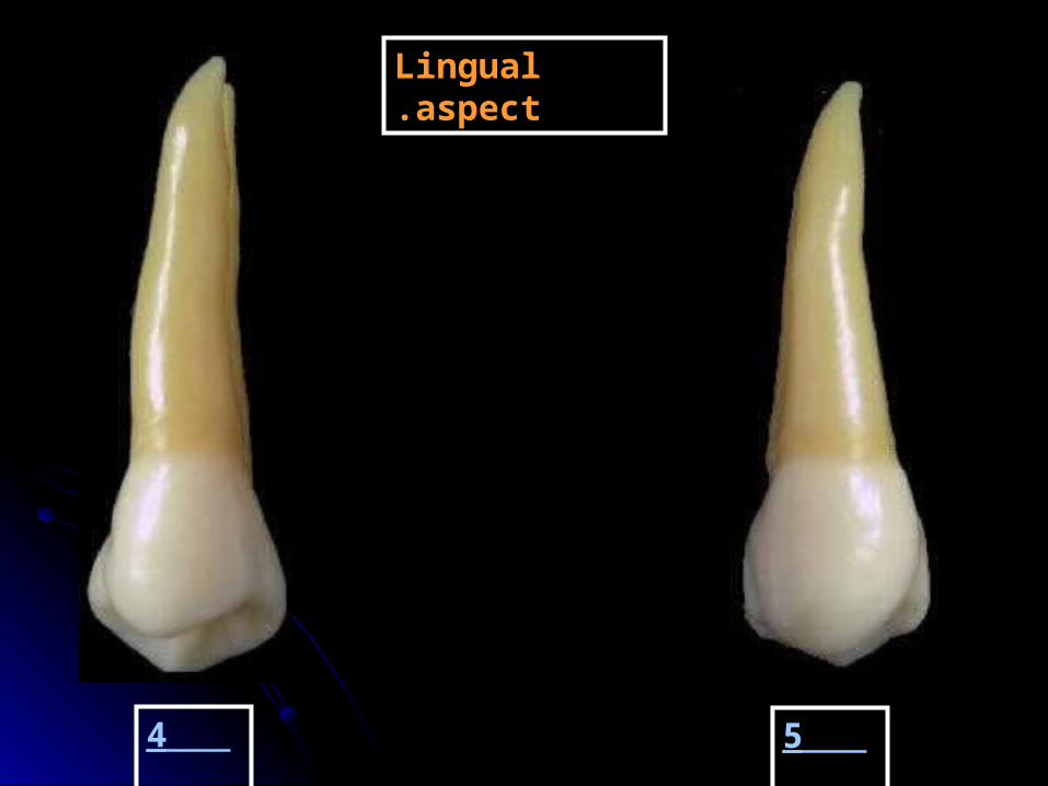
4 5
Lingual aspect.
The mesial aspect
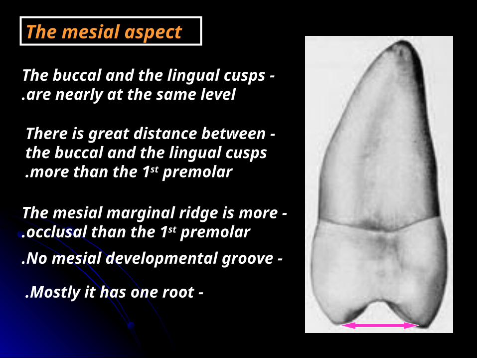
-The buccal and the lingual cusps are nearly at the same level.
-The mesial marginal ridge is more occlusal than the 1st premolar.
-No mesial developmental groove.
-Mostly it has one root.
-There is great distance between the buccal and the lingual cusps more than the 1st premolar.
The distal aspect
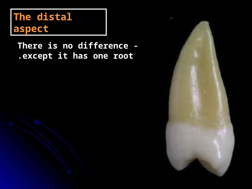
-There is no difference except it has one root.

The occlusal aspect
-It is oval or round shape
-Wide bucco-lingual dimension.
-Short central developmental groove.
-Multiple supplemental grooves.

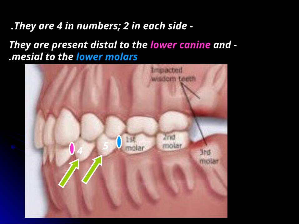
-They are 4 in numbers; 2 in each side.
-They are present distal to the lower canine and mesial to the lower molars.
4 5
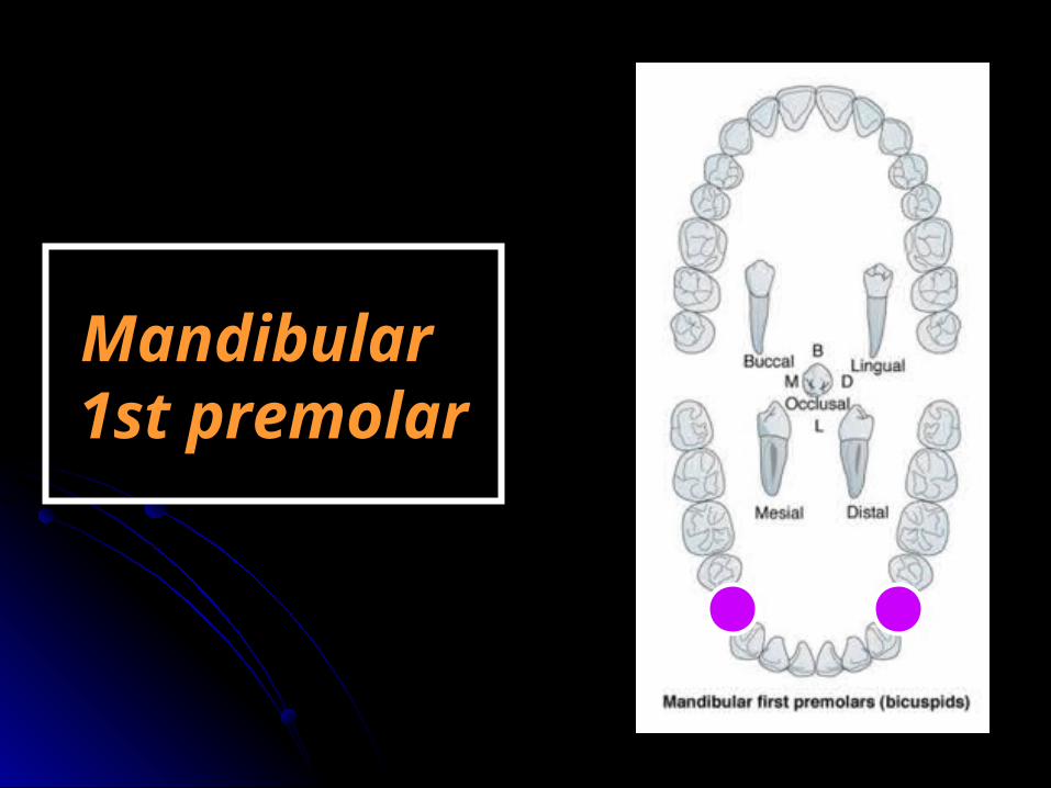
Mandibular Mandibular 1st premolar1st premolar

ChronologyAppearance of the dental organ 7 m.i.uFirst evidence of calcification 13/4-2 years.
Enamel completed 5-6 yearsEruption 10-11 yearsRoot completed 12-13 years
Type and functionThis tooth has the function of tearing and grinding food.
No. of lobesIt has four lobes (three buccal -the middle is well developed (the buccal cusp)- and one lingual (the lingual cusp).

4 5
RelationThe lower 1stt premolar makes contact mesially with the distal surface of the lower canine and distally with the mesial surface of the 2nd premolar.
3
No. of surfaces It has four surfaces and Occlusal aspect.
Buccal Lingual Mesial Distal
OcclusalNo. of roots
It has one root.

The buccal aspect
Geometrical outline
-The short side cervically.-The long side occlusally.
Trapezoid in shape.
The outline -Mesial outline is slightly concave till
the mesial contact area. -The distal outline is slightly concave
till the distal contact area. -The mesial slope is shorter than the
distal slope. -The buccal cusp tip is mesial to the midline.

Surface anatomyThe elevations
-The cervical ridge.
-The buccal ridge.
The depressionsTwo developmental grooves mesial and distal to the buccal ridge.
The root
-The root is cone shape with distal inclination of the apical 3rd.
-The root is shorter than that of the canine.
Lingual aspect
-Similar to the buccal aspect but in a reverse manner and with more lingual convergence.
-The lingual cusp is 2/3 the buccal cusp.
-The mesial and the distal triangular fossae can be seen.
-The lingual surface is spheroid. -Cervical line is slightly convex or
even straight.
-The short lingual cusp can be called well developed cingulum.

-A characteristic feature of the lingual surface is the mesiolingual developmental groove.
The mesial aspect
Geometrical outline -It is rhomboid in shape due to the
lingual inclination.The outline
-The buccal outline is convex from the cervical line to the cusp tip (the maximum convexity at cervical 3rd; the cervical ridge).
-The lingual outline is convex (the maximum convexity at the middle 3rd).
-See the mesio-lingual developmental groove.
-The lingual cusp is 2/3 of the buccal cusp.
-The buccal cusp tip is in line with the root apex; where the lingual cusp tip is in line with the lingual border of the root (lingual inclination).
-The mesial marginal ridge is sloping lingually.
-The contact area is in line with the buccal cusp tip.
The root -It tapers to pointed apex and has
deep developmental groove.

The distal aspect
-Similar to the mesial aspect but differs in:
1 -The distal marginal ridge is straight and perpendicular to the long axis of the tooth.
2 -No developmental groove.
3 -The contact area is broader and more cervically.
4 -The distal cervical line is less curved.

The occlusal aspect
-It is diamond or round shape.
-The buccal cusp has large triangular ridge .
-The small lingual cusp has small triangular ridge.
-The occlusal surface tapers lingually.
-The buccal and lingual triangular ridges connected by transverse ridge.
-Mesial and distal triangular fossae.
-Central developmental groove may cross the transverse ridge.

The pulp cavity
Bucco-lingual section- The pulp chamber is wide with two pulp horns.- The root canal is wide till the middle third then narrow to the apical foramen
Mesio-distal section- Similar to the canine but longer in lower 5 than lower 4.

Mandibular Mandibular 2nd premolar2nd premolar

ChronologyAppearance of the dental organ 8 m.i.uFirst evidence of calcification 21/4-21/2 years.
Enamel completed 6-7 yearsEruption 11-12 yearsRoot completed 13-15 years
Type and functionThis tooth has the function of tearing and grinding food.
No. of lobesThere are two typesIt has four lobes: three buccal and one lingual {2 cusp type}OR five lobes: three buccal and two lingual {3 cusp type}

4 5
RelationThe lower 2nd premolar makes contact mesially with the distal surface of the lower 1st premolar and distally with the mesial surface of the 1st permanent molar.
6

No. of surfaces It has four surfaces and Occlusal aspect.
Buccal Lingual Mesial Distal
OcclusalNo. of roots
It has one root.
The buccal aspect
Geometrical outline
-The short side cervically.-The long side occlusally.
Trapezoid in shape.
The outline -Mesial outline is slightly concave till
the mesial contact area (at occ. 1/3). -The distal outline is slightly concave
till the distal contact area (at occ 1/3). -The mesial slope is shorter than the
distal slope (or may be equal). -The buccal cusp tip is mesial to the midline.

Surface anatomyThe elevations
-The cervical ridge.
-The buccal ridge.
The depressionsTwo developmental grooves mesial and distal to the buccal ridge.
The root
-The root is cone shape with rare distal inclination of the apical 3rd.

Lingual aspect -Similar to the buccal aspect but in
a reverse manner .
-Cervical line is slightly convex or even straight.
☻There may be one or two lingual cusps.
☻The lingual cusp(s) is shorter than the buccal cusp.
- In case of three cusp type: the mesiolingual cusp is larger than the distolingual cusp.
- The two lingual cusps separated by lingual developmental groove
☻Very little lingual convergence.

The mesial aspect
Geometrical outline -It is rhomboid in shape due to the
lingual inclination.
Similar to the lower 4 but differs in:
1- Wider buccolingual.
2- The buccal cusp is just buccal to the root apex. 3- The lingual cusp(s) are well developed.4- The mesial marginal ridge is straight and perpendicular to the long axis.
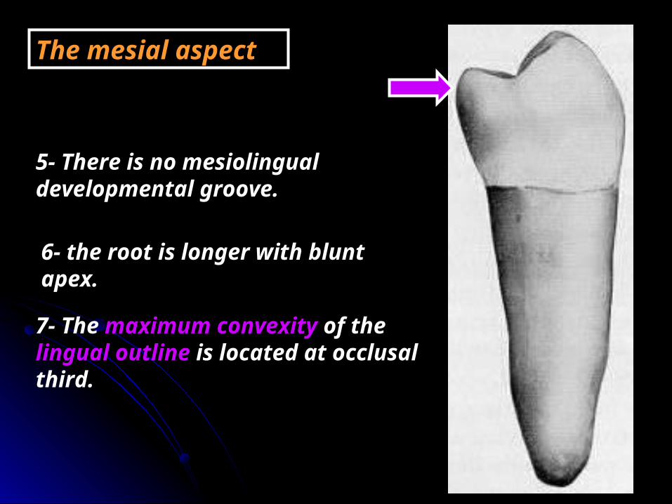
5- There is no mesiolingual developmental groove.
6- the root is longer with blunt apex.
7- The maximum convexity of the lingual outline is located at occlusal third.
The mesial aspect

The distal aspect
- It is similar to the mesial surface but differs in:
1- The distal marginal ridge present more cervically.
2- The tips of both lingual cusps can be seen.
3- In two cusp type there is distolingual developmental depression.

The occlusal aspect
{1} The three cusp type.- The geometrical outline is square.- It has one buccal cusp and two lingual cusps.- The arrangement of the cusps according to the size is: the buccal, the mesiolingual then the distolingual.- Every cusp has triangular ridge. - Y shape developmental groove separating the cusps.- There is central fossa. - Central pit.
- Mesial and distal triangular fossae.
- Mesial and distal marginal ridges.
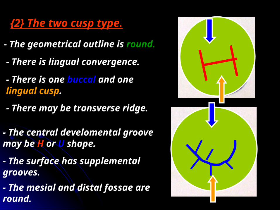
{2} The two cusp type.
- The geometrical outline is round.
- There is lingual convergence.
- There is one buccal and one lingual cusp.
- There may be transverse ridge.
- The central develomental groove may be H or U shape.
- The surface has supplemental grooves.- The mesial and distal fossae are round.


- There are 12 permanent molars (3 in each quadrant).
- They are the largest and strongest teeth in the mouth.
- They have no deciduous predeccessors.
- They are formed of 4 lobes except the lower 1st molar and in some cases of the lower 3rd molar ; formed of 5 lobes.
- They are multirooted teeth where the lower have two roots and the upper have three roots.
- They support and maintain the vertical dimension of the face.
General features

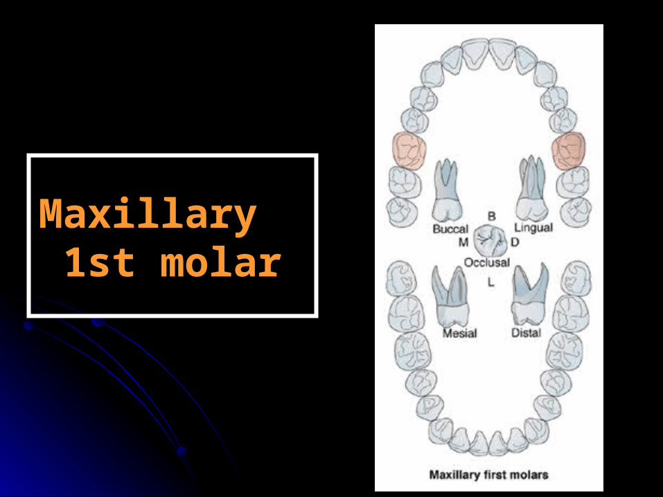
Maxillary Maxillary 1st molar1st molar

ChronologyAppearance of the dental organ 4 m.i.u.First evidence of calcification At birth.Enamel completed 3-4 years.Eruption 6-7 years.Root completed 9-10 years.
Type and functionThis tooth has the function of chewing and grinding food.
No. of lobesIt has four lobes: two buccal and two lingual.

75
RelationThe upper 1st molar makes contact mesially with the distal surface of the upper 2nd premolar and distally with the mesial surface of the 2nd permanent molar.
6
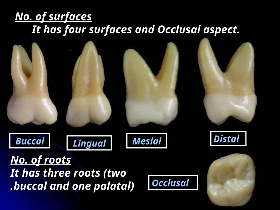
No. of surfaces It has four surfaces and Occlusal aspect.
Buccal Lingual Mesial Distal
Occlusal
No. of roots It has three roots (two
buccal and one palatal).

The buccal aspectGeometrical outlineTrapezoid in shape.
-The short side cervically.-The long side occlusally.The outline
-Mesial outline is straight till the contact area (at the junction of occ. and middle 1/3s).
- Then become convex for the mesial slope of the mesio-buccal cusp.
- The distal outline is convex till the contact area at the middle third.- The distal slope of the disto-buccal cusp is convex.

- The cervical line is straight or slightly convex.- The occlusal outline: the mesiobuccal cusp is broader than the distobuccal cusp.
- The mesio-lingual cusp can be seen between the two buccal cusps.
- The disto-buccal cusp is longer and sharper than the M.B.Cusp
- Buccal developmental groove extends to the middle of the buccal surface separates the two buccal cusps.
- Cervical ridge.Surface anatomy
- The groove may fade out, - Split into 2 shallow grooves. - Or end in a fault pit.
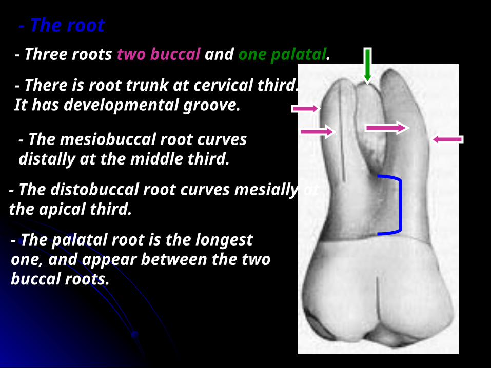
- The root- Three roots two buccal and one palatal.
- There is root trunk at cervical third. It has developmental groove.
- The mesiobuccal root curves distally at the middle third.
- The distobuccal root curves mesially at the apical third.
- The palatal root is the longest one, and appear between the two buccal roots.
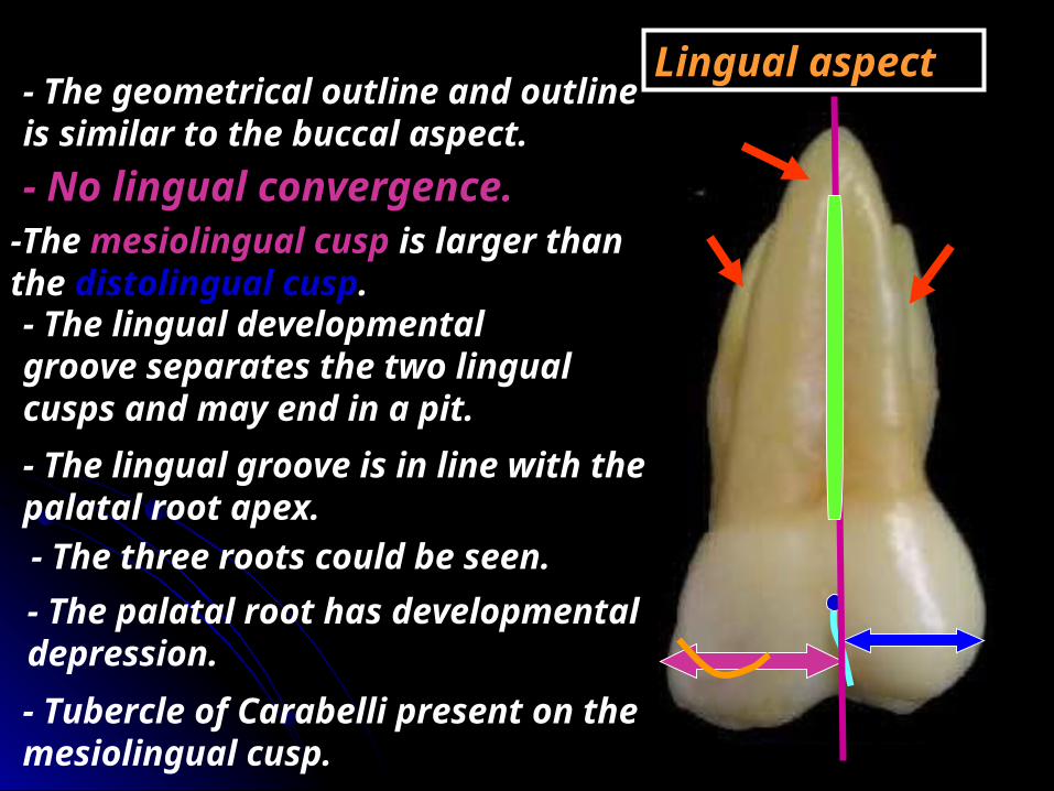
Lingual aspect- The geometrical outline and outline is similar to the buccal aspect.- No lingual convergence.
-The mesiolingual cusp is larger than the distolingual cusp. - The lingual developmental groove separates the two lingual cusps and may end in a pit.
- Tubercle of Carabelli present on the mesiolingual cusp.
- The three roots could be seen.
- The lingual groove is in line with the palatal root apex.
- The palatal root has developmental depression.
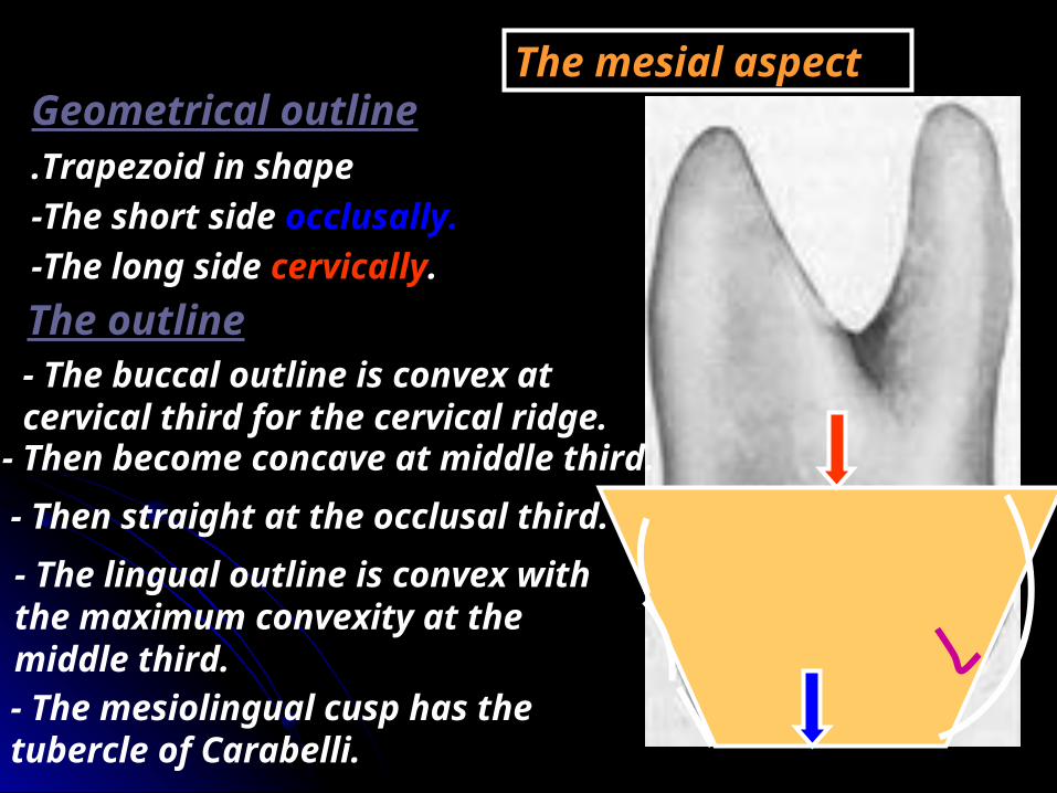
The mesial aspectGeometrical outlineTrapezoid in shape.
-The short side occlusally.-The long side cervically.The outline- The buccal outline is convex at cervical third for the cervical ridge.
- Then become concave at middle third.- Then straight at the occlusal third.- The lingual outline is convex with the maximum convexity at the middle third.- The mesiolingual cusp has the tubercle of Carabelli.
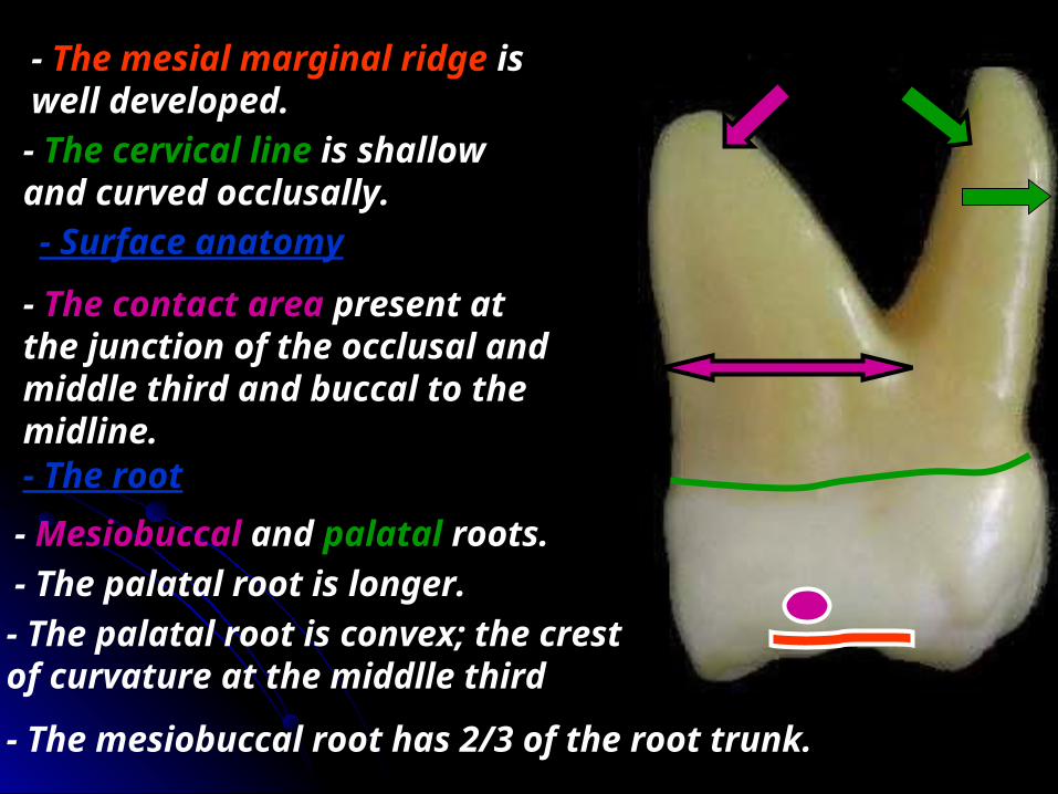
- The mesial marginal ridge is well developed.- The cervical line is shallow and curved occlusally.
- The contact area present at the junction of the occlusal and middle third and buccal to the midline.
- Surface anatomy
- The root- Mesiobuccal and palatal roots.- The palatal root is longer.- The palatal root is convex; the crest of curvature at the middlle third
- The mesiobuccal root has 2/3 of the root trunk.
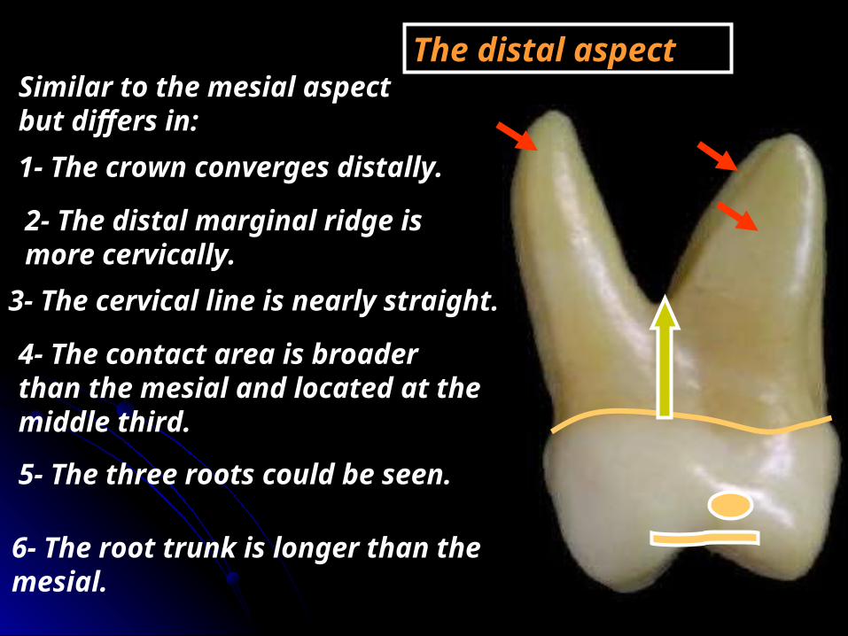
The distal aspectSimilar to the mesial aspect but differs in:1- The crown converges distally.
2- The distal marginal ridge is more cervically.
3- The cervical line is nearly straight.
4- The contact area is broader than the mesial and located at the middle third.
5- The three roots could be seen.
6- The root trunk is longer than the mesial.

The occlusal aspect
- It is rhomboidal in shape.
☻ The mesial outline is longer than the distal.☻The lingual outline is longer than the buccal.
♥ The mesiobuccal and distolingual angles are acute.♥ The mesiolingual and the distobuccal angles are obtuse.
- The four cusps according to the size are:1- The mesiolingual. 2- The mesiobuccal.3- The distobuccal. 4- The distolingual.
☻Tubercle of Carabelli present on the mesiolingual cusp.
14
3 2
B
D
L
M
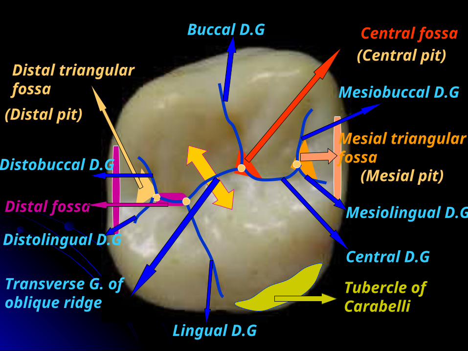
Surface anatomy Central fossa
Distal fossa
Distal triangular fossa
Mesial triangular fossa
(Central pit)
(Mesial pit)
(Distal pit)
Buccal D.G
Mesiobuccal D.G
Mesiolingual D.G
Central D.G
Lingual D.G
Distobuccal D.G
Distolingual D.G
Transverse G. of oblique ridge
Tubercle of Carabelli
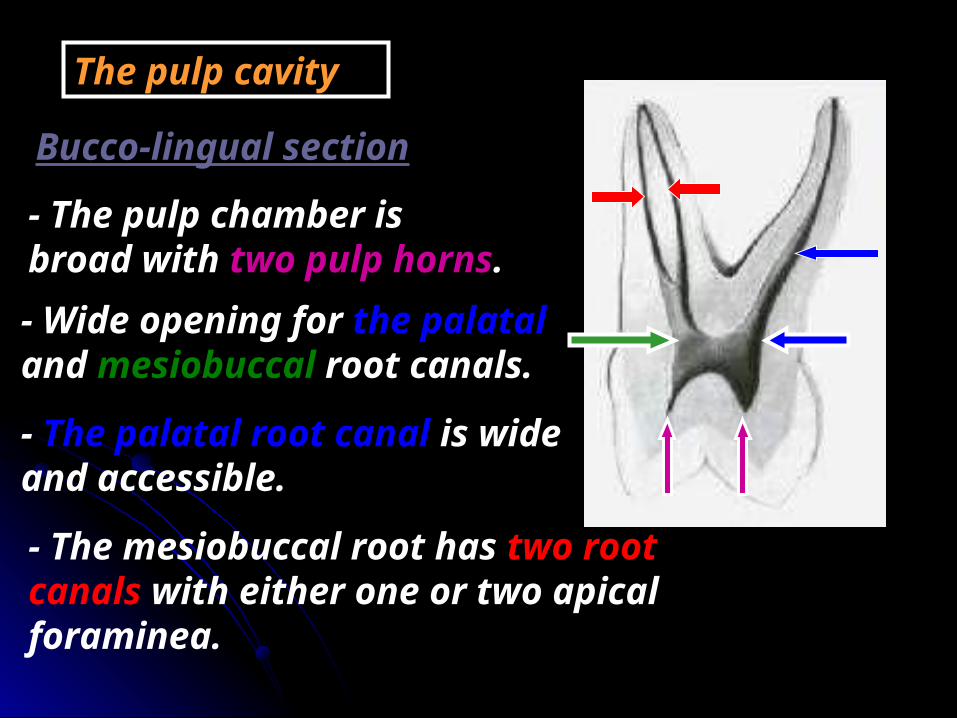
Bucco-lingual section
The pulp cavity
- The pulp chamber is broad with two pulp horns.- Wide opening for the palatal and mesiobuccal root canals.
- The palatal root canal is wide and accessible.
- The mesiobuccal root has two root canals with either one or two apical foraminea.

Mesio-distal section
- The pulp chamber is not wide with two pulp horns.
- The mesiobuccal and distobuccal canals are narrow and tapering to the apex.
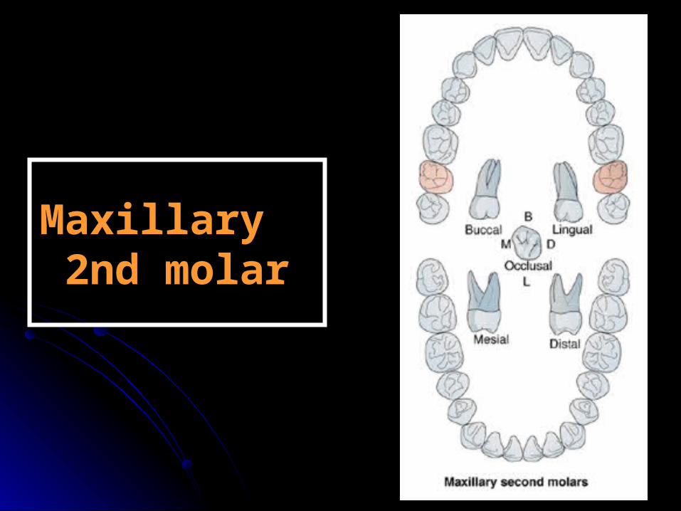
Maxillary Maxillary 2nd molar2nd molar

ChronologyAppearance of the dental organ one yearFirst evidence of calcification 2.5-3 years.Enamel completed 7-8 years.Eruption 12-13 years.Root completed 14-16 years.
Type and functionThis tooth has the function of chewing and grinding food.
No. of lobesIt has four lobes: two buccal and two lingual.

7 8
RelationThe upper 1st molar makes contact mesially with the distal surface of the upper 1st molar and distally with the mesial surface of the 3rd permanent molar.
6

No. of surfaces It has four surfaces and Occlusal aspect.
Buccal Lingual Mesial Distal
Occlusal
No. of roots It has three roots (two
buccal and one palatal).
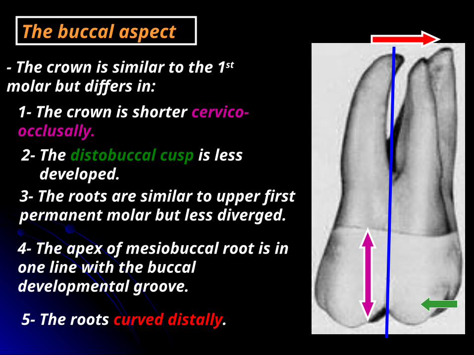
The buccal aspect
- The crown is similar to the 1st molar but differs in:
1- The crown is shorter cervico-occlusally.2- The distobuccal cusp is less
developed.3- The roots are similar to upper first permanent molar but less diverged.
4- The apex of mesiobuccal root is in one line with the buccal developmental groove.
5- The roots curved distally.

Lingual aspect
- Similar to 6 but differs in:
1- The distolingual cusp is smaller and in some cases may be missed.
2-No tubercle of Carabelli on the mesiolingual cusp.
3-The apex of the lingual root is in one line with the cusp tip of the distolingual cusp.

The mesial aspect
Similar to 6 but differs in:
2- No cusp of Carabelli .
1- The crown is shorter

The distal aspect
Similar to 6 but differs in:
The distolingual and the distobuccal cusps are smaller.
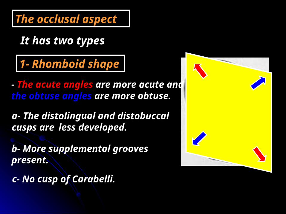
The occlusal aspect
It has two types
1- Rhomboid shape
- The acute angles are more acute and the obtuse angles are more obtuse.
a- The distolingual and distobuccal cusps are less developed.
b- More supplemental grooves present.
c- No cusp of Carabelli.
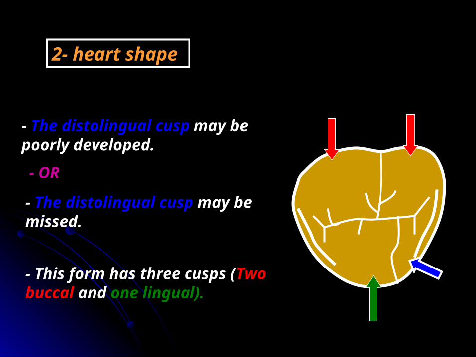
2- heart shape
- The distolingual cusp may be poorly developed.
- The distolingual cusp may be missed.
- This form has three cusps (Two buccal and one lingual).
- OR
Maxillary Maxillary 3rd molar3rd molar
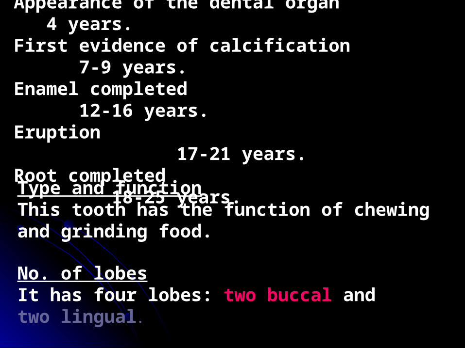
ChronologyAppearance of the dental organ 4 years.First evidence of calcification 7-9 years.Enamel completed 12-16 years.Eruption 17-21 years.Root completed 18-25 years.
Type and functionThis tooth has the function of chewing and grinding food.
No. of lobesIt has four lobes: two buccal and two lingual.
7 8
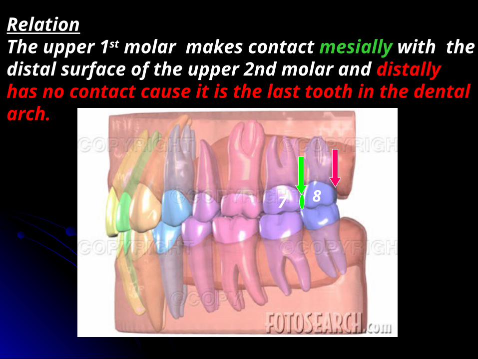
RelationThe upper 1st molar makes contact mesially with the distal surface of the upper 2nd molar and distally has no contact cause it is the last tooth in the dental arch.
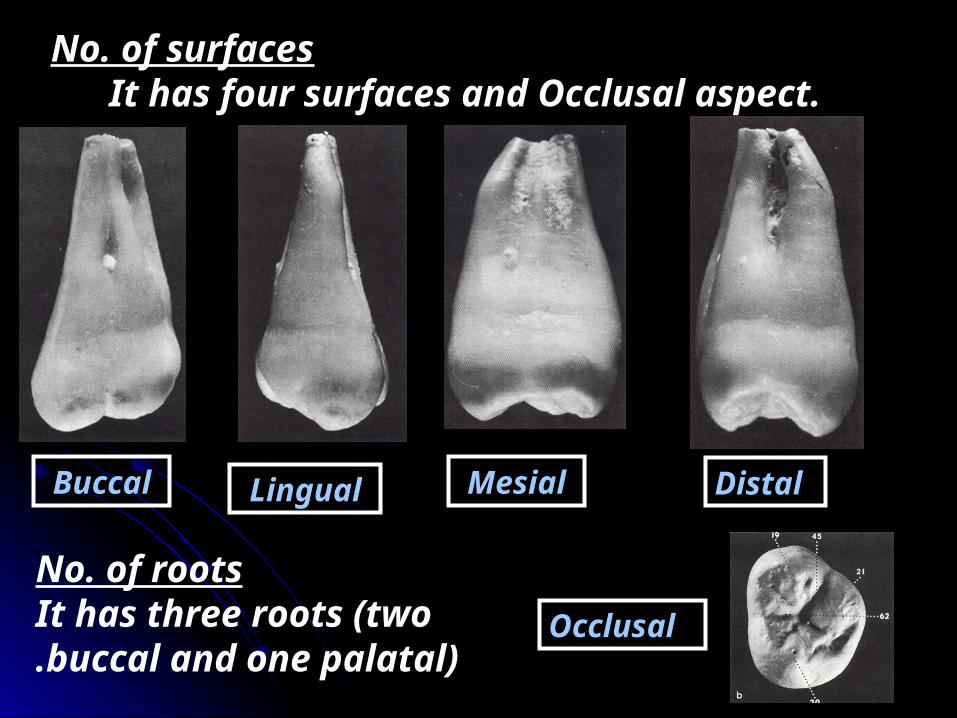
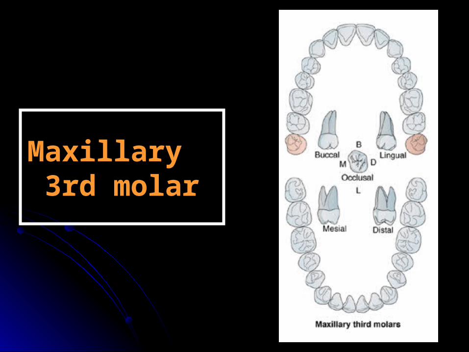
No. of surfaces It has four surfaces and Occlusal aspect.
Buccal Lingual Mesial Distal
OcclusalNo. of roots
It has three roots (two buccal and one palatal).

- The roots are short, poorly developed roots which are curved distally. However, the roots are sometimes so close together that may be completely fused.
☻No standard form observed for this tooth thus, it is hard to describe a typical maxillary third molar.

There are 2 types of the occlusal surface
1- The most common occlusal outline is heart shape, where the tooth has three cusps ( mesiobuccal, distobuccal and lingual cusps).
2- Rhomboid type with four cusps, the distolingual cusp is small and non-functioning cusp. Also the oblique ridge is poorly developed or completely absent.
- Many supplemental grooves are distributed in occlusal surface of the third molar


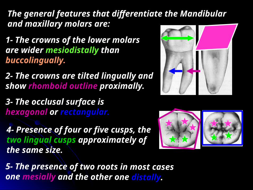
The general features that differentiate the Mandibular and maxillary molars are:
1- The crowns of the lower molars are wider mesiodistally than buccolingually.
4- Presence of four or five cusps, the two lingual cusps approximately of the same size.
3- The occlusal surface is hexagonal or rectangular.
2- The crowns are tilted lingually and show rhomboid outline proximally.
5- The presence of two roots in most cases one mesially and the other one distally.
Mandibular Mandibular 1st molar1st molar

ChronologyAppearance of the dental organ 4 m.i.u.First evidence of calcification At birth.Enamel completed 2.5-3 years.Eruption 6-7 years.Root completed 9-10 years.
Type and functionThis tooth has the function of chewing and grinding food.
No. of lobesIt has five lobes: two buccal, one distal and two lingual.
75
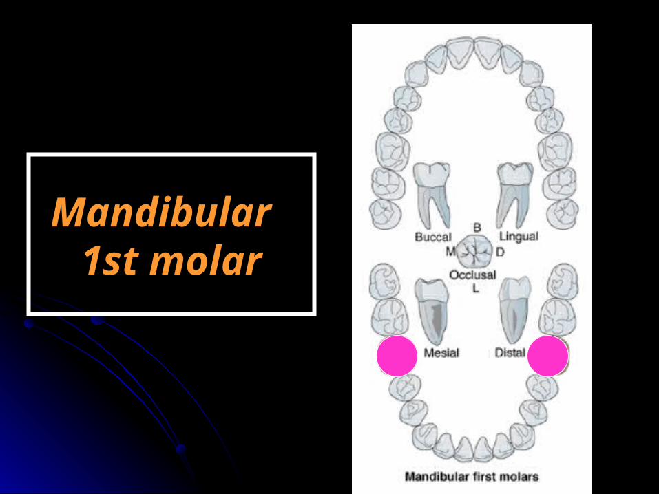
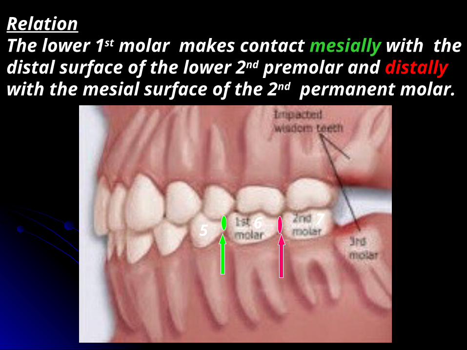
RelationThe lower 1st molar makes contact mesially with the distal surface of the lower 2nd premolar and distally with the mesial surface of the 2nd permanent molar.
6

No. of surfaces It has four surfaces and Occlusal aspect.
Buccal Lingual Mesial Distal
Occlusal
No. of roots It has two roots (one
mesial and one distal).
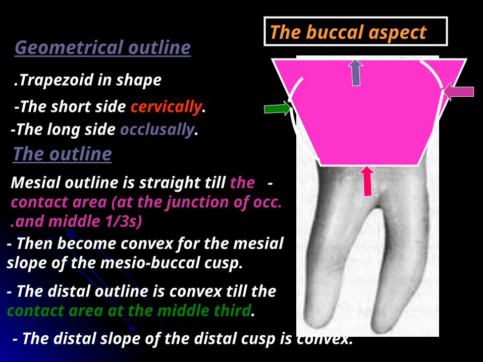
The buccal aspectGeometrical outlineTrapezoid in shape.
-The short side cervically.-The long side occlusally.The outline
-Mesial outline is straight till the contact area (at the junction of occ. and middle 1/3s).
- Then become convex for the mesial slope of the mesio-buccal cusp.
- The distal outline is convex till the contact area at the middle third.- The distal slope of the distal cusp is convex.

-The widest of them is the mesiobuccal then distobuccal and the smallest one is
the distal cusp .
- The cervical line is straight or slightly convex.
-The mesiobuccal developmental groove - Cervical ridge.Surface anatomy
-The occlusal outline is divided into three unequal cusps.
-The mesiobuccal and distobuccal cusp tips are blunt while the distal cusp tip is sharp.
-The distobuccal developmental groove
- The two developmental grooves may fade out near the middle of crown or end in a pit.
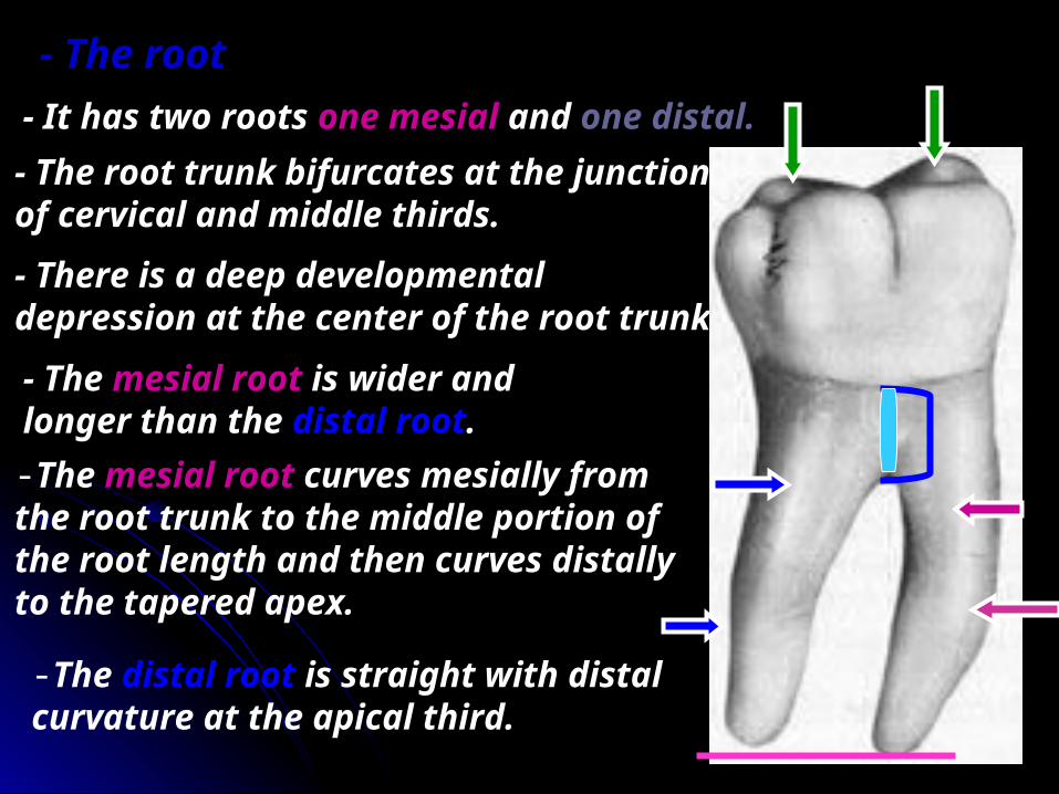
- The root trunk bifurcates at the junction of cervical and middle thirds. - There is a deep developmental depression at the center of the root trunk.
- The root- It has two roots one mesial and one distal.
- The mesial root is wider and longer than the distal root. -The mesial root curves mesially from the root trunk to the middle portion of the root length and then curves distally to the tapered apex.
-The distal root is straight with distal curvature at the apical third.
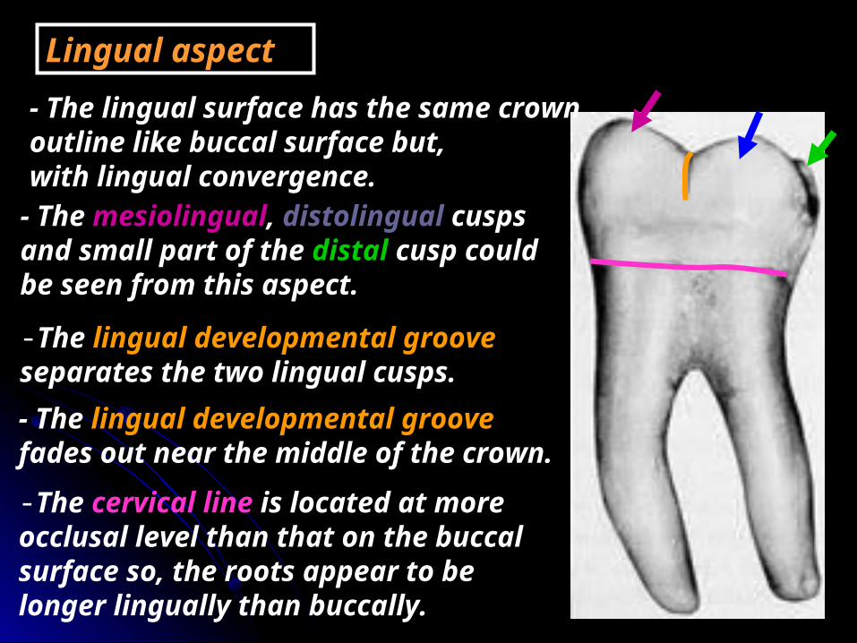
Lingual aspect- The lingual surface has the same crown outline like buccal surface but, with lingual convergence.- The mesiolingual, distolingual cusps and small part of the distal cusp could be seen from this aspect.
-The lingual developmental groove separates the two lingual cusps. - The lingual developmental groove fades out near the middle of the crown. -The cervical line is located at more occlusal level than that on the buccal surface so, the roots appear to be longer lingually than buccally.
The mesial aspect
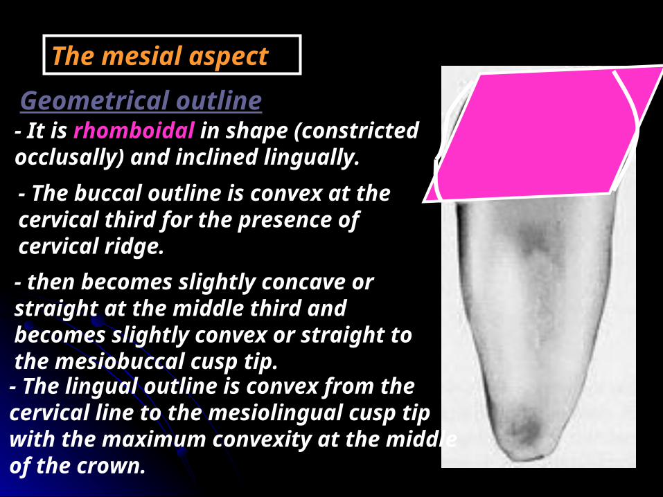
- It is rhomboidal in shape (constricted occlusally) and inclined lingually.
Geometrical outline
- The buccal outline is convex at the cervical third for the presence of cervical ridge. - then becomes slightly concave or straight at the middle third and becomes slightly convex or straight to the mesiobuccal cusp tip.- The lingual outline is convex from the cervical line to the mesiolingual cusp tip with the maximum convexity at the middle of the crown.
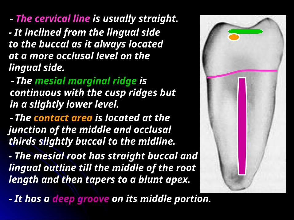
- The cervical line is usually straight. - It inclined from the lingual side to the buccal as it always located at a more occlusal level on the lingual side.-The mesial marginal ridge is continuous with the cusp ridges but in a slightly lower level.-The contact area is located at the junction of the middle and occlusal thirds slightly buccal to the midline.- The mesial root has straight buccal and lingual outline till the middle of the root length and then tapers to a blunt apex.
- It has a deep groove on its middle portion.
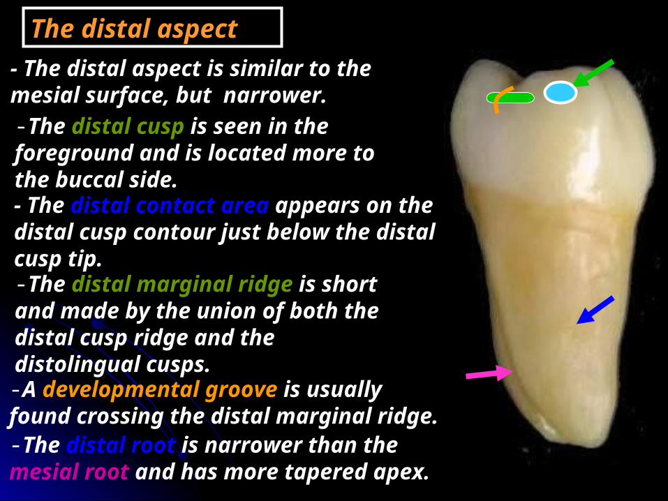
The distal aspect- The distal aspect is similar to the mesial surface, but narrower. -The distal cusp is seen in the foreground and is located more to the buccal side.- The distal contact area appears on the distal cusp contour just below the distal cusp tip. -The distal marginal ridge is short and made by the union of both the distal cusp ridge and the distolingual cusps.-A developmental groove is usually found crossing the distal marginal ridge.-The distal root is narrower than the mesial root and has more tapered apex.
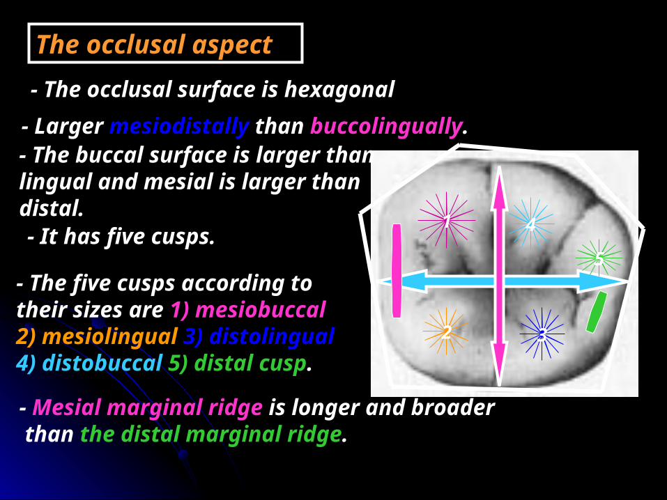
- The occlusal surface is hexagonal
The occlusal aspect
- Larger mesiodistally than buccolingually.- The buccal surface is larger than lingual and mesial is larger than distal.- It has five cusps.
- The five cusps according to their sizes are 1) mesiobuccal 2) mesiolingual 3) distolingual 4) distobuccal 5) distal cusp.
1 4
5
32
- Mesial marginal ridge is longer and broader than the distal marginal ridge.

- The occlusal surface has three fossae:
1- the central fossa
2- mesial triangular fossa
3- distal triangular fossa
central developmental groove
Mesiobuccal developmental groove
Distobuccal developmental groove
The lingual developmental groove
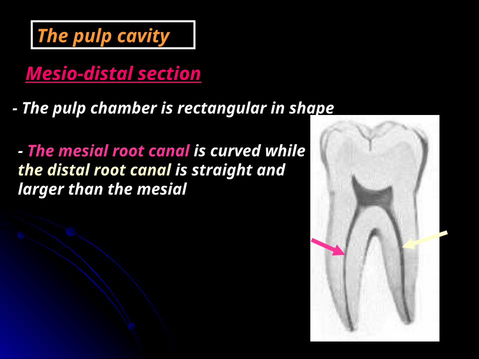
- The pulp chamber is rectangular in shape
The pulp cavity
Mesio-distal section
- The mesial root canal is curved while the distal root canal is straight and larger than the mesial
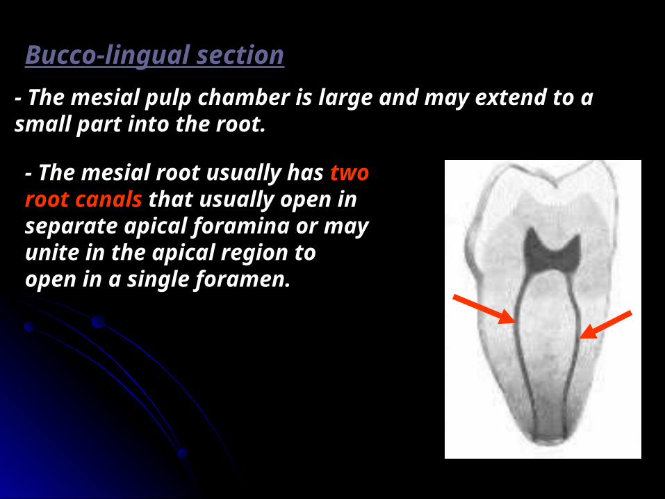
Bucco-lingual section- The mesial pulp chamber is large and may extend to a small part into the root.
- The mesial root usually has two root canals that usually open in separate apical foramina or may unite in the apical region to open in a single foramen.
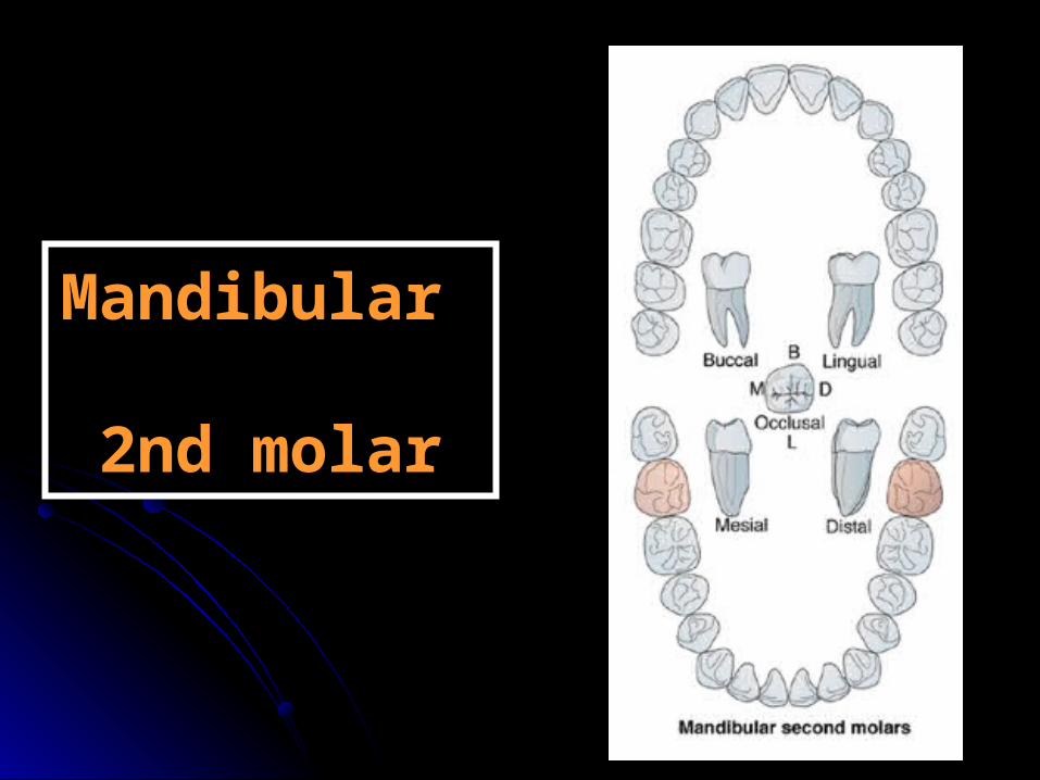
Mandibular Mandibular 2nd molar2nd molar

ChronologyAppearance of the dental organ 1 year.First evidence of calcification 2.5-3 years.Enamel completed 7-8 years.Eruption 11-13 years.Root completed 14-15 years.
Type and functionThis tooth has the function of chewing and grinding food.
No. of lobesIt has four lobes: two buccal and two lingual.

7 86
RelationThe lower 2nd molar makes contact mesially with the distal surface of the lower 1st molar and distally with the mesial surface of the 3rd molar.
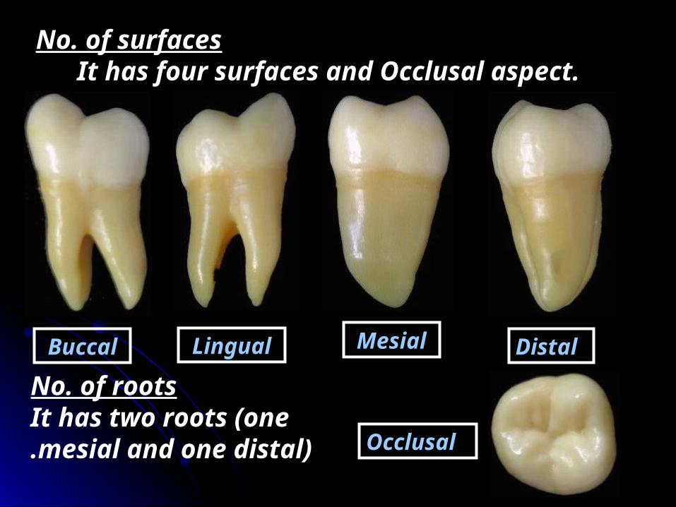
No. of surfaces It has four surfaces and Occlusal aspect.
Buccal Lingual Mesial Distal
Occlusal
No. of roots It has two roots (one
mesial and one distal).

It is similar to the Mandibular first molar except in the following:
1- The crown measurements are generally smaller than that of first permanent molar (smaller mesiodistally and occlusocervically).

2- The crown is usually composed of four developmental lobes. The buccal groove divides the mesiobuccal and distobuccal cusps equally and the lingual groove divides the mesiolingual and distolingual cusps equally.
3- The two roots are smaller, shorter and less divergent

4- The contact areas mesially and distally are located at the same level in the middle third.
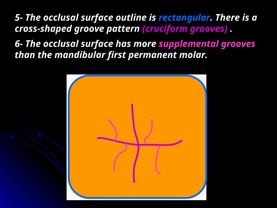
5- The occlusal surface outline is rectangular. There is a cross-shaped groove pattern (cruciform grooves) .6- The occlusal surface has more supplemental grooves than the mandibular first permanent molar.

7- There are three fossae with three developmental pits in their bottom as seen in the Mandibular first molar.
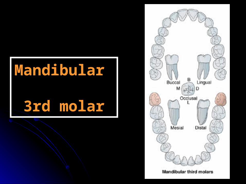
Mandibular Mandibular 3rd molar3rd molar

ChronologyAppearance of the dental organ 4 year.
First evidence of calcification 8-10 years.Enamel completed 12-16 years.Eruption 17-21 years.Root completed 18-25 years.
Type and functionThis tooth has the function of chewing and grinding food.
No. of lobesIt has four lobes: two buccal and two lingual.

7 8
RelationThe lower 3rd molar makes contact mesially with the distal surface of the lower 2nd molar and distally has no contact cause it is the last tooth in the dental arch.

No. of surfaces It has four surfaces and Occlusal aspect.
Buccal Lingual Mesial Distal
Occlusal
No. of roots It has two roots (one
mesial and one distal).

- It is usually smaller in all dimensions than any mandibular molars but, sometimes it may reach the size of the first permanent mandibular molar.- Its crown is variable but two basic types are usually seen:1- The crown resembles the permanent mandibular first
molar with five cusps and similar occlusal pattern with increase number of supplemental grooves.

2- The crown resembles the permanent mandibular second molar, it has 4 cusps and the same occlusal pattern with increase number of supplemental groove.

By: Mohammed Reda SharkesBy: Mohammed Reda SharkesReferences: References: • Notes of Dental anatomy, Physiology And Notes of Dental anatomy, Physiology And
Occlusion. by Prof. Dr. Nahed E.Abo-Azma.Occlusion. by Prof. Dr. Nahed E.Abo-Azma.
• Ash,Major M and stanley j.Nelson,2003. Ash,Major M and stanley j.Nelson,2003. • Wheeler dental anatomy,physiology and Wheeler dental anatomy,physiology and
occlusion.8occlusion.8thth edition. edition. • www.permanent teeth.com teeth.com
Permanent TeethPermanent Teeth


