
J
The Purification and Characterisation of a Prolyl Oligopeptidase
from the Cytosolic Fraction of Bovine Whole Brain
“ V
Thesis Submitted for the Degree of
Doctor of Philosophy
by
Oonagh Dowling B.Sc.
Supervised by
Dr. Brendan O’Connor
School of Biological Sciences
Dublin City University
August 1998

Declaration
I hereby certify that this material, which I now submit for assessment on the program of study leading to the award of Doctor of Philosophy is entirely my own work and has not been taken from the work of others save and to the extent that such work has been cited and acknowledged within the text of my work.S ig n e d : O o W ^ SouJQ'hq D a t e :

For my parents, Theresa and Lawrence, with love and thanks.
Hope
Hope is the thing with feathers That perches in the soul,And sings the tune without the words, And never stops at all,
And sweetest in the gale is heard,And sore must be the storm That could abash the little bird That kept so many warm.
I’ve heard it m the chillest land,And on the strangest sea,Yet, never in extremity,It asked a crumb of me.
Emiliy Dickinson
II

Acknowledgements
My supervisor Dr. Brendan O’Connor, for his enthusiasm and support throughout my project
My lab-mates, past and present, Phil, Damo, Seanin, Ultan, Yvonne and Seamus I couldn’t have
worked with a nicer bunch of people
Other DCU postgrads, Anne Mane (whom I could always count on for coffee and wisdom), Bemie,
we’ll just go for the one. Manning, Sue-Ann and Damien
Richard O’Kennedy for his advice and support over the years
Hans-Ulnch Demuth, Fred Rosche, Michael Werner, Jom Schmidt and the rest of the Halle crew for i
iletting me use their mass spec, all their advice, help, hospitality, samples etc j
!My family, Mum, Dad, Mary, Con, Paul and Laurence for their constant support, both emotional and i
financial throughout my PhD-no words of thanks can express how I feel
Brenda and Marread for their friendship
Last and by no means least, house-mates, 1 confidantes, and dearest friends, Maria, Caroline and
Deirdre I couldn’t imagine what the past four years would have been like without you and I’ll miss
you
I wish to sincerely thank the following
HI

Abbreviations
The following abbreviations a re used throughout this text.
ChAT Choline acetyltransferase
ACTH Adrenocorticotropic hormone
AD Alzheimer’s disease
ADNF-14 Activity-dependent ncurotrophic factor-14
AEBSF 4-(2-Aminoethyl)-benzenesulfonylfluonde
APP Amyloid A4 precursor protein
AVP Arginine vasopressin
BCA Bicinchoninic acid
Bisacryl Bisacrylamide
pNA P-Naphthylamide
Boc Butoxycarbonyl
BPP Bradykinin-potentiating peptide
BSA Bovine serum albumin
Bz Benzyloxyl
CDTA 1,2-Cyclohexanediamine tetraacetic acid
CLIP Corticotropm-like mtermediate peptide
CN 2-Nitrile
Da Daltons
DEAE Diethylammocthyl
DFP Dusofluorophosphate
DMF Dunethylformamide
DMSO D imethy Isu Iphoxide
DPPH Dipeptidyl ainmopeptidase II
DPPIV Dipcplidyl ainmopeptidase IV
DTNB 5 ’ ,5 ’ -Di thio-(-2-mtrobenzoic acid)
DTT Ditluothreitol
EDTA Ethylenediaminetetraacetic acid
EGTA [EUiylenebis(oxyethylenenitnlo)] tetraacetic acid
EH Eadie-Hofstee
Fmoc 9-Fluorenylmethoxycarbonyl
HPLC High perfonnance liquid chromatography
HW Hanes-Woolf
Ki Inhibitor dissociation constant
IV
Km Michaehs-Menten constant
LB Lmeweaver-Burk
LHRH Luteinizing honnone releasing hormone
MALOI-TOF Matrix-assisted laser desorption time of flight
MCA 7-Amino-4-methyl-coumann
MeOH Methanol
MES 2-[N-Morpholino]ethanesulphonic aad
MM Micbaehs-Menten
M/Z Mass/charge ratio
N.D. Not determined
NEM N-Elhylmaleunide
ONp pNitrophenyl ester
PAGE Polyacrylamide gel electrophoresis
PBE Polybuffer exchanger
PCMB p-Chloromercunbenzoate
PDA Photo diode array
PE Prolyl endopeptidase/ Proline endopeptidase
PEG Polyethylene glycol
pGlu Pyroglutamic acid
PMSF Phenylmethylsulphonylfluonde
pNA p-Nitroanihde
PO Prolyl oligopeptidase
PPCE Post-proline cleaving endopeptidase
PS Paradoxical sleep
Pyrr Pyrrolidine
REM Rapid eye movement
Rf Relative mobility
S.D. Standard deviation
SDS Sodium dodecyl sulphate
SEM Standard error mean
SM Sulphamethxazole
Suc Succinyl
TEMED N, N, N, N’-Tetramethyl ethylenediamme
TFA Tnfluoroacetic acid
TRH Thyrotropin releasing honnone
TRH-OH Acid TRH
Tris Tris(hydroxymethyl)amino methane
V

Ve Elution volume
Vo Void volume
v/v Volume per volume
w/v Weight per volume
Xaa- Any amino acid
Yaa- Any ammo acid
Z- N-Benzyloxycarbonyl-
ZIP Z-Pro-prolinal insensitive Z-Gly-Pro-MCA hydrolysing peptidase
ZTTA Z-Thiopro-thioprolmal
ZPP Z-Pro-prolinal
Amino Acid Abbreviations
Ala/A Alanine Leu/L Leucme
Arg/R Arginine Lys/K Lysme
Asn/N Asparagine Met/M Methionine
Asp/D Aspartate Phe/F Phenylalanine
Cys/C Cysteine Pro/P Proline
Gln/Q Glutamine Ser/S Senne
GIu/E Glutamate Thr/T Threonine
His/H Histidine TrpAV Tryptophan
Gly/G Glycine Tyr/Y Tyrosine
Ile/I Isoleucme Val/V Valine
VI

Abstract
Cytosolic bovine brain prolyl oligopeptidase was purified from whole brain using ammonium suphate
precipitation and chnitnjtography with DEAE sepharose, S200 gel filtration, chromatofocusmg and
phenyl sepharose An overall recovery of 23% and purification factor of 253 was achieved
The relative molecular mass of the brain PO, determined by gel filtration chromatography, was found
to be 69 5 kDa This was confirmed by SDS PAGE The purified enzyme was relatively unstable
under assay conditions However the presence of 0 5% w/v BSA improved its stability and the assay
linearity Activity was also inhibited strongly by solvents with DMF being the most inhibitory DMSO
was found to be the optimal solvent in terms of enzyme activity and substrate solubility Optimal
enzyme activity was observed at 37°C with complete inactivation occurring at temperatures of 50°C
or more A pH optimum of 7 4 and a preference for phosphate buffer was found for the enzyme with
complete inactivation of activity below pH 4 5 and above pH 10
The brain PO was confirmed to be a serine protease based on its sensitivity to AEBSF The enzyme
was also inhibited strongly by some cysteine protease inhibitors such as NEM and DTNB and was
activated by DTT Sensitivity to these agents would suggest the presence of a cysteme residue in close
proximity to the active site Some divalent metal salts also exerted some inhibitory effects on activity
with Hg2+, Cu2+, Cd2+, Co2+ and Ni2+ being the most potent(
Substrate spcUilcity studies performed on the purified bovine brain, partially purified bovine serum
and recombinant Flavobacterium meningosepticum PO activity revealed that this oligopeptidase
could hydrolyse a range of proline containing peptides including TRH, LHRH, ADNF-14, substance
P, neurotensin, CLIP, a 45 amino acid residue Gly-Pro-Ala polymer and the APP fragment 708-715
N-blocked prolme containing dipeptides, including Z-Pro-Pro-OH were not hydrolysed by any of the
enzyme The smallest synthetic sequences hydrolysed were an N-blocked tripeptide and a
tetrapeptide
The brain, serum and bacterial activities hydrolysed Z-Gly-Pro-MCA, with Km values of 62 5, 14 6
and 38 5(iM respectively The TRH analog pGlu-His-Pro-MCA was also hydrolysed by all three
activities with higher Km values of 99 8, 52 1 and 73 5(xM for the brain serum and recombinant
Flavobacterium meningosepticum enzyme respectively
A number of prolme-containing peptides were also found to competitively inhibit all three activities
Of these angiotensins I, II and II were the most potent The TRH analogs, Glu2TRH and Phe1TRH
exhibiting the lowest inhibitory potency
VII

Inhibition studies performed using a range of PO-specific inhibitors revealed a-ketobenzothiazole to
be the most potent inhibitor of bovine brain PO with an IC50 value of 63pM The classical PO
inhibitor Z-Pro-prolinal inhibited all three activities with IC50 values of 7-10nM With regard to the
majority of inhibitors tested, the brain, serum and bacterial enzymes were similar m their sensitivity
However the brain and serum activities were approximately 4000 times less sensitive than the
bacterial enzyme to inhibition by the N-blocked dipeptide analog, Z-Phe-Pro-methylketone An
investigation into the time course inhibition of bram and bacterial activities by Z-Phe-Ala-
chloromethylketone found that while the bacterial enzyme was completely inhibited by lxlO'5 M of
this inhibitor alter 60 minutes, the bram enzyme was completely insensitive to inhibition
t
VIII

TABLE OF CONTENTS
Declaration IAcknowledgements HIAbbreviations IVAbstract VII
1. INTRODUCTION
1.1 Proline 1
1.11 Structural Characteristics 1
1.1.2 Physiological Implications of Proline in Peptides 3
1.1.3 Proline-Specific Peptidases 4
1.2 The Discovery of Prolyl Oligopeptidase 5
1.3 Biochemical and Biophysical Characteristicsof Prolyl Oligopeptidase 6
1.31 Molecular Weight 6
1.3.2. Isoelectric Point, pH and Temperature Optima 7
1.3.3 Catalytic Classification of Prolyl Oligopeptidase 913 3 1 Sensitivity to Protease inhibitors 91 3 3 2 Cloning and Sequencing of the Prolyl Oligopeptidase Gene 91 3 3 3 Prolyl Oligopeptidase Catalytic Mechanism Studies 12
14 Prolyl Oligopeptidase Specificity 14
1.4 1 Specificity for Prolme Residues 14
1.4 2 Substrate- Size Limitation 16
1-4 3 Prolyl Oligopeptidase Hydrolysis of Neuro/VasoactivePeptides 17
1.4.4 Prolyl Oligopeptidase Specific Assays 19
1.5 Specific Inhibitors of Prolyl Oligopeptidase 20
1-51 Synthetic Inhibitors 20
1.5.2 Inhibitors of Bacterial origin 22
1.5.3 Endogenous Inhibitors 22
IX
1.6 Distribution 27
1.7 Physiological Relevance of Prolyl Oligopeptidase 30
1.7.1 Prolyl Oligopeptidase Ontogenic Studies-EvidenceFor a Possible Role in Cell Growth 30
1.7.2 Involvement of Prolyl Oligopeptidase in the Pathogenesisof Neurological and Psychiatric Disorders 32
17 2 1 Prolyl Oligopeptidase and Major Depression 3217 2 2 Prolyl Oligopeptidase and Schizophrenia 3317 2 3 Prolyl Oligopeptidase and Neurodegenerative Disorders 3417 2 4 Prolyl Oligopeptidase and Cerebral Ischemia,
Memory and Learning 35
2.0 MATERIALS AND METHODS
2.1 Materials 37
2.2 Fluorescence Spectrophotometry using7-Amino-4-MethyI-Coumarin (MCA) 41
2.2.1 MCA Standard Curves 41
2.2 2 Inner Filter Effects 41
2.3 Protein Determination 41
2.3.1 Absorbance at 280nm 41
2.3.2 Standard BCA Assay 41
2.3.3 Enhanced BCA Assay 42
2.3 4 Biorad Protein Assay 42
2.4 Enzyme Assays 42
2.4 1 Quantitative Measurement of Prolyl OligopeptidaseActivity 42
Quantitative Measurement of Z-Pro-Prolinal Insensitive Z-Gly-Pro-MCA Hydrolysing Activity 43
2.4.3 Non-Quantitative Microplate Procedure forMeasurement of Prolyl oligopeptidase Activity 43
2.4.4 Non-Quantitative Microplate Procedure forMeasurement of Z-Pro-Prolinal Insensitive Z-Gly-Pro-MCA Hydrolysing Activity 43
X

2.5 Partial Purification of Prolyl Oligopeptidasefrom Bovine Serum 43
2.5.1 Bovine Serum Production 43
2.5 2 Phenyl Sepharose CL-4B H I Chromatography I 44
2.5.3 Phenyl Sepharose CL-4B H I Chromatography II 44
2.6 Purification of Prolyl Oligopeptidase fromBovine Brain 44
2.6.1 Bovine Brain Preparation 44
2.6.2 Ammonium Sulphate Precipitation 45
2.6.3 Deae Sepharose Fast flow Anion ExchangeChromatography 45
2 6.4 Sephacryl S -200 Sepharose Gel FiltrationChromatography 45
2.6.5 PBE 94 Chromatofocusing 462 6 6 Phenyl Sepharose Cl -4B Hydrophobic Interactions
Chromatography 46
2.7 Determination of Purity of Purified bovineBrain Prolyl Oligopeptidase 47
2.7.1 Polyacrylamide Gel Electrophoresis 472 7 11 Sample Preparation 472 7 1 2 Preparation of SDS Gels 472 7 13 Visualising Proteins in Polyacrylamide Gel-Silverstainmg 48
2.7.2 Fluorimetnc Assays 49
2.8 Purified Prolyl Oligopeptidase AssayDevelopment 50
2.8.1 Solvents Effects on Purified Prolyl OligopeptidaseActivity and Assay Sensitivity 50
2 8.2 Linearity of Purified Prolyl OligopeptidaseActivity Assay with Respect to Time 50
2 8 2 1 Discontinuous Assay 502 8 2 2 Continuous Assay 50
2 8.3 Linearity of Purified Prolyl Oligopeptidase Assay withRespect to Time Enzyme Concentration 50
2.8.4 Effects of DTT on Prolyl Oligopeptidase Activity AssaySensitivity 51
2 8.5 Effects of EDTA on Prolyl Oligopeptidase Activity AssaySensitivity 51
XI

286
2.9
2.9.12 9 1 12 9 1 1 12 9 1 1 22 9 1 1 3
2 9 1 2
2.9.22 9 2 12 9 2 2
2 9.3
2 9 3 1
2 9 3 2
2.9.4
2.9.5
2.9.6
2 9 6 12 9 6 1 1 2 9 6 12
2 9 6 1 3
2 9 6 22 9 6 2 12 9 6 2 2
2 9 6 2 3
2 9 6 3 2 9 6 3 12 9 6 3 22 9 6 3 3
2.9.7
297 1
Effects of NACL on Prolyl Oligopeptidase Activity Assay Sensitivity 51
Characterisation of Purified Prolyl Oligopeptidase Activity 51
Relative Molecular Mass Determination 51Sephacryl S-200 HR Gel Filtration Chromatography 51Void Volume Determination 51Column Calibration using Molecular Mass Standards 51Determination of Relative Molecular Mass of Purified Enzyme 52
SDS Polyacrylamide Electrophoresis 52
pH Effects 52pH Activity Profile 52pH Inactivation Profile 52
Thermostability Studies on Purified Prolyl Oligopeptidase Activity 53Effects of Assay Temperature on Purified Prolyl Oligopeptidase Activity 53Effects of Preincubation on Purified Prolyl Oligopeptidase Activity at Various Temperatures for Different Times 53
Effects of Divalent Metals Salts on Purified Prolyl Oligopeptidase Activity 53
Effects of Functional Reagents on Purified Prolyl Oligopeptidase Activity 54
Substrate Specificity Studies on Prolyl Oligopeptidase Activity 56
Substrate specificity studies using Reverse Phase HPLC 56Preparation of Stock Substrates and Standards 56Reaction of Substrates with Prolyl Oligopeptidase Enzyme Activity 56Reverse Phase HPLC of Samples 56
Substrate Specificity using MALDT-TOF Mass Spectrometry 58 Preparation of Stock Substrates 58Reaction of Substrates with Purified Prolyl Oligopeptidase Activity 58MALDI -TOF Mass Spectrometry of Samples 58
Kinetic studies 59Km Determination for Z-Gly-Pro-MCA 59Km Determination for pGlu-His-Pro-MCA 59Ki Determination for Proline-Containmg Peptides 59
Effect of Specific Prolyl Oligopeptidase Inhibitors on Purified Prolyl Oligopeptidase Activity 60
Determination of IC50 Values for Specific Prolylxn
Oligopeptidase 60inhibitorsEffects of Z-Phe-Ala-Chloromethylketone on Purified Bovine Brain and Recombinant Prolyl Oligopeptidase Activity with Respect to Time 61
RESULTS
MCA Standard Curves-Quenched and Unquenched 63
Protein Standard Curves 63
Measurement of Prolyl Oligopeptidase and Z-Pro-Prohnal Insensitive Z-Gly-Pro-MCA Hydrolysing Activity 63
Partial Purification of Prolyl Oligopeptidase from Bovine Serum 69Serum Preparation 69Phenyl Sepharose H I Column Chromatography I 69Phenyl Sepharose H I Column Chromatography II 69
Purification of Prolyl Oligopeptidase Activity from the Cystolic Fraction of Bovme Brain 73Bovine Brain Preparation 73Ammonium Sulphate Precipitation 73Deae Sepharose Fast Flow Anion Exchange Chromatography 73 Sephacryl S200 Sepharose Gel Filtration Chromatography 73 PBE 94 Chromatofocusmg 73Phenyl Sepharose H I Column Chromatography 73
Determination of Puntv of Bovine Bram Prolvl Ohgoneptidase 80Polyacrylamide Gel Electrophoresis 80Fluonmetnc Assays for Interfering Peptidase Activity 81
Purified Prolyl Oligopeptidase Activity Assay Development 81Solvent Effects on Purified Prolyl Oligopeptidase Activity and Assay Sensitivity 81Linearity of Purified Prolyl Oligopeptidase Activity with Respect to Time 82Linearity of Purified Prolyl Oligopeptidase Activity with Respect to Enzyme Concentration 82
Effects of DTT on Purified Prolyl Oligopeptidase Activity Assay Sensitivity 82Effects of EDTA on Purified Prolyl Oligopeptidase Activity Assay Sensitivity 82Effects of NaCL on Purified Prolyl Oligopeptidase Activity Assay Sensitivity 82
Characterisation of Purified Prolyl Oligopeptidase Activity 89
Relative Molecular Mass Determination 89Sephacryl S200 HR Gel Filtration Chromatography 89

3.7.1.2 Polyacrylamide Gel Electrophoresis 89
3.7.2 pH Effects 89
3.7.3 Temperature Effects 93
3.7.4 Effects of Divalent Metal Salts on Purified ProlylOligopeptidase Activity 95
3.7.5 Effects of Functional Reagents on Purified ProlylOligopeptidase Activity 96
3.7.6 Substrate Specificity on Purified Prolvl OligopeptidaseActivity 98
3.7.6.1 Substrate Specificity Studies using Reverse Phase HPLC 983.7.6.2 Substrate Specificity with MALDI-TOF Mass Spectrometry 1103.7.6.3 Kinetic Studies 1163.7.6.3.1 Km Determination for Z-Gly-Pro-MCA 1163.7.6.3.2 Km Determination for PGlu-His-Pro-MCA 1163.7.6.3.3 Ki Determination for Proline-Containing Peptides 117
3.7.7 Effects of Specific Inhibitors on Purified Prolvl OligopeptidaseActivity 123
3.7.7.1 Determination of IC50 Values for a Range of SpecificProlyl Oligopeptidase Inhibitors 123
3.7.7.2 Effects of Z-Phe-Ala-Chloromethylketone on Purified BovineBrain and Recombinant Prolyl Oligopeptidase Activitywith Respect to Time 129
4. D IS C U S S IO N
4.1 Fluorescence Spectrophotometry and 7-Amino-4-MethylCoumarin 131
4.2 Quenching and the Inner Filter Effect 131
4.3 Measurements of Z-Gly-Pro-MCA HydrolysingActivity in Crude Brain Supernatant and Serum 132
4.4 Partial Purification of Prolyl Oligopeptidase from BovineSerum 133
4.4.1 Phenyl sepharose CL-4B Hydrophobic Interactionschromatography I and II 133
4.5 Purification of Prolyl Oligopeptidase from the CystolicFraction of Bovine Brain 134
4.5.1 Preparation of the Cystolic Fraction of Bovine Brain 1344.5.2 Ammonium Sulphate Precipitation 1344-5.3 Deae Sepharose Fast Flow Anion Exchange Chromatography 1354.5.4 Sephacryl S200 HR Gel Filtration Chromatography 1354.5.5 PBE 94 Chromatofocusing 1354.5.6 Phenyl Sepharose CL-4B hydrophobic Interactions
Chromatography 1364-5.7 Assessment of Purification Process for Prolyl Oligopeptidase
from Bovine Brain 136
4.6 Prolyl Oligopeptidase Assay DevelopmentXIV
137

137
138139141141
141141142
142
143
143143144144144
145145147
150150150
152
153
155
The Effects of Solvents on Purified Prolyl Oligopeptidase ActivityLinearity of Purified Prolyl Oligopeptidase Activity Assay with Respect to Tune and Enzyme Concentration Effects of DTT on Prolyl Oligopeptidase Activity Effects of EDTA on Prolyl Oligopeptidase Activity Effects of NaCL on Prolyl Oligopeptidase Activity
Characterisation of Purified Prolyl Oligopeptidase ActivityRelative Molecular Mass Determination pH Effects on Purified Prolyl Oligopeptidase Activity Thermostability studies on Purified Prolyl Oligopeptidase ActivityThe Effects of Divalent Metal Salts on Purified Prolyl Oligopeptidase Activity
The Effects of Functional Reagents on Purified ProlylOligopeptidase ActivitySerine Protease InhibitorsCysteine Protease InhibitorsThiol-Reducing AgentsMetal Chelators and Phenanthrohnes
Substrate SpecificityReverse Phase HPLCMALDI-TOF Mass Spectrophotometry
Kinetic StudiesKm Determination for Fluorimetnc Substrates Ki Determination for Prolme-Containing Peptides
•
The Effects of Specific Inhibitors on Prolyl Oligopeptidase Activity
Summary
BIBLIOGRAPHY
XV

1. INTRODUCTION
1.1. Proline
Amongst the twenty amino acids coded for in protein synthesis proline occupies a unique position A
highly conserved residue, and the only mammalian lmrno acid, proline’s unique cyclic structure
influences not only the conformation of peptide chains but restricts their susceptibility to proteases
(Yaron and Naider, 1993, MacArthur and Thorton, 1991)
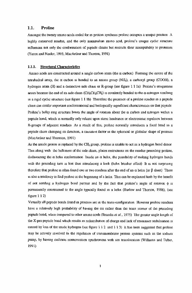
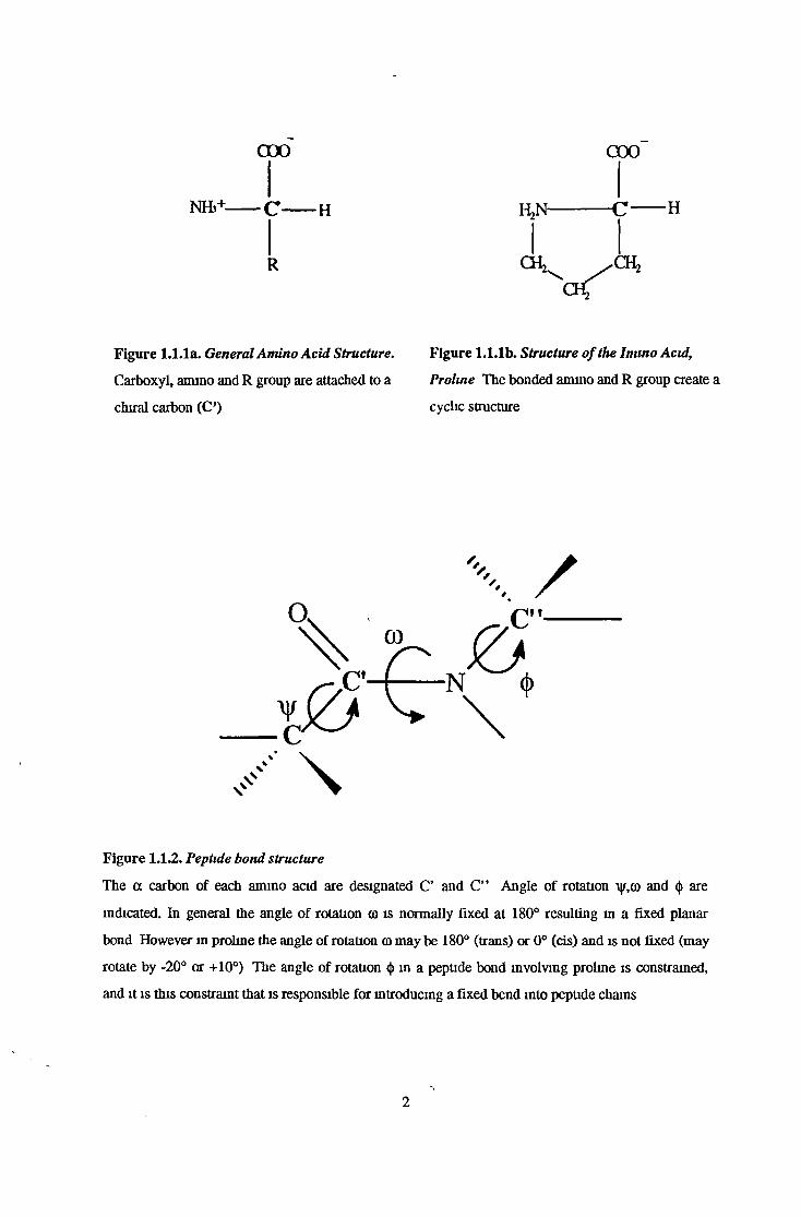
1.1.1. Structural Characteristics
Ammo acids are constructed around a single carbon atom (the a carbon) Forming the centre of the
tetrahedral array, the a carbon is bonded to an amino group (NH3), a carboxyl group (COOH), a
hydrogen atom (H) and a distinctive side chain or R-group (see figure 1 1 la) Proline’s uniqueness
arises because the end of its side chain (CH3CH2CH2) is covalently bonded to the a-nitrogen resulting
in a rigid cyclic structure (see figure 1 1 lb) Therefore the presence of a proline residue in a peptide
chain can confer important conformational and biologically significant characteristics on that peptide
Proline’s bulky rmg structure limits the angle of rotation about the a carbon and nitrogen within a
peptide bond, which is normally only reliant upon steric hindrance or electrostatic repulsion between
R-groups of adjacent residues As a result of this, proline normally introduces a fixed bend m a
peptide chain changing its direction, a causative factor m the spherical or globular shape of proteins
(MacArthur and Thornton, 1991)
As the amide proton is replaced by the CH2 group, proline is unable to act as a hydrogen bond donor
This along with the bulkiness of the side chain, places restrictions on the residue preceding proline,
disfavouring the a helix conformation Inside an a helix, the possibility of making hydrogen bonds
with the preceding turn is lost thus introducing a kink (helix breaker effect) It is not surprising
therefore that proline is often found one or two residues after the end of an a helix (or (3 sheet) There
is also a tendency to find proline at the beginning of a helix This can be explained both by the benefit
of not needing a hydrogen bond partner and by the fact that proline’s angle of rotation <() is
permanently constrained to the angle typically found m a helix (Barlow and Thorton, 1988), (see
figure 1 1 2 )
Virtually all peptide bonds found in proteins are in the trans-configuration However proline residues
have a relatively high probability of having the a s rather than the trans isomer of the preceding
peptide bond, when compared to other amino acids (Brandts et a l , 1975) The greater angle length of
the X-pro peptide bond which results in redistribution of charge and lack of resonance stabilisation is
caused by loss of the lmide hydrogen (see figure 1 1 2 and 1 1 3 ) It has been suggested that proline
may be actively involved in the regulation of transmembrane protein systems such as the sodium
pump, by having cis/trans isomerization synchronous with ion translocation (Williams and Deber,
1991)
1
0 0 0 COO
NHb+ C H HjN C H
R
Figure 1.1.1a. General Amino Acid Structure. Figure 1.1.1b. Structure ofthe Imino Acid,
Figure 1.1.2. Peptide bond structure
The a carbon of each amino acid are designated C’ and C” Angle of rotation \y,co and <j> are
indicated. In general the angle of rotation co is normally fixed at 180° resulting m a fixed planar
bond However in proline the angle of rotation co may be 180° (trans) or 0° (cis) and is not fixed (may
rotate by -20° or +10°) The angle of rotation <|) in a peptide bond involving proline is constrained,
and it is this constraint that is responsible for introducing a fixed bend into peptide chains
Carboxyl, amino and R group are attached to a
chiral carbon (C’)
Prohne The bonded amino and R group create a
cyclic structure
2

CO P2> >N N
Pi V Pi
CO P2
Fig 1.1.3. Cis and Trans-Conformation o f the X-Pro Peptide Bond with a Rotation o f 180°
Around the C-N Imide Bond
This reaction is catalysed by peptidyl-proly cis-trans isomerase (Galat, 1993)
1.1.2. Physiological Implications of Proline in Peptides
A key physiological role played by proline is the protection of biologically active peptides from
enzymatic degradation. As prolrne constitutes 5% of amino acid residue of total brain protein (Lajtha
and Toth, 1974) and is indeed present in many neuropeptides/neurohormones and vasoactive peptides
(VanHoof et a l , 1995, Meintlein, 1988), (see table 1 1 1), prolme-speafic or selective peptidases
should play an important role in the nervous system The presence of prolme may not only determine
the properties of secondary structures necessary for a peptide’s biological activity but may also hinder
any nonspecific proteolytic degradation (Yaron and Naider, 1993)
3

Protein/Peptide
-■> ^
Amino Acid Sequence
Angiotensin II Asp-Arg-Val-Tyr-Ile-His-Pro-Phe
Oxytocin Cys-Tyr-Ile-Gln-Asn-Cys-Pro-Leu-Gly-NHî
Vasopressin Cys-Tyr-Phe-Gln-Asn-Cys-Pro-Arg-Gly-NH2
Bradykinin Arg-Pro-Pro-Gly-Phe-Ser-Pro-Phe-Arg
Substance P Arg-Pro-Lys-Pro-Gln-Gln-Phe-Phe-Gly-Leu-Met
Neuropeptide Y (1-14) Tyr-Pro-Ser-Lys-Pro-Asp-Asn-Pro-Gly-Glu-Asp-Ala-Pro-Ala
Pancreatic polypeptide (1-14) , Ala-Pro-Leu-Glu-Pro-Val-Tyr-Pro-Gly-Asp-Asn-Ala-Thr-Pro
Luteinizing hormone-releasing hormone pGlu-His-Trp-Ser-Tyr-Gly-Leu-Arg-Pro-Gly-NH2
Thyrotropin-releasing hormone pGlu-His-Pro-NH2
Gastrin releasing peptide (1-10) Val-Pro-Leu-Pro-Ala-Gly-Gly-Gly-Thr-Val-
Corticotropin-releasing hormone (1-10) Ser-Glu-Glu-Pro-Pro-Ile-Ser-leu-Asp-Leu-
Calcitoriin (20-32) His-Thr-Phe-Pro-Gln-Thr-Ala-Ile-Gly-Val-Gly-Ala-Pro-NH2
Bradykmm-potentiating peptide pGlu-Gly-Gly-Trp-Pro-Arg-Pro-Gly-Pro-Glu-De-Pro-Pro
Tuftsm Thr-Lys-Pro-Arg
Melanotropin Ser-Tyr-Ser-Met-Glu-His-Phe-Arg-Trp-Gly-Lys-Pro-Val-NH2
Neurotensin pGIu-Leu-Tyr-GIu-Asn-Lys-Pro-Arg-Arg-Pro-Tyr-IIe-Leu
Table 1.1.1. Prohne Containing Neuro- and Vasoactive Peptides in Human
1.13. Proline-Specific Peptidases
Given the unique structural characteristics of proline, it is not unexpected that the presence of proline
m peptide bonds generally makes them resistant to hydrolysis by peptidases, even those of broad
specificity (Yaron and Naider, 1993) However there is now known to exist, a group of proline-
specific peptidases, which have evolved to recognize the pyrrolidine ring of proline. The specificity of
these peptidases is even further limited by both the size of the peptide and the position of the proline
residue. For instance Dipeptidyl ammopeptidase II can only act on peptides of three or four amino
acid residues (Fukasawa et a l , 1983) Ammopeptidase P requires proline to be situated at the N-
termmal penultimate position, but carboxypeptidase P has a requirement for a C-terminal penultimate
prolme residue (Hedeager-Sorenson and Kenny, 1985; Yaron and Berger, 1970)
4

To date eight exopeptidases and one endopeptidase have been identified in mammals as being proline
specific in their action (Cunningham and O’Connor, 1997a), (see table 1.1.2). It is the
endopeptidase,prolyl oligopeptidase that will be the focus of this review.
Proline-SpecificPeptidase
Specificity Reference
Prolyl Oligopeptidase oop«boo 1
Dipeptidyl Peptidase IV o«ooooo12
Dipeptidyl Peptidase II 0900 3
Aminopeptidase P ckxxxx) 4
Prolidase db1 5
Proline Iminopeptidase •oo 6
Prolinase 7
Prolyl Carboxypeptidase 00000*0 8
Carboxypeptidase P oooooib 9
Table 1.1.2 : Mammmalian Proline-Specific Peptidases
Proline= Q , Amino acid= Q , 4^=Site of hydrolysis
1. Recommendations of the Nomenclature Committee of the International Union of Biochemistry and
Molecular Biology, 1992., 2. McDonald et al., 1971; 3. Fukasawa et al., 1983; 4. Yaron and Berger,
1970; 5. Yoshimoto et al., 1983a; 6. Yoshimoto et al., 1983b; 7. Mayer and Nordwig, 1973; 8. Dehm
and Nordwig, 1970; 9. Hedeager-Sorenson and Kenny, 1985.
1.2. The Discovery of Prolyl Oligopeptidase
Enzymatic hydrolysis of proline-containing peptides at either the carboxyl or amino terminus of a
peptide has been known since the 1950s (Davis et al., 1957 and 1953). However the enzymatic
degradation of peptidyl-prolyl-peptide bonds was not observed until much later. In 1971, in a study of
5

the products of uterine oxytocin degradation, an enzyme capable of cleaving the prolyl-leucyl bond of
oxytocin resulting in the release of the dipeptide leucylglycinamide was identified and purified
(Walter et a l , 1971) Further studies on this oxytocin degrading enzyme found that this peptidase,
rather than having a specificity for oxytoan was m fact specific for the ammo acid residue, prolme,
and could mediate cleavage at the carboxyl side of this residue (Walter, 1976, Koida and Walter,
1976). This enzyme has since been termed post-prohne cleaving endopeptidase (PPCE), prolyl or
prolme endopeptidase (PE), (Orlowski el al., 1979, Walter, 1976) Since its initial discovery, several
peptidases isolated and named on the basis of their specificity for a particular bioactive peptide, were
found to be identical to this proline-speafic endopeptidase For instance, TRH deamidase isolated
from rat brain (Rupnow et a l , 1979) and bovine anterior pituitary (Knisatschek and Bauer, 1979),
kinmase B from rabbit brain, which was found to cleave bradykimn (Oliveira et a l , 1976), and endo-
oligopeptidase B, which hydrolysed bradykimn, angiotensin I and II, neurotensin and LHRH
(Camargo et a l , 1984, 1983, Greene et a l , 1982), were all found to be identical to PPCE It is now
accepted that the more correct term for PE/PPCE is prolyl oligopeptidase, a term reccomended due to
the substrate-size limitation of its specificity (Camargo et a l , 1984, Barrett and Rawlings, 1992)
The existance of a new family of senne-type peptidases related to prolyl ohgopeptidase, known as the
S9 or prolyl oligopeptidase family, is now recognized (Barrett and Rawlmgs, 1992)
1.3. Biochemical and Biophysical Characteristics of Prolyl Oligopeptidase
13.1. Molecular WeightPreliminary studies on the molecular weight of PO reported that this peptidase had a dimenc
structure with a molecular weight of 115-140kDa (Mizutam et al., 1984, Koida and Walter, 1976)
However it is now known that mammalian, plant and microbial forms are monomeric with a
molecular weight ranging from 62-77kDa (Kanatani et a l , 1993, Yoshimoto et a l , 1981), (see table
13 1) With the cDNA cloning of porcine brain PO, the reported ammo acid sequence (710 amino
acids) allowed the deduction of a molecular weight for the enzyme of 80 75kDa This was in conflict
with the lower value obtain experimentally, m the same study, of 74 5kDa (Rennex et al., 1991)
Another cDNA cloning study on the human T cell form revealed again a sequence of 710 amino
acids with a deduced molecular weight of 80.75kDa (Shirasawa et a l , 1994). Investigations into the
ammo acid sequence of the Flavobacterum memngosepticum enzyme revealed a complete ammo acid
sequence of 705 ammo acids However this sequence was found to contain a signal peptide of 20
amino acids preceding the mature enzyme. It was confirmed that loss of this signal peptide leads to
the mature form of PO with an estimated molecular mass of 76 782kDa, which correlated well with
previously reported experimental values (Chevallier et a l , 1992)
6
1.3.2. Isoelectric point. pH and Temperature Optima
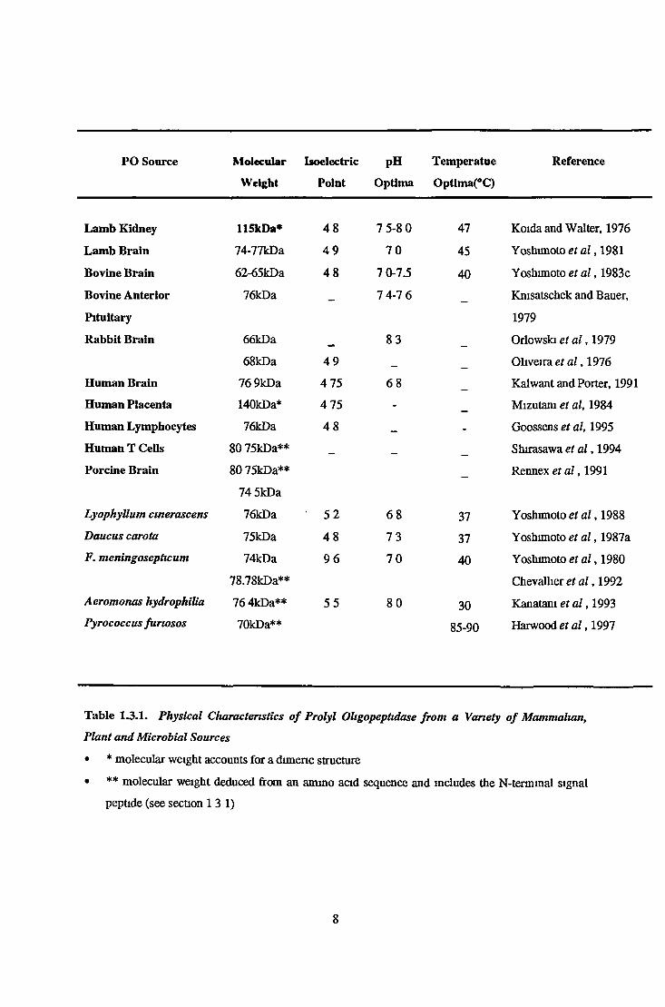
In general mammalian and plant forms of PO have reported to be acidic with isoelectric points of
between 4 5 and 4 9 (Goossens et a l , 1995, Kalwant and Porter, 1991, Yoshunoto et a l , 1983c) The
Flavobacterial memngosepticum form of this peptidase however has a much higher PI of around 9.6
(Yoshimoto et a l , 1980) This is indicative of a difference in the ionizable ammo acid content which
results in a predominantly basic protein Another bacterial form of PO from Aeromonas hydrophiha
was found to have a pi of 5.5, which is again quite high, relative to mammalian forms (Kanatani et
a l , 1993), (see table 1 3 1 )
Generally mammalian, bacterial and plant forms of PO have a broadly neutral pH optima with
reported values ranging from 6.8 to 8 3 (see table 1 3 1 ) Optimal temperatures reported for activity
are generally between 37 and 40°C (Kanatani et a l , 1993, Yoshimoto et a l , 1983c) There has been a
report of a PO from Pyrococcus junosis with a temperature optimum of between 85-90°C (Harwood
et a l , 1997)
7
PO Source Molecular
Weight
Isoelectric
Point
pH
Optima
Temper atue
Optlma(°C)
Reference
Lamb Kidney 115kDa* 4 8 7 5-8 0 47 Koida and Walter, 1976
Lamb Brain 74-77kDa 4 9 7 0 45 Yoshimoto et a l , 1981
Bovine Brain 62-65kDa 4 8 7 0-7.5 40 Yoshimoto et a l , 1983c
Bovine Anterior 76kDa __ 7 4-7 6 Kmsatschek and Bauer,
Pituitary 1979
Rabbit Brain 66kDa 8 3 Orlowski et a l , 1979
68kDa 4 9 _ Oliveira et a l , 1976
Human Brain 76 9kDa 4 75 6 8 Kalwant and Porter, 1991
Human Placenta 140kDa* 4 75 - Mizutam et al, 1984
Human Lymphocytes 76kDa 4 8 _ - Goossens et al, 1995
Human T Cells 80 75kDa** _ Shirasawa et a l , 1994
Porcine Brain 80 75kDa** Rennex et a l , 1991
74 5kDa
LyophyUum cmerascens 76kDa ' 5 2 6 8 37 Yoshimoto et a l , 1988
Daucus carota 75kDa 4 8 7 3 37 Yoshimoto et a l , 1987a
F. meningosepticum 74kDa 9 6 7 0 40 Yoshimoto et a l , 1980
78.78kDa** Chevallier et a l , 1992
Aeromonas hydrophilia 76 4kDa** 5 5 8 0 30 Kanatani et a l , 1993Pyrococcus furiosos 70kDa** 85-90 Harwood et a l ,1997
Table 1.3.1. Physical Characteristics o f Prolyl Oligopeptidase from a Variety o f Mammalian,
Plant and Microbial Sources
• * molecular weight accounts for a dimeric structure
• ** molecular weight deduced from an ammo acid sequence and includes the N-terminal signal
peptide (see section 13 1)
8

1 3 3 . Catalytic Classification of Prolyl Oligopeptidase
1 3 3 .1 . Sensitivity to Protease Inhibitors
One of PO’s distinguishing characteristics is its sensitivity to both serine and cysteine protease
inhibitor reagents. PO has been classified as a serine peptidase based on its sensitivity to DFP, Z-Gly-
Pro-CH2Cl, Ac-Ala-Ala-Pro-CHN2 and to a lesser extent PMSF (Stone et al., 1992; Kalwant and
Porter 1991; Yoshimoto et al., 1987a; Orlowski et al., 1979., Yoshimoto et al., 1977). DFP is a
reagent with a very high specificity for activated catalytically relevant serine residues and the
stoichiometric inhibition of PO by this residue was definitive evidence for the presence of a
catalytically competent active site serine. Mammalian and plant forms of the peptidase have also been
found to be sensitive to cysteine protease inhibitors but at relatively high inhibitor enzyme ratios
which is in contrast to the stoichiometric inhibition observed with DFP (Yoshimoto et al., 1977). It is
plausible that bulky reagents could react with non-catalytically competent cysteine residues at or near
the active site causing a level of steric hindrance which may interfere with catalytic activity. This
hypothesis was confirmed in a series of experiments involving the cysteine protease inhibitors, NEM
and the smaller molecule, iodoacetamide. When PO was treated with these reagents separately 85%
and 50% inhibition was observed with NEM and iodoacetamide respectively. When PO, which had
been treated with iodoacetamide, was exposed to NEM, the peptidase was not further inhibited. This
would seem to indicate the presence of a cysteine residue that is close enough to the active site, to
cause total exclusion of a substrate, when it is complexed to a bulky residue. A smaller reagent such
as iodoacetamide would therefore be able to exert incomplete steric hindrance (Polgar, 1991). There
does seem to be quite a variation in the sensitivity of PO to functional reagents depending on its
source. PO from Flavobacterium meningosepticum and Aeromonas hydrophilia was found to be
resistant to the cysteine protease inhibitor PCMB indicating that no cysteine residue was in close
enough proximity to the active site so as to adversely influence catalytic activity (Kanatani et al.,
1993; Yoshimoto et al., 1980).
1.33.2 Cloning and sequencing of Prolyl Oligopeptidase Gene
Confirmation of PO’s status as a serine protease was obtained through the eventual cloning of the PO
gene and the deduction of its amino acid sequence The amino acid sequence of PO from porcine brain
was reported in 1991. As well as containing 16 half-cystinyl residues, an active site serine, confirmed
on the basis of its inactivation by DFP was identified (Rennex et al., 1991). Essential catalytic
residues have been identified as Ser554 and His680 by reaction with active site-directed reagents
(Rennex et al., 1991; Stone et al., 1991). The amino acid sequence surrounding this active site serine
of PO was found to be Gly-GIy-Ser-Asn-GIy-Gly, distinguishing it from other well characterised
families of serine proteases (Shirasawa et al., 1994; Rennex et al., 1991). The amino acid residues
that surround the active site serine are conserved in each family and are Gly-Asp-fcr-Gly-Gly for the
9

trypsin family, Gly-Thr-Ser-Met-Ala, for the subtilisin family and Gly-Glu-Ser-Tyr-Ala for the
carboxypeptidase Y family (Barrett and Rawlings, 1992; Brenner, 1988). Therefore PO was thought
to represent a new class of serine protease. The order of the triad residues in PO is also distinct from
the other more commonly known serine protease families ie. His57-Aspl02-Serl95 in chymotrypsin,
Asp32-His64-Ser221 in subtilmn and Asp529-Ser554-His680 for PO (Barrett and Rawlings, 1992).
PO has also now been eloned and sequenced from porcine and bovine brain, human T cells,
Flavobacterium meningosepticum and Aeromonas hydrophilia all of which (their primary structures
deduced from nucleotide sequences) show significant sequence homology to each other (Yoshimoto et
al., 1997; Shirasawa et al., 1994; Kanatani et al., 1993., Chevallier et al., 1992; Yoshimoto et al.,
1991; Rennex et al., 1991). This would suggest that the PO protein is highly conserved and is likely
to play an important role in vivo.
While there was initially no significant resemblance between the sequence as a whole or segments
containing the catalytic residues to other known peptidases, it now appears that PO can be placed
amongst a new evolutionary family of serine proteases, known as the S9 family (Recommendations of
the Nomenclature Committee of the International Union of Biochemistry and Molecular Biology,
1992), (see table 1.3.1). The PO sequence shows moderate similarity to DPP IV and an even greater
similarity to the acylaminoacyl-peptidases and protease II previously known as oligopeptidase B. The
greatest similarities between the amino acid sequences of members of this prolyl oligopeptidase
family are seen in the C-terminal third of the sequence which contains the catalytic triad (see figure
1.3.1). The consensus sequence GXSXGGZZ (X is any amino acid and Z is a hydrophobic amino
acid) may be useful in identifying other members of this family. It was only with the discovery of this
family of related peptidases that the Asp residue of the catalytic triad was deduced to be Asp529. Two
aspartic acid residues were found to be conserved between family members, Asp529 and Asp642 (or
Asp641 in slightly shifted alignment). While the environment surrounding the Asp642 varied, the
Asp529 residue was found in a uniformly neutral to mildly hydrophilic segment in all members of the
family allowing the conclusion that Asp529 is most likely a member of the catalytic triad (Barrett and
Rawlings, 1992; Rawlings et al., 1991). While members of the S9 family of serine peptidases display
some sequence homology, with respect to catalytic activity, specificity and subcellular distribution
they are one of the most disparate families of peptidases recognized to date. For instance PO’s action
is confined to oligopeptides (Barrett and Rawlings., 1992), DPP IV, an exopeptidase, cleaves
dipeptides from the N-termini of polypeptides, only when the N-terminus is free (McDonald et al.,
1971), and acyl-aminoacylpeptidase, an omega peptidase preferentially cleaves N-terminal acetyl-
aminoacyl residues from polypeptides (acyl-aminoacyl-peptidase), (Mitta et al., 1989). Protease II an
oligopeptidase cleaves peptide bonds, specifically lysine and arginine residues at the carboxyl end of
amino acids (Kanatani et al., 1991). PO and acyl-aminoacyl peptidase are soluble/cytosolic (Mitta et
al., 1989; Torres et al., 1986) but DPP IV is heavily glycosylated and membrane associated (Misumi
et al., 1992). DPP IV and PO show specificity for prolyl bonds but no such specificity has been
10

observed for acyl-ammoacyl peptidase (Mitta et a l , 1989, Koida and Walter, 1976, McDonald et a l ,
1971)
Structural similarities have also been noted between lipases and members of the prolyl ohgopeptidase
family, more specifically similarities between the short segments comprising the catalytic triad
residues. These similarities include sequence homology, topology and the not entirely open active
site This similarity has provided a possible explanation for the physical rate-limiting catalytic step
likely to be a conformational change In the case of lipases the catalytic triad is covered by a surface
loop or flap and the repositioning of this flap is necessary to render the catalytic site accessible to the
substrate This flap may be conserved in the peptidases of the prolyl oligopeptidase, S9 family
(Polgar, 1992c)
1 N C F D D F Q c A A N G G S N G G L D T K A G H G
2 N C F D D F Q c A A N G G S N G G L D T K A G H G
3 N V F N D F I A A G S G R S N G G L E T N A G H G
4 N T F N D Y L D A C M G G S A G G M D M D S G H G
5 Q D V K D V Q F A V M G G S H G G F Y P K S T H A
6 Q D V K D V Q F A V M G G S H G G F Y P K S N H A
7 L E V E D Q I E A A W G W S Y G G Y Y T D E D H G
8 F E V E D Q I S A A W G W S Y G G Y Y T D E D H G
9 Y E A R D*
Q I S A A F G W S*
Y G G Y F P D S D H S*
ASP 529 SER 554 HIS 680
Figure 13.1. An Alignment o f the Catalytic Triad (Asp-Ser-His) Sequences o f Members o f the
Prolyl Oligopeptidase Family (S9) o f proteases.
* Location of catalytically competent triad residues
1 Human prolyl oligopeptidase (Shirasawa et a l , 1994)
2 Pig prolyl ohgopeptidase (Rennex et a l , 1991)
3 Flavobacterium, meningosepticum prolyl oligopeptidase (Chevallier et al., 1992)
4 Eschericha coli protease II (Kanatam et a l , 1991)
5 Pig acylammoacyl-peptidase (Mitta et a l , 1989)
6 Rat acylammoacyl-peptidase (Kobayashi et a l , 1989).
7 Rat dipeptidyl peptidase IV (Ogata et a l , 1989)
8 Human dipeptidyl peptidase IV (Abbot et a l , 1994).
9. Yeast dipeptidyl aminopeptidase B (Roberts et a l , 1989)
11

♦Subfamily Peptidase Reference
Subfamily A. Oligopeptidase B/Prrtcase II
Prolyl Oligopeptidase
Kanatani et al., 1997
Shirasawa et al., 1994
Subfamily B. Dipeptidyl aminopeptidase A
Dipeptidyl aminopeptidase B
Dipeptidyl peptidase IV
Dipeptidyl peptidase V
Fibroblast activation protein subunit
Seprase
Anna-Arriola and Herskowitz, 1994
Roberts et al., 1989
Abbot et al., 1994
Beauvais et al., 1997
Goldstein et al., 1997
Goldstein et al., 1997
Subfamily C Acyl-aminoacyl peptidase Kobayashi et al., 1989
Table 1.3.1. The S9 or Prolyl Oligopeptidase Family o f Serine Proteases
*This family of peptidases contains deeply divergent groups and is therefore divided in subfamilies
(Rawlings and Barrett, 1993).
13.3.3. Prolyl Oligopeptidase Catalytic Mechanism Studies
Investigations into the mechansitic features of PO catabolism have revealed further distinctions from
the extensively characterised serine protease, trypsin/chymotrypsin and subtilisin, families. Until the
the confirmation of PO’s status as a serine petpidase, it was assumed that serine proteases shared the
same simple pH rate profile controlled by a single ionizing group (histidine) of pKa 7 (Polgar, 1989;
Blow, 1969). Briefly a classical serine protease (chymotrypsin) catalytic reaction has been predicted to
proceed as follows: Acylation: The nucleophilic hydroxyl group of the catalytic serine attacks the
substrate carbonyl group and this reaction is stabilised by general base catalysis, a contribution by the
catalytic histidine side chain, which removes the proton from the attacking oxygen. Deacylation:The
resulting tetrahedral intermediate (covalent enzyme-substrate interaction) then decomposes by general
acid catalysis afforded by the protonated histidine. This results in the return of free enzyme (see figure
1.3.2). It is the catalytically competent histidine residue that facilitates both the formation and
decomposition of the intermediate acyl-enzyme and it is the ionization of this residue that determines
the pH dependence of catalysis (Blow, 1969). These general base/acid reactions are said to be rate
limiting in the case of the chymotrypsin/ subtilisin families and ionic strength has little or no effect
on these proteases. In contrast PO displays a double sigmoidal pH/rate profile indicative of two
12

enzyme forms existing between pH 5 and 9, and its catalysis is sensitive to ionic strength (Polgar,
1992a, Polgar 1992b)
One of the methods routinely used in the studies on the mechanism of senne protease reactions is
the investigation of kinetic deuterium isotqpic effects It was discovered that the acylation /deacylation
steps for a chymotrypsin catalysed reaction, proceeded slower in deuterium oxide by a factor of
around 2-3, indicating that base/ acid catalysis i e a chemical step, is rate limiting for the overall
reaction (Polgar, 1992b) Similar experiments involving PO showed that while the low pH form
displayed a significant deuterium isotope effect, the high, physiologically competent pH form showed
none. This would indicate an isotopically silent step at physiological pH (Polgar, 1991) This
precluded the possibility of a rate limiting conformational change m PO catalysis Further evidence
involved an investigation of the effects of substrates with different chemical leaving groups on PO
reaction rates A substrate with a better leaving group is known to react with a peptidase at a higher
rate provided-the rate limiting step is chemical This was proved to be the case for chymotrypsin with
nitrophenyl ester substrates displaying a much higher reactivity relative to amide substrate
derivatives In the case of PO there appeared to be no significant difference between rate constants
using various derivatives of the substrate, Z-Glv-Pro-Leavmg Groun. on PO catalysis which would
indicate a rate-limiting physical rather than chemical step (Polgar 1992b) Thus some
conformational change m PO can be associated with the catalytic process
Investigations on another member of the prolyl ohgopeptidase family of serine peptidases,
oligopeptidase B (formerly known as protease II), isolated from E Coll, (Pacaud and Richaud, 1975),
have found that this protease has mechanistic features similar to PO While this peptidase is trypsin-
like m its hydrolysis of oligopetides at the carboxy-side of paired basic residues, no kinetic deuterium
isotopic effect was observed for this peptidase indicative of a rate-limiting conformational change
(Polgar, 1997)
13
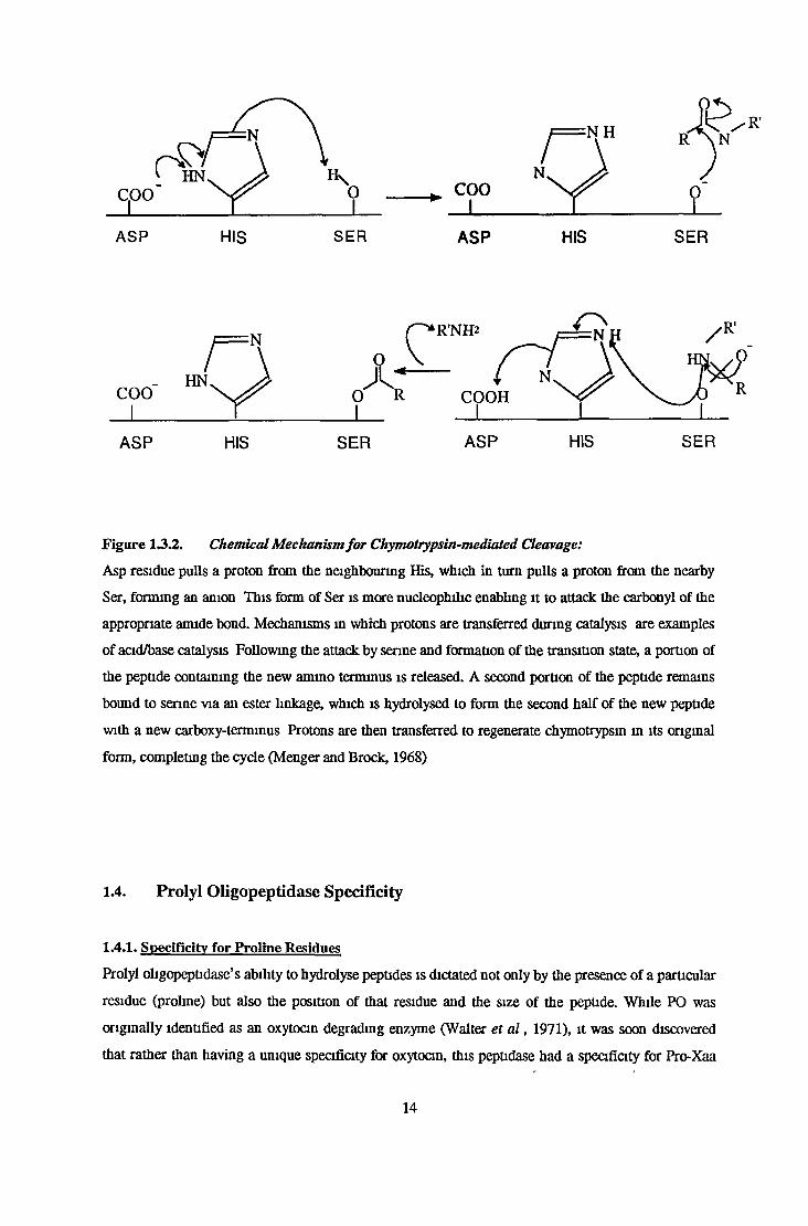
Figure 13.2. Chemical Mechanism fo r Chymotrypsin-mediated Cleavage:
Asp residue pulls a proton from the neighbouring His, which in turn pulls a proton from the nearby
Ser, forming an anion This form of Set is more nucleophihc enabling it to attack the carbonyl of the
appropriate amide bond. Mechanisms m which protons are transferred during catalysis are examples
of acid/base catalysis Following the attack by senne and formation of the transition state, a portion of
the peptide containing the new amino terminus is released. A second portion of the peptide remains
bound to senne via an ester linkage, which is hydrolysed to form the second half of the new peptide
with a new carboxy-terminus Protons are then transferred to regenerate chymotrypsin in its original
form, completing the cycle (Menger and Brock, 1968)
1.4. Prolyl Oligopeptidase Specificity
1.4.1. Specificity for Proline Residues
Prolyl oligopeptidase’s ability to hydrolyse peptides is dictated not only by the presence of a particular
residue (proline) but also the position of that residue and the size of the peptide. While PO was
originally identified as an oxytocin degrading enzyme (Walter et a l , 1971), it was soon discovered
that rather than having a unique specificity for oxytocin, this peptidase had a specificity for Pro-Xaa
14

bonds An exception to this was found to be an inability to act on the Pro-Pro bond (Walter, 1976,
Koida and Walter, 1976) The rate of cleavage of the Pro-Xaa bond was found to be fastest when Xaa
was a hydrophobic residue However the catalytic rate decreased progressively when Xaa was
replaced with basic and acidic residues (Koida and Walter, 1976) An example of this selectivity for
PI’ residues (see figure 14.1.), was observed in the hydrolysis of bradykinin potentiating peptide. PO
hydrolyses BPP, in vitro, at the Pro-Phe, Pro-Gly and Pro-Glu bonds However hydrolysis of the Pro-
Phe bond occurs more rapidly than the Pro-Gly, while the Pro-Glu bond is very slowly hydrolysed .
This action on BPP highlights PO’s preference for hydrophobic residues BPP also possesses a Pro-
Pro bond which was not hydrolysed (Koida and Walter, 1976) PO was found to be unable to cleave a
Pro-Xaa bond when the prolme residue was at the ammo terminus of a peptide, but could hydrolyse
the bonds when Xaa was the carboxyl terminal residue The introduction of a blocking group at the
amino terminus to give Z-Pro-Xaa sequence did not result m hydrolysis Replacement of PI or P2
residues with D-amino acids was found to result in substrates that were resistant to hydrolysis but
replacement of the P3 residue had minor effects In contrast D-amino acid substitution in the PI* and
P2’ positions decreased catalytic activity, but replacement of the P3’ residue elicited no change From
these observations it has been suggested that PO has an extended binding site region of three subsites
• on the ammo side of the scissile bond and two on the carboxyl side (see figure 1 4 1 ) , (Walter and
Yoshimoto, 1978) PO also has an ability to act on alanyl bonds (Ala-Xaa) but at a rate 1/100-1/1000
that of Pro-Xaa bonds, In contrast to this peptidases inability to hydrolyse oligoprolme, it was found
to act on oligoalanine residues (Walter and Yoshimoto, 1978) Replacement of the PI residue with N-
methylalamne and sarcosine resulted m relatively good substrates It can be concluded from these
studies that the SI subsite of PO was designed specifically to fit proline and only accommodated
residues that did not exceed prolines pyrrolidine ring (Nomura et a l , 1986) With respect to the PI’
residue it was found that the phosphorylation of a serine residue in this position increased the rate of
hydrolysis (Rosen et a l, 1991), and the replacement of an Asp residue with Asu (a result of
spontaneous rearrangement of Asp and Asn residues) also resulted m a ten-fold increase m
hydrolysis This could suggest a possible role for PO in the disposal of non-functional proteins from a
cell (Momand and Clarke, 1987)
15
SUBSTRATE
ENZYME
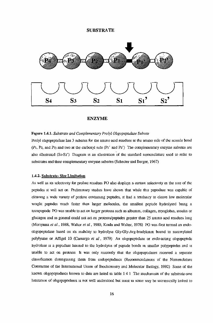
Figure 1.4.1. Substrate and Complementary Prolyl Oligopeptidase Subsite
Prolyl oligopeptidase has 3 subsites for the amino acid residues at the amino side of the scissile bond
(Pi, P2, and P3) and two at the carboxyl side (Pi’ and P2’) The complementary enzyme subsites are
also illustrated (S4-S2’) Diagram is an illustration of the standard nomenclature used to refer to
substrates and their complementary enzyme subsites (Schecter and Berger, 1967)
1.4.2. Substrate- Size Limitation
As well as its selectivity for proline residues PO also displays a certain selectivity m the size of the
peptides it will act on Preliminary studies have shown that while this peptidase was capable of
cleaving a wide variety of proline containing peptides, it had a tendancy to cleave low molecular
weight peptides much faster than larger molecules, the smallest peptide hydrolysed being a
tetrapeptide PO was unable to act on larger proteins such as albumin, collagen, myoglobin, insulin or
glucagon and m general could not act on proteins/peptides greater than 25 ammo acid residues long
(Monyama et a l , 1988, Walter et a l , 1980, Koida and Walter, 1976) PO was first termed an endo-
oligopeptidase based on its inability to hydrolyse Gly-Gly-Arg-bradykimn bound to succmylated
polylysme or Affigel 10 (Camargo et a l , 1979) An oligopeptidase or endo-actmg oligopeptide
hydrolase is a peptidase limited to the hydrolysis of peptide bonds m smaller polypeptides and is
unable to act on proteins It was only reccently that the oligopeptidases received a separate
classification distinguising them from endopeptidases (Recommendations of the Nomenclature
Committee of the International Union of Biochemistry and Molecular Biology, 1992) Some of the
known oligopeptidases known to date are listed in table 1 4 1 The mechanism of the substrate-size
limitation of oligopeptidases is not well understood but must in some way be intrinsically linked to
16

the molecular structure of these peptidases and the high degree of conformational freedom of
oligopeptides Possibly the binding sites of these peptidases could be analogous to receptor binding
sites. Barrett likened the action of oligopeptidases on their peptide substrates to a receptor interacting
with a specific biologically active ligand, for instance the receptor mediated biological activity
mediated by bradykinm is not shown by kininogen (Barrett and Rawlings, 1992)
Peptidase ♦Peptidase Type Reference
Prolyl Ohgopeptidase Senne Monyama et a l , 1988
Oliveira et a l , 1976
Thimet Ohgopeptidase Thimet Barrett and Brown 1990
Nepnlysm Thimet Damelsen et al , 1980
Pitnlysin Metallo Anastasi et a l , 1993
Neurolysm Thimet Senzawa et a l , 1995
Ohgopeptidase M Metallo Krause et a l , 1997
Streptococcal PepB ohgopeptidase Thimet Lm et a l , 1996
Lactococcus lactus PepF ohgopeptidase Metallo Monnet et a l , 1994
Lactococcus cremoris endopeptidase I Metallo Yan et a l , 1987
Lactococcus cremons endopeptidase n Metallo Yan et a l , 1987
E coli Protease II (Ohgopeptidase B) Senne Paucaud et a l , 1975
Table 1.4.1. Oligopeptidases
*With the exception of prolyl ohgopeptidase and E coli protease n , the known oligopeptidases are
metallo/thiol proteases
1.4.3. Prolyl Oligopeptidase Hydrolysis of Neuro/Vasoactive peptides
In vitro PO has been found to hydrolyse a wide variety of prohne containing neuroactive peptides
including TRH, LHRH bradykinm and neurotensin Table 1.4 2 illustrates a range of neuro- and
vasoactive peptides hydrolysed in vitro by PO and their sites of hydrolysis
17

Peptide Sequence Reference
Angiotensin I Asp-Arg-Val-Tyr-Ile-His-Pro-Phe-His-Leu Moriyama et al., 1988
Angiotensin II Asp-Arg-Val-Tyr-Ile-His-Pro-Phe Tate, 1981
BPP pGlu-Gly-Gly-Tip-Pro-Arg-Pro-Gly-Pro-Glu-Ile-Pro-Pro Koida and Walter., 1976
Bradykinin Arg-Pro-Pro-GIy-Phe-Ser-Pro-Phe-Arg Tate, 1981
LHRH pGIu-His-Trp-Ser-Tyr-Gly-Phe-Ser-Pro-Phe-Arg Mendez et al., 1990
TRH pGlu-His-Pro-NH2 Tate, 1981
Neurotensin pGlu-Leu-Tyr-GIu-Asn-Cys-Pro-Leu-Gly-NH2 Tate, 1981
Oxytocin Cys-Tyr-Ile-Gln-Asn-Cys-Pro-Leu-Gly-NH2 Walter, 1976
Substance P Arg-Pro-Lys-Pro-Gln-Gln-Phe-Phe-Gly-Leu-Met Moriyama et al., 1988
AVP Cys-Tyr-Phe-Gln-Asn-Cys-Pro-Arg-Gly Walter, 1976
Melanotropin Ser-Tyr-Ser-Met-Glu-Asn-Lys-Pro-Leu-Gly-NH2 Tate, 1981
Tuftsin Thr-Lys-Pro-Arg Tate, 1981
Table 1.4.2. Physiological Neuro-/Vasoactive peptides Hydrolysed by Prolyl Oligopeptidase in vitro.
Scissile bonds are indicated in bold.
It is still unclear however, what precise role this peptidase plays in the turnover of these peptides in
vivo but the recent development of potent and specific PO inhibitors has contributed to numerous
studies attempting to elucidate the role of PO in neuropeptide turnover.
In the case of TRH degradation in vivo several studies have shown that PO had in fact no influence on
physiological TRH levels. Several studies using Z-Pro-prolinal have shown that in vitro and in vivo
treatment with this inhibitor did not alter TRH levels. There have been similar observations made in
the case of LHRH (Salers et al., 1992; Mendez et al., 1990; Friedman and Wilk, 1989). Ontogenic
studies revealed a poor correlation between TRH, TRH-OH and PO activity (Salers et al., 1992). In
conflict with the conclusion, drawn from these results, that PO had no involvement in the degradation
of TRH in vivo, was the report that the potent PO inhibitor, Y29794, potentiatied the effect of TRH
on release of acetylcholine in rat hippocampus (Nakajima et al., 1992). PO has also been implicated
in the biotransformation of angiotensin I and II to form angiotensin 1-7, a peptide that mimicks the
vasopressin-releasing and cardiovascular effects of angiotensin II (Schiavone et al., 1988). The
production of angiotensin 1-7 in canine brain homogenates was found to be inhibited by Z-Pro-
prolinal (Welches et al., 1991). The portal plasma of sheep was discovered to contain a significantly
higher level of angiotensin 1-7, than in jugular plasma. It has been speculated that a high level of PO,
found in the median eminence, could contribute to the metabolism of angiotensin I delivered by
18

arterial blood (Lawrence et a l , 1992) Administration of the PO specific inhibitor, JTP-4819 to aged
rats was found to increase substance P immunoreactivity However there was found to be no
significant change in AVP immunoreactivity, suggesting that it had no involvement in the hydrolysis
of this peptide (Toide et a l , 1995)
1.4.4. Prolvl OIigopeptida.se Specific Assays
Since POs initial discovery in human uterine extracts, radiometric, spectrophotometnc and
spectrofiuorimetnc assays have been developed and used in this peptidase’s detection Following its
discovery, known substrates of PO oxytocin and arginine-vasopressin were used in native or
radiolabelled forms to detect PO activity (Walter et a l , 1971, Walter, 1976) Today the vast majority
of PO assays are based on the synthetic substrate N-blocked-Gly-Pro linked to a colounmetric or
fluorimetric label In 1979 Yoshimoto et a l , reported on the synthesis and use of Z-Gly-Pro-MCA (7-
ammo-4-methylcoumarin) a fluorescent substrate which had a Km of 20|iM for PO purified from
lamb kidney, while Orlowski et a l , synthesised Z-Gly-Pro-SM (sulphamethoxazole) a colounmetric
substrate. This basic structure (N-blocked-Gly-Pro-colounmetnc/fluonmetnc label) has changed little
over tune even though the solvents needed to solubilise these substrates were known to adversely
effect POs activity (Knisatschek et a l , 1980) In an investigation, that examined a range of
fluonmetnc substrates, it was reported that glycine was in fact a poor choice of amino acid for the P2
position as PO could accommodate bulky residues and had a preference for positively charged groups
(Noula et a l , 1997) This confirms the suggestion that there,are one or more negative charges at or
near the active site which are responsible for electrostatic attraction or repulsion between PO and
charged substrates (Polgar, 1992a) Other high affinity substrates developed by Noula et al., include
Z-Lys-Pro-NH-Meq, Z-Lys(Boc)-Pro-NH-Meq and Z-Glu-Pro-NH-Meq with Km values of 2.1, 2 9
and 5 OftM respectively for the porcine kidney enzyme Another potential reason why further
substrate development studies are required, was the discovery of a second distinctive Z-Gly-Pro-MCA
hydrolysing activity in bovine serum (Cunningham and O’Connor, 1997b) As this peptidase is
insensitive to Z-Pro-prolinal (hence its name Z-Pro-prolinal insensitive peptidase or ZIP), this
specific PO inhibitor can be incorporated into the assay as a method of distinguishing these two
activities It is however imperative that further substrate specificity studies on this new peptidase be
completed in order to develop specific, high affinity substrates for both peptidases
19
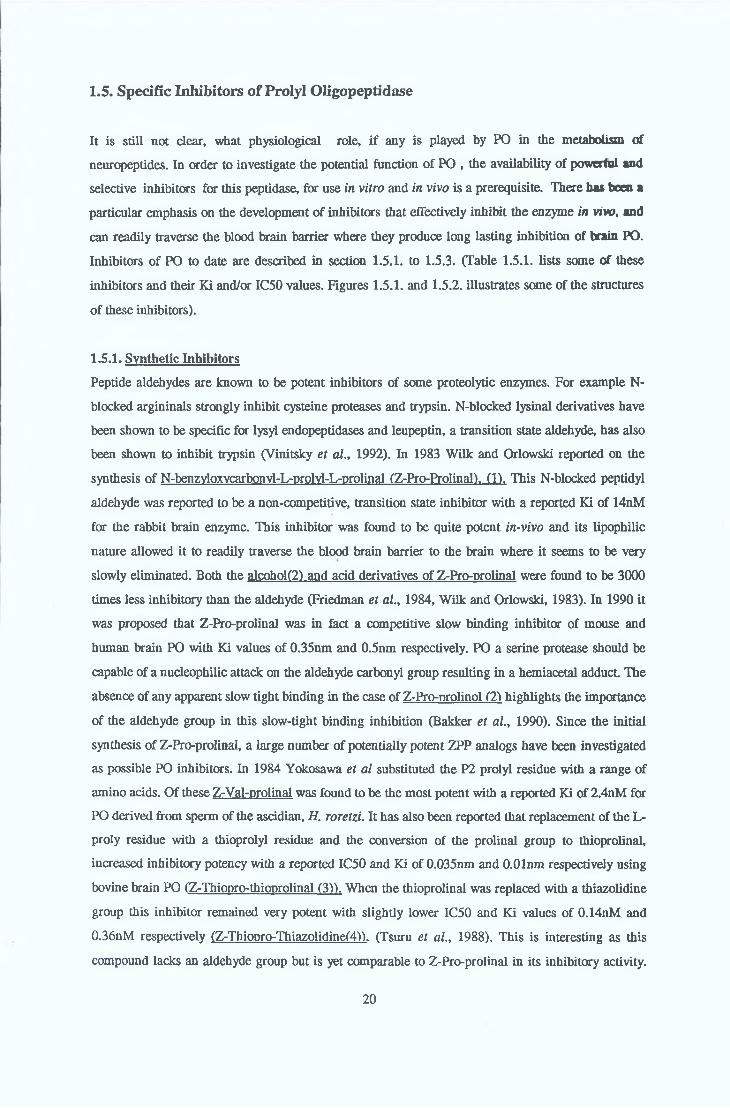
1.5. Specific Inhibitors of Prolyl Oligopeptidase
It is still not clear, what physiological role, if any is played by PO in the metabolism of
neuropeptides. In order to investigate the potential function of PO , the availability of powerful and
selective inhibitors for this peptidase, for use in vitro and in vivo is a prerequisite. There has boca a
particular emphasis on the development of inhibitors that effectively inhibit the enzyme in vivo, and
can readily traverse the blood brain barrier where they produce long lasting inhibition of brain PO.
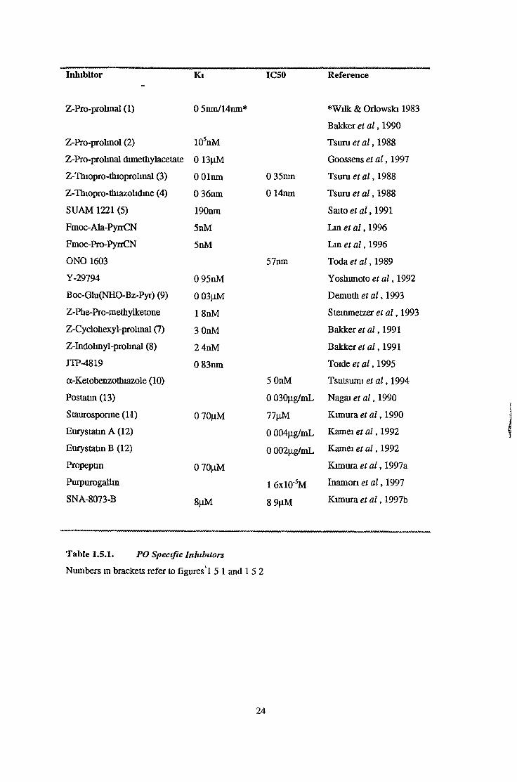
Inhibitors of PO to date are described in section 1.5.1. to 1.5.3. (Table 1.5.1. lists some of these
inhibitors and their Ki and/or IC50 values. Figures 1.5.1. and 1.5.2. illustrates some of the structures
of these inhibitors).
1.5.1. Synthetic Inhibitors
Peptide aldehydes are known to be potent inhibitors of some proteolytic enzymes. For example N-
blocked argininals strongly inhibit cysteine proteases and trypsin. N-blocked lysinal derivatives have
been shown to be specific for lysyl endopeptidases and leupeptin, a transition state aldehyde, has also
been shown to inhibit trypsin (Vinitsky et al., 1992). In 1983 Wilk and Orlowski reported on the
synthesis of N-benzvIoxvcarbonvl-L-prolvl-L-prolinal (Z-Pro-ProlinaD. (1). This N-blocked peptidyl
aldehyde was reported to be a non-competitive, transition state inhibitor with a reported Ki of 14nM
for the rabbit brain enzyme. This inhibitor was found to be quite potent in-vivo and its lipophilic
nature allowed it to readily traverse the blood brain barrier to the brain where it seems to be very
slowly eliminated. Both the alcohol(2) and acid derivatives of Z-Pro-prolinal were found to be 3000
times less inhibitory than the aldehyde (Friedman et al., 1984, Wilk and Orlowski, 1983). In 1990 it
was proposed that Z-Pro-prolinal was in fact a competitive slow binding inhibitor of mouse and
human brain PO with Ki values of 0.35nm and 0.5nm respectively. PO a serine protease should be
capable of a nucleophilic attack on the aldehyde carbonyl group resulting in a hemiacetal adducL The
absence of any apparent slow tight binding in the case of Z-Pro-nrolinol (2) highlights the importance
of the aldehyde group in this slow-tight binding inhibition (Bakker et al., 1990). Since the initial
synthesis of Z-Pro-prolinal, a large number of potentially potent ZPP analogs have been investigated
as possible PO inhibitors. In 1984 Yokosawa et al substituted the P2 prolyl residue with a range of
amino acids. Of these Z-Val-prolinal was found to be the most potent with a reported Ki of 2.4nM for
PO derived from sperm of the ascidian, H. roretzi. It has also been reported that replacement of the L-
proly residue with a thioprolyl residue and the conversion of the prolinal group to thioprolinal,
increased inhibitory potency with a reported IC50 and Ki of 0.035nm and O.Olnm respectively using
bovine brain PO (Z-Thiopro-thioprolinal (3)). When the thioprolinal was replaced with a thiazolidine
group this inhibitor remained very potent with slightly lower IC50 and Ki values of 0.14nM and
0.36nM respectively (Z-Thiop^o-Thiazolidine(4,)). (Tsuru et al., 1988). This is interesting as this
compound lacks an aldehyde group but is yet comparable to Z-Pro-prolinal in its inhibitory activity.
20

In studies using Z-pro-proknal and derivatives it was found that a main chain length of the inhibitor
corresponding to 3 subsites, SI, S2 and S3 is nost suitable for inhibitory activity. This is a good
reflection of the fact that PO is very active towards low molecular weight peptides but inert towards
high molecular mass peptides For a potent inhibitor specific for PO, a restricted chain length in the
main chain interacting with the SI, S2, S3 subsites of the enzyme was critical, with a hydrophobic
pocket in the S3 subsite of the enzyme which seemed to interact hydrophobically with a certain N-
blocked group of the inhibitor, such as the benzyloxycarbonyl group How the introduction of sulphur
atom affects the enzyme-inhibitor interaction is unknown', but somehow it either induces stenc fitness
in the enzyme-inhibitor interaction or it introduces an electron attracting or donating force which
results in a firm interaction between the enzyme and the inhibitor (Tsuru et al., 1988)
Prodrugs based on Z-Pro-prolmal and its derivatives have also been synthesised Because the
aldehyde group of this inhibitor is very reactive, it was disguised with an acetal group which could be
converted in the stomach to the aldehyde moiety Interestingly these acetals were also potent in their
inhibition of PO in vitro with a reported IC50 for Z-Pro-prolmal dimethyl acetate of 0 13|iM
(Goossens et a l , 1997) Many other potent PO inhibitors many of which are variations of the N-
blocked dipeptide chain have been developed. The most potent of these include SUAM 1221 (5). a
phenyl butanoyl prolyl-pyrrolidine derivative with a reported IC50 of 190nM and a derivative of this,
(replacement of proline to thioproline) which had an IC50 67nM (6), (Saito et al 1991). The
conversion of the pyrrolidine group of Z-Pro-pyrrolidine, to a fluoropyrrolidine to give Z-Pro-
fluoropvrrolidme. resulted in a compound with a Ki of 0 8nM (Goossens et a l , 1997)
Fmoc-aminoacvlnvrrolidine-2-nitriles have also been found to possess potent inhibitory activity
against PO. Both Fmoc-Ala-Pro-PvrrCN and Fmoc-Pro-PvrrCN had Ki values of 5nM for PO and
were found to be very stable, cell permeable and crossed the blood brain barrier (Li et a l , 1996)
Natural substrates of PO, oxytocin and vasopressin, are already inhibitors of this peptidase These
peptide hormones have intramolecular disulphide bridges between cysteine residues and it has been
suggested that a thiol-disulphide exchange between the substrate and a thiol group in or near the
active site PO could be a mechanism for inhibition Inhibitors mimicking these natural substrates,
which incorporated an NH-O-acyl moiety (N-peptidyl-O-acyl hydroxylamines are known inhibitors of
thiol enzymes (Bromme et a l , 1989)) have been prepared, the most potent of them being Boc-
Glu(NHO-Bz)Pvr (9). (Demuth et a l , 1993)
A wide variety of dipentidvl-a keto heterocvcles (10). both one-rmg and two-ring types, have also
proved themselves to be potent PO inhibitors It was suggested that the potency of these compounds
was dependent on the presence of an sp2 nitrogen atom which is able to form a hydrogen bond at a p-
position from the ketone moiety (Tsutsumi et a l , 1994)
A reversible and competitive non-peptide PO inhibitor Y-29794. was reported by Nakajima et a l , m
1992 This inhibitor was found to be potent (Ki=0 95nM) highly specific and easily penetrated the
blood-brain barrier
21

Pentidvldiazomethanes and neotidvlchloromethanes have also been repeated to have inhibitory
activity against PO. These agents have contributed tremendously use in the study of the active site of
PO, confirming its status as a serine protese POs position as a serine protease was clarified by its
inhibition with DFP and Z-Gly-Pro-CH2Cl (Yoshimoto et a l, 1977) Active site directed
chloromethanes are thought to alkylate active site histidines of serine proteses and also cysteine
residues m various enzymes The competitive PO inhibitor Ac-Ala-Ala-Ala-CILCl was found, out of
23 histidine residues, to selectively alkylate His-680, providing evidence that this residue is mdeed
the catalytic histidine. This agent also alkylated some PO cysteine residues
(Stone et al., 1991) Modification of at least one of these cysteine residues may be at least partly
responsible for inactivation of the enzyme. In this respect PO could be similar to certain members of
the subtilism serine protease family that are inactivated by modification of a non-essential cysteine
residue within the active site
Peptidyl diazomethanes have been useful in confirming POs status as a senne protease It was
initially thought that peptidyl diazomethanes were reagents that specifically inhibited cysteine
proteases by alkylation of the active site cysteine residues (Shaw, 1990). Some senne protesases
however are now known (members of the subtilism family) to be inhibited by these diazomethane
derivatives through the formation of a covalent bond with the active site histidine (Ermer et a l ,
1990) Peptidyl diazomethanes such as Ac-Ala-Ala-Pro-CHN7 have been found to be reversible slow
tight binders of PO with the formation of a covalent complex between the active site senne and the
diazomethane (Stone et a l , 1992)
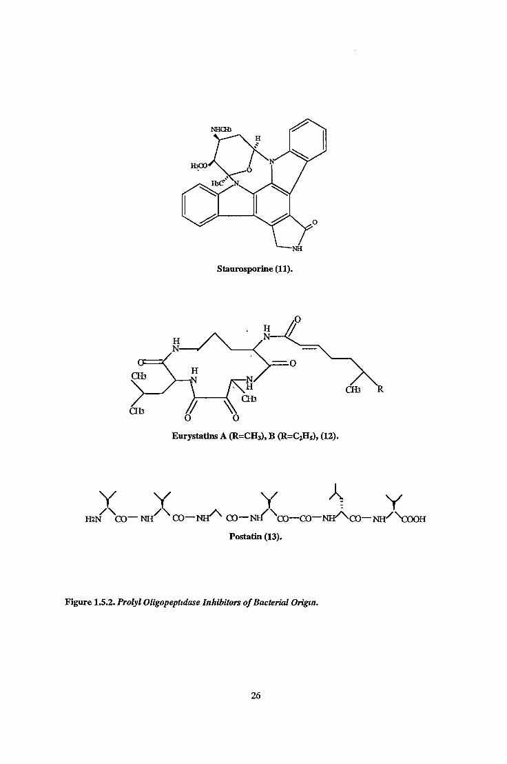
1.5.2. Inhibitors of Bacterial Origin.
A number of compounds which are bacterial in origin ware found to possess quite potent PO
inhibitory activity (see table 1.5 1) These include bacitracin. staurosnonnedl). (Kimura et a l ,
1990), nostatin (13). (Tsuda et a l , 1996), eurvstatms A and B (12). (Toda et al., 1992), propeptm
(Kimura et a l , 1997a), purpurogallin (Inamon et a l , 1997), lipohexm (Heinze et al 1997) and SNA-
80736 (Kimura et a l , 1997b), a stereoisomer of the antibiotic fugianmycin B A variety of denvatives
of postatin have been chemically synthesised in an attempt to obtain a greater inhibitory potency and
selectivity Two postatin analogs containing its charactenstic (S)-3-amino-2-oxovaleryl moiety were
found to have IC50s of 5 8ng/mL and 8 2ng/mL (in contrast to postatin’s 0 030|xg/mL) and unlike
postatin had no inhibitory effect on cathepsin B activity(Tsuda et a l , 1996)
1.5.3. Endogenous Inhibitors
Since POs initial discovery, a number of naturally occurring inhibitors have been purified from a
variety of mammalian tissues including porcine pancreas (Yoshimoto et a l , 1982), neonatal rat
22
pancreatic islet cell extracts (Salers, 1994), rat brain (Soeda et a l , 1985) and bovine brain (Ohmori et
a l , 1994) Distribution studies performed using porcine and rat tissues found this inhibitor to be
uniformly distributed with highest levels in the pancreas (Yoshimoto et a l, 1982) The molecular
weight of inhibitors purified from various tissues ranged from 6 5 to 7.0 kDa. Ammo acid analysis on
the purified inhibitor from bovine brain revealed it to be identical to segment 38-55 of glial fibrillary
acidic protein and had a Ki of 8 6fiM (Ohmori et a l , 1994, Maruyama et a l , 1996) It is known that
some protease inhibitors released from glial cells (nexms) are involved m neunte growth regulation
This coupled with the observation that staurosporine another PO inhibitor was found to stimulate
neunte growth in a rat cell line suggests a possible role for PO in cell growth (Kimura et a l , 1990)
This will be dicussed further in section 17 1
Other endogenous inhibitors of PO from rat liver cytosol include coenzyme A, its acyl denvatives
and acyl carnitine (Yamakawa et a l , 1990)
23
Inhibitor Ki IC50 Reference
Z-Pro-prohnol (2) 105nM
Z-Pro-prolinal dimethylacetate 0 13|0.M
Z-Thiopro-thioprolmal (3) 0 Olnm 0 35nm
Z-Thiopro-thiazolidine (4) 0 36nm 0 14nm
SUAM 1221 (5) 190nm
Fmoc-Ala-PyrrCN 5nM
Fmoc-Pro-PyrrCN 5nM
O NO 1603 57nm
Y-29794 0 95nM
Boc-Glu(NHO-Bz-Pyr) (9) 0 03|jM
Z-Phe-Pro-methylketone 1 8nM
Z-Cyclohexyl-prolmal (7) 3 OnM
Z-Indolinyl-prolinal (8) 2 4nM
JTP-4819 0 83nm
a-Ketobenzothiazole (10) 5 OnM
Postatin (13) 0 030|ig/mL
Staurosponne (11) 0 70^M 77|iM
Eurystatm A (12) 0 004p.g/mL
Eurystatm B (12) 0 002p.g/mL
Propeptin 0 70(iM
Purpurogalhn ! 6x10'5M
SNA-8073-B 8|iM 8 9|J.M
Z-Pro-prolinal (1) 0 5nm/14nm* *Wilk & Orlowski 1983
Bakker et a l , 1990
Tsuru et a l , 1988
Goossens et a l , 1997
Tsuru et a l , 1988
Tsuru et a l , 1988
Saito et a l , 1991
Lin et a l , 1996
Lin et a l , 1996
Toda et a l , 1989
Yoshunoto et a l , 1992
Demuth et a l , 1993
Stemmetzer et a l , 1993
Bakkeret a l , 1991
Bakker et a l , 1991
Toide et a l , 1995
Tsutsumi et a l , 1994
Nagai et a l , 1990
Kimuraei a l , 1990
Kamei et a l , 1992
Kamei et a l , 1992
Kimura et a l , 1997a
Inamon et a l , 1997
Kimura et a l , 1997b
Table 1.5.1. PO Specific Inhibitors
Numbers m brackets refer to figures ! 5 1 and 15 2
24

Z-Pro-Prolinal (1). Z-Pro-Prolinol (2).
Z-Thiopro-Thioprolmal (3). Z-Thiopro-Thiazolidine (4).oCH2CH2CH2CO — N
CO— N
SUAM 1221 (5).
H2CH2CH2CO— N
CO-N
SUAM 1221 (thiapro derivative), (6).
Z-IndoIinyI-ProIinaI(8).
BOC-GIu(NHO-Bz-Pyr),(9). a-Ketobenzothiazole (10).
Figure 1.5.1. Prolyl Oligopephdase Specific Synthetic Inhibitors
25
ÀY Y Y '> YHi n ' ^CO— N H ^ ^ CO— N H ^ CO— N H ^ \) 0 — CO- N H ^ C O — NH/ \x )O H
Postatili (13).
Figure 1.5.2. Prolyl Oligopeptidase Inhibitors o f Bacterial Origin.
26

1.6. DistributionSince its discovery in human uterine homogenates (Walter et a l , 1971), prolyl oligopeptidase has
been identified in a wide variety of organisms; mammalian, plant and microbial Distribution studies
performed in human (Kato et a l , 1980), rat (Yoshimoto et a l , 1979), rabbit (Orlowski et a l , 1979)
and mouse tissue sources (Dauch et a l , 1993) have revealed a ubiquitous tissue distribution with
uniformly high levels in the brain Significant differences in peripheral tissues levels reflect a
substantive species to species variation (see table 1 6 1 ) With respect to peripheral organs, highest
levels of PO m humans were observed m skeletal muscle, testis and kidney, with low levels m aorta,
heart tissue and serum (Kato et a l , 1980) In contrast, rabbit levels were lowest in skeletal muscle
and highest in the intestine, lung and spleen (Orlowski et a l , 1979) In rat highest PO activity was
found in kidney and liver while the pancreas and small intestine had little or no activity (Yoshimoto
et a l , 1979)
Consistently high levels were detected in the brain of all species examined, with some variation in
regionalization of activity (see table 1 6 2 ) Cortical levels in all species examined were high In
human bram the frontal cortex had highest levels with the thalamus showing lowest levels It is
important to note that the thalamus levels were less than 10-fold lower than cortical levels, not as
great a difference in comparison to the trend m levels in peripheral tissues (Kato et a l , 1980) In
rabbit bram highest levels were found in the endorhinal cortex, hippocampus and striatum, with the
frontal and parietal cortex also having relatively high levels Lowest levels were observed m rabbit
medulla and pons (Orlowski et a l , 1979) In bovme brain high PO levels were detected m the caudate
nucleus and thalamus with low levels m the anterior pituitary ( Tate, 1981)
Such uniform tissue distribution of a peptidase activity is highly supportive of a role in peptide
metabolism in a particular organism This coupled with PO’s unique specificity for internal proline
bonds makes it an ideal candidate for further study
The majority of subcellular localization sudies performed to date on PO have found it to be primarily
cytosolic in nature in a variety of sources including rabbit, rat and bovme bram, hamster
hypothalamus and murine macrophages, PO having been either co-localized with LDH and ChAT,
two cytoplasmic marker enzymes or having stayed in soluble fraction under conditions that would
sediment membrane fraction, lysosomes and other subcellular granules (Torres et a l , 1986, Greene
and Shaw, 1983, Dresdner et a l , 1982, Tate, 1981) In a Sarcophagia peregrina embryonic cell line
PO was found to be entirely localised in the nucleus using lmmunofluorescent staining (Ohtsuki et
a l , 1997) In a follow up study on mouse Swiss 3T3 cells activity was detected m the cytoplasm and
nuclei (Ishmo et a l , 1998) There have been reports however of a particulate form of this peptidase
(Camargo et a l , 1984, Dalmaz et a l , 1986) and m 1996, O’Leary et a l , reported on the purification
of a particulate prolyl endopeptidase type peptidase from the synaptosomal membranes of bovine
bram As well as its membrane location this PO type activity displayed some unique characteristics,
such as an insensitivity to serine protease inhibitors and sensitivity to the metal chelator 1,10-
27

phenanthrohne. Because of POs cytosolic location its role in neuropeptide metabolism and
physiological significance is obscured It is generally assumed that the action of neuropeptides is
catalysed by membrane bound ectoenzymes and very little is known about the internalisation of these
peptides for degradation. The brain cytosol is a rich source of peptide degrading enzymes Resident
exo- and oligopeptidases are thought likely to participate in the terminal stages of intracellular
protein degradation However their physiological significance and their role in neuropeptide
metabolism is unknown There is the possibility that cytosolic peptidases may control the intracellular
generation of biologically active peptides They may function to degrade peptides delivered to the cell
by receptor mediated endocytosis or possibly degrade peptides which have been released
mtracellularly from damaged vesicles Alternatively they may control levels of peptides targeted for
the nucleus for signalling Whatever the role of this cytosolic prolyl oligopeptidase, its ability to act
on a number of physiologically relevant proline-contammg neuropeptides, warrants that this
peptidase be subject to further study.
28

Tissue Human1 Rabbit2
Skeletal Muscle 1 1
Testis 0 82
Kidney 0 57 170
Submandibular Gland 0.55
Pancreas 045 2.04
Liver 0 36 2 9
Tonsil Gland 028
Thyroid 0 27
Stomach 0 26
Small Intestine 0 25 413
Urinary Bladder 0 23
Thymus 0 20
Adrenal Gland 018
Lung 0 17 3 75
Spleen 015 3 08
Diaphragm 015 24 2
Uterus 0 12 2 42
Heart 009 2 29
Serum 0 0001
Table 1.6.1. Comparison ofPO Distribution in Human and Rabbit Peripheral Organs.
Activities expressed as a factor of levels m skeletal muscle 1 Assayed with Suc-Gly-Pro-MCA, Kato
et al., 1980,2 Assayed with Z-Gly-Pro-SM, Orlowski et a l , 1979
29

Hum an B rain Region1 Activity R abbit B rain Region Activity2
Frontal cortex 1 Frontal cortex 1
Nucleus caudatus 0 4 6 Parietal cortex 0 94
Pallidum interior 0 19 Entorhmal cortex 13
Pallidum ex tenor 0 20 Occipital cortex 0 77
Thalamus antenores 0 008 Stratum 11
medialis 0 1 4 Hippocampus 116
laterales 0 20 Cerebellum 084
Pulvinar 0 26 Midbram 065
Hypothalamus 0 19 Medulla 0 51
Locus coeruleus 0 26 Pons 049
Hypothalamus 0 86
Table 1.6.2. Distribution o f Prolyl Oligopeptidase in Human Brain
Activities expressed as a factor of levels in frontal cortex
1 Assayed with Suc-Gly-pro-MCA, Kato et a l , 1980,2 Assayed with Z-Gly-Pro-SM, Orlowski et a l ,
1979
1.7. Physiological Relevance of Prolyl Oligopeptidase
Because of its ubiquitous distribution and ability to act on a large number of biologically relevant
peptides (Wilk, 1983, Kato et a l , 1980), it is generally believed that prolyl oligopeptidase could
contribute to a number of important biological functions Studies involving the physiological
relevance of prolyl oligopeptidase m vivo usually involve an observation of abnormal levels of activity
pertinent to some particular disease state or the induction of some physiological change after the
administration of specific prolyl oligopeptidase inhibitors
1.7.1. Prolyl Oligopeptidase Ontogenic Studies-Evidence for a Possible Role in Cell Growth
Since the first ontogenic studies were completed on prolyl oligopeptidase evidence has accumulated
implicating this peptidase in a possible role in cell growth and proliferation
Various ontogenic studies performed in relation to PO in a variety of mammalian tissues have
revealed a common pattern in its expression during mammalian development and suggest that it is a
30

(
developmentally regulated peptidase Corresponding to the first couple of weeks of life, a period of
cell growth and development, is a dramatic increase in PO levels This is followed by a slow decline
to normal adult levels In rat brain, lung, kidney and liver and mouse brain. PO levels increased
dramatically during the first two weeks of life followed by a gradual decrease to adulthood levels
(Dauch et a l , 1993, Fuse et a l , 1990). A similar trend was noted in a separate study on rat pancreas
when it was found that PO levels increased during the first 2-3 weeks of life before dropping to adult
levels (Salers et a l , 1992) In prenatal investigations performed using primary cultures of rat cerebral
cortex, PO activity was seen to augment dramatically during the first week of growth, corresponding
to a period of rapid neuronal differentiation and development. After the second week of growth, levels
dropped to adult levels (Szappanos et al., 1994) In an investigation of regenerating rat liver cells an
increased PO activity was noted corresponding to the early stage of regeneration Accompanied by
this increased activity was a decrease in an endogenous PO inhibitor Treatment of cells with Z-Gly-
Pro-CH2 resulted in inactivation of intracellular PO which was accompanied by a suppression of the
regeneration process (Yamakawa et a l , 1994) Using cultured embryonic cells and imagmal discs of
Sacrphaga peregnna (flesh fly) it was found that the specific PO inhibitor ZTTA inhibited DNA
synthesis and thus cell/disc differentiation and proliferation Furthermore PO was found to be
exclusively located m the nucleus suggesting an endogenous nuclear substrate (Ohtsuki et a l , 1997
and 1994) . In a follow up study using mouse Swiss 3T3 cells findings regarding ZTTA mediated
inhibition of cell proliferation were confirmed (Ishino et al 1998)
Perhaps the most striking case of cell growth and tissue proliferation is the growth of cancer cells It
is now widely believed that peptidases do have an important role to play in tumour development and
progression (Gottesman, 1990), and studies have established a link between elevated PO levels and
cancerous tumours Increased PO levels have to date been found in lung tumours, prostate, breast and
sigmoid malignancies (Goossens et al 1996a, Sedo et a l , 1991) Further evidence connecting PO to
cancer PO was found to be localised on chromosome 6q22 (Goosens et a l , 1996b) It is a well
established fact that a region on chromosome 6 containing this locus is susceptible to rearrangements
resulting in loss of tumour suppressor genes (Trent and Ziegler, 1992), which could by some
mechanism lead to elevated PO activity
What is evident from all of the investigations on PO levels during development and cell growth, is
the possibility that the peptidase may be intrinsically linked to cell growth, development and
specifically DNA synthesis To further clarify PO’s role in cell growth, possible endogenous
substrates and specifically nuclear substrates of this peptidase must be identified.
31

1.7.2. Involvement of Prolvl Oligopeptidase tn The Pathogenesis of Neurological and
Psychiatric Disorders.
Over the last three decades over forty small peptides have been identified in the mammalian central
nervous system (Krieger, 1984; Donlon, 1995). As the distribution, neurochemical,
electrophysiological and pharmacological effects of this group of neuromodulators have been studied
it has become increasingly evident that these peptide containing neural circuits may be pathologically
altered in neuropsychiatric disorders such as major depression, Alzheimer’s disease and
schizophrenia (Nemeroff and Bissette, 1985). Perturbations in certain peptidase activities could be
linked to abnormal levels of these bioactive peptides and in turn lead to neuroendocrine and
neurochemical disturbances.
To date a lot of interest has been generated in prolyl oligopeptidases possible role in certain
neurological and psychiatric disorders because of its distribution in the brain (Tate, 1981; Kato e t a l . ,
1980), and its broad specificity of action on proline-containing bioactive peptides i n v i t r o (Wilk,
1983; Kato e t a l . , 1980; Koida and Walter, 1976).
1.7.2.1. Prolyl Oligopeptidase and M ajor Depression
Major depression is characterized by numerous neuroendocrine and immunological disturbances that
theoretically could be linked to prolyl oligopeptidase activity. The following are just some of the
neuroendocrine/ immunological hallmarks o f major depression.
• Increased concentrations of certain neuropeptides such as TRH (Kirkegaard et al., 1979), and
Substance P (Rimon e t a l . , 1984), in cerebrospinal fluid.
• Hypothalmic-Pituitary-Adrenal axis hyperactivity leading to increased secretion of certain
neuropeptides such as arginine vasopressin, corticotropin releasing hormone, P-endorphin and
adrenocorticotrophic hormone (ACTH), (Maes e t a l . , 1993a, 1991; VonBardeleben e t a l . , 1985).
• Systemic immune activation characterised by T cell activation, B cell proliferation and increased
levels o f phagocytic cells in peripheral blood ( Maes, e t a l . , 1995a, 1994, 1993a).
• An increased production of inflammatory cytokines such as interleukin-ip, inteleukin-6 and
interferon y which may underlie both immune activation and an acute phase response (Maes e t
a l . , 1993a).
• Moderate autoimmune response (Maes e t a l . , 1993b, Gastpar and Muller, 1981).
A possible role for PO in the pathophysiology of major depression stems from not only its ability to
act on neuropeptides such as TRH and substance P i n v i t r o but also suggestions of its possible
involvement in the pathogenic mechanisms of inflammatory and autoimmune responses (Aoyagi e t
32

I
a l , 1984, Shoji et al 1989) and its possible involvement m the modification of Tcell function Several
studies have already shown that bidirectional communication exists between the immune and
neuroendocrine systems due to shared signal molecules A number of neuropeptide receptors have
been identified on cells of the immune system and the functions of these cell* can be effected by
neuropeptides For instance substance P receptors have been found on lymphocytes and are believed
to regulate the functions of these cells (Li et a l , 1998) These possible characteristics of PO in vivo
prompted studies on a possible link between prolyl oligopeptidase and depression In 1994 decreased
PO levels were observed in serum of major depressed patients in comparison to normal controls
(Maes et a l , 1994) In fact PO activity was found to be inversely related to the seventy of depression
Further studies attempting to establish a possible role for prolyl oligopeptidase m the
pathophysiology of major depression established that treatment of major depressed patients with
antidepressants resulted in a significant increase m PO activity Furthermore one of these
antidepressants fluoxetine was found, in vitro, to enhance PO isolated from human platelets (Maes et
a l , 1995b)
It is still uncertain if diminished PO activity m major depression is involved m the neuroendocrine
and immune pathophysiology of this disorder or whether prolyl oligopeptidase levels are simply a
marker for this condition However the possibility of POs involvement m the modulation of
neuroactive peptides in vivo, its role in immune response generation and the significant enhancement
of its activityj following antidepressant therapy warrants further investigations into the role of PO in
major depression
1.7.2.2. Prolyl Oligopeptidase and Schizophrenia
Studies on neuropeptide concentrations m patients with schizophrenia have found significant
alterations in levels of these neurally active peptides in companson to normal controls Of these
neuropeptides, possible PO substrates in vivo, substance P, neurotensin, TRH, p-endorphin and
arginine vasopressin were all found to be significantly altered (Nemeroff et a l , 1987) It has been
speculated that disorders m peptide metabolizing enzymes may be present in schizophrenia (Wiegant
et a l , 1988, Beckmann et a l , 1984) Low molecular weight hyperpeptiduria is a characteristic in
schizophrenia and may be associated with diminished peptidase activity (Reichelt et a l , 1981) It has
also been shown that certain antipsychotic drugs can induce the activity of some peptidases in vivo
therefore increasing peptide metabolism (Traficante and Tumball, 1982)
An increased level PO activity was observed in serum samples from schizophrenic patients in
companson to normal controls (Maes et a l , 1995b) While this might seem to contradict the
observation of low molecular weight hyperpeptiduria in schizophrenia, it has been speculated that a
selective reduction of some peptidases together with an increase in the activity of other peptidases
such as PO may be a characteristic of this disorder In addition it was found that treatment with the
33
antipsychotic drug valproate reduced PO activity (Maes e t a l . , 1995b). Further evidence for a possible
role for PO in schizophrenia includes observed decreases in cerebrospinal fluid arginine vasopressin
and neurotensin and postmortem brain TRH. However there has been conflicting reports of an
increase in neurotensin and substance P in postmortem brain of schizophrenic patients (Nemeroff e t
a l , 1987).
The evidence for a possible role for PO in psychotic disorders such as schizophrenia and mania is still
unclear. However the results suggest that PO could be used as a marker in these conditions.
1.7.2J . Prolyl Oligopeptldase and Neurodegenerative Disorders
Many studies have been completed on the possible role of PO in neurodegenerative disorders such
Huntington’s, Parkinson’s and Alzheimer’s disease. In 1984 Pittaway e t a l observed a decreased PO
activity in post-mortem brain of subjects with Huntington’s disease. At the time it was speculated
that decreased PO activity could be reflective of neuronal cell damage particularly with the evidence
that loss of PO activity took place where degeneration was most pronounced (basal ganglia, frontal
cortex and subtantia nigra). In a complementary study (Hagihara and Nagatsu., 1987) a decrease in
PO activity was observed in CSF fluid of Parkinsonian patients. Extensive work has been done in the
past decade on the possible role o f PO in the pathogenesis o f Alzheimer’s disease. In 1990 Guiroy
found that the highly potent and PO specific inhibitor, Z-Pro-prolinal induced neuronal degeneration
in primary rat hippocampus neurons i n v i v o , specifically causing an accumulation of phosphorylated
neurofilaments. This observation coupled with evidence that PO has a preference for substrates with a
phosphoserine in the PI position and thus may act on phosphorylated peptides in vivo (Rosen e t a l . ,
1991), implies a possible contribution by PO to the pathogenesis of neurodegenerative disorders such
as Alzheimer’s disease. Several studies involving the measurement of PO levels in post-mortem
brain, serum and CSF from AD subjects are in agreement with this data, all reporting a decrease in
PO level (Mantle e t a l . , 1996; Yoshida e t a l . , 1996). The question still remains however as to whether
PO levels are simply reflective of neuronal cell damage or whether PO can be implicated in the
disease process through its degradation of neuropeptides i n v i v o or in its possible role in cell stress
response. It seems unlikely however that PO is simply a “marker” for cell damage as it was observed
that PO activity uniformly decreased in both grey and white matter of post-mortem brains of AD
subjects (Mantle e t a l . , 1996). Results of studies completed on serum however must be treated with
caution due to the discovery of a second Z-Gly-Pro-MCA hydrolysing, Z-Pro-prolinal insensitive
peptidase (Cunningham and O ’Connor, 1997b).
Other studies on the link between PO and Alzheimer’s disease have attempted to establish a
connection between PO activity and the generation of amyloid A4 protein. Amyloid A4 protein has
been found in the deposits in senile plaques characteristic of Alzheimer’s disease. This 42 amino acid
peptide has a tendency to self aggregate and induce neuronal apotosis (Yamaguchi e t a l . , 1988;
34
Glenner and Wong, 1984). Amyloid A4 is derived from a larger transmembrane amyloid precursor
protein (Haass e t a l . , 1992). It is thought proteolytic processing of this precursor protein at residues
672 and 714 could lead to the formation of the pathogenic amyloid A4 protein (Checler e t a l . , 1995).
Using a model synthetic substrate, Suc-Ile-Ala-MCA, which is homologous to the C-terminal portion
of the A4 peptide, a peptidase identified as prolyl oligopeptidase was isolated from rat brain (Ishiura
e t a l . , 1990). Additional evidence involved the effect o f PO specific inhibitors on amyloid A4 protein
in cell culture. The specific and potent PO inhibitor ONO-1603 was found to have neuroprotecrtive
effects on rat cerebellar granule cells as well as increasing levels o f muscarinic cholinergic neurons
which are thought to induce normal secretion and processing of amyloid precursor protein (secreted
APP is thought to have neuroprotective and neurotrophic functions). It is conceivable that induction
of muscarinic cholinergic neurons could normalise the processing and secretion of APP and suppress
the production of neurotoxic amyloid A4 peptides (Katsube e t a l . , 1996). Another inhibitor study
using the highly specific PO inhibitor, JTP-4819, found that cleavage of the model substrate Suc-Ile-
Ala-MCA was inhibited on treatment of N-6108-15 neuronal cell line with this inhibitor. Induced
amyloid A4 formation in these cultures was retarded upon addition of this inhibitor to cultures
(Shinoda e t a l . , 1997).
Another study that reinforces the link between PO and amyloidgenesis in the brain found that
amyloid A4-like immunoreactivity in hippocampus of senscence accelerated mouse matched PO-like
immunoreactivity (Funkunari e t a l . , 1994).
While all the evidence accumulated appears to suggest that PO plays some role in the
neuropathogenesis of Alzheimer’s disease, specifically through its role in the processing of amyloid
precursor protein current evidence suggests that this peptidase is an oligopeptidase incapable of
cleaving peptides greater than 25 amino acid residues. There is also the question of PE’s cytosolic
location. It is possible that internalised APP could be initially processed by an unknown “B-secretase”
at the N-terminal portion of amyloid A4 protein. This fragment could then be acted upon by PE to
give the final neurotoxic, amyloidogenic amyloid A4 peptide (Ishiura e t a l . , 1990).
1.7.2.3. Prolyl Oligopeptidase and Cerebral Ischemia, Memory and Learning
Ever since the development of potent and specific PO inhibitors, several studies have established a
possible link between PO and cerebral ischemia effects, memory and learning. A number of PO
substrates i n v i t r o , including TRH, Substance P and AVP are known to exert a neuroprotective effect
through their ability to potentiate ACh release, thus improving a cholinergic imbalance, and have
been shown to improve the performance of animals in memory and learning tasks (Tanaka e t a l . ,
1996; Shibata e t a l . , 1992; Griffiths, 1987; DeWied, 1984; Schlesinger e t a l . , 1983). TRH has also
been known to prevent excessive release of the neurotoxic glutamate, a characteristic of ischemic
episodes (Renaud e t a l . , 1979).
35

Several specific PO inhibitors have been found to prevent the induction of scopolamine or lesionmg
induced ischemia, including Z-Pro-prolmal, Z-Val-prohnal, Z-Gly-Pro-CIfcQ and ZTTA (Shishido
et al., 1996, Yoshimoto et a l , 1987b). The inhibitor JTP-4819 has been found to enhance the
acquisition and retrieval of memory processes, and enhance ACh release in aged rats (Toide et a l ,
1995). This inhibitor also reversed an age-related decrease of cortical and hippocampal substance P
and TRH levels, possibly through the prevention of their inactivation (Shinoda et al., 1995)
It is evident from all of the studies on PO’s possible involvement in neuropsychlatnc and
neurodegenerative disorders, whether its role is simply as a “marker” for cell injury or whether it is
directly involved, that further studies are needed to finally clarify its function in the brain
36

2. MATERIALS AND METHODS

2.1. Materials
Sigma Chemical Company (Poole. Dorset, England):
2-Iodoacetamide
2-Mercaptoethanol
8-Hydroxyqumoline
1 ,10-Phenanthroline
Ala-MCA
Ammonim persulphate
Aprotinin
Arg-MCA
Bacitracin
Benzamidine
Bisacrylamide
Blue Dextran
Bovine Serum Albumin
Cadmium Sulphate
Calcium Sulphate
CTDA
Cellulose type 50
Chymotrypsin Inhibitor
Cobalt Sulphate
Dithiothreitol
DTNB
EDTA
EGT A
Glycme
Imidazole
Iodoacetate
Leupeptin
Magnesium Sulphate
Manganese Sulphate
MCA
Mercuric Sulphate
MES
MW-GF-200 Marker Kit
N-Acetylimidazole
N-Decanoyl Co-A
N-Ethylmaleimide
Nickel Sulphate
PCMB
pGlu-His-Pro
Phenylmethylsuphonylfluonde
Potassium Phosphate (dibasic)
PotassiumPhosphate (monobasic)
Pro-MCA
Puromycm
SDS
Silver Stam High MW Standard Kit
Silver Stam Kit
Sodium Chlonde
TEMED
TRH-OH
Tnzma Base
Trypsin Inhibitor from Soybean
Zinc Sulphate
Bachem Feinchemikalein AG (Bubendorf. Switzerland):
Activity-Dependent Neurotrophic Factor-14 Substance P
Bradykinin TRH
(G1u2)-TRH TRH-OH
Gly-Gly-Pro-Ala Z-Gly-Pro-MCA
37
(Gly-Pro-Ala)n Polymer Z-Gly-Pro-Ala
Gly-Pro-MCA Z-Pro-Ala
Leu-Gly Z-Pro-Gly
LHRH Z-Pro-Leu-Gly
Lys-Ala-MCA Z-Pro-Pro
(PheJ)-TRH Z-Pro-prolinal-dimethylacetate
pGlu-His-Pro-MCA
BDH Chemicals Ltd. (Poole. Dorset. England):
Acetone
Acetomtnle
Acrylamide
Ammonia Solution
Bromophenol Blue
Calcium Chloride
Citric Acid
Copper Sulphate
Dimethylformamide
Dimethylsulphoxide
Dioxane
Glacial Acetic Acid
Glycerol
Hydrochloric Acid
Methanol
Polyethylene Glycol 6000
Urea
Zinc Chloride
Merck Chemical Company (Frankfurt Germany):
Ammonium Sulphate Sodium Hydroxide
Sodium Hydrogen Phosphate
Pharmacia Fine Chemical Company (Uppsala. Sweden):
Activated Thiol Sepharose CL-4B Phenyl Sepharose CL-4B
Blue Sepharose Fast Flow Polybuffer Exchanger 94
Chelating Sepharose Fast Flow Polybuffer 74
DEAE-Sepharose Fast Flow Sephacryl S200
Bio-Rad Laboratories (Hercules. California. USA):
Biogel HT Hydroxylapatite
Bio-Rad Protein Assay Dye Reagent Concentrate
38

Kevpak Meats (Clonee. Co. Meath. Ireland):
Bovine whole brain
Bovine whole blood
Aldrich Chemical Company (Poole. Dorset. England):
1 .7-Phenanthroline
4 .7-Phenanthroline
Tnfluoroacetic acid
Calbiochem-Novabiochem (UK) Ltd. (Nottingham. England):
AEBSF Angiotensin III
Angiotensin I Neurotensin
Angiotensin II Pepstatin
Pierce Chemical Company (Illinois. USA):
BCA Reagent
Mount Sinai School of Medicine (New York. Courtesy of Dr. S. Wilk):
Fmoc-Ala-Pro-Nitnle
Fmoc-Pro-Pro-Nitnle
Z-Pro-Prolmal
University College Galway (Courtesy of Dr. G. O’Cuinn):
Gly-Ala-Phe
Gly-Pro-Ala
University Renes Descartes (Paris. France. Courtesy of Prof. B. Roques):
Kelatorphan
39

Institute of Microbial Chemistry fTokvo. Japan. Courtesy of Dr. M. Nagai): Postatin
Pfizer (Groton. CT. USA. Courtesy of Dr. S. FaracO:Z-Indohnyl Prolinal
Z-Cyclohexyl Prolinal
Meiii Seika Kaisha. LTD (Yokohama. Japan. Courtesy of Dr. S. TsutsumI):
oc-Ketobenzothiazole
Z-Pro-Prolmal
Hans-Knoell Institue of Natural Product Research (Halle. Germany. Courtesy of Dr. H.U.
Demuth):Amyloid Precursor Protein 37-44 Z-Phe-Ala-Chloromethylketone
Boc-Glu(NHO-Bz)-Pyrr
ACTH 18-35
Z-Phe-Pro-Melhylketone
Recombinant Flavobactenummenigosepticum PO
Nagasaki University (Japan. Courtesy of Prof. T. Yoshimoto):Z-Thiopropyl Prolinal
40
2.2. Fluorescence Spectrophotometry using 7-Amino-4-Methyl-Coumarin
(MCA)
22 .1 . MCA Standard Curves
250nM stock MCA solution in 8% DMSO was prepared in lOOmM potassium phosphate pH 7.4 at
37°C. This stock solution was stored at 4°C for up to one month. Lower MCA concentrations were
prepared using lOOmM potassium phosphate pH 7.4 as diluent MCA standard curves were prepared
by combining 100|iL of lOOmM potassium phosphate pH 7.4, 400(xL of appropriate MCA
concentration and lmL of 1.5M acetic acid. Standard curves between the ranges 0-10|iM and 0-
100nM MCA were prepared in triplicate. Fluorimetric analysis of these samples was achieved using a
Perkin Elmer LS50 Fluorescence Spectrophotometer at excitation and emission wavelengths of
370nm and 440nm respectively. Excitation slit widths were maintained at lOnm while emission slit
widths were adjusted to produce fluorimetric intensities appropriate for the range being analysed.
22.2. Inner Filter Effect
The inner filter or ‘quenching’ effect of enzyme samples was determined by combining lOO iL of
enzyme sample, 400 |jL of appropriate MCA dilution and lm L 1.5M acetic acid. These samples were
prepared in triplicate and assayed as outlined in section 2.2.1.
2.3. Protein Determination
2.3.1. Absorbance at 280nm
The absorbance of proteins based on the Xmax of tryptophan residues at 280nm was used as a
qualitative method of determining protein levels in post-column chromatography fractions during
purification procedures. A Shimadzu UV 160A Spectrophotometer was used to determine this
absorbance.
23 2 . Standard BCA Assay
The standard BCA protocol was used to quantify protein concentrations of samples between the range
of 0-2mg/mL. Samples were dialysed for 12 hours against lOmM potassium phosphate pH 7.4 to
remove any interfering substances. Samples with a protein concentration above 2mg/mL were diluted
to a suitable protein concentration with lOmM potassium phosphate pH 7.4. 200(JL of BCA reagent
was added to lOpL of sample in triplicate in 96 well microplate and incubated for 30min at 37°C. 0-
41

2mg/mL BSA standard curves were prepared in triplicate The absorbance of each well was
determined at 560nm using a Titertek Multiscan PLUS plate reader
233 . Enhanced BCA AssayThe enhanced BCA assay protocol was used to quantify protein concentrations of am ple» that could
not be determined by the less sensitive standard BCA assay protocol. This assay was performed as
described in section 2.3.2 using a sample/standard volume of 20|iL and an incubation temperature
of 60°C. 0-500jxg/mL BSA standard curves were prepared m triplicate
23.4. Biorad Protein Microassav
The Biorad protein microassay was used to quantify protein concentrations that could not be
determined by the less sensitive enhanced BCA assay protocol Samples were dialysed for 12 hours
against lOmM potassium phosphate pH 7 4 to remove any interfering substances 200nL of Biorad
reagent was added to 800pL of sample in triplicate and incubated for 5 minutes at room temperature
BSA standard curves m the range 0-25fj.g/mL were also prepared in triplicate The absorbance of each
sample was determined at 595nm using a Shimadzu UV 160A Spectrophotometer
2.4. Enzyme Assays
2.4.1. Quantitative Measurement of Prolvl Oligopeptidase ActivityZ-Gly-Pro-MCA degrading activity was determined according to a modification of the original
procedure of Yoshimoto et a l , (1979) lOmM Z-Gly-Pro-MCA substrate stock was prepared in 100%
DMSO lOOmM potassium phosphate with 5mM DTT and 0 5mM EDTA, pH 7 4 at 37°C was added
slowly to 600|iL DMSO and 200nL substrate stock to a final volume of lOmL This gave a final
substrate concentration of 200pM in 8% DMSO (v/v) 400nL of substrate solution was added to
lOOjiL of sample in triplicate This reaction mixture was incubated for 30 minutes at 37°C Both
samples and substrate were premcubated at 37°C to allow them to reach thermal equilibrium The
reaction was terminated by the addition of lmL of 1 5M acetic acid. Negative controls were prepared
by the addition of lmL of acetic acid to the sample pries' to the addition of substrate These controls
were also incubated at 37°C for 30 minutes MCA released from the substrate was determined
fluorimetrically as described in section 2 21 Fluorimetric intensities obtained for each sample were
converted to picomoles MCA releaesd per mm per mL using standard curves prepared as outlined in
section 2 21 Enzyme units were defined as pmoles of MCA released per mm at 37°C
42

2.4.2. Quantitative Measurement of Z-Pro-ProHnal-Insensitive Z-Glv-Pro-MCA Hvdrolvsing
Activity
Z-Gly-ProMCA substrate was prepared as outlined in section 2 4 1 . 400|iL of substrate solution was
added to 80uL of sample which had been preincubated for 15 minutes at 37°C with 20uL of 5x1 O M
Z-Pro-prolinal (10% v/v methanol) This reaction mixture was terminated by the addition of lmL of
1.5M acetic acid. Negative controls were prepared as in section 2.4 1 incorporating Z-Pro-prolinal
MCA released was determined fluonmetncally as described m section 2 2.1. Fluonmetric mtensities
obtained for each sample were converted to picomoles MCA released per mm per mL usmg standard
curves (incorporating Z-Pro-prolinal), prepared as outlined m section 2 2.1 Enzyme units were
defined as picomoles MCA released per mm at 37°C
2.43. Non-Ouantitative Microplate Procedure for Measurement of Prolyl Oligopeptidase ActivityThis assay was developed to assist m the identification of post column chromatography PO containing
fractions Substrate was prepared as outlined m section 2 4 1 lOOpL of substrate solution was added
to 100(iL of sample m each well and the microplate was incubated at 37°C for 30 minutes The
reaction was terminated by the addition of 100|jL of 1 5M acetic aad and MCA liberated was
measured usmg a Perkin Elmer LS50 Fluorescence Spectrophotometer with a plate reader
attachment
2.4.4. Non-Ouantitative Microplate Procedure for Measurement of Z-Pro-Prolinal Insensitive Z-Glv-Pro-MCA Hvdrolvsing Activity
As outlined m section 2 4 3 , except 100(iL of substrate was added to 20|iL of SxlO^M Z-Pro-prolmal
and IOOjjL of sample.
2.5. Partial Purification of Prolyl Oligopeptidase from Bovine Serum
2.5.1. Bovine serum production
Bovine whole blood was collected from a freshly slaughtered animal This was then stored at 4°C for
24 hours to allow clot formation The remaining unclotted whole blood was decanted and centrifuged
at 60OOrpm (4000g) for 1 hour at 4°C using a Beckman J2-MC centrifuge fitted with a pre-cooled
JA21 rotor The supernatant/serum was removed, pooled and divided into 20mL aliquots for storage
at -17°C.
43

2.5.2. Phenvl Sepharo.se CL-4B Hydrophobic Interactions Chromatography I
A 40mL Phenyl Sepharose CL-4B hydrophobic interactions column (2.5x6cm) was equilibrated with
200mL (5 bed volumes) of 200mM ammonium sulphate in lOOmM potassium phosphate, SmM DTT,
0.5mM EDTA, pH 7.4. Solid ammonium sulphate was dissolved in 20mL of bovine serum lo give a
final concentration of 200mM. This sample was applied to the column which was then washed with
150 mL of equilibration buffer. Bound protein was eluted with a distilled water wash. 5mL fractions
were collected throughout the procedure at a flow rate of lmL/min. The entire procedure was carried
out at 4°C. Fractions were assayed for PO and ZIP activity using the fluorimetric assays as outlined in
sections 2.4.3 and 2.4.4. respectively. Protein levels in each fraction were determined by measuring
their absorbance at 280nm. Fractions containing PO activity were pooled and enzyme activity and
protein levels were quantified using the fluorimetric assay as outlined in section 2.4.1 and the
standard BCA assay as outlined in section 2.3.2. Samples were dialysed against lOmM potassium
phosphate buffer pH 7.4, (section 2.3.2) prior to protein analysis.
2.5.3. Phenvl Sepharose CL-4B Hydrophobic Interactions Chromatography H
A 20mL Phenyl Sepharose CL-4B hydrophobic interactions column (2.5x3cm) was equilibrated with
lOOmL of 1M ammonium sulphate in lOOmM potassium phosphate, 5mM DTT, 0.5mM EDTA, pH
7.4. Solid ammonium sulphate was dissolved in the post phenyl sepharose pool I ( pH was maintained
at 7.4 using 1M NaOH), to give a final concentration of 1M. This sample was centrifuged for 30
minutes at 15,000rpm at 4°C using a Beckman J2-MC centrifuge fitted with a pre-cooled JA20 rotor.
The resulting supernatant was applied to the column which was then washed with lOOmL of
equilibration buffer. Bound protein was eluted with a lOOmL linear gradient of 1-OM ammonium
sulphate, lOO-lOmM potassium phosphate and 0-15% v/v glycerol, pH 7.4. The column was then
washed with 60mL of lOmM potassium phosphate with 15% (v/v) glycerol, 5mM DTT, 0.5mM
EDTA, pH 7.4. Fractions were collected, assayed and pooled as outlined in section 2.5.2.
2.6. P urifica tion o f P ro ly l O ligopeptidase from Bovine B ra in
All steps were performed at 4°C unless otherwise stated.
2.6.1. Bovine Brain Preparation
Bovine whole brain was obtained from a freshly slaughtered animal. This brain was divided into 50g
slices for storage at -80°C. A 50g brain slice was defrosted and homogenised, using a Sorvall Omni
Mixer in 200mLs of ice-cold lOOmM potassium phosphate buffer with 5mM DTT, 0.5mM EDTA, pH
7.4. The resulting homogenate was centrifuged for 45 minutes at 15,000rpm using a Beckman J2-MC
44

refridgerated centrifuge fitted with a JA-20 rotor. The supernatant (SI) was retained. The remaining
pellets were resuspended in lOOmLs ice-cold distilled water (osmotic shock step to remove any
occluded prolyl oligopeptidase) and centrifuged as before. The supernatant (S2) was combined with
the first supernatant (SI). The combined supernatants were then ultracentriluged at 38,OOOrpm
(100,000g) using a Beckman L8-M Ultracentrifuge fitted with a 70Ti rotor. The resulting
supernatants were pooled and divided into 40mL aliquots for storage at -80°C
2.62. Ammonium Sulphate Precipitation
Solid ammonium sulphate was added to 40mLs of post ultracentrifugation aliquot with constant
stirring to give 45% saturation (pH was adjusted with 1M NaOH). After 1 hour of constant stirring
this aliquot was centrifuged for 45 minutes at 15,000rpm (see section 2.6.1). The resulting
supernatants (SI) were pooled and solid ammonium sulphate was added as before to give 75%
saturation. After 1 hour of constant stirring the aliquot was centrifuged as before. The pellets (P2)
were resuspended in a total volume of 5mL Tris-HCl with 5mM DTT, 0.5mM EDTA, pH 8.0 at
2.63. DEAE Sepharose Fast Flow Anion Exchange Chromatography
The post ammonium sulphate PO aliquot was dialysed for 12 hours against 3L of 50mM Tris-HCI
buffer with 5mM DTT and 0.5mM EDTA pH 8.0 at 4°C. A 20ml DEAE sepharose column
(2.5x3cm) was equilibrated with lOOmL of 50mM Tris/HCl with 5mM DTT, 0.5mM EDTA pH 8.0.
The dialysed sample was applied to the column which was washed with lOOmLs of equilibration
buffer. Bound activity was eluted with a lOOmL linear 0-350mM NaCl gradient in Tris-HCl buffer,
5mM DTT, 0.5mM EDTA pH 8.0 followed by a 60mL 350mM NaCl wash in 50mM Tris-HCl, 5mM
DTT, 0.5mM EDTA pH 8.0. 5mL fractions were collected and assayed for PO activity using the
qualitative microplate assay procedure as outlined in section 2.4.1. Protein determinations for each
fraction were made qualitatively by measuring their absorbance at 280nm. Protein levels in post
DEAE pool were determined quantitatively using the BCA standard microplate assay protocol as
outlined in section 2.3.2 after overnight dialysis against 5L of 25mM potassium phosphate pH 7.4.
All equilibration, loading, washing and elution steps were carried out at lmL/min.
2.6.4. Sephacrvl S-200 Sepharose Gel Filtration Chromatography
Post DEAE sepharose pool was concentrated to 2mL by reverse osmosis using polyethylene glycol.
Glycerol was added to reverse dialysed sample to give a 10% v/v final concentration. A 230mL
Sephacryl S200 column (2.5cmx47cm) was equilibrated with 500mL of 25mM imidazole-HCl with
45
5mM DTT and 0.5mM EDTA, pH 6 3. The reverse dialysed sample was applied, using a synnge to
the top of the resin and the column was washed with 300mL of equilibration buffer 5mL fractions
were collected and assayed for PO activity using the microplate procedure as outlined in section 2.4.3
Samples of fractions were dialysed overnight against lOmM potassium phosphate buffer pH 7 4 for
protein determinations using the BCA enhanced microplate assay as outlined m section 2.3 3 PO
activity containing fractions ware pooled. Equilibration and elution steps were earned out at
lmL/min.
2.6.5. PBE94 ChromatofocusingPost sephacryl S200 HR pool was dialysed for 3 hours against 2L of 25mM imidazole/HCl with 5mM
DTT, 0 5mM EDTA, pH 6 3. Buffer was changed after 1 and 2 hours A 35mL PBE 94 column
(1 5cmx20cm) was equilibrated with 1L of 25mM mudazole/HCl with 5mM DTT, 0 5mM EDTA,
pH 6 3, followed by a 20mL wash with 1 8 dilution of polybuffer 74, with 5mM DTT, 0 5mM EDTA,
pH 4 5 The buffer head of the column was allowed to fall to just above the top of the resin and
dialysed post sephacryl S200 HR pool was applied The column was then washed with 250mL of 1 8
diluted polybuffer 74, 5mM DTT 0 5mM EDTA, pH 4 5 5mL fractions were collected and assayed
for PO activity using the microplate procedure as outlined in section 2 4 3. Samples of fractions were
dialysed against lOmM potassium phosphate buffer, pH 7 4 and protein was determined using the
Biorad microassay procedure as outlined in section 2 3.4 PO containing fractions were pooled
Equilibration, loading and elution steps were earned out at lmL/min
2.6.6. Phenvl Sepharose CL-4B Hydrophobic Interactions Chromatography.
A 20mL phenyl sepharose CL-4B Hydrophobic Interactions column (2 5x3cm) was equilibrated with
lOOmL of 50mM potassium phosphate buffer with 5mM DTT, 0 5mM EDTA and 1M ammonium
sulphate, pH 7 4. Solid ammonium sulphate was added to the post chromatofocusing pool to bring it
to a final concentration of 1M Sample was applied to column which was then washed with 50mL of
equilibration buffer Bound activity was eluted with a 5QmL linear of 1-OM ammonium sulphate, 50-
lOmM potasium phosphate, 0-15% v/v glycerol gradient, followed by a 60mL wash of lOmM
potassium phosphate buffer with 5mM DTT, 0 5mM EDTA and 15% v/v glycerol, pH 7 4 5mL
fractions were collected and assayed for PO activity using the microplate procedure as outlined in
section 2 3 3 Samples of fractions were dialysed against lOmM potassium phosphate buffer pH 7 4
for protein measurements using the Biorad microassay procedure as outlined in section 2 3 4 PO
containing fractions were pooled and stored with 0 5% w/v protease-free BSA at -80°C until further
use Equilibration step was carried out at 2mL/mm while loading washing and elution steps were
earned out at lmL/min
46

2.7. Determination of Purity of Purified Bovine Brain Prolyl Oligopeptidase
2.7.1. Polyacrylamide Gel ElectrophoresisSDS polyacrylamide gel electrophoresis was used to determine the purity of the post phenyl sepharose
PO pool. Electrophoresis was also used to determine the molecular weight of this enzyme.
2.7.1.1. Sample PreparationSamples generated over the duration of the bovine brain PO purification (crude brain homogenate to
post phenyl sepharose pool) were used in electrophoresis These samples were dialysed for 24 hours
against 1L of 62 5mM Tns/HCl, pH 6 8 with buffer changes at 3, 6 and 12 hours Dialysed samples
were then diluted with an equal volume of solubilisation buffer (62.5mM Tns/HCl, pH6 8, 20% v/v
glycerol, 8% w/v SDS, 10% v/v 2-mercaptoethanol and 0 01% w/v bromophenol blue Silverstam
high molecular weight standards were also prepared These standards consisted of . Carbonic
Anhydrase (29,000Da), Fumarase (48,500Da), Bovine Serum Albumin (66,000Da), Phosphorylase B
(97,000Da) and p-Galactosidase (116,000Da) Both samples and markers were boiled for 60 seconds
and kept on ice until use
2.7.1.2. Preparation of SDS gels
10% resolving gel and 3.75% stacking gel were prepared according to table 2 71 using the following
stock solutions which were prepared m deionised water.
Solution Composition
Resolving Gel Buffer 3M Tns/HCl, pH 8.8
A 16Qmmxl60mmxlmm 10% stacking gel overlayed with a 3 75% stacking gel was cast m an Atto
vertical electrophoresis system. 20pL of sample or standard was loaded into the wells and gels were
electrophonsed for 4 hours at a constant current of 25mA/gel
Stacking Gel Buffer
Acryl/Bisacryl Stock
Ammonium Persulphate
SDS
Running Buffer
0 5M Tns/HCl, pH 6 8
30% w/v Acrylamide, 0 8% Bisacryl
1 5% Ammonium Persulphate
10% w/v SDS
0 025M Tns/HCl, 0 192M Glycine, 0 1% SDS, pH 8 3
47

Solution Volume Required for
10% Resolving Gel
Volume Required for
3.75% Stacking Gel
Acryl/Bisacryl Stock lOmL 2 5mL
Resolving Gel Buffer 3 75mL —
Stacking Gel Buffer _ 5mL
SDS 300(iL 200nL
Deionised Water 14 45mL 11 3mL
Ammonium Persulphate 1 5mL lmL
TEMED 15pL 15pL
Table 2.7.1. SDS Gel Electrophoresis Gel Preparation
2.7.13 . Visualising Proteins in Polyacrylamide Gel-Silver Staining
Polyacrylamide gels were stained using a Sigma (AG-25) Silver Stam kit, which was based on the
method of Heukeshoven and Demick (1985) Table 2.7 2 outlines the steps involved It should be
noted that a number of steps m the original silver staining procedure including a reducing step after
development were found to be unnecessary An image of the stained gel was obtained using a UVP
ImageStore 7500 with a UVP white/UV translummator camera unit driven by ImageStore 7500
software
Step Reagent Volume Time
Fixing 30% v/v Ethanol,
10% v/v Glacial Acetic Acid
3x300mL 3x20 mm
Rmsmg Deionised water 3x300mL 3x10 min
Silver Staining Silver Nitrate 300mL 30 mm
Rinsing Deionised Water 300mL 10 sec
Developing Sodium carbonate/Formaldehyde 150mLx2 8mmx2
Development Stop 1% v/v Glacial Acetic Acid 300mL
Table 2.7.2. Silver Staining Procedure
48

2 7.2. Fluorimetric Assays
Purified bovine brain PO activity was assayed for various pepudase activities as outlined in section
2.4 1, replacing 200nM Z-Gly-Pro-MCA substrate with various quenched fluorimetric substrates
These substrates, with details of their preparation are listed m table 2 7 3 All assays were performed
in triplicate and suitable negative controls were included
Substrate Activity Detected Preparation
Ala-MCA* Alanine Ammopeptidase Buffer
Arg-MCA2 Arginine Ammopeptidase Buffer
Gly-Arg-MCA3 Dipeptidyl Ammopeptidase I Buffer
Gly-Pro-MCA4 Dipeptidyl Ammopeptidase IV Buffer
Leu-MCA5 Leucine Ammopeptidase Buffer
Lys-Ala-MCA6 Dipeptidyl Ammopeptidase II Buffer
pGlu-MCA7 Pyroglutamyl Ammopeptidase I 4% v/v DMSO
*pGlu-His-Pro-MCA8 Pyroglutamyl Ammopeptidase II 4% v/v DMSO
Z-Arg-MCA9 Trypsin 8% v/v DMSO
*Z-Gly-Pro-MCA10 Z-Pro-Prohnal Insensitive Prolyl 8% v/v DMSO
OligopepUdase
Table 2.7.3. Fluorimetric substrates used to determine the presence or absence of
contaminating peptidase activity.
The buffer used m all preparations was lOOmM potassium phosphate, pH 7 4 This was heated to
37°C prior to dissolution of substrate Where required, DMSO was used to dissolve substrate prior to
addition of buffer
* Assay was performed in the presence and absence of Z-Pro-prolinal, which was incorporated m
order to inhibit PO activity and thus eliminate the possibility of interference (see section 2 4 2 )
1 Mantle et a l , (1983) 10 Cunningham and O’Connor (1998)
2 Barrett and Kirschke, (1981)
3 Chan et a l , (1985)
4 Checler et a l , (1985)
5 Kagga et a l , (1998)
6 Nagatsu et a l , (1985)
7 Cummins and O’Connor (1996)
8 O’Leary and O’Connor, (1995)
9 Nishikata et a l , (1985)
49

2.8. Purified Prolyl Oligopeptidase Assay Developement
2.8.1. Solvent Effects on Purified Prolvl Oligopeptidase Activity and Assay SensitivityThe effect of different concentrations of various solvents on PO activity, and substrate solubility in
these solvents was investigated. lOmM stock of Z-Gly-Pro-MCA was prepared in 100* DMSO, DMF, dioxane, methanol and ethanol This stock substrate solution was diluted with lOOmM potassium phosphate buffer with 5mM DTT and 0 5mM EDTA, pH 7 4 and appropriate solvent to
give a final concentration of 200jiM substrate with 5-10% v/v solvent. Purified PO was assayed m
triplicate with these substrates as outlined in section 2 41
2.8.2. Linearity of Purified Prolvl Oligopeptidase Activity Assay with Respect to Time
2.8.2.1. Discontinuous Assay
Purified PO activity which had been stored in the presence and absence of 0 5% w/v BSA, was
assayed as outlined m section 2 4 1 Reactions were stopped with lmL of 1 5M acetic acid after 10,
20, 30,40,50 and 60 mm Plots of fluorescent intensity versus tune were prepared
2.8.2.2. Continuous AssayPurified PO activity (with 0 5% v/v BSA) was diluted 1/5 with lOOmM potassium phosphate buffer
with 0 5% v/v BSA, pH 7 4 and was premcubated for 15 mm at 37°C 100|iM and 200jtM Z-Gly-
Pro-MCA in 4 and 8% v/v DMSO respectively was also premcubated until it reached thermal
equilibrium 300nL of enzyme was added to 1 2mL of substrate in a cuvette holder which was kept at
37°C Enzyme activity/MCA liberated was continuously monitored over 40 minutes, with readings
taken every 2 seconds Plots of fluorescent intensity versus time were prepared
2 8.3. Linearity of Purified Prolvl Oligopeptidase Assay with Respect to Enzyme Concentration
A range of dilutions of purified PO were prepared in 50mM potassium phosphate buffer with 5mM
DTT, 0 5mM EDTA, pH 7 4 Enzyme dilutions were assayed in triplicate as outlined in section
2 4 1
50
2.8.4. Effect of DTT on Prolvl Olieopeptidase Activity Assay Sensitivity2mL aliquots of 200(iM Z-Gly-Pro-MCA , 8% v/v DMSO were prepared incorporating O-lOOmM
DTT. Purified PO activity was assayed in triplicate as outlined in section 2.4.1
2.8.5. Effect of EDTA on Prolvl Olieopeptidase Activity Assay Sensitivity
2mL aliquots of 200jiM Z-Gly-Pro-MCA, 8% v/v DMSO, were prepared incorporating 0-15QmM
EDTA and purified PO activity was assayed in triplicate as outlined in section 241.
2.8.6. Effect of NaCl on Prolvl Ollgopeptidase Activity Assay Sensitivity
2mL aliquots of 200jiM Z-Gly-Pro-MCA, 8% v/v DMSO were prepared incorporating 0-1 5M NaCL
and PO activity was assayed in triplicate as outlined in section 241
2.9. Characterisation of Purified Prolyl Oligopeptidase Activity
2.9.1. Relative Molecular Mass DeterminationThe relative molecular mass of prolyl oligopeptidase was determined using gel filtration
chromatography and polyacrylamide gel electrophoresis
2.9.1.1. Sephacryl S-200 HR Gel Filtration ChromatographyA 230mL (2 5cm x 47cm) Sephacryl S-200 HR gel filtration column was used for this procedure
Column was run at 4°C at a flow rate of lmL/min for the entire experiment
2 9111. Void Volume Determination
Sephacryl S200 column was equilibrated with 300mL of 50mM potassium phosphate buffer with 150mM NaCl, pH 7 4. 2mL of a 2mg/mL blue dextran solution containing 10% v/v glycerol was applied directly onto the column resin, under buffer, using a syringe Column was washed with equilibration buffer until blue dextran had visibly eluted 2 5mL fractions were collected throughout the procedure The absorbance of each fraction was measured at 620nm and the volume at which this absorbance reached a maximum was taken to be the void volume (Vo)
2 9112. Column Calibration using Molecular Mass Standards
Sephacryl S-200 column was prepared and equilibrated as outlined in section 2 9 1 1 1 2mL of eachstandard containing 10% v/v glycerol was applied onto column which was then washed with
!
51

equilibration buffer 2.5mL fractions were collected and assayed for protein using the standard BCA
assay as outiined in section 2 3 2 The elution volume (Ve) for each standard was taken as the wlume
in which the absorbance at 595nm reached a maximum. A calibration curve of Log molecular mass
versus Ve/Wo was prepared. Protein standards used in this procedure were as follows Cytochrome C
(12,400Da), Carbonic Anhydrase (29,000Da), Bovine Serum Albumin (66,000Da), Alcohol
Dehydrogenase (150,000 Da) and p-Amylase (200,000Da)
2 9.11 3. Determination of Relative Molecular Mass of Purified Enzyme
Sephacryl S-200 column was prepared and equilibrated as outlined in section 2 9 111. 2mL of
purified prolyl oligopeptidase sample with 10% v/v glycerol was applied onto column which was then
washed with equilibration buffer 2.5mL fractions were collected and assayed for PO activity using
the fluonmetric assay outlined in section 2.4 1 The elution volume (Ve) for the enzyme was taken as
the volume at which maximum PO activity was observed The molecular mass of the enzyme was
determined using the calibration curve constructed m section 2 9 1 1 2
2.9.1.2. SDS Polyacrylamide Gel Electrophoresis
The SDS polyacrylamide gel electrophoresis of samples and standards has already been outlined in
section 2 7.1 In order to determine the molecular mass of the purified enzyme a calibration curve of
the Log molecular mass versus the Relative Mobility (Rf) of each standard was constructed The Rf
value was determined by dividing the distance migrated by each standard, by the distance migrated by
the bromophenol blue dye front It was then possible, from this calibration curve to estimate the
molecular mass of the punfed enzyme, using its calculated Rf value.
2.9.2. pH Effects
2.9.2.I. pH Activity Profile
lOmL of post phenyl sepharose PO pool was dialysed overnight against distdled water 50pL of
dialysate was premcubated at 37°C for 15 minutes with 50fiL of appropriate 50mM buffer at various
pHs 200(jM Z-Gly-Pro-MCA was prepared in a range of 50mM buffer systems at various pH values
outlined m table 2 9 1 Enzyme activities were determined in triplicate as outlined m section 2 4 1 ,
using substrate prepared m a pH range of 2 5-10 5
2 9 2 2 . pH Inactivation Profile.
lOmL of post phenyl sepharose PO pool was dialysed overnight against distilled water 50fiL of
dialysate was preincubated at 37°C for 15 minutes with 50(jL of 50mM buffer at various pHs (2 5-
10 5) 200fiM Z-Gly-Pro-MCA was prepared as outlined in section 2 4 1 using 50mM potassium
52

{
phosphate buffer pH 7 4. Enzyme activities were determined m triplicate as outlined in section 2 4 1
The various buffer systems used are outlined in table 29 1.
Buffer pH Range
Citric Acid / Dibasic Potassium Phosphate 2 5-6.0
Potassium Phosphate 6 0-8 0
Tns/HCl 8 0-9.0
Glycine / NaOH 9.0-100
Table 2.9.1. Buffer Species and Ranges used in pH Activity and Inactivation Profiles
2.93. Thermostability Studies on Purified Prolvl Oligopeptidase Activity
2.93.1. Effect of Assay Temperature on Purified Prolyl Oligopeptidase Activity
Purified PO activity was assayed at 4, 20, 37, 45, 50 and 60°C, by a modification of the assay
described in section 24 1 . Both the enzyme sample and the substrate used were premcubated at the
appropriate temperature for 15 minutes prior to the assay
2.93.2. Effect of Preincubation of Purified Prolyl Oligopeptidase Activity at Various
Temperatures for Different Tunes
Purified PO activity was premcubated fa- 0, 15, 30, and 45 min. at 20, 37, 40, 50 and 60°C and
assayed as outlined in section 241 .
2.9.4. Effect of Divalent Metal Salts on Purified Prolvl Oligopeptidase Activity
Purified PO activity was dialysed for 18hours against 1 5L of either 20mM potassium phosphate
buffer, pH 7.4 or 20mM Tris/HCl pH 7.4 (see table 2 9.2 ), with a buffer change after 12 hours 2mM
stocks of divalent metal salts were prepared as outlined m table 2.9 2. pH of stock solutions were
adjusted to 7.4 using 20mM monobasic or dibasic potassium phosphate where appropriate or 1 5M
HC1 when Tns buffer was used 50|iL of dialysate was premcubated for 15 minutes with 50pL of
53
/

metal salt and assayed as outlined m section 2 4 1 with the exception that DTT and EDTA were
omitted from substrate. Suitable negative and positive controls were prepared and standard curves
incorporating each divalent metal salt were prepared to allow for any quenchmg effects
Metal Salt Concentration (mM) Preparation and Dialysis Buffer
CaS04 2 20mM Tris/HCl
CdS04 2 20mM Tns/HCl
C0SO4* 2 20mM Tns/HCl
CuS04 2 20mM Potassium Phosphate buffer
FeCl3 2 20mM Potassium Phosphate buffer
HgS04** 2 20mM Tns/HCl
MgS04 2 20mM Potassium Phosphate buffer
MnS04 2 20mM Tns/HCl
N1SO4 2 20mM Tns/HCl
ZnS04 2 20mM Tns/HCl
Table 2.9.2. Preparation o f Divalent Meted Salts
*Metal salt dissolution assisted by boiling water bath
**Metal salt initially dissolved m 1 5M HC1 and then diluted to appropriate concentration with Tns
buffer
2.9.5. Effect of Functional Reagents on Purified Prolvl Oligppentidase Activity
40mL of purified PO activity was dialysed for 18 hours against 2 5L of lOQmM potassium phosphate
buffer, pH 7 4 Dialysis buffer was changed after 6 and 12 hours 50|iL of dialysed enzyme sample
was preincubated at 37°C for 15 minutes with 50jiL of appropriate functional reagent Functional
reagents were prepared as outlined in table 2 9 3 After the incubation period, enzyme assays were
carried out in triplicate as outlined m section 2.4 1 Suitable negative and positive controls were also
prepared Positive controls incorporated 50(xL of relevant diluent without functional reagent, for
example 5% v/v acetone, to replace the functional reagent Quenched standard curves incorporating
each relevant functional reagents were also prepared Results were converted into enzyme units/mL
and expressed as a percentage of positive control activity (100%)
54

Compound Class Compound Name Concentration Preparation1
Cysteine Protease Iodoacetamide 20mM Buffer
Inhibitors Iodoacetate 20mM Buffer
N-Ethylmalemide (NEM) 20mM Buffer
Dithiobisnitrobenzoic acid (DTNB) 20mM Buffer
Cvsteine Protease Dithiothreitol (DTT) 20mM Buffer
Activators 2-Mercaptoethanol 20mM Buffer
Serine Protease Aminoethyl-Benzenesulfonylfluoride 20mM Buffer
Inhibitors (AEBSF)
Phenylmethanesulfonylfluoride (PMSF) 2mM 5% v/v Acetone
Metallo Protease EDTA 20mM Buffer
Inhibitors EGTA 20mM Buffer
CDTA 20mM Buffer
8-Hydroxyquinoline 20mM 5% v/v Acetone2
Imidazole 20mM Buffer
1 , 10-Phenanthroline 20mM 5% v/v Acetone
1 ,7-Phenanthroline 20mM 5% v/v Acetone
4, 7-Phenanthroline 20mM 5% v/v Acetone2
Miscellaneous Aprotinin lmg/mL Buffer
Bacitracin 2mg/mL Buffer
Benzamidine 20mM Buffer
Carnitine 20mM Buffer
N-Decanoyl Coenzyme A 2mg/mL Buffer
Pepstatin lmg/mL 5% v/v Acetone
Soyabean Trypsin Inhibitor 2mg/mL Buffer
Table 2.9.3. Preparation o f Functional Reagents
\A11 functional reagents were prepared in 50mM potassium phosphate buffer with or without 5% v/v
acetone. Reagents prepared in acetone were initially dissolved in 100% acetone and adjusted to stock
concentration upon addition of 50mM potassium phosphate buffer. Adjustment of pH to 7.4 was
achieved through use of 50mM monobasic and dibasic potassium phosphate buffer which were added,
where appropriate during preparation o f stock solutions.
2 Complete dissolution was achieved by heating in a boiling water bath.
55

2.9.6. Substrate Specificity Studies on Purified Prolyl Oligopeptidase Activity
The substrate specificity of purified bovine brain PO activity for a range of both natural and synthetic
proline containing peptides was determined using reverse phase HPLC, MALDI-TOF mass
spectrometry and fluonmetry Some comparative studies wore earned out using the purified bovine
brain, F memngosepticum, porcine kidney and partially purified bovine serum prolyl oligopeptidase
activities
2.9.6.1. Substrate Specificity Studies Using Reverse Phase HPLC
The HPLC system used consisted of a Beckman System Gold Programmable Dual Pump Module 126,
Photodiode Array Detector Module 128 and Autosampler 507 The column used was a Beckman
Ultrasphere C-8 reverse phase column (4 6mmx200mm) and a Beckman Ultrasphere C18 reverse
phase guard column (4 6mmx45mm) All solvents and buffers used in sample preparation and
chromatography were filtered and degassed Ultrapure water was used in buffer preparation
2 9 6 1 1 Preparation of Stock Substrates and Standards
3mL of 1.3mM substrate/standard was prepared as outlined m table 2 9 4 Bnefly samples were
initially dissolved m 300(iL of appropnate solvent (when required), and diluted to 3mL with lOOmM
potassium phosphate buffer pH 7 4
2 9 61 2. Reaction of Substrates With Prolvl Oligopeptidase Enzyme Activities
50pL of enzyme sample was added to 200ijL of substrate The reaction was allowed to proceed at
37°C for 24 hours Reactions were then terminated by the addition of 25pL of 5% (v/v) TFA. Suitable
negative controls and blanks were prepared.
2 9 6 1 3 Reverse Phase HPLC of Samples
Mobile phases for the HPLC consisted of solvent A, 98 8% MeOHLO 2% (v/v) TFA and solvent B,
98 8% Ultrapure water 0 2% (v/v)TFA The reverse phase C8 column was equilibrated with 8mL of
15% solvent A/85% solvent B 20|iL of sample was injected onto the column followed by a 15mL
15%-70% linear gradient of solvent A This was followed by a lOmL wash of 70% solvent A/30%
solvent B A flow rate of lmL/mm was used throughout the procedure The absorbance of the eluent
was continuously monitored at 214nm
56

Substrate/Fragment Concentration (mM) Solubility
LHRH 1 3 10% MeOH1
TRH 1 3 10% MeOH1
TRH-OH 1 3 10% MeOH1
G1u2TRH 1 3 10% MeOH1
Phe2TRH 1 3 10% MeOH1
TRH-Gly 13 10% MeOH1
Bradykmin 13 10% MeOH1
Substance P 1 3 10%v/v 1 5M Acetic Acid'
tADNF-14 13 Buffet3
Angiotensin I 13 Buffer3*
Angiotensin II 13 Buffer3*
Angiotensin HI 13 Buffer3*
Arg8-Vasopressm 13 Buffer3
H-Gly-Pro-OH 13 10% MeOH1
H-Gly-Gly-Pro-Ala-OH 13 10% MeOH1
H-Gly-Ala-Phe-OH 1 3 10% MeOH1
Z-Pro-Pro-OH 1.3 10% MeOH1
Z-Pro-Gly-OH 13 10% MeOH1
Z-Pro-Ala-OH 13 10% MeOH1
Z-Pro-Leu-Gly-OH 13 10% MeOH1
H-Leu-Gly-OH 1 3 10% MeOH1
Z-Gly-Pro-Ala-OH 13 10% MeOH1
Table 2.9.4. Preparation OfHPLC Substrate Solutwns:
1 Substrate/fragment was initially dissolved m 300[iL of methanol and was diluted to 1 3mM by the
addition of lOOmM potassium phosphate buffer pH 7.4
2 Substrate/fragment was initially dissolved m 1 5M acetic acid and diluted to 1 3M by the addition of
lOOmM potassium phosphate buffer pH 7.4.
3Substrate/fragment was dissolved in 100% lOOmM potassium phosphate buffer pH 7 4 *Sonication
was required to get complete dissolution.
tADNF 14. Activity Dependent Neurotrophic Factor 14
57
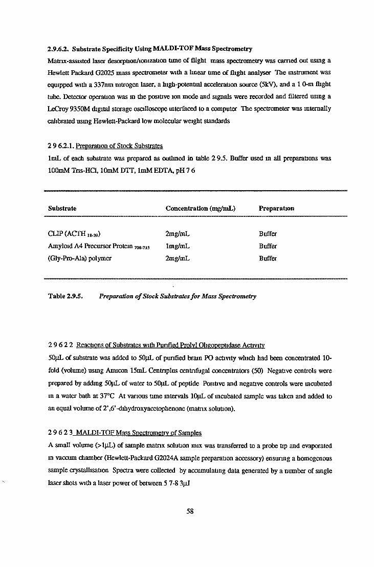
2.9.6J2. Substrate Specificity Using MALDI-TOF Mass Spectrometry
Matnx-assisted laser desorptionViomzation time of flight mass spectrometry was earned out using a
Hewlett Packard G2025 mass spectrometer with a linear time of flight analyser The instrument was
equipped with a 337nm nitrogen laser, a high-potential acceleration source (5kV), and a 1 0-m flight
tube. Detector operation was m the positive ion mode and signals were recorded and filtered using a
LeCroy 9350M digital storage oscilloscope interfaced to a computo' The spectrometer was internally
calibrated using Hewlett-Packard low molecular weight standards
2 9 6.2.1. Preparation of Stock Substrates
lmL of each substrate was prepared as outlined in table 2 9.5. Buffer used m all preparations was
lOOmM Tns-HCl, lOmM DTT, ImM EDTA, pH 7 6
Substrate Concentration (mg/mL) Preparation
CLIP (ACTH 18.39) 2mg/mL Buffer
Amyloid A4 Precursor Protein 708-715 lmg/mL Buffer
(Gly-Pro-Ala) polymer 2mg/mL Buffer
Table 2.9.5. Preparation o f Stock Substrates for Mass Spectrometry
2 9 6 2 2 Reactions of Substrates with Punfled Prolvl Oligppeptidase Activity
50pL of substrate was added to 50pL of punfied brain PO activity which had been concentrated 10-
fold (volume) using Amicon 15mL Centnplus centrifugal concentrators (50) Negative controls were
prepared by adding 50|iL of water to 50(iL of peptide Positive and negative controls were incubated
m a water bath at 37°C At various time intervals lOjiL of incubated sample was taken and added to
an equal volume of 2’,6’-dihydroxyacetophenone (matnx solution).
2 9 6 2 3 MALDI-TOF Mass Spectrometry of Samples
A small volume (>l|iL) of sample matrix solution mix was transferred to a probe tip and evaporated
in vaccum chamber (Hewlett-Packard G2024A sample preparation accessory) ensuring a homogenous
sample crystallisation Spectra were collected by accumulating data generated by a number of single
laser shots with a laser power of between 5 7-8 3|iJ
58

2.9.63. Kinetic StudiesSubstrate specificity studies, on bovine brain, serum and F meningosepticum PO activities, based on
kinetic analysis were performed.
2.9 6.3.1. Km 1>tprmm»non for Z-Glv-Pro-MCA
The Michaelis Mental Constant (Km) was determined for the reaction of purified bovine brain,
bovine serum and F meningosepticum. PO activity, with the substrate Z-Gly-Pro-MCA A range of
concentrations (0-200jiM) of Z-Gly-Pro-MCA were prepared from a 200nM stock solution, 8% v/v
DMSO, lOOmM potassium phosphate buffer, SmM DTT, 0 SmM EDTA, pH 7.4 Stock was prepared
as described in section 2.4.1. Enzyme activities were determined in triplicate and suitable negative
controls were prepared, (section 2 4 1 ) The Km and Vmax values for this substrate were determined
using Michaelis- Menten, Eadie-Hoftsee, Lineweaver-Burk, Hanes-Woolf and Direct Linear Plot
analysis
2 9 63 2 Km Determination for pGlu-His-Pro-MCA
The Michaelis-Menten Constant (Km) was determined for the reaction of of bovine brain, bovine
serum and F. meningosepticum PO activity, with the fluorimetnc substrate pGlu-His-Pro-MCA A
range of concentrations of pGlu-His-Pro-MCA (0-200|iM) were prepared usmg lOOmM potassium
phosphate buffo:, 5mM DTT, 0.5mM EDTA, pH 7.4 as diluent Enzyme activities w oe determined m
triplicate m a similar method as outlined m section 2 4.1 Suitable negative controls were included
The Km and Vmax values for this substrate were determined usmg Michaelis-Menten, Eadie-Hoftsee,
Lineweaver-Burk, Hanes-Woolf and Direct Linear Plot analysis
2 9 6 3 3 Ki Determination for Proline-Contaimng Peptides
The effects of a variety of prolme-containing peptides on Km values obtained for purified brain,
serum and recombinant PO activities for the substrate Z-Gly-pro-MCA was determined The peptides
and their preparation is outlined m table 2.9.6. A 400jiM stock solution of Z-Gly-Pro-MCA, 16% v/v
DMSO, was prepared as outlined m section 2.4 1 From this stock solution a range of dilutions were
prepared (0-400pM) using lOOmM potassium phosphate buffer, 5mM DTT, 0 5mM EDTA, pH 7 4 as
diluent 2mL of appropriate peptide was added to an equal volume of substrate dilution PO activity
was assayed m triplicate using these substrate mixtures as outlined m section 2 4 1 The Km and
apparent Km values were determined usmg the Lineweaver-Burk analysis model
59

Peptide Stock
Concentration
Assay
Concentration
Preparation
LHRH ImM 400|XM 5% MeOH1
TRH 500[iM 200fiM 5% MeOH1
GIu2-TRH 400nM 160jxM 5% MeOH1
Phe -TRH 500pM 200nM 5% MeOH1
Bradykmm 400fjM 160JIM 5% MeOH1
Substance P 400pM 160(iM 5% v/v 1 5M acetic acid1
Angiotensin I 250jiM 100(iM Buffer2*
Angiotensin II 250pM 100nM Buffer2*
Angiotensin III 250jiM IOOjjM Buffer3*
Arga-Vasopressin 250|iM 100pM Buffer2*
Neurotensin 250pM 100nM 5% MeOH1
Table 2.9.6. Preparation o f Peptides For Kinetic Studies , >
1Substrate/fragment was initially dissolved in methanol and was diluted to appropriate concentration
by the addition of lOOmM potassium phosphate buffer, 5mM DTT, 0 5mM EDTA pH 7 4 (final
methanol concentration of 5% v/v) 2 Substrate/fragment was dissolved in 100% lOOmM potassium
phosphate buffer, 5mM DTT, 0 5mM EDTA, pH 7 4 3 Substrate/fragment was initially dissolved m
1 5M acetic acid and diluted to 1 3M by the addition of lOOmM potassium phosphate buffer pH 7 4
*Sonication was required to achieve complete dissolution.
2.9.7. Effect of Specific Prolyl Oligopeptidase Inhibitors on Purified Prolvl Oligopeptidase
Activity
2.9.7.1. Determination of IC50 Values for Specific Prolyl Oligopepeptidase Inhibitors
A range of dilutions of specific PO inhibitors was prepared as outlined in Table 2 9 7 A stock
substrate of 400(iM Z-Gly-Pro-MCA, 16% v/v DMSO m lOOmM potassium phosphate buffer pH 7 4
with lOmM DTT and ImM EDTA was prepared and preincubated at 37°C until completely dissolved
and thermal equilibrium was reached 2mL of substrate stock was added to 2mL of appropriate
inhibitor giving a final substrate concentration of 200jxM with 8% DMSO v/v PO activity (Bacterial
60

brain and serum), using substrate: inhibitor mixtures was assayed in triplicate as outlined in section
2.4.1. Suitable negative and positive controls were prepared.
Inhibitor Stock Concentration(M)
Preparation Assay Concentration (M)
Z-Pro-Prolinal 5xl0 '2 100% v/v MeOH Ixl0 '5- lx l0 '13
Z-Pro-Prolinal-Dimethylacetate
lxlO'2 100% v/v MeOH lxlO 'M xlO '13
Cyclohexyl-Prolinal 2.008xl0‘2 50% v/v Dioxane Ix l0 '3- l x l 0 14
Z-Indolinyl-Prolinal 8.6xl0'3 50% v/v Dioxane Ix l0 '4- lx l0 '14
Fmoc-Ala-Pyrr-Nitrile 5xl0 '3 20% v/v DMSO I—*
X >—i O H-‘ X H-* O ïo
Fmoc-Pro-Pyrr-Nitrile 2.6xl0'5 100% v/v Dioxane Ix l0 ‘3- lx l0 '12
Z-Phe-Pro-Methylketone 1.6xl0'2 Buffer Ix l0 '3x lx l0 ‘14
Postatin 8.1xl0'3 20% v/v Dioxane Ix l0 '4- lx l0 '13
a-Ketobenzothiazole 6 .9xl0'5 50% v/v Dioxane Ix l0 -5x lx l0 '15
Table 2.9.7. Preparation o f Prolyl Oligopeptidase Specific Inhibitors.
Stock concentrations of inhibitors were prepared in appropriate solvents before dilution with lOOmM
potassium phosphate buffer, pH 7.4 to give appropriate dilutions. Dilutions were prepared two-fold
concentrated and then diluted with 400pM Z-Gly-Pro-MCA, 16% v/v DMSO, prepared as outlined
in section 2.4.1. Purified PO activity was assayed in triplicate using substrate/inhibitor mixture as
outlined in section 2.4.1., incorporating suitable negative and positive controls.
2.9.7.2. Effect of Z-Phe-Ala-Chloromethylketone on Purified Bovine Brain and Recombinant
Prolyl Oligopeptidase Activity With Time
A lxlO^M solution of Z-Phe-Ala-CMK was prepared in 50%v/v acetonitrile. Using this as the
inhibitor mixture the following reaction mixtures were prepared: 122^L of bovine brain/recombinant
PO activity, 24|iL of inhibitor mixture and 94pL of 50mM hepes buffer with ImM DTT, ImM
EDTA, 150mM NaCl, pH 7.6. Positive controls with 24p.L of 50%v/v acetonitrile instead of inhibitor
mixture were also prepared. These reactions mixtures were left at room temperature (20°C) and at
61

appropriate time intervals up to 470 minutes 12jiL of this reaction mixture was taken and assayed
fluorimetncally as follows 6|xL of SOjtM Z-Gly-Pro-MCA was added to reaction mixture with 102(iL
of 50mM hepes buffer with ImM DTT, ImM EDTA and 150mM NaCl, pH 7 6 After 1 hour at 37°C
100(iL of this solution was added to 500nL of 1M sodium acetate/HCl, 5% v/v DMSO, pH 4 4 and
fluorescent intensities were measured at excitation and emission wavelengths of 370nm and 440nm
respectively Results were expressed as a percentage of activity of samples taken at a time of 0
minutes
62

3. RESULTS

3. Results
3.1. MCA Standard Curves-Quenched and UnquenchedMCA standard curves were prepared as outlined in section 2 21 and 2 2 2 Plots of fluorimetric
in tensity versus MCA concentration are presented in figures 3.1.1. to 3.1.3. Figures 3 1.1. and 3 1.2
illustrate the quenching effect of undiluted and diluted soluble bovine brain supernatant on
fluorescence. Figure 3.1 3 illustrates the quenching effect of undiluted serum Table 31 is a
summary of the slopes obtained from each curve.
3.2. Protein Standard CurvesProtein standard curves incorporating BSA were prepared as outlined m sections 2 3 2, 2.3 3 and
2 3 4 Figures 3 2 1 and 3 2 2 , plots of absorbance at 560nm versus BSA concentration, represent
BCA standard and enhanced microplate procedure Figure 3 2 3 , absorbance at 595nm versus BSA
concentration, represents the Biorad microassay procedure
3.3. Measurement of Prolyl Oligopeptidase and Z-Pro-Prolinal Insensitive Z-
Gly-Pro-MCA Hydrolysing Activity
Figures 3 3.1 and 3 3 2 represent PO and ZIP activity in bovine serum and bo vine brain supernatant
which were measured as outlined in section 2 4.1 and 2 4 2 Bovine serum was found to contain 277
units of PO activity and 51 units of ZIP activity per mL of serum (5542 and 1020 units of PO and
ZIP activity respectively in 20mL of serum) Brain supernatant was found to contain 45882 units of
PO activity per mL (1835292 units in 40mL of supernatant) but no ZIP activity was detectable. Units
of activity are picomoles of MCA released per minute at 37°C
63
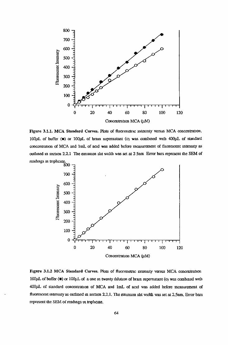
Concentration MCA (jiM)
Figure 3.1.1. MCA Standard Curves. Plots of fluorimetrie intensity versus MCA concentration.
lOOjiL of buffer (•) or lOOpL of brain supernatant (o> was combined with 400jiL of standard
concentration of MCA and lmL of acid was added before measurement of fluorescent intensity as
outlined in section 2.2.1 The mission slit width was set at 2 5nm Error bars represent the SEM of
Concentration MCA (jiM)
Figure 3.1.2 MCA Standard Curves. Plots of fluorimetrie intensity versus MCA concentration
lOOpL of buffer (•) or 100|iL of a one in twenty dilution of brain supernatant (o) was combined with
400pL of standard concentration of MCA and lmL of acid was added before measurement of
fluorescent intensity as outlined in section 2.2.1. The emission slit width was set at 2.5nm. Error bars
represent the SEM of readings in triplicate.
64

Concentration MCA (|iM)
Figure 3.1.3. MCA Standard Curves. Plots of fluorimetrie intensity versus MCA concentration
lOOpL of buffer (•) or lOOpL of a serum (o) was combined with 400|iL of standard concentration of
MCA and lmL of acid was added before measurement of fluorescent intensity as outlined m section
The emission slit width was set at 10 Onm Error bars represent the SEM of readings in triplicate
Figure Unquenched Quenched
3 1 1 7 662 5.968
3 12 7 570 7 570
3 1 3 68 207 46 924
Table 3.1. Slope Determinations for Quenched and Unquenched Standard Curves
65

Concentration BSA (mg /ml)
Figure 3.2.1. BSA Standard Curve Plot of absorbance at 560nm versus BSA concentration obtained
usmg the BCA assay proccedure as outlined m section 2 3 2 Error bars represent the SEM of
triplicate readings
Concentration BSA (mg /ml)
Figure 3.2.2. BSA Standard Curve Plot of absorbance at 560nm versus BSA concentration obtained
using the BCA enhanced assay procedure as outlined in section 2 3 3 Error bars represent the SEM
of triplicate readings
66

Concentration BSA (jig /ml)
Figure 3.2.3. BSA Standard Curve. Plot of absorbance at 595nm versus BSA concentration obtained
using the Biorad microassay procedure as outlined in section 2 3 4 Error bars represent the SEM of
triplicate readings
67

*2?•?Ia£
8e+3 -,
6e+3 -
4e+3 -
2e+3 -
Oe+O
Activity* without ZPP
Total ActivityBovine Serum (20mL)
Figure 3.3.1.
2 g + g ‘ Activity without ZPP
le+6 -*£*?I
Ile+6 —
5e+5 -
0e+0
Activity* with ZPP
Bovine Brain Supernatant (40mL)
Figure 3.3.2.
Figure 3 3.1. and 3.3.2. Measurement of PO and ZIP Activity in Serum and Bram
Supernatant. Enzyme activities of samples were measured as outlined in section 2 4 1 and 2 4 2
respectively Total Activity* refers to activity expressed in terms of total enzyme units 1 e Units or
pmoles MCA released per mmute at 37°C ZPP represents 5xlO-4M Z-Pro-Prolmal This inhibitor
was incorporated into the assay as outlined in section 2 4 2
68
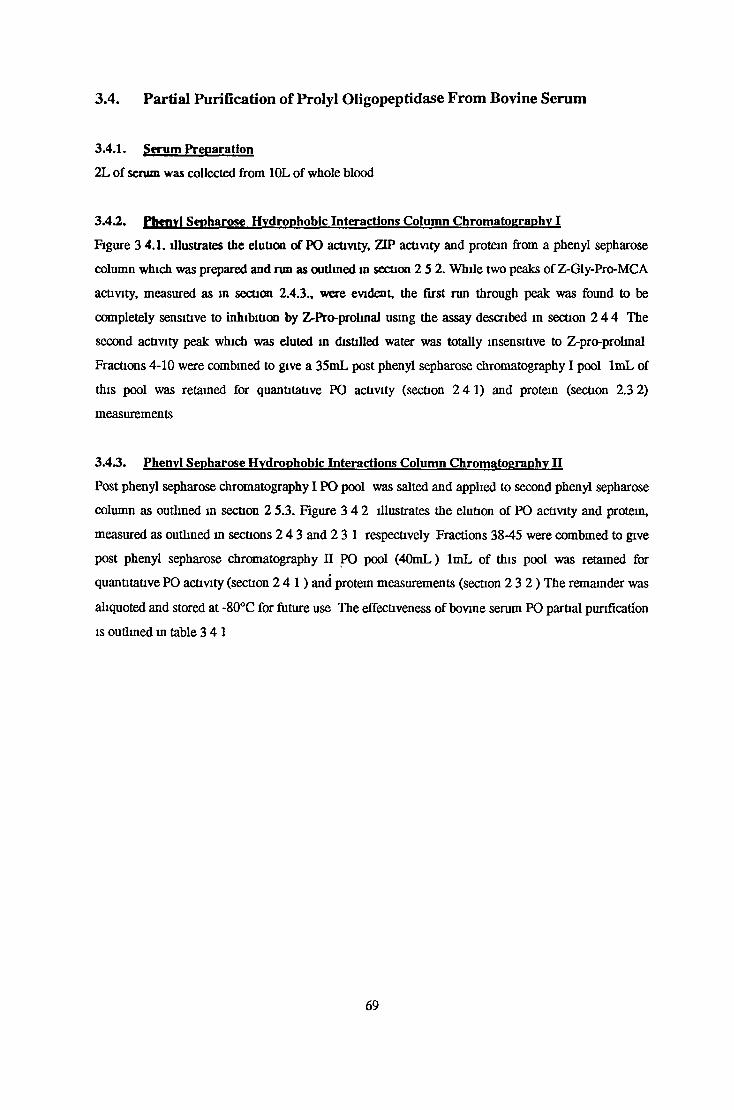
3.4. Partial Purification of Prolyl Oligopeptidase From Bovine Serum
3.4.1. Serum Preparation
2L of serum was collected from 10L of whole blood
3.4.2. Phcnvl Seoharose Hydrophobic Interactions Column Chromatography I
Figure 3 4.1. illustrates the elution of PO activity, ZIP activity and protein from a phenyl sepharose
column which was prepared and run as outlined in section 2 5 2. While two peaks of Z-Gly-Pro-MCA
activity, measured as m section 2.4.3., were evident, the first run through peak was found to be
completely sensitive to inhibition by Z-Pro-prolinal using the assay described in section 2 4 4 The
second activity peak which was eluted in distilled water was totally insensitive to Z-pro-prolmal
Fractions 4-10 were combined to give a 35mL post phenyl sepharose chromatography I pool lmL of
this pool was retained for quantitative PO activity (section 2 41 ) and protein (section 2.3 2)
measurements
3.4.3. Phenvl Sepharose Hydrophobic Interactions Column Chromatography H
Post phenyl sepharose chromatography I PO pool was salted and applied to second phenyl sepharose
column as outlined in section 2 5.3. Figure 3 4 2 illustrates the elution of PO activity and protein,
measured as outlined in sections 2 4 3 and 2 31 respectively Fractions 38-45 were combined to give
post phenyl sepharose chromatography II PO pool (40mL) lmL of this pool was retained for
quantitative PO activity (section 2 4 1 ) and protein measurements (section 2 3 2 ) The remainder was
aliquoted and stored at -80°C for future use The effectiveness of bovine serum PO partial purification
is outlined in table 3 41
69

s3ü§goSC3xs
13ooasS
250 -
200 -
150
100
50 -
0 -
300 n i - 3.00
2 75
h 2 50
225
- 2.00
- 175
- 150
- 1.25
100
- 0 75
- 0 50
- 0 25
- 0 00
Fraction Number
Figure 3.4.1 Elution Profile of Z-Gly-Pro-MCA Degrading Activity from Phenyl Sepharose
Hydrophobic Interaction Chromatography I. 20mL of bovine serum, containing 200mM
ammonium sulphate, was applied to column as outlined m section 2 5 2 The column was eluted
isocratically with distilled water containing no ammonium sulphate Fractions were assayed for
protem ( ) using the absorbance at 280nm as outlined m section 2 3 1 , PO activity (•) and ZIP
activity was assayed as outlined in sections 2 4 3 and 2 4 4 respectively
70
Abso
rban
ce
@ 28
0nm
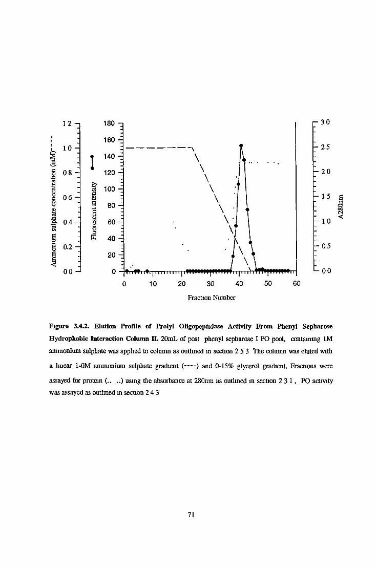
III,kss
1 2 —1
10 -
0 8 -
0 6 -
£ , 0 4 -
ss
§8&es■a3
g 0.2
00 - 1
1 - 3 0
- 25
- 20
- 15
- 10
- 0 5
*-00
Fraction Number
Figure 3.4.2. Elution Profile of Prolyl Oligopeptidase Activity From Phenyl Sepharose
Hydrophobic Interaction Column H. 20mL of post phenyl sepharose I PO pool, containing 1M
ammonium sulphate was applied to column as outlined m section 2 5 3 The column was eluted with
a linear 1-0M ammonium sulphate gradient (-----) and 0-15% glycerol gradient Fractions were
assayed for protein (.. ..) using the absorbance at 280nm as outlined m section 2 3 1 , PO activity
was assayed as outlined in section 24 3
71
A28
0nm

Sample Total Protein (mg) Total activity (units) Specific Activity (Umts/mg) Purification factor Recovery (%)
Serum 1,210 4 5,542 4 57 1 100
Phenyl Sepbarose I 344,9 3,002 8.70 190 54
Ammonium Sulphate 305 6 2,873 9 40 2 06 52
Phenyl Sepharose II 45 9 2,059 449 9 82 37
Table 3.4.1. Partial Purification ofPO from Bovine Serum. Units are expressed as pmoles MCA released per minute at 37°C Pools were assayed
for PO activity as outlined in section 2 4 1 Protein was estimated using the BCA standard microplate assay as outlined m section 2 3 2
72

3.5. Purification of Prolyl Oligopeptiadase Activity from the Cytosolic
Fraction of Bovine Brain
3.5.1. Bovine Brain Preparation
50g brain slice yielded 240mL of post ultracentrifugation cytosolic brain supernatant
3.4.2. Ammonium Sulphate Precipitation
45% and 75% ammonium sulphate precipitation steps were performed as outlined m section 2 6 2
PO activity remained in the supernatant (38mL) following the 45% step and centrifugation PO
activity was precipitated after the 75% step
3.53. DEAE Sepharose Fast Flow Anion Exchange Chromatography
Following dialysis 7mL of post ammonium sulphate precipitate PO pool was applied to DEAE
sepharose column as outlined in section 2 6 3 Figure 3 5 1 shows the elution profile of PO activity in
a linear 0-350mM NaCl gradient Fractions 32-36 were combined to give the post DEAE sepharose
chromatography pool (30mL). lmL of this pool was retained for activity (section 2 4 1 ) and protein
measurements (2 3.2.).
3.5.4. SephacrvI S200 Sepharose Gel Filtration Chromatography
Following concentration by reverse osmosis 2mL of the post DEAE sepharose chromatography pool
was applied to a sephacryl S200 gel filtration column as outlined in section 2 6 4 Figure 3 5 2 shows
the elution of PO activity and protein from the column, qualitatively measured as m sections 2 4 3
and 2.3 3. respectively. Fractions 31-37 were combined to give the post sephacryl S200 gel filtration
chromatography pool lmL of this pool was retained for quantitative PO activity and protein
measurements as outlined in sections 2 4 1 and 2 3 3 respectively
3.5.5. PBE 94 Chromatofocusing
Post sephacryl S200 gel filtration chromatography pool was dialysed and applied to chromatofocusing
column as outlined in section 2.6 5 PO activity was eluted m a decreasing, linear, 6-4 pH gradient
PO activity eluted approximately at pH 4 8 (see figure 3 5 3). Fractions 37-43 were combined to give
the post chromatofocusing pool (35mL) lmL of this pool was retained for quantitative PO activity
and protein measurements (see sections 2 4 1 and 2 3.4 respectively)
3.5.6. Phenvl Sepharose Hydrophobic Interactions Column Chromatography
Post chromatofocusing pool was salted and applied to phenyl sepharose column as outlined in section
2 6 6 Figure 3 5 4 shows the elution of PO activity during a lOmM potassium phosphate buffer with
15% v/v glycerol wash Fractions 25-38 were combined to give the post phenyl sepharose
73

chromatography pool (25mL) lmL of this pool was retained for quantitative protein and activity
measurements as outlined in sections 2 4.1 and 2 3 4 respectively The effectiveness of the
purification is shown m table 3 5 1
74

400 - ,
300 -
200 -
ir£SSto§I 100
0 - 1
seO sI/*)08
!co
3
r- 800
- 600
- 400
- 200
Fraction Number
Figure 3.5.1. Elution profile of Prolyl Oligopeptidase from DEAE Sepharose Anion Exchange
Cromatography. 7 5mL of post ammonium sulphate precipitation dialysate was applied to column at
pH 8 0 as outlined in section 2 6 3 The column was eluted with a linear 0-350mM sodium chloride (-
) gradient Fractions were assayed for PO activity (•) as outlined m section 2 4 3. and protein
using the BCA standard microplate assay procedure as described in section 2 3 2
75

scioo\ir>
8§
aw%COOs3E
I
Fraction number
Figure 3.5.2. Elution Profile of Prolyl Oligopeptidase Activity From Sephacryl S200 Sepharose
Gel Filtration Chromatography. 2mL of reverse dialysed post DEAE sepharose pool with 10% (v/v)
glycerol was applied to column as outlined m section 2.6 4 The column was eluted with 25mM
imidazole-HCl with 150mM NaCl, pH 6 3 Fractions were assayed for PO activity (•) as outlined m
section 2 4 3 Protein was determined using the BCA enhanced microassay procedure as outlined in
section 2 3 3 Vo represents the void volume (105mL).
76

tua,
6.5 -I
6 0
55 -
5 0 -
4 5 -
4 0 -
35 J
Se0VOin©8
1C/3
%
Fraction Number
Figure 3.5.3. Elution Profile of Prolyl Oligopeptidase Activity from PBE 94 Chromatofocusmg
Column. 35mL of post sephacryl S200 aliquot was applied to column at pH 6 0 Column was then
eluted with a linear 6 0-4 0 pH gradient Fractions were assayed for PO activity (•) as outlined m
section 2 4 3 Protein ( ... ) was measured as outlined in section 2.3 4 The pH of each fraction was
also monitored
77

s3h§caJ3Ck
638 sI
1 5 0 - i
125 -
100 -
0 75 -
0 50 -
0 25 -
000 -
-0 25
acino\m®8§■ICO
Fraction number
Figure 3.5.4. Elution Profile of Prolyl Oligopeptidase Activity From Phenyl Sepharose
Hydrophobic Interactions Column. Post chromatofocusing pool was applied to column m 1M
ammonium sulphate Prolyl oligopeptidase activity was eluted with a linear 1-0M ammonium
sulphate (---- ) gradient, followed by a lOOmL wash with lOmM potassium phosphate buffer with
5mM DTT, 0 5mM EDTA and 15% (v/v) glycerol Fractions were assayed for PO activity ( • ) and
protein (----- ) according to sections 2 4 3 and 23 4 respectively
78

Sample Total Protein (mg) Total Activity (units) Specific Activity (Units/mg) Purification Factor Recovery (%)
’Brain Cytosol 122 26 1835292 15011 1 100
’Ammonium sulphate 24 95 1578408 63263 4 2 86
’DEAE Anion Exchange 10 69 927209 - 86736 5 8 51
2Sephacryl S200 6 57 534215 81311 5 4 29
3PBE94 0 19 433978 2319498 154.5 24
3Phenyl Sepharose O il 419227 3811155 253 9 23
Table 3.5.1. Purification of Prolyl Oligopeptidase from Bovine Brain Units are expressed as pmoles MCA released per minute at 37°C
Pools were assayed for PO activity as described in section 2 41 Protein was estimated using1 BCA Standard microplate assay
(section 2 3 2) ,2 BCA Enhanced microplate assay (section 2 3 3 ) and 3Biorad protein microassay (section 2 3 4 )
79
3.5.7. Determination of Purity of Bovine Brain Prolvl Oligppeptidase
3.5.7.1. Polyacrylamide Gel Electrophoresis
SDS polyacrylamide electrophoresis was performed as outlined in section 2 7 1 Figure 3 5 5
represents the silver stained gel image showing the various steps in bovine brain PO purification and
molecular weight markers
Lane Number Sample
1 DE AE Sepharose
2 S200
3 PBE94
4 Phenyl Sepharose
5 Markers
A B-Galactosidase (116kDa)B Fumarase (48 5kDa)C Carbomc Anhydrase (29kDa)
B
Figure 3.5.5 SDS-Polyacrylamide Gel Electrophoresis For Bovine Brain PO Purification
Silver stained polyacrylamide gel prepared as outlined in section 2 7 1 , illustrates the purification
process with a major and minor band m the post phenyl sepharose pool Location of the major band
relative to the molecular weight bands indicates a molecular weight of 66,000 Daltons
80
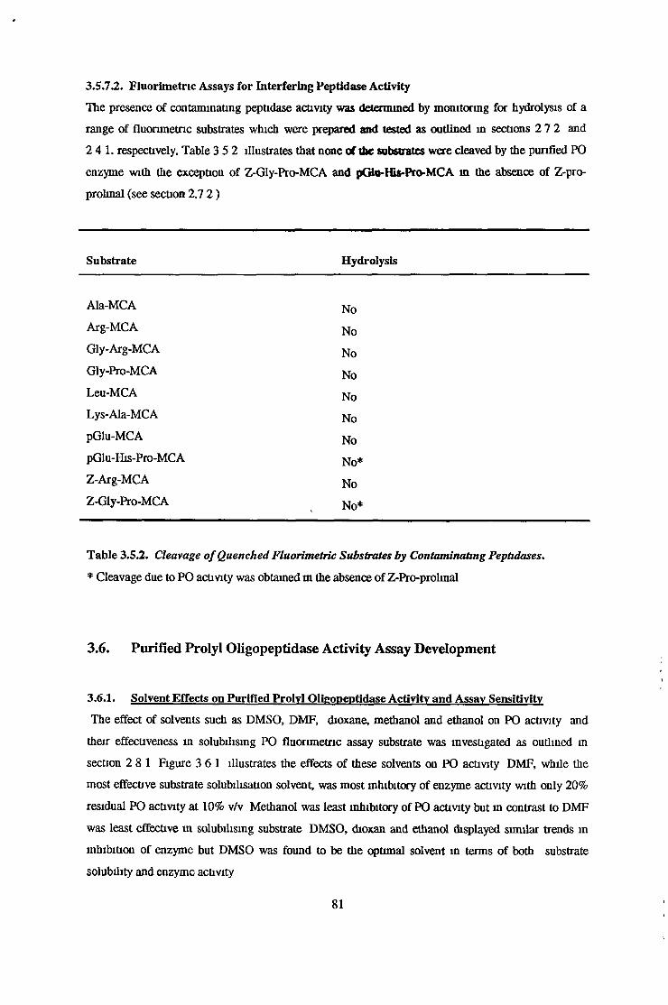
3.5.72,. Fluorimetnc Assays for Interfering Peptidase Activity
The presence of contaminating peptidase activity was determined by monitoring for hydrolysis of a
range of fluorimetnc substrates which were prepared and tested as outlined in sections 2 7 2 and
2 4 1 . respectively. Table 3 5 2 illustrates that none of the substrates were cleaved by the purified PO
enzyme with the exception of Z-Gly-Pro-MCA and pGtu-His-Pro-MCA in the absence of Z-pro-
prohnal (see section 2.7 2 )
Substrate Hydrolysis
Ala-MCA NoArg-MCA NoGly-Arg-MCA NoGly-Pro-MCA NoLeu-MCA NoLys-Ala-MCA NopGlu-MCA NopGlu-His-Pro-MCA No*Z-Arg-MCA NoZ-Gly-Pro-MCA No*
Table 3.5.2. Cleavage o f Quenched Fluorimetnc Substrates by Contaminating Peptidases.
* Cleavage due to PO activity was obtained m the absence of Z-Pro-prolinal
3.6. Purified Prolyl Oligopeptidase Activity Assay Development
3.6.1. Solvent Effects on Purified Prolvl Oligopeptidase Activity and Assay Sensitivity
The effect of solvents such as DMSO, DMF, dioxane, methanol and ethanol on PO activity and
their effectiveness in solubilising PO fluorimetnc assay substrate was investigated as outlined in
section 2 81 Figure 3 61 illustrates the effects of these solvents on PO activity DMF, while the
most effective substrate solubilisation solvent, was most inhibitory of enzyme activity with only 20%
residual PO activity at 10% v/v Methanol was least inhibitory of PO activity but in contrast to DMF
was least effective m solubilising substrate DMSO, dioxan and ethanol displayed similar trends in
inhibition of enzyme but DMSO was found to be the optimal solvent in terms of both substrate
solubility and enzyme activity
81
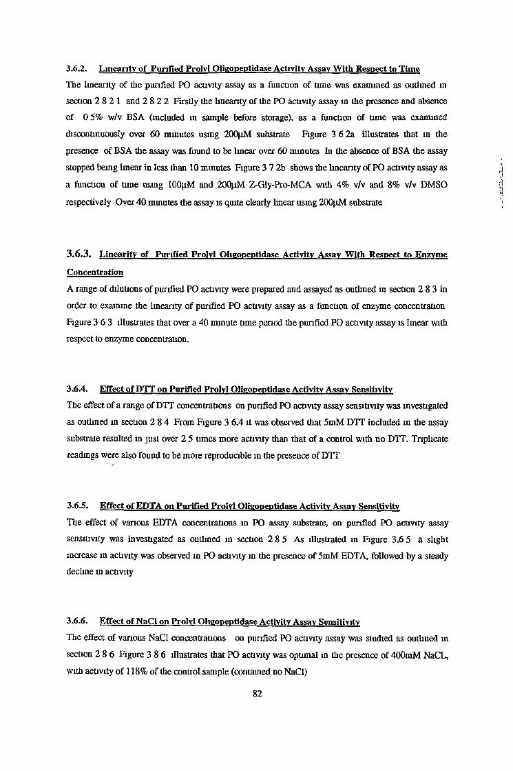
3.6.2. Linearity of Purified Prolvl Qiigopeptidase Activity Assay With Respect to TimeThe linearity of the purified PO activity assay as a function of time was examined as outlined m
section 2 8 21 and 2 8 2 2 Firstly the linearity of the PO activity assay in the presence and absence
of 0 5% w/v BSA (included m sample before storage), as a function of time was examined
discontinuously over 60 minutes using 200jiM substrate Figure 3 6 2a illustrates that m the
presence of BSA the assay was found to be linear over 60 minutes In the absence of BSA the assay
stopped being linear in less than 10 minutes Figure 3 7 2b shows the linearity of PO activity assay as
a function of tune using 100[xM and 200}iM Z-Gly-Pro-MCA with 4% v/v and 8% v/v DMSO
respectively Over 40 minutes the assay is quite clearly linear using 200nM substrate
3.6.3. Linearity of Purified Prolvl Qiigopeptidase Activity Assay With Respect to Enzvme
Concentration
A range of dilutions of purified PO activity were prepared and assayed as outlined m section 2 8 3 in
order to examine the linearity of purified PO activity assay as a function of enzyme concentration
Figure 3 6 3 illustrates that over a 40 minute time period the purified PO activity assay is linear with
respect to enzyme concentration.
3.6.4. Effect of DTT on Purified Prolvl Qiigopeptidase Activity Assay SensitivityThe effect of a range of DTT concentrations on purified PO activity assay sensitivity was investigated
as outlined in section 2 8 4 From Figure 3 6.4 it was observed that 5mM DTT included in the assay
substrate resulted in just over 2 5 times more activity than that of a control with no DTT. Triplicate
readings were also found to be more reproducible in the presence of DTT
3.6.5. Effect of EDTA on Purified Prolvl Qiigopeptidase Activity Assay Sensitivity
The effect of various EDTA concentrations in PO assay substrate, on purified PO activity assay
sensitivity was investigated as outlined m section 2 8 5 As illustrated m Figure 3.6 5 a slight
increase in activity was observed in PO activity in the presence of 5mM EDTA, followed by a steady
decline in activity
3.6.6. Effect of NaCl on Prolvl Qiigopeptidase Activity Assay Sensitivity
The effect of various NaCl concentrations on purified PO activity assay was studied as outlined in
section 2 8 6 Figure 3 8 6 illustrates that PO activity was optimal m the presence of 400mM NaCL,
with activity of 118% of the control sample (contained no NaCl)
82
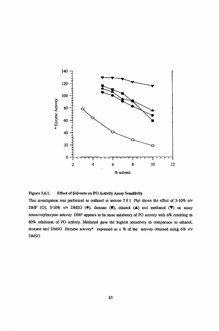
% solvent
Figure 3.6.1. Effect of Solvents on PO Activity Assay Sensitivity
This investigation was performed as outlined in section 2 81 Plot shows the effect of 3-10% v/v
DMF (O), 5-10% v/v DMSO (•) , dioxane (■), ethanol (A) and methanol (T) on assay
sensitivity/enzyme activity DMF appears to be most inhibitory of PO activity with 6% resulting m
60% inhibition of PO activity. Methanol gave the highest sensitivity m comparison to ethanol,
dioxane and DMSO Enzyme activity* expressed as a % of the activity obtained using 6% v/v
DMSO
83

s
§§oe
Time (minutes)
Time (seconds)
Figures 3.6.2a. and 3.6.2b. Linearity of Purified PO Assay With Respect To Time.
Figure 3 6 2a. shows linearity of purified PO enzyme assay with ( • ) and without (O) 0 5% w/v BSA
over 60 minutes Figure 3 6 2b shows linearity of purified PO assay with 100nM and 200|iM
substrate
84

%>&cêoaN[S
Relative concentration of purified PO
Figure 3.6.3. Linearity of Prolyl Oligopeptidase Assay With Respect to Enzyme Concentration.Plot of enzyme activity versus enzyme concentration Enzyme activity refers to % of activity in
undiluted sample
85

DTT concentration (mM)
Figure 3.6.4. Effect of DTT on Prolyl Oligopeptidase Assay Sensitivity.
Plot shows effect of O-lOOmM DTT on PO activity Investigation was earned out as outlined m
section 2 64 Enzyme activity* expressed as % of activity m the absence of DTT
86

EDTA concentration (mM)
Figure 3.6.5. Effect of EDTA on PO Assay Sensitivity
Plot shows the effect of EDTA on PO assay sensitivity/activity Experiment was performed as outlined
m section 2 6 5 Enzyme activity* expressed as % of activity in the absence of EDTA
87
NaCl concentration (mM)
Figure 3.6.6. Effect of NaCl on PO Assay Sensitivity
Plot show the effect of NaCl on the sensitivity of PO assay. Experiment was performed as outlined m
section 2.8 6 PO activity/ assay sensitivity increases at NaCl concentrations up to 400mM and
declines at higher NaCl concentrations Enzyme activity* is an expression of the % of activity in the
absence of NaCl
88

3.7. Characterisation of Purified Prolyl Oligopeptidase Activity
3.7.1. Relative Molecular Mass Determination
3.7.1.1. Sephacryl S200 HR Gel Filtration Chromatography
The relative molecular mass of the purified PO activity was determined using a calibrated Sephacryl
S200 gel filtration column which was prepared and run as described m section 2 9 1 1 Using a plot of
Log(molecular mass) versus elution volume (molecular mass standards)/void volume as illustrated m
Figure 3 7.11 the molecular mass of PO was found to be 69 440 kDa.
3.7.1.2. Polyacrylamide gel Electrophoresis
Using electrophoresis the relative molecular mass of the purified PO activity was determined as
described m section 2 9 1 2 From a plot of Log (molecular mass) vs Rf value (distance travelled by
mass standard or unknown/distance travelled by bromophenol blue dye front), illustrated in figure
3 7 1 2 the relative molecular mass of purified PO activity was found to be 69 501 kDa The gel used
in the molecular mass determination is shown in figure 3 5 5
3.7.2. pH Effects
The effects of assaying PO activity at a range of pHs and premcubating the enzyme at a range of pHs,
prior to assaying for activity at pH 7 4 was investigated as described in sections 2 9.2 1 and 2 9.2 2
respectively It can be concluded from figure 3 7.2a. that the optimum pH for assay of PO activity is
7 4 using potassium phosphate buffer When PO activity was preincubated at a range of different pHs,
using different buffer systems, and then assayed at pH 7 4 (potassium phosphate) it was found that the
enzyme was almost completely inactivated at pHs of 4 5 or less and 10 0 or greater Premcubation of
the enzyme at pHs between 5 and 9 prior to assay at pH 7 4 resulted in no loss of activity This is
illustrated in figure 3.7 2b
89

60 -i
55 -
3 5 — i i i i | i i i i | i"i i i | i i i i | i i i i | i n i
13 14 t 15 16 17 18 19
W e / V o
Figure 3.7.I.I. Sephacryl S-200 Molecular Mass Calibration Curve. Plot of Log molecular mass
versus Ve/Vo where Ve is the elution volume of the molecular mass standard ( • ) or unknown (PO
activity), (o) and Vo is the void volume (105mL) Ve and Vo were determined as outlined in sections
2.9 1.11 and 2 91 1.2. Linear regression analysis of the data obtained from the molecular mass
standards produced the following calibration equation'
Log(molecular mass) = -2 6(Ve/Vo) + 9 86
From this equation the molecular weight of PO was found to be 69 44kDa
90
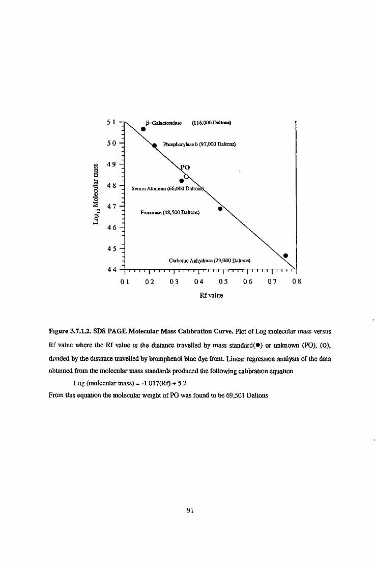
Rf value
Figure 3.7.I.2. SDS PAGE Molecular Mass Calibration Curve. Plot of Log molecular mass versus
Rf value where the Rf value is the distance travelled by mass standard(*) or unknown (PO), (0),
divided by the distance travelled by bromphenol blue dye front Linear regression analysis of the data
obtained from the molecular mass standards produced the following calibration equation
Log (molecular mass) = -1 017(Rf) + 5 2
From this equation the molecular weight of PO was found to be 69,501 Daltons
91

pH
pH
Figures 3.7.2a. and 3.7.2b. Effect of pH on Prolyl Oligopeptidase Activity. Plots show the effect of
pH on PO activity Investigations were carried out as in sections 2 9.2 1 and 2 9 2 2 Figure 3 7 2a
represents the activity of PO when assayed at different pHs It is evident that PO activity has a broad
pH/activity range with a pH optima of 7 4 in potassium phosphate Figure 3 7 2b represents the
inactivation profile of PO (pH at which PO activity is completely inactivated even when assay
performed at pH 7 4) PO activity is completely inactivated below pH 4 5 and 10 0 Buffer systems
used were citrate/phosphate ( • ) , phosphate (O), tris/HCl (A) and glycine/NaOH (♦ ) Enzyme
activity* expressed as % of activity obtained in potassium phosphate, pH 7 4
92

3.73 . Temperature Effects
The effect of assaying PO activity at different temperatures and preincubating the enzyme at a range
of temperatures, for different times, prior to to assaying at 37°C was investigated as outlined m
sections 2 9 3 1 and 2.9 3 2 respectively The optimal temperatue for tfae assay of PO activity was
determined to be 37°C. This is illustrated in figure 3 7.3a Figure 3 7.3b. illustrates that PO activity
was completely inactivated by preincubation at 50°C and 60°C, for 15 minutes or more, prior to assay
at 37°C. Preincubation of PO samples at 37°C and 40°C at various times up to 45 minutes, bad little
effect on activity but samples left at room temperature (20°C) for more than 30 minutes lost greater
than 30% of activity upon assay at 37°C
93
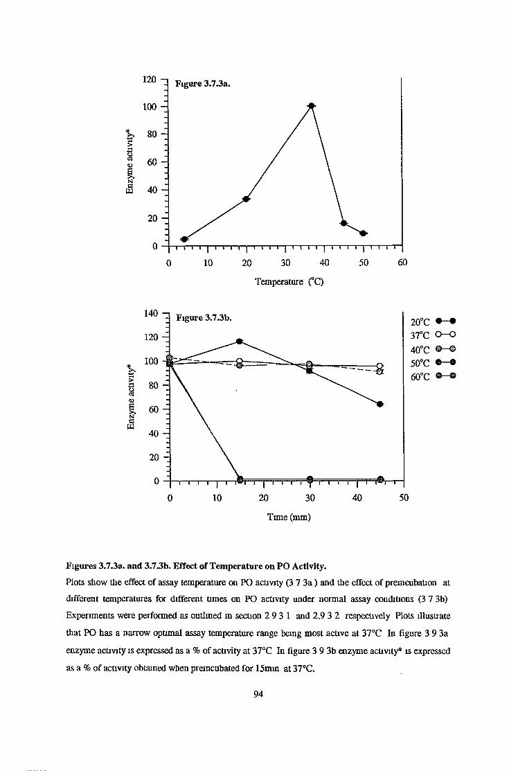
Temperature (°C)
%>3cà
NaW
20°C37°C 0 —0
4 O o n
50°C A ^ A V V60°C
Time (mm)
Figures 3.7.3a. and 3.73b. Effect of Temperature on PO Activity.
Plots show the effect of assay temperature on PO activity (3 7 3 a ) and the effect of premcubation at
different temperatures for different times on PO activity under normal assay conditions (3 7 3b)
Experiments were performed as outlined m section 2 9 3 1 and 2.9 3 2 respectively Plots illustrate
that PO has a narrow optimal assay temperature range being most active at 37°C In figure 3 9 3a
enzyme activity is expressed as a % of activity at 37°C In figure 3 9 3b enzyme activity* is expressed
as a % of activity obtained when preincubated for 15mm at 37°C.
94
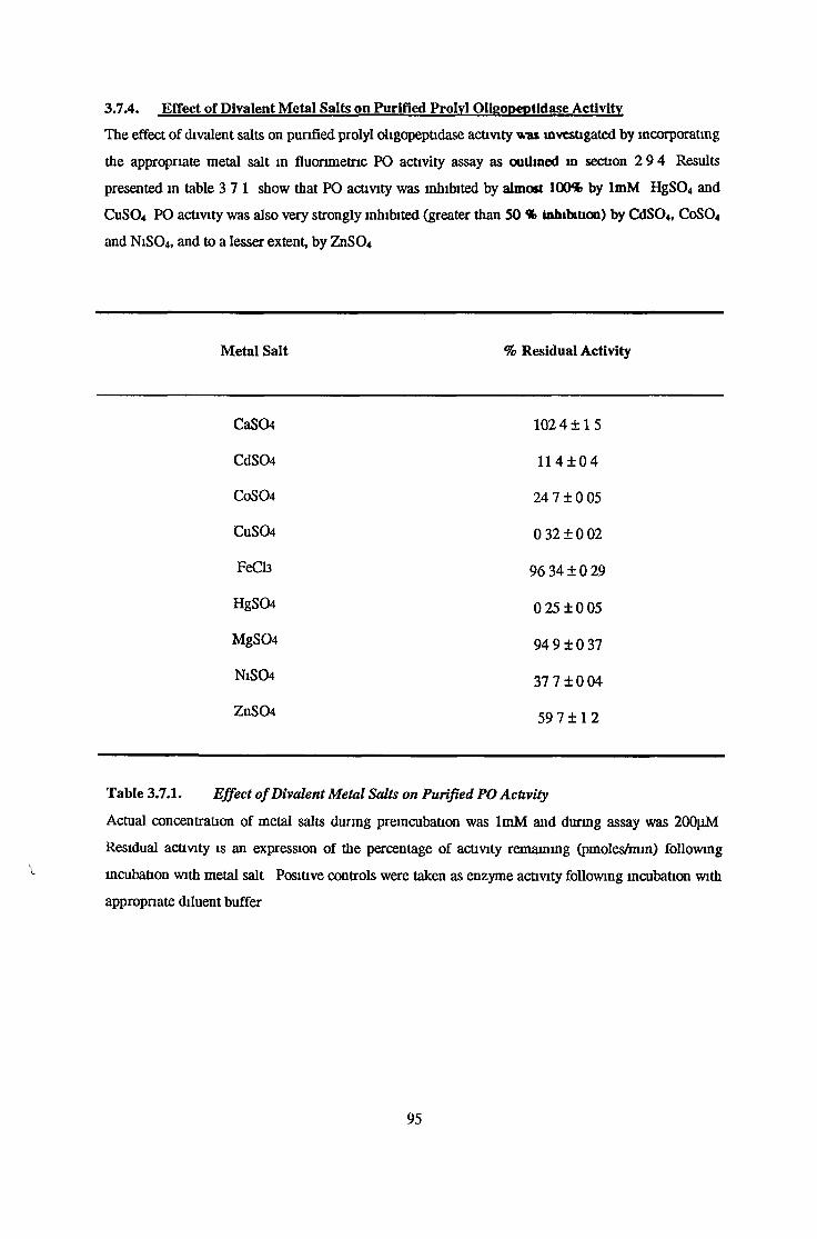
3.7.4. Effect of Divalent Metal Salts on Purified Prolyl Oligopeotidase Activity
The effect of divalent salts on purified prolyl oligopeptidase activity was investigated by incorporating
the appropriate metal salt in fluorimetrie PO activity assay as outlined in section 2 9 4 Results
presented in table 3 7 1 show that PO activity was inhibited by almost 100* by ImM HgS04 and
CuS04 PO activity was also very strongly inhibited (greater than 50 % iahitnuoo) by CdSO«, C0 SO4
and N1SO4 , and to a lesser extent, by ZnS04
Metal Salt % Residual Activity
CaS04 102 4 1 1 5
CdS04 11 4 ± 0 4
C0 SO4 24 7 ± 0 05
CuS04 0 32 ± 0 02
FeCl3 96 34 ± 0 29
HgS04 0 25 ± 0 05
MgS04 94 9 ± 0 37
N1SO4 377 + 0 04
ZnS04 59 7 ± 1 2
Table 3.7.1. Effect o f Divalent Metal Salts on Purified PO Activity
Actual concentration of metal salts during premcubation was ImM and during assay was 200jiM
Residual activity is an expression of the percentage of activity remaining (pmoles/mm) following
incubation with metal salt Positive controls were taken as enzyme activity following incubation with
appropriate diluent buffer
95

3 7.5. Effect of Functional Reagents on Purified Prolvl Oligopeptidase Activity
The effect of various functional reagents on purified PO activity was investigated as outlined in
section 2 9 5 Table 3 7 2. illustrates the percentage residual activity after premcubation with
appropriate functional reagent The percentage residual activity is an expression of enzyme activity
(units/mL) as a percentage of positive control activity (100%) In table 3 7 2 concentrations of
functional reagents are the actual concentrations during the incubations Of the reagents tested the
cysteine protease inhibitors, DTNB (10, 5mM and 0 05mM) NEM (lOmM) and senne protease
inhibitor, AEBSF (lOmM) were found to be the most potent inhibitors of PO activity, resulting in
complete inhibition PO activity was also significantly inhibited by the cysteine protease inhibitors
iodoacetamide(10mM) and to a lesser extent lodoacetate (lOmM) with only 14 84% and 40 40%
activity remaining respectively Activity was also inhibited by over 50% by lOmM 1,10
phenanthroline, bacitracin and N-Decanoyl CoA and to a lesser extent by 1,7 and 4,7 phenanthroline
(lOmM) A two-fold increase in PO activity following incubation with lOmM DTT was observed
96

Functional Reagent % Residual Activity
lOinM 5mM ImM O.lmM 0.05mM
Iodoacetamide 14.84 26.11 63.40
Iodoacetate 40.40 59.67 98.98
N-Ethylmalemide (NEM) 0.36 1.16
Dithiomtrobenzoicacid (DTNB) 0 0 0.46
Dithiothreitol 2013 106 83
2-Mercaptoethanol 85.09 81.43
AEBSF 0 1736
PMSF 89.27 95.11
EDTA 112 65 111.77
EGTA 95 81 9845
CDTA 94 38 10014
8-Hydroxyqumohne 89 71 107 99
Imidazole 89 05 98.73
1 ,10-Phenanthrohne 43 08 56 27
1, 7-Phenanthrolme 74 35 87 38 96 10
4 , 7-Phenanthroline 70 90 82 42 87 17
Benzamidme 93 12 94.41
Camitme 101 88 10098
lmg/mL 0.5mg/mL 0.1mg/mL 0.05mg/mL
Bacitracin 21.59 30.19
Pepstatm 89 15 103 47
Soybean Trypsin Inhibitor 77 75 89 57
Aprotmm 86 00 93 42
Puromycm 92 71 98 17
N-Decanoyl CoA 46.05 81 54 83.84
Table 3.7.2. Effect of Functional Reagents on Prolyl Oligopeptidase Activity
Functional reagents were prepared according to section 2 9 5 % Residual activity is an expression of
the remaining activity relative to positive controls Concentrations given were the actual
concentrations during the 15 minute pre-mcubation

3.7.6. Substrate Specificity Studies on Purified Prolvl Oligonentidase Activity
Substrate specificity studies on purified bovine brain prolyl oligopeptidase activity were performed as
described m section 2 9 6 Using reverse phase HPLC and MALDI-TOF mass spectrometry, the
cleavage of some proline containing peptides was monitored qualitatively Fluoresence spectrometry
was used to determine kinetic parameter for fluorescent substrates and prolme containing peptides
Some comparative investigations were earned out using partially punfied bovine serum PO activity
and purified recombinant Flavobacterium memngosepticum PO activity
3.7.6.I. Substrate Specificity Studies using Reverse Phase HPLC
Reverse phase HPLC was used to determine whether brain, serum and F memngosepticum PO
activities cleaved a variety of prohne containing peptides, listed in table 2 9 6 1 Reactions of peptides
with enzyme samples and reverse phase HPLC was carried out as outlined m sections 2 9 6 1 2 and
2 9 6 1 3 respectively The optimal wavelength for detection of peptides and cleavage products was
found to be 214nm Table 3 7 3 illustrates the peptides tested and whether or not they were cleaved
by the various PO activities Figures 3 7 6 1 1 to 3 8 6 1 10 represent chromatograms of absorbance
at 214nm versus retention time (minutes) for ADNF-14, angiotensin I, angiotensin II, angiotensin III,
Arg8-vasopressin, bradykmm, Glu2-TRH, Gly-Gly-Pro-Ala, TRH and TRH-Gly respectively for brain
and F memngosepticum activities
98

Peptide Cleavge Detected
Brain PO Serum PO F. memngosepticum
PO
ADNF-14 Yes N D N D
Angiotensin I Yes Yes Yes
Angiotensin II Yes Yes Yes
Angiotensin III Yes Yes Yes
Arg8-Vasopressm No N D Yes
Bradykmm Yes Yes Yes
H-Gly-AIa-Phe-OH No No No
H-Gly-Gly-Pro-Ala-OH No No Yes
H-Gly-Pro-OH No No No
G1u2TRH Yes Yes Yes
Phe2TRH Yes N D Yes
TRH-Gly Yes Yes Yes
TRH Yes Yes Yes
LHRH Yes Yes Yes
Substance P Yes Yes Yes
Z-Gly-Pro-Ala-OH Yes Yes Yes
Z-Pro-Ala-OH No No No
Z-Pro-Gly-OH No No No
Z-Pro-Leu-Gly-OH No No No
Z-Pro-Pro-OH No No No
Table 3.7.3. Substrate Specificity studies-Reverse Phase HPLC ofProhne Containing Peptides
Reverse Phase HPLC was earned out on peptides which had been incubated with brain, serum and
bacterial PO activities as oudined m section 2 9 6.1 Table states whether or not cleavage was
obtained N D -not determined
99

scr}-
10
? 0 8 H¿3c 8 £3 0 6 -> 00 <u c
cs®8§o</3
0 4
02 -
00 - 1
r- 80
70
S<0
tP
40
Time (mm)
Figure 3.7.6.I.I. Chromatogram of absorbance at 214nm versus retention time in minutes for ADNF-
14 Substrate specificity studies performed as outlined in section 2 9 6 1 l i e presence of ADNF-14
before (-—) and after (—) incubation with purified bovine brain PO, is illustrated Cleavage product
*, although not well resolved from ADNF-14 peak, is also shown
1 0 0

1 4 - i
? 12 tiI 10
Ëa os<uaI °-6c
ï 04 8S 02t</)3 00
snrfHesfl<L>s
Time (min)
14
f 12S8 10Ëa os0)aS 064jj
S 04 8§ 02 "SC/33 00
o§3
Time (min)
Figures 3.7.6.1.2a and 3.7.6.1.2b. Chromatograms of absorbance at 214nm versus retention time in
minutes for Angiotensin I Substrate specificity studies performed as outlined m section 2 9 6 1 The
presence of Angiotensin I before (— ) and after (—) incubation with purified bovme bram PO
(3 7 6 1 2a ) bacterial PO (3 7 6 1 2b ) is illustrated Cleavage products * , are also shown
1 0 1
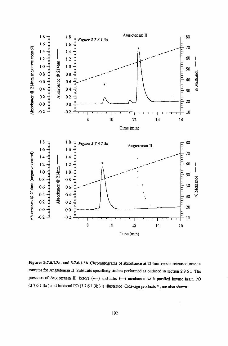
Ügo
u3W><¡Ja6c•<Tts®8
1 8 16
1 4
12 10 0 8
0 6
0 4
02
00 -0 2
osfcS>
Time (min)
oaa
í>so
6arj-Hes©ss£>V-iOco"<
18
1 6
1 4
12100 8
0 6
0.4
0200
-02
<Us
Time (mm)
Figures 3.7.6.1.3a. and 3.7.6.1.3b. Chromatograms of absorbance at 214nm versus retention time in
minutes for Angiotensin n Substrate specificity studies performed as outlined in section 2 9 6 1 The
presence of Angiotensin II before (— ) and after (—) incubation with purified bovine brain PO
(3 7 6 1 3a ) and bacterial PO (3 7 6 1 3b ) is illustrated Cleavage products * , are also shown
1 0 2

0 10
1 0 8!>3 S1 s ss-'t
1 2
es®8
!V)<!
0 6
0 4
02
00
-0 2
70
10
Time (mm)
0 ü1
gDû<Üci 04Tj-
1“CS
■fco
1 2
10
0 8
0 6
02
00
-0 2
70
10
T im e (m in)
Figures 3.7.6.1.4a. and 3.7.6.1.4b. Chromatograms of absorbance at 214nm versus retention tune m
minutes for Angiotensin III Substrate specificity studies performed as outlined m section 2 9 6 1 The
presence of Angiotensin III before (— ) and after (—) incubation with purified bovine brain PO
(3 7 6 1 4a ) and bacterial PO (3 7 6 1 4b ) is illustrated Cleavage products * , are also shown
103

£3oovG,eGTfr-Hes®8
01
1 2
10
0 8
0.6
0 4
02
00
-0 2
M
Time (min)
1 2 —1
? 10
I o sSÎa 060)tí¡3(3TT1—4CÍ
0 4 -
02 -
8S oo -3 ■s s3 - 0 2 J
OstS >
Time (min)
Figures 3.7.6.1.5a. and 3.7.6.1.5b. Chromatograms of absorbance at 214nm versus retention time m
minutes for Arg8-vasopressm Substrate specificity studies performed as outlined m section 2 9 6 1
The presence of Arg'-vasopressin before (— ) and after (— ) incubation with purified bovme brain
PO (3 7 6 1 5a.) and bacterial PO (3 7 6.1 5b ) is illustrated Cleavage products * , are also shown
1 0 4

g3 0 6 SI 0 4T—<® 02
II 00
1 2
? 10
i 08
I
Time (mm)
§s8
12
10
0 8aI 0 6G
S 0 4(N(§) 0 2 81 00O003 - 0 2
O§■§s
Time (min)
Figures 3.7.6.1.6a. and 3.7.6.1.6b. Chromatograms of absorbance at 214nm versus retention time in
minutes for Bradykinin Substrate specificity studies performed as outlined in section 2 9 6 1 The
presence of Bradykinin before (— ) and after (—) incubation with purified bovine brain PO
(3 7 6 1 6a ) and bacterial PO (3 7 6 1 6b ) is illustrated Cleavage products * , are also shown
1 0 5

«8*cEs•*rcs
%K/i<:
0 8 - 1
0 6 -
0.4 -
02 -
0 0 - 1
6e»■*(S<§>8
•o<
I
Time (minutes)
0 hi1 !> 3 S’ 3acTf
1-Hcs<§)8
1ç/5£><
0 8 - i
0 6 -
0 4
02 -
00 - 1
Time (min)
Figures 3.7.6.1.7a and 3.7.6.1.7b. Chromatograms of absorbance at 214nm versus retention time in
minutes for Glu2-TRH Substrate specificity studies performed as outlined m section 2 9 6 1 The
presence of GIu2-TRH before (— ) and after (—) incubation with purified bovine brain PO
(3 7 6 1 7a ) and bacterial PO (3 7 6 1 7b ) is illustrated
1 0 6

isgo
38PsBaes
8
!co3
1 0 - I
0 8 -
0 6 -
0 4 -
0 2 -
00 -
-0 2
Ss■'tiHes
8
-0 2 i i i i | i i i i | i i i i | i i i ~i "|"i i i i | i i i i | i i i i | i i i i2 4 6 8 10 12 14 16
Time (mm)
ois§g38PSBaes®8
lO - i
0 8 -
0 6 -
0.4 -
02 -
00 -
3 -02
l O —i Figure 3 7 61 8b.
0 8 -
SBrr
es
8
1</)
0 6 -
6 8 10 12 14 16
Time (mm)
80
r 70
- 60 |
: I- 50
- 3 0
- 20
10
Figures 3.7.6.1.8a and 3.7.6.1.8b. Chromatograms of absorbance at 214nm versus retention time m
minutes for Gly-Gly-Pro-Ala Substrate specificity studies performed as outlined in section 2 9 6 1
The presence of Gly-Gly-Pro-Ala before (— ) and after (—) incubation with purified bovine brain
PO (3 7.6 1 8a ) bacterial PO (3 7 6 1 8b ) is illustrated The major cleavage product * , Gly-Gly-Pro,
is also shown
107

1 2 - i
0 10 to1 08X3S) 0 6oS€ 044i-Hes©85 00%00
3 -02
02 -
•SI
Time (mm)
1 2 - i
9 “ 10b8 0 8j>Si 0.60GVm '1 04
t-Hes<§>8§ 00%w -02
02 -
5os
Time (mm)
Figures 3.7.6.1.9a. and 3.7.6.1.9b. Chromatograms of absorbance at 214nm versus retention time m
minutes for TRH Substrate specificity studies performed as outlined m section 2 9 6 1 The presence
of TRH before (— ) and after (—) incubation with purified bovine bram PO (3 7 6 1 9a ) and
bacterial PO (3 7 6 1 9b ) is illustrated The major cleavage product * , TRH-OH, is also shown
1 0 8

¿3G
o
gS§ecVw'
Sct j-»“Hcs
©8
1C/33
16
14
12
1.0
08
06
04
02
00
-0 2
s
20
Time (mm)
oa§O>2?&S
t-H<N®8
!co3
1 6
14
12
10
08
06
04
02
00
-0 2
64?rHCS®83Hco<:
Figures 3.7.6.1.10a. and 3.7.6.1.10b. Chromatograms of absorbance at 214nm versus retention time
in minutes for TRH-Gly Substrate specificity studies performed as outlined m section 2 9 6 1 The
presence of TRH-Gly before (— ) and after (—) incubation with purified bovine bram PO
( 3 7 6 1 10a ) and bacterial PO (3 7 6 1 10b ) is illustrated The major cleavage product * , TRH-OH,
is also shown
109

3.7 6.2. Substrate Specificity With MALDI-TOF Mass Spectrometry
MALDI-TOF Mass Spectrometry was used to determine whether purified brain PO cleaved ACTH ]8_
3 9 , (Gly-Pro-Ala) polymer and amyloid precursor protein fragment 7 0 8 -7 15 It was also intended to
confirm if bovine brain PO behaved m a similar manner to the bacterial and the porcine kidney
forms of the enzyme Mass spectrometry was performed as outlined in section 2 9 6 3 Brain PO was
found to cleave ACTHi8. 39 at 2 positions to give ACTH18.24, ACTH2 5 .3 9 and ACTH2 5 .3 6 Cleavage
sites were marked by the presence of prolme PO was also found to cleave the (Gly-Pro-Ala) polymer
over time Amyloid fragment was cleaved to give APP7 0 8 -713 Table 3 7 4 summarises the cleavage
sites deduced for these peptides and figures 3 7 6 2 1 - 3 7 6 2 3 represent the mass spectra (relative
intensity versus mass/charge ratio) obtained Comparisons with the bacterial and porcine enzymes are
discussed m section 4 7 6 2
Peptide Cleavage
IAPP708-715 Gly-Gly-Val-Val-Ile-Ala-Thr-Val
ICLIP (ACTH18.39) Ar|-Pro-Val-Lys-Val-Tyr-Pro-Asn-Gly-Ala-Glu-Asp-Glu-Ser-Ala-Glu-Ala-Phe-
Pro-Leu-Glu-Phe
(Gly-Pro-Ala)i5 (Gly-Pro-Ala-Gly-Pro-Ala-Gly-Pn>Ala)5
Table 3.7.4. Prolyl Ohgopeptidase Cleavage Sites as Determined by MALDI-TOF Mass
Spectrometry
APP Amyloid A4 Precursor Protein
CLIP Corticotropin Like Lobe Intermediate Peptide
1 1 0
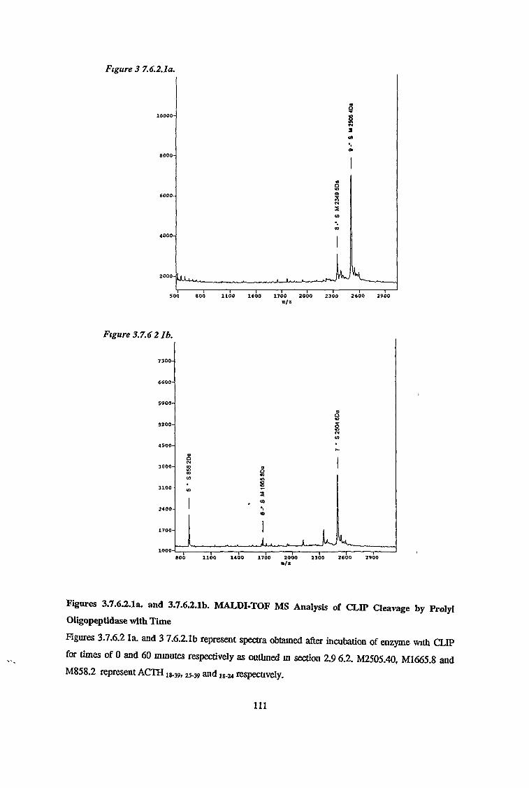
Figure 3 7.6.2.1a.
Figures 3.7.6.2.1a. and 3.7.6.2.1b. MALDI-TOF MS Analysis of CLIP Cleavage by Prolyl
Oligopeptidase with Time
Figures 3.7.6.2 la. and 3 7.6.2.1b represent spectra obtained after incubation of enzyme with CLIP
for times of 0 and 60 minutes respectively as outlined in section 2.9 6 .2 . M2505.40, M1665.8 and
M858.2 represent ACTH 18-39, 2 5 -3 9 and ig.2 4 respectively.
I l l
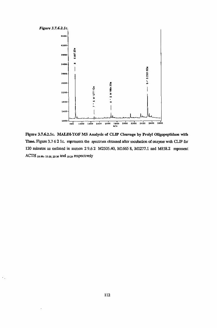
Figure 3.7.6.2.1c. MALDI-TOF MS Analysis of CLIP Cleavage by Prolyl Oligopeptidase with
Time. Figure 3.7 6 2 lc . represents the spectrum obtained after incubation of enzyme with CLIP for
120 minutes as outlined in section 2 9.6 2 M2505.40, M1665 8, M1277.1 and M858.2 represent
ACTH i8-39> 2 5-39,2 5 -3 6 and 18 -2 4 respectively
1 1 2

m/sFigure 3.7.6.2.2b
n /z
Figure 3.7.6.2.2a and 3.7.6.2.2b. MALDI-TOF MS Analysis of APP 708.7 x5 Cleavage by Prolyl
Oligopeptidase with Time. Figures 3 7.6 2.2a and 3 7.6.2.2b. represents the spectra obtained after
incubation of enzyme with APP for 0 and 120 minutes respectively as outlined in section 2.9 6.2
M715.8 and M515.7 represent APP7 0 8 -7 1 5 and 7 0 8 -7 13 respectively.
113
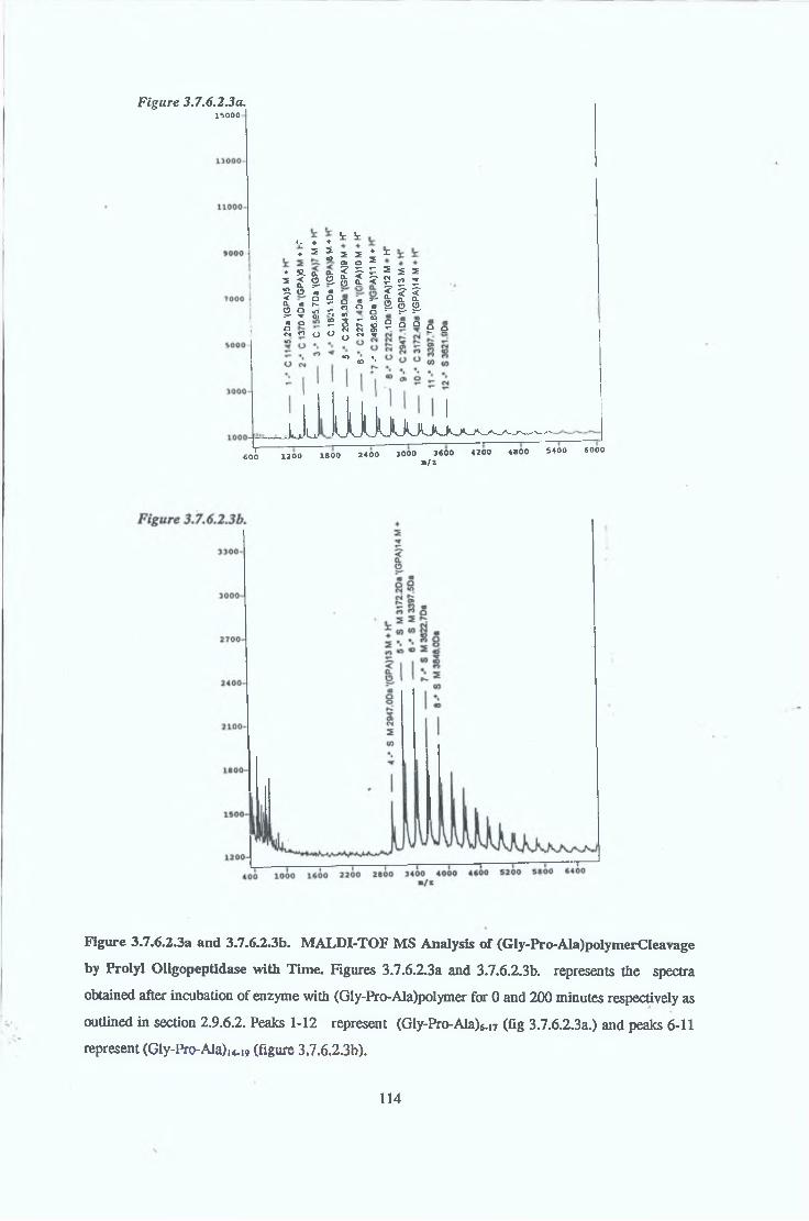
Figure 3.7.6.2.3a.15000
T~600
L ♦ ♦♦ 2 I
♦ S. a q.S J 2 2 •s. o ' . «< Q O CL <0 N ro o o r; ^ o*« d ?Qn o 0 o
t iS 2 22.0H Ia <o s-’o Q «<D O3 *- fl0-o r~- toN N 5n N ^“ <o **
♦ xs s sH. rt *III2 a o e q
*n
Ivj. La JUUuUuoJ^^X200 1 8 0 0 2 4 0 0 3000 3600 4200 4800 5400 6000
B/Z
Figure 3.7.6.2.3a and 3.7.6.2.3b. MALDI-TOF MS Analysis of (Gly-Pro-Ala)polymerCleavage
by Prolyl Oligopeptidase with Time. Figures 3.7.6.2.3a and 3.7.6.2.3b. represents the spectra
obtained after incubation of enzyme with (Gly-Pro-Ala)polymer for 0 and 200 minutes respectively as
outlined in section 2.9.6.2. Peaks 1-12 represent (Gly-Pro-Ala)^ (fig 3.7.6.2.3a.) and peaks 6-11
represent (Gly-Pro-Ala),«.,, (figure 3.7.6.2.3b).
114

Figure 3.7.6.2 3c
n/z
Figure 3.7.6.2.3c. MALDI-TOF MS Analysis of (Gly-Pro-Ala)poIymerCleavage by Prolyl
Oligopeptidase with Time.
Figure 3 7.6.2.3c and represents the spectrum obtained after incubation of enzyme with (Gly-Pro-
Ala)polymer for 300 minutes as outlined in section 2 9.6 2. Peaks 4-8 represent (Gly-Pro-Ala)14-19
1 1 5

\
3 7 6 3 1 Km Determination for Z-Glv-Pro-MCA
Michaelis Menten Constants were determined for the reactions of purified bovine bram, bovine serum
and recombinant bacterial PO activities using the substrate Z-Gly-Pro-MCA as outlined m section
2 9 6 3 1 Km values were determined using Michaelis Menten, Lineweaver-Burk, Eadie Hoftsee and
Hanes-Wolf analysis Figures 3 7 6 3 1 1 to 3 7 6 3 1 3 represent data obtained experimentally as
Lmeweaver-Burk plots for bram, serum and bacterial PO respectively Table 3 7 5 illustrates the Km
results
3 7 6 3 2 Km Determination for nGlu-His-Pro-MCA
Michaelis Menten Constants were determined for the reactions of purified bovine brain, bovine serum
and recombinant bacterial PO activities using the substrate pGlu-His-Pro-MCA as outlined in section
2 9 6 3 1 Km values were determined using Michaelis Menten, Lmeweaver-Burk, Eadie Hoftsee and
Hanes-Wolf analysis Figures 3 7 6 3 2 1 to 3 7 6 3 2 3 represent data obtained experimentally as
Lmeweaver-Burk plots for bram, serum and bacterial PO respectively Table 3 7 6 3 1 illustrates the
Km results
3.7.6.3. Kinetic Studies
Substrate Brain PO
MM LB HW EH
Serum PO
MM LB HW EH
Recombinant PO
MM LB HW EH
Z-Gly-Pro-MCA
pGlu-His-Pro-MCA
39 5 62 5 48 5 59 1
82 3 99 8 91 3 92 3
10 6 14 6 11 5 13 5
42 3 52 1 48 6 47 8
21 2 38 5 17 5 24 7
52 9 73 5 64 3 66 3
Table 3.7 5. Km values (¡lM) obtained for Fluonmetnc Substrates Z-Gly-Pro-MCA and pGlu-
His-Pro-MCA
Km values were obtained for brain, serum and recombinant bacterial PO activities MM-Michaelis-
Menten, LB-Lineweaver-Burk, EH-Eadie-Hoftstee, HW-Hanes-Wolf
116

3 7 6 3 3 Ki Determination for Prolme-Containing Peptides
Ki values were determined using brain serum and recombinant PO for a range of prolme-contaimng
peptides as outlined in section 2 9 6 3 3 , using the fluorimetric substrate Z-Gly-Pro-MCA Inhibitor
constants (Ki values) were determined using Lmeweaver-Burk analysis and are outlined m table
3 7 6 Figures 3 7 6 3 3 1 to 3 7 6 3 3 5 represent Lineweaver-Burk reciprocal plots for angiotensin II,
AVP, TRH, LHRH and neurotensin using bpvme brain activity
Inhibitor ♦Concentration
(|iM)
Brain
(1)
KifuM )
Serum
(2)
Recombmant
(3)
TRH 200 90 91 83 3 642
LHRH 400 30 02 - -
Bradykmin 160 33 3 12 0 133 5
Neurotensin 100 212 3 8 155 3
Arg8-Vasopressm 100 77 8 33 333 3
Angiotensin I 100 14 3 3 0 -
Angiotensin II 100 12 7 1 8 19 56
Angiotensin III 100 23 1 2 7 17 8
Glu2TRH 160 2797 2 221.4 1285 4
Phe2TRH 200 493 2 128 80 63191
Table 3.7.6. Ki Values Determined for a Range o f Prolme-Containing Peptides
Ki values were determined using Z-Gly-Pro-MCA as substrate All peptides were found to be
competitive inhibitors of PO and Ki values were estimated from Km and apparent Km values (Km in
the presence of (inhibitor/substrate)
* Actual concentration of peptide during assay
1 1 7

Fig 3 7 6 3 1 1
1/Substrate concentration (pM'1)
Fig- 3 7 6 3 1 2.
00140 -j 0 0120 0 0100 i 0 0080 -j 0 0060 4 010020 ^
p r r i r r iT i p hQQQOi 1 1 1 1 1 1 1 1 1 1 1 1 1 1 1 1 1 1 1 1 1 1 1 1 1 1 1 1
-0 08 -0 04 0 00 0 04 0 08
1/substrate concentration (pM 1)
Fig 3 7 6 3 1 3
0 014 - 3
0 012 0 010 4
I 0 008 4 | 0 006 4
0 004 4
0 00
-0 04-0 02 0 00 0 02 0 04 0 06 0 08 0 10 0 12
1/substrate concentration ((-iM-1)
Figures 3.7.6.3.1.1 to 3.7.6.3.I.3. Kinetic Analysis of brain, serum and bacterial PO respectively
using Z-Gly-Pro-MCA
Lmeweaver-Burk reciprocal plots of reaction velocity versus Z-Gly-Pro-MCA concentration Km
results illustrated in table 3.7 6.3 1
118

1/substrate concentration (fiM"1)
1/substrate concentration ((jM1)
1/substrate concentration (¡oMT1)
Figures 3.7.6.3.2.1 to 3.7.6.3.2.3. Kinetic Analysis o f brain, serum and bacterial PO respectively
using pGlu-HiS'Pro-MCA
Lmeweaver-Burk reciprocal plots of reaction velocity versus Z-Gly-Pro-MCA concentration. Km
results illustrated in table 3 7 6 3 1
119
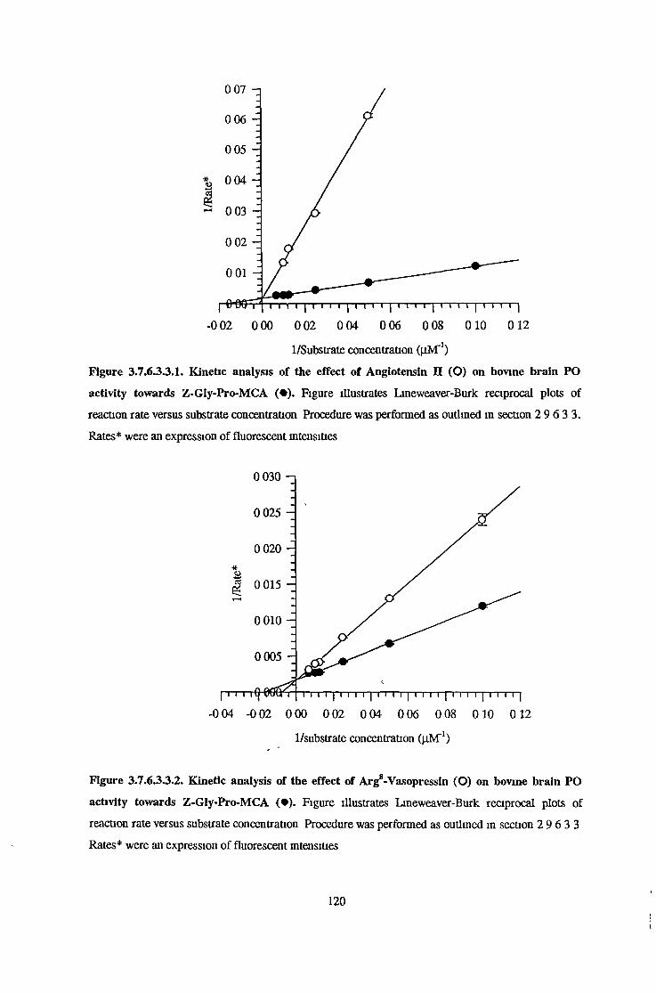
1/Substrate concentration ((iMT1)
Figure 3.7.6.3.3.I. Kinetic analysis of the effect of Angiotensin II (O) on bovine brain PO
activity towards Z-GIy-Pro-MCA ( • ) . Figure illustrates Lineweaver-Burk reciprocal plots of
reaction rate versus substrate concentration Procedure was performed as outlined in section 2 9 6 3 3.
Rates* were an expression of fluorescent intensities
-0 02 0 00 0 02 0 04 0 06 0 08 0 10 0 12
1/substrate concentration (nM 1)
Figure 3.7.6.3.3.2. Kinetic analysis of the effect of Arg*-Vasopressin (O) on bovine brain PO
activity towards Z-Gly-Pro-MCA ( • ) . Figure illustrates Lineweaver-Burk reciprocal plots of
reaction rate versus substrate concentration Procedure was performed as outlined in section 2 9 6 3 3
Rates* were an expression of fluorescent intensities
1 2 0
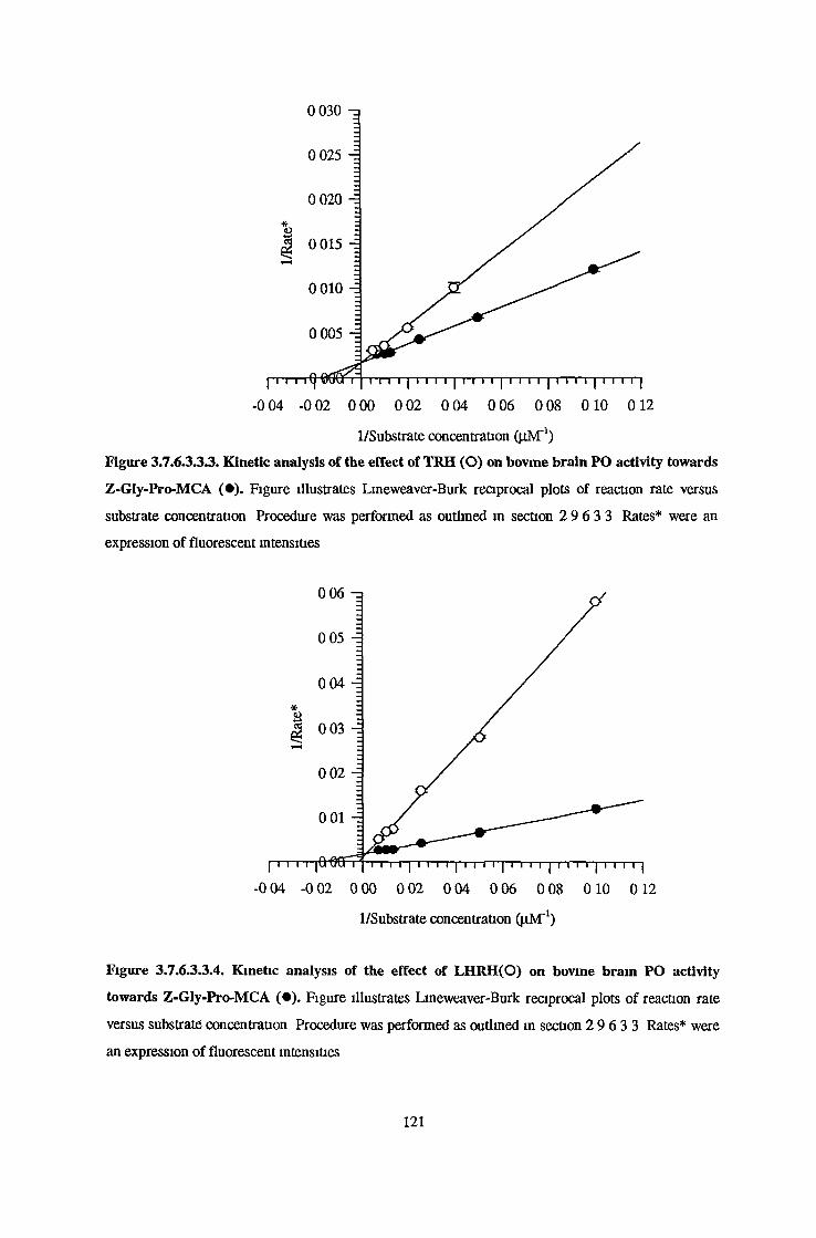
0 030 -a
1/Substrate concentration (pM'1)
Figure 3.7.6.3.3.3. Kinetic analysis of the effect of TRH (O) on bovine brain PO activity towards
Z-GIy-Pro-MCA ( • ) . Figure illustrates Lmeweaver-Burk reciprocal plots of reaction rate versus
substrate concentration Procedure was performed as outlined in section 2 9 6 3 3 Rates* were an
expression of fluorescent intensities
1/Substrate concentration ( j jM '1)
Figure 3.7.6.3.3.4. Kinetic analysis of the effect of LHRH(O) on bovine brain PO activity
towards Z-Gly-Pro-MCA ( • ) . Figure illustrates Lmeweaver-Burk reciprocal plots of reaction rate
versus substrate concentration Procedure was performed as outlined in section 2 9 6 3 3 Rates* were
an expression of fluorescent intensities
1 2 1
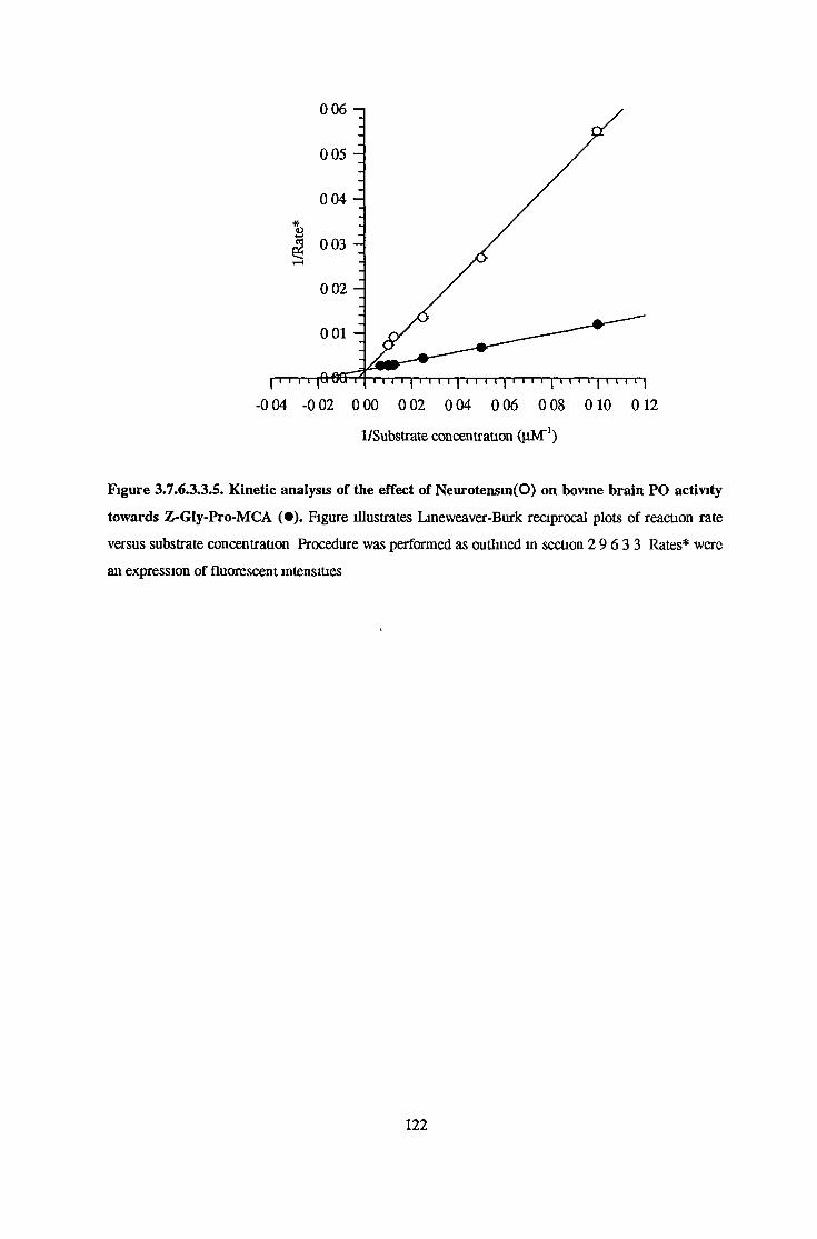
-0 04 -0 02 0 00 0 02 0 04 0 06 0 08 0 10 0 12
1/Substrate concentration (pM'1)
Figure 3.7.6.3.3.5. Kinetic analysis of the effect of Neurotensin(O) on bovine brain PO activity
towards Z-Gly-Pro-MCA ( • ) . Figure illustrates Lineweaver-Burk reciprocal plots of reaction rate
versus substrate concentration Procedure was performed as outlined in section 2 9 6 3 3 Rates* were
an expression of fluorescent intensities
1 2 2

3.7.7. Effect of Specific Inhibitors on Purified Prolyl Oligopeptidase Activity
3.7.7.1. Determination of IC50 Values For a Range of Specific Prolyl Oligopeptidase Inhibitors
The effect of various dilutions of specific inhibitors on purified bovine brain, bovine serum and
recombinant flavobactenal PO activities was investigated as outlined m section 2 9.7 1 Figures
3.7.7.1.1 to 3 7 7.1.9. illustrate the effect of these inhibitors on purified bovme brain PO activity
Table 3 7 7. summarises the IC50s determined for brain, serum and recombinant activities, a-
Ketobenothiazole displayed the most potent inhibitory activity, with an IC50 of 6 SlxlO'^M for the
bram activity All three PO activities were strongly inhibited by Z-Pro-prolmal, Z-indolmyl prohnal,
Fmoc-Ala-Pro-mtrile and Fmoc-Pro-Pro-mtrile However in the case of Fmoc-Pro-Pro-nitrile the
IC50 obtained for the recombinant activity was almost 10-fold higher than the IC50s obtained for the
mammalian activities
Inhibitor
Brain
icsonvn
Serum Bacterial
Z-Pro-prolmal lOnM lOnM 7nM
Z-Pro-prolinal-Dimethyhylacetate 44|aM 4pM 32jiM
Cyclohexyl-prolinal 649nM ND ND
Z-Indohnyl-prohnal lOnM lOnM 29nM
Fmoc-Ala-Pyrr-Nitnle lOnM lOnM 66nM
Fmoc-pro-Pyrr-Nitnle 89nM 88nM 551nM
Z-Phe-Pro-Methylketone 13(iM llpM 45nM
Postatm 17|iM 1 3pM 8pM
a-Ketobenzothiazole 63pM ND ND
Table 3.7.7. IC50 Values Determined for Proline Specific Peptidase Inhibitors
The effect of specific inhibitors on purified bovine bram, serum and recombinant flavobactenal PO
activities was investigated as outlined m section 2 9 7 1 Results are expressed as IC50s (M) which
were taken as the concentration of inhibitor required to inhibit enzyme activity by 50% Enzyme
activities* were expressed as percentages of activities without inhibitor
123

>%
NCW
120 - i
100
80
60
40
20
0
-20
Figure 3 7.7.11
T Tle-10 le-8 le-6
Z-Pro-Prolinal concentration (M)
le-4
%>a&
NaW
120
100
80
60
40
20
0 -d
-20
Figure 3 7 7.1.2
T Tle-12 le-10 le-8 le-6 le-4 le-2
Z-Pro-Prolmal- Dimethylacetate concentration (M)
Figures 3.7.7.I.I. and 3.7.7.I.2. Effect of Z-Pro-prolinal and Z-Pro-proIinal dimethylacetate on
bovine brain prolyl oligopeptidase. Figures 3 7.7 1 1 and 3 7 7 1 2 illustrate the effect of Z-Pro-
prolmal and Z-Pro-prolinal dimethyl acetate respectively on purified bovme brain PO IC50 values
determined for each inhibitor are illustrated in table 3.7 7 Experiments were performed as outlined m
section 3.7 7 1 Enzyme activity* is an expression of the % of activity obtained in the absence of
inhibitor
124
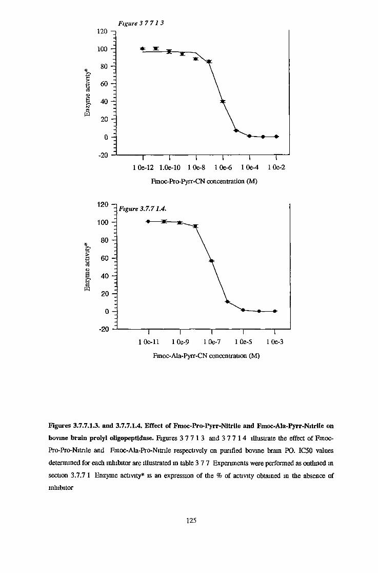
Figure 3 7 7 1 3120 - i
100
80 -
60
I 40
§ : 20 -
0 J
-20
>§
>a
N
(S
120 -n
100 -
80
60
40
20
0
-20
I I I I t I10e-12 1.0e-10 1 0e-8 1 Oe-6 1 0e-4 1 Oe-2
Fmoc-Pro-Pyrr-CN concentration (M)
Figure 3.7.71.4.
T T1 0 e - ll 10e-9 1 Oe-7 1 Oe-5 1 0e-3
Fmoc-Ala-Pyrr-CN concentration (M)
Figures 3.7.7.I.3. and 3.7.7.I.4. Effect of Fmoc-Pro-Pyrr-Nitrile and Fmoc-AIa-Pyrr-Nitrile on
bovine brain prolyl oligopeptidase. Figures 3 7 7 1 3 and 3 7 7 1 4 illustrate the effect of Fmoc-
Pro-Pro-Nitnle and Fmoc-Ala-Pro-Nitrile respectively on purified bovine brain PO. IC50 values
determined for each inhibitor are illustrated in table 3 7 7 Experiments were performed as outlined in
section 3.7.7 1 Enzyme activity* is an expression of the % of activity obtained in the absence of
inhibitor
1 2 5
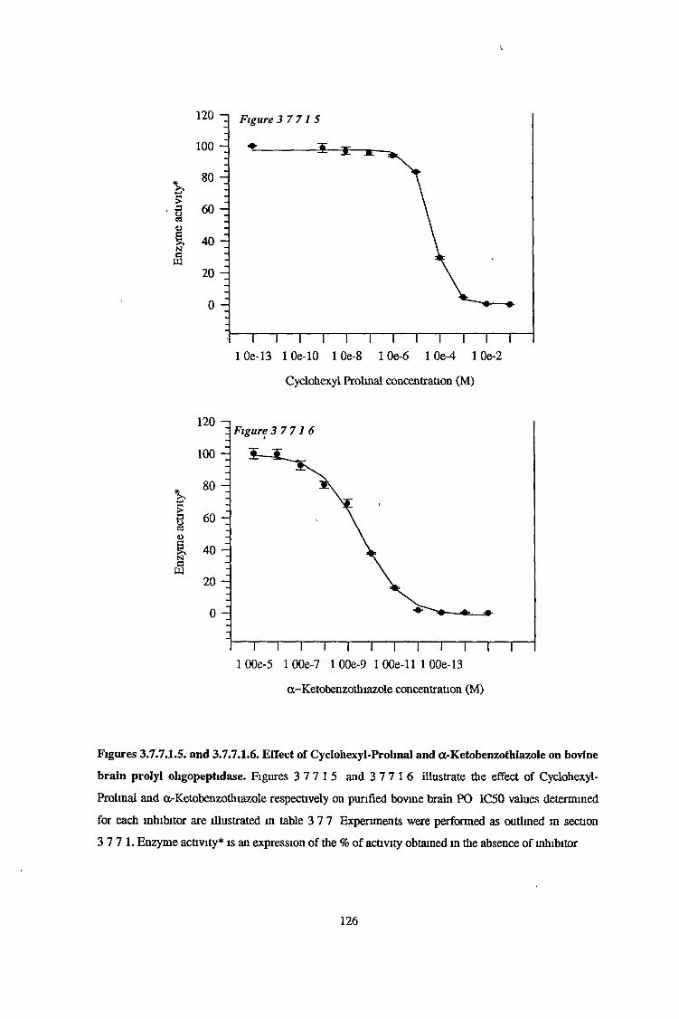
I
>a
NSW
100
80
60
40
20
0
1 2 0 Figure 3 7 7 1 5
1 1 1 1 1 1 1 1 1 1 1----10e-13 1 0e-10 1 0e-8 1 Oe-6 1 0e-4 1 Oe-2
Cyclohexyl Prolmal concentration (M)
>a
a-Ketobenzothiazole concentration (M)
Figures 3.7.7.I.5. and 3.7.7.I.6. Effect of Cyclohexyl-Prohnal and a-Ketobenzothiazole on bovine
brain prolyl oligopeptidase. Figures 3 7 7 1 5 and 3 7 7 1 6 illustrate the effect of Cyclohexyl-
Prolmal and a-Ketobenzothiazole respectively on purified bovine brain PO IC50 values determined
for each inhibitor are illustrated in table 3 7 7 Experiments were performed as outlined m section
3 7 7 1. Enzyme activity* is an expression of the % of activity obtained in the absence of inhibitor
126

&?artu
*
NcW
120 -q
100 -
80
60
40
20
0 -d
-20
Figure 3 7 7 1 .7 .
T T Tle-13
*&£J33oN
120 - i
100
80
60
40
20
0 -l
-20
le-11 le-9 le-7 le-5
Postatin concentration (M)
Figure 3 7.7.1.8.
le-3
I I I1 Oe-12 1 0e-10 1 0e-8
T1 0e-6 1 0e-4
Z-Phe-Pro-CMK concentration (M)
10e-2
Figures 3.7.7.I.7. and 3.7.7.I.8. Effect of Postatin and Z-Phe-Pro-Chloromethylketone on bovine
bram prolyl oligopeptidase. Figures 3 7 7 17. and 3 7 7.1 8 illustrate the effect of Postatin and Z-
Phe-Pro-Chloromethylketone respectively on purified bovine brain PO IC50 values determined for
each inhibitor aré illustrated in table 3 7 7 Experiments were performed as outlined in section
3 7 7 1. Enzyme activity* is an expression of the % of activity obtained in the absence of inhibitor
127

*>.4-»>£>ort<uNC
ID
120 - ]
100
80
60
40
20
0
-20
Figure 3 7 7 1 9
T Tle-12 le-10 le-8 le-6 le-4
Z-Indolinyl Prolinal concentration (M)
le-2
Figure 3.7.7.I.9. Effect of Z-Indolinyl-Prolinal on bovine brain prolyl oligopeptidase. Figure
3 7 7 19 illustrates the effect of Z-Indolinyl-Prolinal on purified bovine brain PO IC50 values
determined for each inhibitor are illustrated in table 3 7 7 Experiments were performed as outlined in
section 3 7 7 1 Enzyme activity* is an expression of the % of activity obtained m the absence of
inhibitor
128

3.1.1.2. Effect of Z*Phe-AIa-ChloromethyIketone on Purified Bovine Brain and Recombinant
Prolyl Oligopeptidase Activity with Time
The effect of Z-Phe-Ala-Chloromethylketone on purified bovine brain and recombinant
Flavobacterium meningosepticum prolyl oligopeptidase activity was investigated as outlined in
section 2 8 7 2 Enzyme activity, expressed as a percentage of activity in the absence of inhibitor, over
time is illustrated m figure 3 7 7.2.1 The inhibitor which was found to completely inhibit the
recombinant activity after 45 minutes, had no effect on the bovine brain form of the enzyme
129

Time (minutes)
Figure 3.7.7.2.I. Effect of Z-Phe-Ala-CMK on purified Bovine Brain and recombinant
Flavobacterium meningosepticum Prolyl Oligopeptidase Activity.
The effect of Z-Phe-Ala-CMK on bovine brain (•) and recombinant PO (■) activities over time was
investigated as outlined in section 2 8 7 2 Enzyme activities* were expressed as a percentage of
activities measured at 0 minutes Positive controls with no inhibitor for brain (o) and recombinant (□)
samples were mcluded in the experiment
130

4. DISCUSSION

4.1. Fluoresence Spectrophotometry and 7-Amino-4-Methyl Coumarin
In order to dctcct low levels of peptidases, the use of high affinity substrates is essential Early studies
on PO made use of radiometric substrates such as radiolabelled oxytocin (Walter et a l , 1971)
Fluonmetnc assays offer increased safety and ease of use over radiometric assays and increased
sensitivity and selectivity over colorimetric assays (Rendell, 1987) The enzyme assays outlined in
section 2 4 arc all based on the use of an internally quenched MCA molecule attached to an N-
blocked dipeptide, Z-Gly-Pro This substate was first synthesised by Yoshimoto et a l , 1979, and was
reported to have a higher affinity for PO and the greatest sensitivity over substrates such as Z-GIy-
Pro-ONp Km and kcat values of 20nM and 557 3s'1 were reported for this substrate, by this group
Fluoresence is a form of luminesence involving the re-emission of previously absorbed radiation In
the assay system for PO, this peptidase mediates peptide bond cleavage at the carboxyl end of proline
leading to the liberation of MCA Electromagnetic radiation at a wavelength of 370nm is absorbed by
MCA elevating it to an excited state Then on returning to the ground state, radiation of 440nm is
emitted
4.2. Quenching and the Inner Filter Effect
With regard to fluoresence a variety of forms of quenching can affect sensitivity leading to
misleadingly low fluoresence For instance self quenching is the result of fluoresmg molecules
colliding and losing their excitation energy by radiationless transfer Colhsional impurity quenching
involves the formation of a complex between excited MCA and a ground state impurity again
resulting in radiationless energy transfer This type of quenching can be usually attributed to the
presence of dissolved oxygen or compounds containing heavy atoms such as halogens (Willard et a l ,
1988) It is a form of quenching that is the result of the presence of impurities that absorb either the
exciting or emitted radiation, leading to a reduction of the fluorescent power that is particularly
problematic in the case of crude biological samples such as serum and brain extracts This type of
quenching is also known as the “inner filter” effect In order to compensate for this effect it was
necessary in the preparation of MCA standard curves to replicate assay conditions Therefore serum
and brain supernatant were incorporated into MCA dilutions during the preliminary assays of crude
samples for PO and ZIP activities (for preparation see section 2 2 ) Figures 3 1 1 and 3 1 3
illustrate the quenching effect of neat brain supernatant and serum on MCA fluoresence This
influence is also reflected m the slopes obtained for the quenched and unquenched curves In the case
of neat bram supernatant a 23% reduction in the slope value was observed at an emission slit width
of 2 5nm, but in the case of serum a 31% decrease m the slope value was observed at an emission slit
width of 5nm (see table 3 1 ) Another possibility m the minimisation of the quenching effect is
dilution of the sample This proved successful in the case of bram supernatant as the PO activity
131

present was too high to measure at the lowest emission slit width of 2.5nm. Therefore a one in twenty
dilution was necessary to make the measurement of liberated MCA. This dilution also eliminated the
quenching effect as identical slopes were obtained for standard curves prepared with buffer and a
working dilution of brain supernatant (see table 3.1.). Although dilution of serum would have been
feasible to minimise the quenching effect, it would have meant expanding the emission slit width.
Expanding the emission slit width would consequently broaden the bandwidth over which the
fluorimeter integrates light emitted from a particular sample. For instance for an emission wavelength
and slit width of 440nm and 2.5nm respectively, the fluorimeter integrates light radiated from 438.75
to 441.25nm. If the slit width is increased to lOnm, light radiated from 435-445nm is integrated.
Although an increase in slit widths would undoubtedly lead to an increase in sensitivity this would be
accompanied by a parallel increase in inner filter effect interference.
Although the quenching effect of bovine brain supernatant on fluoresence was eliminated by dilution,
quenched standard curves were still prepared throughout purification and characterisation
procedures. Many compounds other than biological samples can contribute to quenching. These
include buffers such as Tris-HCl and imidazole-HCl. Decreasing the pH of these buffers can cause a
corresponding decrease in slope. A variety of functional reagents and divalent metals can also
contribute to quenching. For instance slopes obtained for standard curves prepared in the presence of
lOmM 8-hydroxyquinoline, a metal chelator, are 57% lower than those calculated for standard curves
prepared with buffer. As a precautionary measure quenched standard curves were prepared for all
functional reagents and metal salts.
4.3. Measurement of Z-GIy-Pro-MCA Hydrolysing Activity in Crude Bovine
Brain Supernatant and Serum.
Z-Gly-Pro-MCA hydrolysing activity was measured in crude bovine brain supernatant and bovine
serum as outlined in sections 2.4.1 and 2.4.2. in a modification of procedures described previously
(Yoshimoto et al., 1979; Cunningham and O’Connor, 1997b). Assays on both samples were
performed in the presence and absence of SxlCT'M Z-Pro-prolinal, the classical, specific PO inhibitor
(Wilk and Orlowski., 1983). Previously it was concluded that Z-GIy-Pro-MCA was a substrate
specific for PO detection. Many studies, particularly clinical evaluations of PO levels in serum, have
used this fluorimetric substrate but have failed to incorporate a specific PO inhibitor to validate that
all Z-Gly-Pro-MCA hydrolysing activity was entirely attributable to PO (Maes et al.,1994, 1995b).
However the existence of a second Z-Gly-Pro-MCA hydrolysing activity, which was completely
insensitive to Z-Pro-prolinal was recently reported by this laboratory (Cunningham and O’Connor.,
1997b). PO and this ZPP insensitive peptidase were clearly distinguishable on the basis of their
behaviour during cation exchange chromatography and sensitivity to DTT, the ZPP insensitive
peptidase activity being unaffected by this reducing agent’s presence. Therefore in the case of serum
132
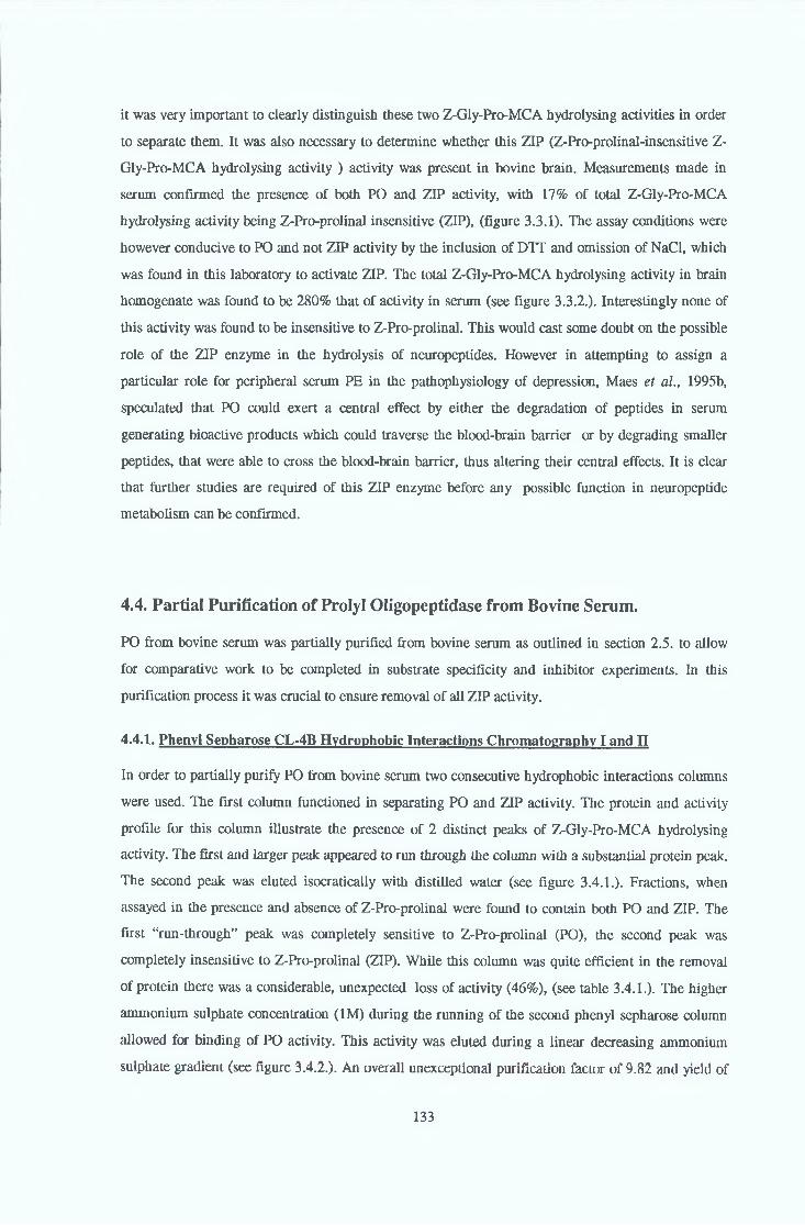
it was very important to clearly distinguish these two Z-Gly-Pro-MCA hydrolysing activities in order
to separate them. It was also necessary to determine whether this ZIP (Z-Pro-prolinal-insensitive Z-
Gly-Pro-MCA hydrolysing activity ) activity was present in bovine brain. Measurements made in
serum confirmed the presence of both PO and ZIP activity, with 17% of total Z-Gly-Pro-MCA
hydrolysing activity being Z-Pro-prolinal insensitive (ZIP), (figure 3.3.1). The assay conditions were
however conducive to PO and not ZIP activity by the inclusion of DTT and omission of NaCl, which
was found in this laboratory to activate ZIP. The total Z-Gly-Pro-MCA hydrolysing activity in brain
homogenate was found to be 280% that of activity in serum (see figure 3.3.2.). Interestingly none of
this activity was found to be insensitive to Z-Pro-prolinal. This would cast some doubt on the possible
role of the ZIP enzyme in the hydrolysis of neuropeptides. However in attempting to assign a
particular role for peripheral serum PE in the pathophysiology of depression, Maes et al., 1995b,
speculated that PO could exert a central effect by either the degradation of peptides in serum
generating bioactive products which could traverse the blood-brain barrier or by degrading smaller
peptides, that were able to cross the blood-brain barrier, thus altering their central effects. It is clear
that further studies are required of this ZIP enzyme before any possible function in neuropeptide
metabolism can be confirmed.
4.4. Partial Purification of Prolyl Oligopeptidase from Bovine Serum.
PO from bovine serum was partially purified from bovine serum as outlined in section 2.5. to allow
for comparative work to be completed in substrate specificity and inhibitor experiments. In this
purification process it was crucial to ensure removal of all ZIP activity.
4.4.1. Phenyl Sepharose CL-4B Hydrophobic Interactions Chromatography I and II
In order to partially purify PO from bovine serum two consecutive hydrophobic interactions columns
were used. The first column functioned in separating PO and ZIP activity. The protein and activity
profile for this column illustrate the presence of 2 distinct peaks of Z-Gly-Pro-MCA hydrolysing
activity. The first and larger peak appeared to run through the column with a substantial protein peak.
The second peak was eluted isocratically with distilled water (see figure 3.4.1.). Fractions, when
assayed in the presence and absence of Z-Pro-prolinal were found to contain both PO and ZIP. The
first “run-through” peak was completely sensitive to Z-Pro-prolinal (PO), the second peak was
completely insensitive to Z-Pro-prolinal (ZIP). While this column was quite efficient in the removal
of protein there was a considerable, unexpected loss of activity (46%), (see table 3.4.1.). The higher
ammonium sulphate concentration (1M) during the running of the second phenyl sepharose column
allowed for binding of PO activity. This activity was eluted during a linear decreasing ammonium
sulphate gradient (see figure 3.4.2.). An overall unexceptional purification factor of 9.82 and yield of
133

37% were obtained overall for this partial purification (table 3 4 1 ) It was however evident that PO
was quite unstable as precautions, such as the inclusion of DTT m all buffers, were taken during the
purification procedure.
4.5. Purification of PO from the Cytosolic Fraction of Bovine Brain.
4.5.1. Preparation of the Cytosolic Fraction of Bovine Brain
Although there have been some reports of membrane forms of PO (O’Leary et a l , 1996, Dalmaz et
a l , 1986), this peptidase is thought to be primarily cytosolic m its location (Torres et a l , 1986,
Dresdner et a l , 1982) Preparation of the cytosolic fraction of brain involved a series of
homogenisation and centrifugation steps An osmotic shock step was included at this stage to
facilitate the removal of any PO that had occluded m the cell membrane (Dresdner et a l , 1982) A
final ultracentrifugation step was performed on the combined supernatants to separate any possible
remaining membrane material such as microsomes (see section 2 6 1 ) A report on a membrane form
of PO from bovine brain (O’Leary et a l , 1996) found that this form of the peptidase had features
quite distinct from the cytosolic form such as a sensitivity to thiol reagents and metal chelators which
would classify it as a thimet peptidase Therefore it was necessary to ensure its complete removal
Microsomes are derived primarily from endoplasmic recticulum Upon cell breakage byIhomogenisation, the endoplasmic recticulum disrupts and its fragments tend to undergo vesculation
The resulting membranous vesicles are termed microsomes It is well known that ultracenrifugation
for 60 minutes at 100,000g results in the sedimentation of these microsomes, ensuring their removal
(Lambert, 1989)
4.5.2. Ammonium Sulphate Precipitation.
The ammonium sulphate precipitation step performed on the post ultracentrifugation brain
supernatant served both as a crude clean up step and a means of concentrating the brain extract prior
to column chromatography (see section 2 6 2 ) The optimal cut with respect to yield and the degree
of purification was found to be 45-75% ammonium sulphate with an overall purification factor of 4 2
and yield of 86% (table 3 5 .1 ) An ammonium sulphate step often features in PO purification
protocols with varying degrees of success One protocol which utilised quite a narrow cut of 40-65%
reported a 99% yield and a purification of 3 25 (Tate, 1981) In contrast to this Yoshimoto et a l ,
1983c, performed a 50-80% cut, achieving only a 47% yield and a five-fold purification factor These
results reflect the losses made in activity in order to achieve a higher purification factor
134

4 .53 . DEAE Sepharose Fast Flow Anion Exchange Chromatography
With the knowledge that mammalian PO is primarily an acidic protein with a pi of around 4 75-4 9
(Yoshimoto et a l , 1981, Oliveira et a l , 1976, Kalwant and Porter, 1991) anion exchange
chromatography was choosen as a suitable step (see section 2 6.3.) Cation exchange chromatography
would have required a pH below 4 5, which would have been quite detrimental to activity As can be
seen m the activity-protem profile in figure 3 5 1 A significant portion of protein ran through the
column with another substantial portion removed during the linear increasing NaCl gradient used to
remove bound PO activity. However an unremarkable purification factor of 1 4 was achieved for the
individual step with an overall yield of 58 7 (table 3 5 1 ) This is a fairly dramatic loss considering
that the peptidase was not exposed to any substantial pH extremes or particularly high NaCl
concentrations
4.5.4. Sephacrvl S-200 Gel Filtration Chromatography
Following ion-exchange chromatography the PO pool was concentrated and applied to a sephacryl
S200 column (see section 2 6 4) Although this column was not a very effective step with regard to
purification, assessed from its actmty-protem profile (figure 3 5 2), the decreased purification factor
and yield of 57% (see table 3 5 1), it eliminated the need for an overnight dialysis step into
unidazole-HCl, pH 6 3, which was found to be extremely harsh on PO activity
4.5.5. PBE94 Chromatofocusing
Because of the difficulties faced in finding a suitable and specific chromatographic procedure (see
section 4 5 7 ) , chromatofocusing was chosen due to its wide applicability, potentially very high
resolution and its suitability for the late stages of purification (Roe, 1989) In fact chromatofocusing
has been used successfully in previous purification protocols involving PO (Rennex et al., 1991,
Browne and O’Cumn 1983) Binding of PO was achieved at pH 6 3 and was eluted during a pH
gradient between pH 4 7-4 9 (see section 2 6 5 for procedure) This roughly confirms the pi value of
PO to be between this pH range Fairly good resolution of protein was obtained with this step (figure
3 5 3 ), and supnsmgly activity was not adversely effected by the pH extremes with a recovery and
purification factor of 83% and 28 5 for the step itself and an overall recovery and purification factor
of 24% and 155 (see table 3 5 1 )
135
Phenyl sepharose was chosen as a final column firstly for its ability to separate PO from the
potentially harmful effects of the poly- or amphoteric buffer used u» the elution of the
chromatofocusing column and secondly as a strategy to stabilise and prepare PO for storage (see
section 2.6 6 for procedure) These factors and its value as a purification step made it ideal for a final
step As can be seen from the actraty-protem profile (see figure 3.5.4) a significant protein peak ran
through the column Attempts to resolve the bound PO peak from other bound protein through the use
of a combined ammonium sulphate, potassium phosphate (decreasing) and glycerol (increasing)
gradient were unsuccessful as can be seen in the activity-protem profile PO and all other protein
eluted in lOmM potassium phosphate, pH 7 4 with 15% v/v glycerol, an ideal media for freezing A
purification factor and recovery of 1 64 and 95% was obtained for this particular step (see table
3 5 1 )
4.5.6. Phenvl Sepharose CL-4B Hydrophobic Interactions Chromatography
4.5.7. Assessment of Purification Process of Prolyl Qligppeptidase from Bovine Brain.
Overall the purification of PO from bovine brain proved to be a very difficult procedure to develop
due to the difficulty m resolving PO from other contaminating protem and secondly the difficulty in
maintaining PO activity during steps such as gel filtration The overall purification factor for the
purification was 253 9 with a recovery of 23% which was quite acceptable considering the losses
sustained during the initial chromatographic steps All the usual precautions were taken during this
purification scheme with all columns run at 4°C and the inclusion of DTT m all buffers All columns
were run consecutively with minimum time lapses between steps The SDS-PAGE silverstamed gel
illustrated m figure 3 5 5 illustrates the presence of a major band corresponding to the molecular
mass protein standard BSA which has a molecular weight of 66,000Da This mass was confirmed
using a calibrated S200 column (see section 3 7 1 ) A faint band was also observed, with a molecular
weight of around 50kDa. A range of fluonmetnc assays designed for the detection of other peptidases,
for example ZIP, PAP I and DAP IV, did not detect any of these peptidases (see table 3 5 2)
A variety of purification regimes were attempted in addition to the procedures oudined in sections
2 6 2-2 6 6 including hydroxylapatite (both commercial and synthesised in our laboratory), a variety
of gel filtration resins and dye-hgand resms, metal chelate chromatography and activated thiol
sepharose. This latter resin was used in an attempt to exploit PO’s dependence on DTT Although not
an absolute requirement for activity, DTT activated PO activity indicating the presence of thiol
groups. There have also been a variety of reports relating to mammalian PO’s sensitivity to cysteine
peptidase inhibitors (Kalwant and Porter, 1991, Tate, 1981, Yoshimoto et a l , 1977). Usually the
136

difficulty with thiol sepharose is the removal of the reducing agent, DTT, in a short time period The
enzyme thiol groups must be reduced, with all the disulphide groups converted to thiols. The
reducing agent must be removed quickly in order to minimise reversion of thiol groups to disulphide
groups prior to chromatography Using a method previously developed m our laboratory (Cummins
and O’Connor, 1996) for DTT removal it was found that the resin had a very poor affinity for PO and
its resolution from contaminating protein was impossible. Another group (Yoshimoto et a l , 1983)
reported on the successful use of another thiol group affinity resin m the purification of bovine brain
PO, PCMB-T sepharose
Metal chelating chromatography was used to again exploit the presence of cysteine residues m PO
Protein adsorption occurs by the binding of the thiol groups of surface amino acids to transition
metals such as zinc (Roe, 1989) Unfortunately no binding of PO or no resolution of PO from other
protein took place using zinc and calcium It is possible that these cysteine residues may be somewhat
buried within the enzyme 3-dimensional structure but this would not explain PO’s sensitivity to
cysteine peptidase inhibitors and some transition metals
Another method which has been used previously by other groups with some success is hydroxylapatite
chromatography (Kalwant and Porter, 1991). Chromatography with this resin was found not to be
reproducible due to its sensitivity to phosphate concentrations and the very low flow rates necessary to
achieve binding, which were too slow to maintain enzyme activity
4.6. Prolyl Oligopeptidase Assay Development
Once PO was purified it was necessary to re-evaluate the assay procedure and determine the optimal
conditions for PO activity and assay linearity. It was necessary therefore to investigate the effects of
solvents and possible activators on enzyme activity and also, the effect of substrate concentration and
enzyme concentration on linearity with respect to time For an enzyme assay to be reproducible and
quantitative, substrate hydrolysis must be linear with respect to time and substrate concentration It is
important to point out that all enzyme assay development studies were performed on purified PO
When the assay was found to be optimal the purification was performed with this new assay All
figures presented m table 3 5 1 were obtained using the optimised assay
4.6.1. The Efect of Solvents on Purified Prolyl Oligopeptidase Activity
Because of Z-Gly-Pro-MCA’s insolubility in aqueous media and the known inhibitory effects of
solvents on this peptidase (Kmsatschek et a l , 1980), it was necessary to find a solvent optimal for
137

substrate solubility and PO activity The relationship between enzymic hydration and catalytic activity
is well known. Wat»- is involved m all noncovalent interactions, such as hydrogen bonding, ionic
interactions, van der Waals and hydrophobic forces, that help to maintain the catalytically active
enzyme conformation (Zaks and Klibanov, 1988) However some enzymes do function in a naturally
hydrophobic environment such as a cell membrane and m some cases can tolerate or even be
activated by low solvent concentations, for instance trypsin activity was found to be enhanced in the
presence of 10% v/v acetomtnle and dioxane (Batra and Gupta, 1994). In the case of solvent-enzyme
inhibition there is a correlation between the loss of enzyme activity and the polarity of the solvent in
question Highly polar solvents sudi as dimethylformamide can interact with an enzyme and
associated water molecules to drastically reduce or destroy catalytic activity (Arnold, 1990) Using the
procedure outlined in section 2 8 1 it was set out to determine not only which solvent was least
detrimental on enzyme activity, but also optimal for substrate solubility While DMF was found to be
the most effective solvent with respect to substrate solubility it was most inhibitory of enzyme activity
On the other hand, while methanol was the least inhibitory on activity, it was not very efficient for
substrate dissolution. Ethanol, dioxane and DMSO were similar in their inhibitory effects on activity,
but DMSO was found to be most effective for substrate solubility (see figure 3 6 1 ) The optimal
concentration of DMSO for the working assay concentration of 200pM was 8% v/v Although the
substrate tended to precipitate on removal from a 37°C water bath at this DMSO concentration, 10%
DMSO would have been too detrimental to enzyme activity and stability It is worth noting that none
of the solvents tested even slightly activated'PO at low concentrations This however may be due to
POs naturally hydrophilic environment withm the cell cytoplasm DMSO is thought to disrupt the
secondary structure of a protein causing it to unfold In a study of the effects of DMSO on myoglobin
and concanavalin A, low DMSO concentrations had no effect on these protein’s gross secondary
structure, but their thermal stability was reduced (Jackson and Mantsch, 1991) It was therefore
necessary to minimise any possible adverse effects DMSO could have on PO during characterisation
experiments
2.6.2. Linearity of Purified Prolyl Oligopeptidase Activity Assay with Respect to Time and Enzyme Concentration
In order for an enzyme activity to be quantifiable it is necessary to ensure linearity with respect to
time and enzyme concentration The progress curves of most enzyme reactions with respect to time
are generally of the form illustrated in figure 4 1
138

Figure 4.1. Linearity of Product Formation with Respect to Time
The time course is linear to begin with but the rate of product formation declines at longer times.
There are many reasons for such departure from linearity but in the case of PO and Z-Gly-Pro-MCA
non-linearity was thought to be primarily due to substrate depletion and instability of the purified
enzyme during assay. This is highlighted by figures 3.6.2a. and 3.6.2b. Firstly activity was monitored
over time using purified PO samples which had been stored in the presence and absence of BSA. In
the absence of BSA the PO reaction rate departed from linearity in less than ten minutes. This
departure from linearity is indicative of PO’s instability in dilute protein solutions. The addition of
BSA to the purified sample restores linearity, more than likely by stabilisation of the enzyme. It is
also evident from continuous assays performed with 100(jM and 200[iM Z-GIy-Pro-MCA and
monitored over forty minutes that the higher concentration of substrate was necessary to ensure
linearity. At this point it was decided to decrease the assay time of one hour, used previously in this
laboratory (O’Leary et al., 1996; Cunningham and O’Connor, 1997b) to thirty minutes. Using the
conditions determined from these time-course experiments i.e. 200|iM substrate, 0.5% w/v BSA, the
assay was determined to be linear over thirty minutes with respect to enzyme concentration (see figure
3.6.3).
4.6.3. Effect of DTT on Purified Prolvl Oligopeptidase Activity
Numerous reports have shown PO to be activated by the thiol-reducing agent DTT (Kalwant and
Porter, 1991; Orlowski et al., 1979; Walter, 1976). This is in agreement with the findings that
mammalian and plant forms of PO were sensitive to certain thiol peptidase inhibitors such as PCMB,
NEM, and 2-iodoacetamide (Kalwant and Porter, 1991; Yoshimoto et al., 1987; Tate, 1981). As these
inhibitors were not as potent in eliciting inhibition in comparison to DFP it was suggested PO had a
cysteine residue close enough to the active site to influence activity under certain conditions (Polgar,
139

1991) PO activity was found to increase by over 250% in the presence of 5mM DTT However this
peak in activity was followed by a steady decline between lOmM and lOOmM DTT (see figure 3 6 3 )
DTT is a thiol reducing agent and can function to reduce disulphide bridges to thiol groups (see
figure 4 .2 ) At a particular concentration DTT could serve to slightly unfold a proiem, by disruption
of these disulphide bridges, making the active site more accessible to a substrate. If used in excess
DTT could totally disrupt an enzyme structure and this would explain the decline in PO activity at
high DTT concentrations (figure 3 6.3.). Alternatively DTT could activate a cysteine residue that is
m close enough proximity to the active site, to assist nucleophilic attack upon a substrate by the
catalytically competent cysteine residue
Another interesting characteristic afforded by the mclusion of DTT m the assay was an improvement
in the reproducibility of triplicates and therefore was considered an essential component of the assay
system
R--------CHz------S S CH2 R’
Disuphide-lmked chains H OH
HS Ofe C C CH2 SH
OH H
Dithiothreitol
R CH2 SH + HS CH2 R’
Separated reduced chains
Figure 4.2. Reduction o f Disulphide bridges to Thiols by DTT
140
4.6.4. Effect of EDTA on Purified Prolyl Oligopeptidase Activity
The reasons for inclusion of EDTA into the assay system were twofold. Firstly metal cations can
contribute to the oxidative breakdown of DTT by the formation of free radicals and the inactivation of
sulphydryl groups. There have also been numerous reports of inhibition of PO by divalent metals such
as Hg2* and Cu2* (Kalwant and Porter, 1991; Yoshimoto et al., 1987; Tate, 1981). Therefore the
inclusion of EDTA minimised the effects of any metal contamination on enzyme activity. As can be
seen in figure 3.6.5., 0.5mM EDTA slightly increased PO activity by around 4%. This increase was
followed by a steady decline in activity at EDTA concentrations between lOmM and 150mM.
4.6.5. Effect of NaCl on Purified Prolvl Oligopeptidase Activity
NaCl has been reported to frequently stabilise protein structure by preferential hydration, water being
involved in all noncovalent interactions that help maintain the catalytically active enzyme
conformation (Timasheff and Arakawa, 1989). Prolyl oligopeptidase unlike the members of other
serine protease families is very sensitive to ionic strength, its activity enhanced by high
concentrations of NaCl (Polgar, 1991). This was confirmed by results obtained for bovine brain PO
(figure 3.6.6.) with activation of the enzyme followed by a steady decline in activity at concentrations
greater than 400mM. Although this inhibiton was also observed by Polgar, 1991, it may have been in
part due to insolubility of the substrate observed at high salt concentrations. Because of the difficulties
in dissolving Z-Gly-Pro-MCA in the presence of NaCl, it was not included in the assay system.
4.7. Characterisation of Purified Prolyl Oligopeptidase Activity
4.7.1. Relative Molecular Mass Determination
The Relative molecular mass of IK) was determined by Sephacryl S-200 gel filtration chromatography
and SDS-PAGE as outlined in section 2.9.1. From molecular mass calibration curves prepared from
these experiments the molecular weight for PO was confirmed to be 69-70kDa and monomeric
(figures 3.7.1.1. and 3.7.1.2). This concurs very well with the majority of investigations which have
reported a molecular weight of between 62-80kDa (table 1.3.1.). It is worth noting that some of the
higher molecular weights quoted were deduced from an amino acid sequence and did not correct for
the presence of an N-tenninal signal sequence which is removed to give the mature peptidase
(Shirasawa et al., 1994; Chevallier et al., 1992). Two early studies reported molecular weights of
115kDa and 140kDa for PO from lamb kidney and human placenta respectively and suggested that
the enzyme was dimeric (Koida and Walter, 1976; Mizutani et al., 1984). It is now widely accepted
however that PO is monomeric and a later study on the lamb kidney enzyme confirmed the molecular
141

weight to be m fact 74-77kDa (Yoshimoto et a l , 1981). Further investigations of the human placental
peptidase confirmed the moleclar weight to be 79kDa (Kalwant and Porter, 1991)
4.7.2. PH Effects on Purified Prolvl Oligopeptidase Activity
In order to investigate the effect of pH and different buffers on PO activity two different pH
experiments were performed The first, described m section 2.9.2.1 served to simply determine the
optimum pH and buffer for PO activity From figure 3 7 2a. it is evident that PO activity was
detectable between 4 5 and 9 0 with optimal activity at pH 7 4 in potassium phosphate This is not
surprising considering PO is found naturally at a physiological pH of 7.4 and is similar to the
findings of other studies (table 13 1 ) However activity m Tns-HCl is 23% lower at pH 8 0 than
that measured in phosphate, indicating a general preference for phosphate buffer The second pH
experiment, described m section 2 9 2 2 was used to determine the pHs at which PO was irreversibly
inactivated prior to assay at pH 7 4 From figure 3 7 2b, it is evident that PO was inactivated outside
the pH range 5-9 5 . This was unexpected as during the purification PO was eluted m an amphoteric
buffer of pH 4 5 with only a 5% loss m activity (table 3 5 1 ) This discrepancy may have been due to
the protocol followed in this experiment which involved a premcubation at 37°C for 15 minutes with
the appropriate buffer However during the purification the enzyme was kept at 4°C prior to assay
There may also have been variations in PO’s sensitivity to the citrate/phosphate used in this
procedure and the polybuffer used to elute the chromatofocusmg column.
4.7.3. Thermostability Studies on Purified Prolvl Oligopeptidase Activity
Similar to pH studies the effect of temperature on PO was investigated using two different strategies
as outlined m section 2 9 3 Firstly the optimal temperature at which to perform the PO activity assay
was determined as described m secton 2 9.3 1 , which clearly indicated that 37°C was the optimal
temperature for activity (figure 3 7 3a). Again however this was not surprising considering
physiological temperature is 37°C. This result was m agreement with other studies done on
mammalian, plant and bacterial forms of the peptidase (see table 13 1) Another temperature
experiment was performed to determine the effect of premcubation of PO at various temperatures for
different times on activity prior assay at 37°C It was found that a premcubation at temperatures of
50°C or more for tunes of 15 minutes or greater caused complete inactivation of the peptidase activity
(see figure 3 7 3b)
142
4.7.4. The Effect of Divalent Metal Salts on Purified Prolvl Oligopeptidase Activity
The effect of divalent metal salts on PO activity was investigated as outlined in section 2 9 4 Previous
studies on PO have shown an inhibition of mammalian, plant and bacterial PO by a variety of metal
ions such as Hg2+, Zn2+ and Cu2* (Kalwant and Porter, 1991, Yoshimoto et a l , 1987, Walter, 1976)
PO activity was severely inhibited by Hg2+, Cu2+ and Cd2+. Activity was also inhibited substantially by
Co2*, Ni2+ and Zn2*, but little or no inhibition was observed with Fe2+, Mg2+ and Ca2+, (see table
3 7 1) In contrast to another study which investigated the effects of metal salts on PO from bovine
brain, carrot and F meningosepticum Ni2+ was quite potent m its inhibition of bovine brain PO with
over 60% inhibition However this study reported only a 10% inhibition of bovme brain activity
(Yoshimoto et a l , 1987) Zn2+ was also found to be not quite as inhibitory as other reports with only
40% inhibition This is m contrast to some studies that reported complete inactivation of PO activity
(Yoshimoto et a l , 1987, Orlowski et al, 1979). It is worth noting at this stage that the preparation of
these divalent salts solutions required strict monitoring of pH, otherwise any inhibition observed
could well have been contributed to by pH extremes Also quenched standard curves incorporating all
metal ions were prepared to allow for quenching effects
4.7.5. The Effect of Functional Reagents on Purified Prolvl Oligopeptidase Activity.
An investigation into the effects of a variety of functional reagents was investigated as outlined m
section 2 9 5 Table 3 7 2 lists the effects of a variety of classes of functional reagents, at different
concentrations, on PO activity The most significant results are discussed as follows
4.7.5.1. Serine Protease Inhibitors
PO has been classified in the past as a serine peptidase on the basis of its sensitivity to the classical
serine protease inhibitor, DFP and analysis of its ammo acid sequence (Rennex et a l , 1991, Kalwant
and Porter, 1991; Yoshimoto et al., 1987a, 1983c, Tate, 1981) In this experiment AEBSF, an
irreversible inhibitor of serme proteases such as trypsin and chymotrypsm, was used as a safer, more
stable and soluble alternative to the use of DFP (Markwardt et a l , 1974) ImM AEBSF completely
inhibited PO and a ten-fold lower concentration inhibited PO by over 80% confirming the presence of
an active site serme residue Another study completed in this laboratory on bovine serum PO reported
a lower inhibitory potency for AEBSF, with only 80% inhibition at lOmM (Cunningham and
O’Connor, 1998) In contrast to this PMSF only inhibited PO by 10 73 % at ImM (table 3 7 2 ) . This
lack of sensitivity to PMSF is a peculiarity that has been found, by other investigators, with all forms
143

of PO, mammalian, plant and microbial (Cunningham and O’Connor, 1998; Yoshimoto et al., 1987;
Orlowski et al., 1979).
Other serine protease inhibitors aprotinin and bezamidine had minor effects on activity with aprotinin
causing maximum inhibition of 14% at 5mM and benzamidine inhibiting PO by 7% at lOmM (uNe
3.7.2.). Other studies have also found benzamidine to have litde or no effect on PO activity
(Cunningham and O’Connor, 1998; Browne and O’Cuinn, 1983).
4.7.5.2. Cysteine Protease Inhibitors
Inhibition of mammalian and plant forms of PO by cysteine protease inhibitors has been well
documented (Kanatani et al 1993; Yoshimoto et al., 1987; Yoshimoto et al., 1981; Orlowski et al.,
1979). However this inhibition seems to be significant only at high concentrations of a particular
functional reagent with the size of a reagent determining its inhibitory potency. It has been speculated
that PO may have a non-catalytically competent cysteine residue in close vicinity to the active site.
Larger reagents were found to exert more of an inhibitory effect on PO activity possibly due to
exclusion of a substrate from the active site due to steric hindrance (Polgar, 1991) Reagents such as
PCMB, DTNB and NEM should therefore be more inhibitory of PO activity than the smaller
iodoacetamide. This effect was confirmed by experiments on the bovine brain PO, with iodoacetate
and iodoacetamide exerting moderate inhibition, 60 and 80% respectively at lOmM, considering the
high concentrations used. NEM and DTNB were far more effective inhibitors with complete
inhibition of PO at concentrations of lOmM (see table 3.7.2).
4.7.5.3. Thiol-reducing Agents
The effect of DTT on PO activity has been discussed previously (see section 4.6.3.) In this procedure
DTT (lOmM) when preincubated with PO, caused just over a two-fold enhancement of activity (table
3.7.2). This level of activation is significantly lower than that reported for bovine serum
(Cunningham and O’Connor, 1998). This discrepancy may however be attributed to the absence of
DTT in the substrate buffer. Upon addition of the substrate the total DTT concentration was diluted to
2mM, and it is quite possible that a higher DTT concentration is required for the duration of the assay
in order to be effective. The extent of the activation observed is however in accordance with results
obtained for section 2.8.4.
4.7.5.4. Metal Chelators and Phenanthrolines
EDTA and 8-hydroxyquinoline (ImM) were both found to slightly activate PO activity, possibly
because of their ability to chelate metal cations, such as zinc and nickel, which were previously
found to inhibit PO (see table 3.7.1.). These metals may simply be present in an assay component as
trace contaminants. Of the three phenanthrolines assessed only 1, 10-phenanthroline is a chelator.
All three of these compound were, however found to inhibit PO (see table 3.7.2). Because these three
144
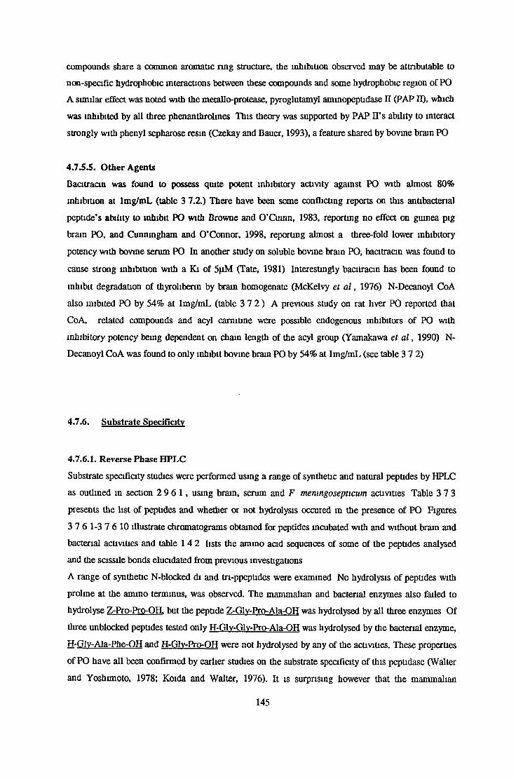
compounds share a common aromatic ring structure, the inhibition observed may be attributable to
non-specific hydrophobic interactions between these compounds and some hydrophobic region of PO
A similar effect was noted with the metallo-protease, pyroglutamyl aminopeptidase II (PAP II), which
was inhibited by all three phenanthrolines This theory was supported by PAP II’s ability to interact
strongly with phenyl sepharose resin (Czekay and Bauer, 1993), a feature shared by bovine brain PO
4.7.5.5. Other Agents
Bacitracin was found to possess quite potent inhibitory activity against PO with almost 80%
inhibition at lmg/mL (table 3 7.2.) There have been some conflicting reports on this antibacterial
peptide’s ability to inhibit PO with Browne and O’Cumn, 1983, reporting no effect on guinea pig
brain PO, and Cunningham and O’Connor, 1998, reporting almost a three-fold lower inhibitory
potency with bovine serum PO In another study on soluble bovine brain PO, bacitracin was found to
cause strong inhibition with a Ki of 5pM (Tate, 1981) Interestingly bacitracin has been found to
inhibit degradation of thyroliberin by brain homogenate (McKelvy et a l , 1976) N-Decanoyl CoA
also mibited PO by 54% at lmg/mL (table 3 7 2 ) A previous study on rat liver PO reported that
CoA, related compounds and acyl carnitine were possible endogenous inhibitors of PO with
inhibitory potency being dependent on chain length of the acyl group (Yamakawa et a l , 1990) N-
Decanoyl CoA was found to only inhibit bovine brain PO by 54% at lmg/mL (see table 3 7 2)
4.7.6. Substrate Specificity
4.7.6.1. Reverse Phase HPLC
Substrate specificity studies were performed using a range of synthetic and natural peptides by HPLC
as outlined in section 2 9 6 1 , using brain, serum and F memngosepticum activities Table 3 7 3
presents the list of peptides and whether or not hydrolysis occured m the presence of PO Figures
3 7 6 1-3 7 6 10 illustrate chromatograms obtained for peptides incubated with and without brain and
bacterial activities and table 1 4 2 lists the ammo acid sequences of some of the peptides analysed
and the sassile bonds elucidated from previous investigations
A range of synthetic N-blocked di and tn-ppeptides were examined No hydrolysis of peptides with
proline at the amino terminus, was observed. The mammalian and bacterial enzymes also failed to
hydrolyse Z-Pro-Pro-OH. but the peptide Z-Glv-Pro-Ala-OH was hydrolysed by all three enzymes Of
three unblocked peptides tested only H-Glv-Glv-Pro-Ala-OH was hydrolysed by the bacterial enzyme,
H-Glv-Ala-Phe-OH and H-Glv-Pro-OH were not hydrolysed by any of the activities. These properties
of PO have all been confirmed by earlier studies on the substrate specificity of this peptidase (Walter
and Yoshimoto, 1978; Koida and Walter, 1976). It is surprising however that the mammalian
1 4 5
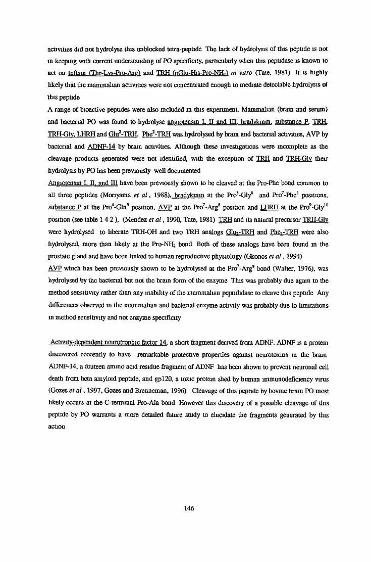
activities did not hydrolyse this unblocked tetra-peptide The lack of hydrolysis of this peptide is not
in keeping with current understanding of PO specificity, particularly when this peptidase is known to
act on tuftsin (Thr-Lvs-Pro-Arg) and TRH (pGlu-His-Pro-NHi) in vitro (Tate, 1981) It is highly
likely that the mammalian activities were not concentrated enough to mediate detectable hydrolysis of
this peptide
A range of bioactive peptides were also mcluded in this experiment Mammalian (brain and serum)
and bacterial PO was found to hydrolyse angiotensin I. II and IQ. bradvkinm. substance P. TRH.
TRH-Glv. LHRH and Glu2-TRH. Phe2-TRH was hydrolysed by brain and bacterial activities, AVP by
bacterial and ADNF-14 by brain activities. Although these investigations were incomplete as the
cleavage products generated were not identified, with the exception of TRH and TRH-Glv their
hydrolysis by PO has been previously well documented
Angiotensin I. II. and III have been previously shown to be cleaved at the Pro-Phe bond common to
all three peptides (Monyama et a l , 1988). bradvkinm at the Pro3-Gly8 and Pro7-Phes positions,
substance P at the Pro4-Gln5 position, AVP at the Pro7-Arg8 position and LHRH at the Pro’-Gly10
position (see table 1 4 2 ) , (Mendez et a l , 1990, Tate, 1981) TRH and its natural precursor TRH-Glv
were hydrolysed to liberate TRH-OH and two TRH analogs GIu?-TRH and Ph&rTRH were also
hydrolysed, more than likely at the Pro-NH2 bond Both of these analogs have been found in the
prostate gland and have been linked to human reproductive physiology (Gkonos et a l , 1994)
AVP which has been previously shown to be hydrolysed at the Pro7-Arg8 bond (Walter, 1976), was
hydrolysed by the bacterial but not the brain form of the enzyme This was probably due again to the
method sensitivity rather than any inability of the mammalian peptididase to cleave this peptide Any
differences observed in the mammalian and bacterial enzyme activity was probably due to limitations
in method sensitivity and not enzyme specificity
Activity-dependent neurotrophic factor 14. a short fragment derived from ADNF. ADNF is a protein
discovered reccently to have remarkable protective properties against neurotoxins m the brain
ADNF-14, a fouteen amino acid residue fragment of ADNF has been shown to prevent neuronal cell
death from beta amyloid peptide, and gpl20, a toxic protein shed by human immunodeficiency virus
(Gozes et a l , 1997, Gozes and Brenneman, 1996) Cleavage of this peptide by bovine brain PO most
likely occurs at the C-termmal Pro-Ala bond However this discovery of a possible cleavage of this
peptide by PO warrants a more detailed future study to elucidate the fragments generated by this
action
146

4.7.6.2. MALDI-TOF-Mass Spectrometry
MALDI-TOF MS was performed to firstly determine whether or not the brain form of PO hydrolysed
the CLIP fragment, ACTHi8.39, a Gy-Pro-Ala polymer and an amyloid A4 precursor protein fragment
708-715. The fragments generated from this reaction were also determinable by their mass/charge
ratio (m/z). Secondly the action of the bovine brain activity was compared to the F memngosepticum
and porcine kidney form of the peptidase The experiments were performed as outlined m section
2.9 6.2 Table 3 7 4. lists the PO hydrolysis sites of this these three peptides and figures 3 7.6 2.1 to
3 7 6 2 3 represent the mass spectra obtained at various times during the incubations
Bovine brain PO was found to hydrolyse the CLIP fragment at the Pro2 4-Asn2 5 and Pro^-Leu37 bonds,
generating the fragments ACTHis-2 4, 2 5 .3 9 and 25.3« (see figure 3 7 6 2 1 ) These fragments are thought
to be physiologically relevant in terms of paradoxical sleep. In another study the porcine kidney
enzyme had been found to hydrolyse the same bonds (Demuth, H. U , Unpublished data), (see
figure 4 7 2)
ACTH18 .3 9 or CLIP has been shown to exert a very selective paradoxical or REM sleep inducing
effect The full CLIP sequence is not required for this PS inducing effect, as fragments ACTH18 .2 4
and ACTH20-24 can increasePS to a similar extent While CLIP and A C T H i8.24 can only increase the
number of PS incidents, the fragment A C T H 20-24 can increase both the duration and number of
these episodes It has been proposed that the concerted action of PO and DAP IV results m the
release of these three fragments (Wetzel et a l , 1997) Figure 4 7 2 illustrates the proposed PO and
DAP IV sites of hydrolysis on ACTH18 -39 that leads to formation of these fragments
The bacterial activity was found to hydrolyse the Ala27-Glu28 and the Pro24-Asn25 but not the Pro36-
Leu37 bonds (see figure 4 7 2) This difference m specificity between bacterial and mammalian forms
of PO has not been observed previously and is suggestive of some structural difference between the
enzyme’s subsites However further investigation of these differences using a range of synthetic
substrates would have to be earned out m order to determine exactly what subsites are involved
The Gly-Pro-Ala polymer, (Gly-Pro-Ala)i5 was also found to be hydrolysed over time by bovine brain
PO Unfortunately due to time limitations complete hydrolysis was not observed However figure
4 7.3a and 4 7 3b represent the hydroysis of this polymer over time by the porcine kidney and F
memngosepticum forms of PO Hydrolysis of this polymer by PO is in conflict with the current
opinion that PO specificty is limited to smaller peptides of 25 amino acid residues or less (Monyama
et a l , 1988, Camargo et a l , 1979) and its classification as an oligopeptidase (Barrett and Rawlings,
1992) as the Gly-Pro-Ala polymer has 45 amino acid residues. This unexpected result means that a
more detailed study on PO’s ability to hydrolyse larger synthetic and natural peptides is required
POs ability to cleave this (Gly-Pro-Ala) polymer is of tremendous importance when considenng its
ability to cleave the amyloid A4 precursor protein fragment 70 8.7 1 s It was found that the bovine brain
147

PO hydrolyses this fragment at the Ala7 1 3 -Thr7 1 4 bond. This ability to cleave this fragment could be
of physiological relevance Amyloid A4 protein, which is found in deposits in senile plaques of
patients with Alzheimer’s disease and Down’s syndrome, is derived from the larger amyloid A4
precursor protein This 42 amino acid peptide is believed to be intrinsically linked to the
pathogenesis of this disease because of its ability to aggregate and induce neuronal cell death
(Yamaguchi et a l , 1988) It is widely believed that proteolytic processing of the precursor protein
could lead to the formation of amyloid A4 protein. Reccent studies using model synthetic substrates
have suggested that PO and a multicatalytic proteinase (MCP) , ingensin, are responsible for
hydrolysis of the precursor protein to generate the carboxyl and ammo terminal ends of amyloid A4^
42 peptide respectively This hydrolysis is represented in figure 4 7 4. It has been argued however
that PO’s classification as an oligopeptidase would rule out any possible role in amyloid A4
generation, even if a previous proteolytic action had occured giving rise to the N-temrunal portion of
this peptide However this possibility must be considered with the knowledge that PO is capable of
hydrolysing of the 45 amino acid polymer, (Gly-Pro-Ala)i5
DAPTVE Q bo»vine Brain vine Brain
Bacterial
CLDP20.24 CLIP25.36
r
o
CUP 18-24 CLIP25-39
^ Proline
O Alanine
^^Bacterial
Figure 4.7.2. Proposed Prolyl Oligopeptidase Hydrolysis Sites on ACTH18-39
Bacterial PO from F memngosepticum was found to hydrolyse the Pro2 4-Asn2 5 and Ala2 7-Glu28
bonds and porcine kidney DAP IV the Pro19-Val2 0 bond of CLIP18-24 (Unpublished data courtesy of
Hans Ulrich Demuth, Hans-Knoll Institute for Natural product Research, Halle, Germany) Bovine
brain sites were determined as outlined in scction 2 9 6 3
148
Figure 4.7.3a.
T=0
r=30niin
l'ii
lili... T=lXO*nint <--------1-------- '--------r
JULllllJili.il ,,l .L -X i, T=300min
.. I I 1 L L II L II L—I------ '-------1------ 1------ 1------- 1------1—1000 2000 3000 4000
M/Z
T=l<U)l>rs
5000
Figure 4.7.3b.
&
ulu.T=0
JJLi_______ T=30mint 1 r
T=60min
T=180min
t r n 1 r
A w i.T=100hrs
1-------r-1000 2000 3000
u n4000
-T ------------------ 1-----------
5000
Figure 4 .73 Hydrolysis of Gly-Pro-Ala Polymer by Porcine Kidney (figure 4 7 3a.) and F
menmgosepticum (figure 4 7 3b) Prolyl Oligopeptidase with Tune * Represents (Gly-Pro-Ala)15,
M/Z=mass/charge ratio
______________ Amyloid Precursor Protein 1.742_________________
MCP? PO?
1 42
Amyloid A4 Peptide 1-42
Figure 4.7.4. Proposed Mechanism of Amyloid Precursor Hydrolysis Resulting in Amyloid A4
Peptide Formation The amyloid A4 peptide is generated from the larger amyloid precursor
protein APPi.742 MCP (multicatalytic proteinase) and PO (prolyl oligopeptidase) have been identified
as possible enzyme candidates, responsible for APP proteolyltic processing (Yamaguchi et a l , 1988)
4.7.6.3. Kinetic Studies
4 7 6 3 1 Km Determination for Fluorimetnc Substrates
Michaelis Menton Constants were determined for brain, serum and bacterial PO activities as outlined
in section 2 9 6 3 1 using Z-Gly-Pro-MCA and the TRH derivative pGlu-His-Pro-MCA What is
apparent from the Km values determined (see table 3 7 5 ) is firstly the difference in affinities
between the three PO activities for a particular substrate The bovine brain enzyme had a lower
affinity for both fluorimetnc substrates with Km values of 62 5|iM for Z-Gly-Pro-MCA and 99.8|iM
for pGlu-His-Pro-MCA. The Km found for Z-Gly-Pro-MCA is considerably higher than the value of
20(iM reported when this substrate was first synthsised (Yoshimoto et a l , 1979), but is comparable to
other reports since (Cunningham and O’Connor, 1998, O’Leary and O’Connor, 1995) PO from all
sources had a higher affinity for this N-blocked dipeptide than the TRH deivative pGlu-His-Pro-MCA
4 7 6 3 2 Ki Determinations for Proline-Containing Peptides
The substrate specificity of the mammalian and bacterial forms of PO was investigated by examining
the effect of a range of proline-containing peptides on the Km value determined for Z-Gly-Pro-MCA
1 5 0

Angiotensins I, II and III were found to be potent competitive inhibitors of all three enzyme activities
(see table 3 7 6) This is m agreement with a previous study on porcine muscle PO (Monyama et a l ,
1988)
All three activities displayed a very low affinity for the TRH analogs Phe2TRH and Glu2TRH. This
low speafiaty suggests that PO may not play an important physiological role in then- degradation
(see table 3 7 6) In contrast to this both mammalian forms exhibited a strong specificity towards TRH
with Ki values (91pM for brain and 83(iM for serum) approximate to their Km values for the
fluonmetnc substrate Z-Gly-Pro-MCA (see table 3.7 6) These values are much lower than the value
(680jiM) reported previously for the serum enzyme (Cunningham and O’Connor, 1998) and slightly
higher than the Ki value reported for the rat brain enzyme (18 9|iM), (Hersh, 1981) The competitive
action of TRH on the bacterial enzyme activity towards Z-Gly-pro-MCA produced a Ki of 642(iM,
illustrating a 20 fold decrease in specificity This could be reflective of the fact that the bacterial
activity could have no possible natural function in the degradation of TRH. This could also explain
the bacterial enzyme’s much lower specificity for bradykinm neurotensin and AVP, when compared
to the mammalian activities
Both the mammalian activities had high specificities for bradykinm, neurotensin and AVP with Ki
values lower or, in the case of AVP and brain PO, approximate to their specificity towards Z-Gly-Pro-
MCA (see table 3 7 6) The Ki values determined for the serum enzyme for all these peptides were at
least one order of magnitude lower than values reported previously for the serum enzyme
(Cunningham and O’Connor, 1998) It is not fully understood why the serum PO activity had a
slightly higher specificity than the brain PO for all the substrates tested However it is unlikely that
this difference is due to a differences in speafiaty as serum levels in vivo are much lower than the
appreaable levels found m brain (Kato et a l , 1980) These high levels in the brain are thought to
reflect the significance of the brain enzyme’s ability to regulate levels of prolme-contaming
neuropeptides in vivo
In view of these results and results obtained from other substrate speaficity studies (see sections
4 7 6 1 and 4 7 6 2 ) it is clear that PO is an enzyme with a very broad speaficity for proline-
contaming peptides However the physiological significance of this peptidase and its ability to
hydrolyse peptides in vivo still remains to be eluadated
151

4.7.7. The Effect of Specific Inhibitors on Prolvl Oligopeptidase Activity
A range of known specific PO inhibitors were tested on bovine brain, serum and F. meningosepticum
prolyl oligopeptidase activity as outlined in section 2.9.7. The results are presented in the form of
IC50 values in table 3.7.7.
The most potent inhibitor tested was a-ketobenzothiazole with an IC50 walue of 63pM for the bovine
brain enzyme. This value is in agreement with the value of 42pM, obtained for the bovine serum
enzyme (Cunningham and O’Connor 1998). However both of these inhibitor values are considerably
lower than that reported by Tsutsumi et al., 1994, for the porcine kidney enzyme (table 1.5.1).
The poorest inhibitor tested was Z-Pro-prolinal dimethylacetate, with IC50s ranging from 32-44pM
for mammalian and bacterial forms of PO. This inhibitory potency was over 30 times lower than that
reported previously (Goossens et al., 1997) and highlights the importance of the aldehyde group of Z-
Pro-prolinal in PO inhibition. Z-Pro-prolinal while not the most potent inhibitor tested was found to
have IC50 values of 10, 9 and 7nM for brain, serum and bacterial PO activities respectively. These
values are higher than the value of 0.74nM quoted by Tsuru et al., 1988, using bovine brain PO.
Cunningham and O’Connor, 1998, reported an IC50 of 16nM using the bovine serum enzyme.
Another Z-Pro-prolinal derivative, Z-indolinyl-prolinal was found to be similar in potency to Z-pro-
prolinal with IC50s of lOnM for both of the mammalian enzymes, with the bacterial enzyme value
being over four-fold higher than that of Z-pro-prolinal. However these values are similar to those
obtained by Bakker et al., 1991 (see table 1.5.1). It should be noted that a value of 45pM has been
reported previously for the bovine serum enzyme (Cunningham and O’Connor, 1998).
Cyclohexyl-prolinal was found to be a rather poor inhibitor of the bovine brain enzyme relative to Z-
pro-prolinal and Z-Indolinyl-prolinal with an IC50 value (649nm) considerably higher than
previously reported (see table 1.5.1).
N-blocked dipeptide derivatives using an Fmoc blocking group, Fmoc-Ala-Pyrr-nitrile and Fmoc-
Pro-Pyrr-nitrile had identical IC50s for the mammalian enzymes. For both of these inhibitors
however the bacterial enzyme was found to be just over six times less sensitive to their inhibitory
activity (see table 3.7.7).
Postatin, an inhibitor of bacterial origin was quite poor in its inhibition of the mammalian and the
bacterial enzymes with IC50s in the micomolar range (table 3.7.7), considerably higher than
previously reported values of 35nM for the bovine serum enzyme (Cunningham and O’Connor,
1998).
Z-Phe-Pro-methylketone was probably the most interesting inhibitor tested, not because of its
inhibitory potency, but the large discrepancy in the sensitivity of mammalian and bacterial forms of
PO observed. While both the brain and serum enzymes had IC50s of 13 and ll|iM respectively the
bacterial enzyme was nearly 3000 times more sensitive to inhibition by this compound.
A timecourse investigation into the effects of Z-Phe-Ala-chloromethylketone found that while the
bacterial enzyme was completely inhibited by 1x10 5M of this inhibitor after 60 minutes, the bovine
152

brain enzyme exhibited no sensitivity to this inhibitor Chloromethylketones, previously thought to
be specific for cysteine residues, have been shown to act on PO by binding to the active site serine
residue (Stone et a l , 1992) Inhibition of bacterial PO confirms this action on the serine residue, as
bacterial forms of PO have been previously shown to be insensitive to cysteine protease inhibitors It
is not understood why a compound of this class could distinguish between different forms of PO In a
study of the comparuoo of inhibition of F meningosephcum and bovine brain by prolinal containing
peptide inhibitors, Boc-Pro-prolinal, and Z-Pyr-prolinal were found to be 1000 times more inhibitory
of the bacterial enzyme possibly due to sane structural difference around S2 and S3 subsites
(Yoshimoto et al., 1985) The bactenal activity may have a higher affinity for the peptide portion of
this inhibitor, Z-Phe-Ala, in particular the alanine rsidue at the S I’ subsite. In studies on the
hydrolysis of CLIP the F. menmgosepticum activity, unlike the brain activity, hydrolysed the Ala-Glu
bond (see section 4 7 6 2) Although previous studies have found that the bacterial and mammalian
forms of PO have been shown to have quite similar substrate specificities for synthetic substrates and
natural peptides, and have similar subsite widths (Yoshimoto et a l , 1980, 1977), it can be now
suggested that mammalian and bacterial forms of PO could have slightly different subsite
stereospecificities Further studies are required m order to clarify these possible differences
4.8. Summary
PO was purified from the cytosolic fraction of bovine whole brain Following preparation of this
fraction, PO activity was purified using ammonium sulphate precipitation, DEAE sepharose,
sephacryl S200 gel filtration, chromatofocusmg and phenyl sepharose chromatography, resulting m a
253-fold purification and a 23% recovery of enzyme activity
Bovine serum PO was partially purified from bovine serum for comparative substrate specificity and
inhibitor studies The purification consisted of chromatography on two separate phenyl sepharose
columns, run under different conditions). The first column served to remove ZIP activity A 10-fold
purification factor and 37% recovery was achieved
Prior to characterisation work the PO fluorimetnc activity assay was optimised This assay was found
to be linear over an assay period of 30 minutes, using 200pM Z-Gly-Pro-MCA prepared in 8% v/v
DMSO 5mM DTT and 0 5mM EDTA were also included to increase assay sensitivity The inclusion
of 0 5% w/v BSA served to stabilise the enzyme for storage and during the assay While NaCl was
found to activate activity and increase the sensitivity of the assay, its adverse effect on substrate
solubility warranted its exclusion from the assay procedure
153

The relative molecular mass of the brain activity, determined by gel filtration chromatography and
SDS PAGE , was found to be 69 5kDa This activity was found to be optimal at 37°C The optimal
pH for activity was found to be 7 4 in phoshate buffer
The brain enzyme was identified as a serine protease based on its sensitivity to AEBSF PO activity
was strongly inhibited by the cysteine protease inhibitors DTNB and NEM While the enzyme was
activated by DTT, this thiol reducing agent was not essential for catalytic activity This would suggest
the presence of a non-catalytically competent cysteine residue in close proximity to the active site
Enzyme activity was also strongly inhibited by some metal ions, including Hg2+, Cu2+, Co2+ and Ni2+
The brain, serum and Flavobacterium menmgosepticum PO activities hydrolysed the substrates Z-
Gly-Pro-MCA (Km values of 63jjM, 15|J.M and 39|iM respectively) and pGlu-His-Pro-MCA (Km
values of lOOpM, 52jj.M and 74|iM respectively All three activities hydrolysed a range of proline-
containing synthetic and natural peptides, including angiotensin I, II and III, TRH, bradykimn,
neurotensin, APP7 0 8-7 i5 , a Gly-Pro-Ala polymer and CLIP A number of these peptides also
competitively inhibited PO activity towards Z-Gly-Pro-MCA The most potent, with respect to brain
serum and bacterial activities, were angiotensin I, II and III which had Ki values lower than the Km
value for Z-Gly-Pro-MCA
Brain, serum and recombinant activities were inhibited by a range of PO specific inhibitors with a-
ketobenzothiaazole, Z-mdolmyl-prolmal and Fmoc-Ala-Pyrr-nitrile exerting the greatest inhibitory
activity The bacterial enzyme was found to be 4000 times more sensitive to Z-Phe-Pro-methylketone
than the brain enzyme lxlO'5 M Z-Phe-Ala-chloromethylketone completely inhibited the bacterial
activity, but the brain enzyme was completely insensitive to this inhibitor
In conclusion the characteristics of PO purified from bovine brain are similar to those reported
previously for other mammalian forms of this enzyme However further investigations are required to
determine the exact specificity of this enzyme particularly with concern to peptide size With the
development of highly potent and specific inhibitors, it should be possible to elucidate the
physiological role of PO in peptide degradation and in the pathogenesis of certain neurological
disease states
154

5. BIBLIOGRAPHY

Abbot, C A , Baker, E , Sutherland, G R. and McCaughan, G W (1994), Genomic organisation,
exact localisation and tissue expression of the human CD26 (dipeptidyl peptidase IV) gene,
Immunogenetics, 40, 331-338
Anastasi, A , Knight, C G and Barrett, A J (1993), Characterisation of the bacterial
metalloendopeptidase pitrilysin by use of a continuous fluorescence assay, Biochemical Journal, 290,
601-607
Anna-Amola, S S and Herskowitz, I (1994), Isolation and DNA sequence of the STE1 gene
encoding dipeptidyl aminopeptidase, Yeast, 10, 801-810
Aoyagi, T , Wada, T and Tanaka, T (1984), Decreased serum levels of various hydrolytic enzymes
m patients with rheumatoid arthritis, Biochemistry International, 8, 529-535
Arnold, F H. (1990), Engineering enzymes for non aqueous solvents, Trends in Biotechnology, 8,
244-249
Bakker, A V , Jung, S , Spencer, R. W , Vinick, F J and Faraci, S W (1990), Slow tight-binding
inhibition of prolyl endopeptidase by benzyloxycarbonyl-prolyl-prolinal, Biochemical Journal, 271,
559-562
Bakker, A V , Daffeh, J , Jung, S , Vincent, L W , Nagel A A , Spencer, R. W , Vinick, F J and
Faraci, S W (1991), Novel in vitro and in vivo inhibitors of prolyl endopeptidase, Bioorgamc and
Medicinal Chemistry Letters, 1, 585-590
Barlow, D J and Thornton, J M (1988), Helix geometry in proteins, Journal of Molecular Biology,
201,601-619
Barrett, A J and Kirschke, H. (1981), Cathepsin-B, cathepsin-H and cathepsin-L, Methods in
Enzymology, 80, 535-561
Barret, A J and Brown, M A (1990), Chicken liver Pz-peptidase, a thiol dependent metallo-
peptidase, Biochemical Journal, 271, 701-706
Barrett, A J. and Rawlings, N D (1992), Oligopeptidases, and the emergence of the ohgopeptidase
family, Biol Cherrt Hoppe Seyler, 373, 353-360
155

Batra, R. and Gupta, N (1994), Enhancement of enzyme activity in aqueous-organic solvent
mixtures, Biotechnology Letters, 16, 1059-1064
Beckmann, H., Saavedra, J M and Gattaz, W.F (1984), Low angiotensin-converting enzyme activity
(kminase II) in cerebrospinal fluid of schizophrenia, Biological Psychiatry, 19.679-684
Beauvais, A , Monod, M , Debeaupuis, J. P , Diaquin, M , Kobayashi, H. and Latge, J P (1997),
Biochemical and antigenic characterisation of a new dipeptidyl-peptidase isolated from Aspergillus
fumigatus, The Journal of Biological Chemistry, 272,6238-6244
Blow, D M (1969), Role of a buried acid group m the mechanism of chymotrypsin, Nature, 175,
337-340
Blumberg, S , Teichberg, V I , Charli, J L , Hersh, L B and McKelvy, J F (1980), Cleavage of
substance P in vitro by prolyl endopeptidase was found to generate two biologically active substance P
fragments by cleavage of the Pro4-Pbe5 bond, Brain Research, 192,477-491
Brandts, J F , Halvorson, H. R. and Brennan, M. (1975), Consideration of the possibility that the
slow step in protein denaturation reactions is due to cis-trans isomerisation of proline residues,
Biochemistry, 14,4953-4963
Brenner, S (1988), The molecular evolution of genes and proteins a tale of two serines, Nature, 334,
528-530
Bromme, D , Schierhom, A , Kirschke, H., Wiederanders, B , Barth, A , Fittkau, S and Demuth, H.
U (1989), Potent and selective inactivation of cysteine proteinases with N-peptidyl-O-acyl
hydroxylammes, Biochemical Journal, 263, 861-866
Browne, P and O’Cuinn, G (1983), An evaluation of the role of pyroglutamyl peptidase, a post-
prohne cleaving enzyme and a post-proline dipeptidylaminopeptidase, each purified from the soluble
fraction of guinea-pig brain, in the degradation of thyroliberin in vitro, European Journal of
Biochemistry, 137,75-87
Camargo, A C. M , Caldo, H. and Reis M L (1979), Susceptibility of a peptide derived from
bradykinm to hydrolysis by brain endo-ohgopeptidases and pancreatic proteinases, The Journal of
Biological Chemistry, 254, 5304-5307
156

Camargo, A C M , Caldo, H. and Emson, P C (1983), Degradation of neurotensin by rabbit brain
endo-oligopeptidase A and endo-ohgopeptidase B (proline-endopeptidase), Biochemical and
Biophysical Research Communications, 116,1151-1159
Camargo A C M , Almeida, M L C and Emerson P C (1984), Involvement of endo-
oligopeptidases A and B in the degradation of neurotensin by rabbit brain, Journal of
Neurochenustry, 42, 1758-1761
Campagnole-Santos, M L , Diz, D I , Santo, R .A S , Khosla, M C , Brosnihan, K B and Ferrario,
C M (1989), Cardiovascular effects of angiotensm-(l-7) injected into the dorsal medulla of rats,
American Journal of Physiology, 257, H-324-H329
Chan, S A T., Toursark, K , Sweeny, J P. and Jones, T H. D (1985), Dipeptidyl ammopeptidases
and ammopeptidases in Dictyostelium discoideum, Biochemical and Biophysical Research
Communications, 127, 962-968
Checler, F , Vincent, J P and Kitabgi (1985), Inactivation of neurotensin by rat brain synaptic
membranes partly occurs through cleavage at the arg8-arg9 peptide bond by a metalloendopeptidase,
Journal of Neurochenustry, 45,1509-1513
Checler, F (1995), Processing of the beta-amyloid precursor protein and its regulation m Alzheimer’s
disease, Journal of Neurochenustry, 65,1431-1444
Chevallier, S , Goeltz, P , Thibault, P , Banville, D and Gagnon, J (1992), Characterisation of a
prolyl endopeptidase from Flavobacterium memngosepticum, The Journal of Biological Chemistry,
267, 8192-8199
Cummins, P M and O’Connor, B (1996), Bovine brain pyroglutamyl ammopeptidase (type-1)
purification and characterisation of a neuropeptide-inactivating peptidase, International Journal of
Biochemistry and Cell Biology, 28, 883-893
Cunningham, D F and O’Connor, B (1997a), Proline specific peptidases, Biochinuca et Biophysica
Acta, 1343, 160-186
157

Cunningham, D F and O’Connor, B (1997b), Identification and initial characterisation of a N-
benzyloxycarbonyl-prolyl-prolinal (Z-Pro-prolinal)-msensitive 7-(N-benzyloxycarbooyl-gl>wl-prolyl-
amido)-4-methylcoumarm (Z-Gly-Pro-NH-Mec)-hydrolysing peptidase in bovine serum. European
Journal of Biochemistry, 244,900-903
Cunningham, D F and O’Connor, B (1998), A study of prolyl endopeptidase in bovine serum and
its relevance to the tissue enzyme, Biochemistry and Cell Biology, In Press
Czekay, G and Bauer, K (1993), Identification of thr thyrotropin-releasing hormone-degrading
ectoenzyme as a metallopeptidase, Biochemical Journal, 290,921-926
Dalmaz, C , Netto, C A , Volkmer, N , Dias, R. D and Izquierdo I (1986), Distribution of proline
endopeptidase activity in sub-synaposomal fractions of rat hypothalamus, Brazilian Journal of
Medical Biology and Research, 19,685-690
Danielsen, E M , Vyas, J P and Kenny A J (1980), A neutral endopeptidase in the microvillar
membrane of pig intestine Partial purification and properties, Biochemical Journal, 191, 645-648
Dauch, P , Masuo, Y , Vincent, J P and Checler, F (1993), A survey of the cerebral regionalization
and ontogeny o f eight exo- and endpeptidaseis m munnes, Peptides, 14, 593-599.
Davis, N C and Smith, E L (1953), Partial purification and specificity of immodipeptidase, The
Journal of Biological Chemistry, 200, 373-384
Davis, N C and Smith, E L (1957), Purification and some properties of prolidase of swine kidney,
The Journal of Biological Chemistry, 224,261-275
Dehm, P and Nordwig, A (1970), The cleavage of prolyl peptides by kidney peptidases Isolation of
a microsomal carboxypeptidase from swine kidney, European Journal of Biochemistry, 17, 372-377
Demuth, H. U , Schlenzig, D , Schierhom, A , Grosche, G , Chapot-Cartier, M P and Gripon, J C
(1993), Design of (w-N-(0-acyl)hydroxyamid)aminodicarboxylic acid pyrrolidines as potent
inhibitors of prohne-specific peptidases, FEBS, 320, 23-27
DeWied, D (1984), The importance of vasopressin in memory, Trends in Neurosciences, 7, 62-64
158

Donlon, J (1995), The production of biologically active peptides in brain tissues, in Metabolism of
Brain Peptides, O’Cuinn, G (ed ), CRC Press, New York, 1-12
Dresdner, K., Barker, L A , Orlowski, M and Wilk, S (1982), Subcellular distribution of prolyl
endopeptidase and cation sensitive neutral endopeptidase in rabbit brain, Journal of Neurochemistry,
38,1151-1154
Ermer, A., Baumann, H., Steude, G , Peters, K , Fittkau, S , Dolaschka, P ,Genov, N C (1990),
Pepude diazomethyl-ketones are inhibitors of subtilisin-type serine proteases, Journal of Enzyme
Inhibition, 4, 35-42
Friedman, T C , Orlowski, M and Wilk, S (1984), Prolyl endopeptidase inhibition m vivo by N-
benzyloxycarbonyl-prolyl-prohnal, Journal of Neurochemistry, 42,237-241
Friedman, T C and Wilk, S (1989), The effect of inhibitors of prolyl endopeptidase and pyrglutamyl
peptide hydrolase on TRH degradation in rat serum, Biochemical and Biophysical Research
Communications, 132, 787-794
Fukasawa, K , Fukasawa, K M , Hiraoka, B Y and Harada, M (1983), Purification and properties
of dipeptidyl peptidase II from rat kidney, Bwchimica et Biophysica Acta, 745, 6-11
Funkunari, A , Kato, A , Sakai, Y , Yoshimoto, T , Ishiura, S , Suzuki, K and Nakajima, T (1994),
Colocalization of prolyl endopeptidase and amyloid p-peptide m brains of senescence-accelerated
mouse, Neuroscience Letters, 176, 201-204
Fuse, Y , Polk, D H., Lam, R. W , Reviczky, A L and Fisher, D A (1990), Distribution and
ontogeny of thyrotropin-releasing hormone degrading enzymes in rats, American Journal of
Physiology, 259, E787-E791
Galat, A (1993), Peptidylprohne ci v-iran.Msomerases immunophilins, European Journal of
Biochemistry, 216, 689-707
Gastpar, M and Muller, W (1981), Autoantibodies in affective disorders, Progress in
Neuropsychopharmacology and Biological Psychiatry, 5,91-96
Gkonos, P. J , Kwok, C. K„ Block, N. L. and Roos, B. A. (1994), Identification of the human seminal
TRH-like peptide pGlu-Phe-Pro-NH2 in normal human prostate, Peptides, 15, 1281-1283
159

Glenner G G. and Wong C W (1984), Initial report of the purification and characterisation of a
novel cerebrovascular amyloid protein, Biochemical and Biophysical Research Communications. 120.
885-890
Goldstem, L A., Ghersi, G , Pineiro-Sanchez, M L , Salamone, M , Yeh, Y., Hessate, D and Cben,
W T (1997), Molecular cloning of seprase a serine integral membrane protease from human
melanoma, Biochimica et Biophysica Acta, 1361,11-19
Goossens, F , DeMeester, I , VanHoof, G , Hendriks, D , Vriend, G and Scharpe, S (1995), The
purification, characterisation and analysis of primary and secondary structure of prolyl oligopeptidase
from human lymphocytes Evidence that the enzyme belongs to the a/p hydrolase fold family,
European Journal of Biochemistry, 233,432-441
Goossens, F , DeMeester, I , Vanhoof, G and Scharpe, S (1996a), Distribution of prolyl
oligopeptidase m human peripheral tissues and body fluids, The European Journal of Clinical
Chemistry and Biochemistry, 34,17-22
Goossens, F. J , Wauters, J. G , Vanhoof, G C , Bossuyt, P J , Schatteman, K. A , Loens, K and
Scharpe, S L (1996b), Subregional mapping of the human lymphocyte prolyl oligopeptidase gene
(PREP) to human chromosome 6q22, Cytogenetics and Cell Genetics, 74,99-101
Goossens, F , VanHoof, G , DeMeester, I , Augustyns, K , Borloo, M , Tourwe, D and Haemers, A
(1997), Development and evaluation of peptide-based prolyl oligopeptidase inhibitors Introduction of
N-benzyloxycarbonyl-prolyl-3-fluoropyrrolidine as a lead in inhibitor design, European Journal of
Biochemistry, 250,177-183
Gottesman, M M (1990), Introduction Do proteases play a role in cancer’, Seminar Of Cancer
Biology, 34, 17-22
Gozes, I and Brenneman, D E (1996), Activity-dependent neurotrophic factor an extracellular
neuroprotective chaperonm, Journal of Molecular Neuroscience, 7,235-244
Gozes, I , Bardea, A , Bechar, M , Pearl, O , Reshef, A , Zamostiano, R , Davidson, A , Rubinraut, S ,
Gilaldi, E , Fndkm, M and Brenneman, D E (1997), Neuropeptides and neuronal survival
neuroprotective strategy for Alzheimer’s disease. Annals of the New York Academy of Sciences, 814,
161-166
160

Greene, L J , Spadaro, A C C , Martins, A R , DeJesus, W D P and Camargo, A C M (1982),
Brain endo-oligopeptidase a post-proline cleaving enzyme that inactivates angiotensin I and II,
Hypertension, 4, 178-184
Greene, G D J and Shaw, E. (1983), A prolyl endopeptidase from murine macrophages, its assay
and specific inactivation. Archives of Biochemistry and Biophysics, 255, 331-337
Griffiths, E C , Bans, C , Visser, T J and Klootwyk, W (1985), Thyrotropm-releasmg hormone
inactivation by human postmortem brain, Regulatory Peptides, 10,145-155
Griffiths, E C (1987), Clinical applications of thyrotropin-releasing hormone, Clinical Science, 73,
449-457
Guiroy, D , C (1990), Proteinase inhibitors leupeptm and Z-prolyl-prohnal induce neuronal
degeneration in primary rat hippocampal neurons in vitro, Neurobiology and Aging, 11, 288
Haas, C , Koo, E H., Mellon, A , Hung, A Y and Selkoe, D J (1992), Targeting of cell-surface 0-
amyloid precursor protein to lysosomes alternative processing into amyloid-bearing fragments,
Nature, 357, 500-503
Hagihara, M and Nagatsu, T (1987), Post-proline cleaving enzyme in human cerebrospinal fluid
from control patients and Parkinsonian patients, Biochemical Medicine and Metabolic Biology, 38,
387-391
Harris, E L V (1989), Initial planning, In Protein purification methods A practical approach,
Hams, E. L V. and Angal, S (eds), ERL Press, 1-64
Harwood, V J , Denson, J D , Robinson-Bidle, K A and Schreider, H. J (1997), Overexpression
and characterisation of a prolyl endopeptidase from the hyperthermophilic archaeon, Pyrococcus
furiosus, Journal of Biochemistry, 179, 3613-3618
Hedeager-Sorenson, S and Kenny, A J (1985), Proteins of the kidney microvillar membrane
Purification and properties of carboxypeptidase P from pig kidneys, Biochemical Journal, 229, 251-
257
161

Heinze, S., Ritzau, M.f Ihn, W., Hulsmann, H., Schlegel, B., Domberge, K., Heck, W. F„ Zerlin, M.,
Christner, C., Grafe, U., Kullertz, G. and Fischer, G. (1997), Lipohexin, a new inhibitor of proiyi
endopeptidase from Moeszia lintneri (HKI-004) and Paecilomyces sp. (HKI-0055; HKI-0096). 1.
Screening, isolation and structure elucidation, The Journal o f Antibiotics, 50, 379-383.
Hersh, L. B. (1981), Immunological, physical and chemical evidence for the identity of brain and
kidney post-proline cleaving endopeptidase, Journal o f Neurochemistry, 37, 172-178.
Heukshoven, J. and Demick, R. (1985), Simplified method for silver staining of proteins in
polyacrylamide gels and the mechanism of silverstaining, Electrophoresis, 6,103-112.
Inamori, Y., Muro, C., Sajima, E., Katagiri, M., Okamoto, Y., Tanaka, H., Sakagami, Y. and
Tsujibo, H. (1997), Biological activity of purpuogallin, Bioscience, Biotechnology and Biochemistry,
61, 890-892.
Ishino, T., Ohtsuki, S., Homma, K. and Natori, S. (1998), cDNA cloning of mouse prolyl
endopeptidase and its involvement in DNA synthesis by Swiss 3T3 cells, Journal o f Biochemistry,
123, 540-545.
Ishiura, S., Tsukahara, T., Tabira, T„ Shimizu, T., Arahata, K. and Sugita, H. (1990), Identification
of a putative amyloid A4-generating enzymeas prolyl endopeptidase, FEBS Letters, 260,131-133.
Jackson, M. and Mantsch, H. H. (1991), Beware of proteins in DMSO, Biochimica et Biophysica
Acta, 1078, 231-235.
Kagga, M. M., Laurent, F., Doh, A., Luffau, G., Yvore, P. and Pery, P. (1998), Purification of a
leucine aminopeptidase from Eimeria falciformis, Vetinary Research, 29,107-111.
Kalwant, S. and Porter, A. G. (1991), Purification and characterization of a human brain prolyl
endopeptidase, Biochemical Journal, 276, 237-244
Kamei, H., Ueki, T„ Obi, Y., Fukagawa, Y. and Oki, T. (1992), Protective effect of eurystatins A and
B, new prolyl endopeptidase inhibitors, on scopolamine-induced amnesia in rats, Japanese Journal o f
Pharmacology, 60, 377-380.
Kanatani, T., Masuda, T., Shimoda, T„ Misoka, F., Sheng Lin, X., Yoshimoto, T. and Tsuru, D.
(1991), Protease II from Escherichia coli: sequencing and expression of the enzyme gene and
characterisation of the expressed enzyme, Journal o f Biochemistry, 110, 315-320.
162
Kanatani, A , Yoshunoto, T , Kitazono, A ,Kokubo, T and Tsuru, D (1993), Prolyl endopeptidase
from Aeromonas hydrophilia Cloning sequencing and expression of the enzyme gene and
characterisation of the expressed enzyme, Journal of Biochemistry, 113,790-796
Kato, T„ Okada, M and Nagatsu, T (1980), Distribution of post-proline cleaving enzyme m human
brain and penpheral tissues, Molecular Cell Biology, 32, 117-121
Katsube, N , Sunaga, K , Chuang, D , and Ishitani, R (1996), ONO-1603, a potential antidementia
drug shows neuroprotective effects and increases m3 muscarinic receptor mRNA levels in
differentiating rat cerebellar granular neurons, Neuroscience Letters, 214,151-159
Kimura, K-i , Kauaguchi, N , Yoshihama, M and Kauanishi, G (1990), Staurosponne, a prolyl
endopeptidase inhibitor, Agricultural and Biological Chemistry, 54, 3021-3022
Kimura, K-i , Kanou, F , Takahashi, H , Esumi, Y , Uramoto, M and Yoshihama, M (1997a),
Propeptin, a new inhibitor of prolyl endopeptidase produced by Microbispora, The Journal of
Antibiotics, 50, 373-378
Kimura, K-i , Kanou, F , Koshino, H., Uramoto, M and Yoshihama, M ,(1997b), SNA-8073-B, a
new isotetracenone antibiotic inhibits prolyl endopeptidase I Fermentation, isolation and biological
properties, The Journal of Antibiotics, 50, 291-296
Kirkegaard, C F , Hummer, L and Rogowski, P (1979), Increased levels of TRH in cerebrospinal
fluid from patients with endogenous depression, Psychoneuroendocrinology, 4, 277-235
Kmsatschek, H. and Bauer, K (1979), Characterisation of “thyroliberin-deamidating enzyme” as a
post-proline cleaving enzyme partial purification and enzyme-chemical analysis of the enzyme from
anterior pituitary tissue, The Journal of Biological Chemistry, 245, 10936-10943
Kmsatschek, H , Klemhauf, H and Bauer, K (1980), Specific fluorogemc substrates for the
thyrotropin-releasing factor-deamidating post-proline cleaving enzyme, FEBS Letters, 111, 157-161
Kobayashi, K , Lin, L W , Yeadon, J E , Klickstein, L B and Smith, J A (1989), Cloning and
sequence analysis of rat liver cDNA encoding acyl-peptide hydrolase, The Journal of Biological
Chemistry, 264, 8892-8899
163

Koida, M. and Walter, R. (1976), Post-proline cleaving enzyme: purification of tthe endopeptidase by
affinity chromatography, The Journal of Biological Chemistry, 251, 7593-7599.
Krause D. R , Piva, T. J., Brown, S. B. and Ellem, K. A. (1997), Characterisation and localization of
mitochondrial oligopeptidase (MOP) (EC 3.4.24.16) activity in the human cervical adenocarcinoma
cell line HeLa, Journal of Cell Biochemistry, 66, 297-308.
Krieger, D.T. (1984), Brain peptides: What, where and why?, Science, 222,975-983.
Lajtha, A. and Toth, J. (1974), Post mortem changes in cerebral free amino acid pool, Brain
Research, 76, 546-551.
Lambert, A. (1989), Clarification and extraction, In Protein Purification Methods: A Practical
Approach, Harris, E. L. V. and Angal, S. (eds), IRL Press, Oxford, 101-108.
Lawrewnce, A. C., Clarke, I. J. and Campell, D. J. (1992), Increased angiotensin-(l-7) in
hypophyseal-portal plasma of conscious sheep, Neuroendocrinology, 5 5 ,105-114.
Li, J., Wilk, E. and Wilk, S. (1996), Inhibition of prolyl oligopeptidase by Fmoc-
aminoacylpyrrolidine-2-nitriles, Journal of Neurochemistry, 66, 2105-2111.
Li, J. P., Douglas, S. D. and Ho, W. Z. (1998), Human lymphocytes express substance P and its
receptor, Journal of Neuroimmunology, 86, 80-86.
Lin, B., Averett, W. F., Novak, J., Chartham, W.W., Hollingshead, S. K , Coligan, J. E., Egan, M. L.
and Pritchard, D. G. (1996), Characterisation of Pep B, a group B streptococcal oligopeptidase,
Infectious Immunology, 64, 3401-3406.
MacArthur, M. W. and Thorton, J. M. (1991), Influences of proline residues on proteinconformation,
Journal of Molecular Biology, 218, 397-412.
Maes, M„ DeJonckheere, C. and Vandervorst, C. (1991), Abnormal pituitary function during
melancholia: reduced a-melanocyte-stimulating hormone secretion and increased intact ACTH non-
supression, Journal of Affective Disorders, 22, 149-157.
Maes, M., Scharpe, S. and Meltzer, H. Y. (1993a), Relationships between interleukin-6 activity, acute
phase proteins and HPA-axis function in severe depression, Psychiatry Research, 49, 11-27.
164

Maes, M„ Meltzer, H Y and Jacobs, J (1993b), Autoimmumty m depression increased
antipbospbolipid autoantibody Uters, Acta Psychiatry Scandinavia, 87, 160-166
Maes, M , Goossens, F , Scharpe, S , Meltzer, H. Y , D’Hondt, P. and Cosyns, P (1994), Lower
serum prolyl endopeptidase enzyme activity in major depression further evidence that peptidases play
a role in the pathophysiology of depression, Biobgical Psychiatry, 35, 545-552
Maes, M. (1995a), Evidence for an immune response in major depression a review and hypothesis,
Progress in Neuropsychopharmacology and Biological Psychiatry, 19, 11-38
Maes, M , Goossens, F , Scharpe, S , Calabrese, J , Desnyeler, R. and Meltzer, H-Y (1995b),
Alterations in plasma prolyl endopeptidase activity in depression, mania and schizophrenia effects of
antidepressants, mood stabilizers and antipsychotic drugs, Psychiatry Research, 58,217-215
Mantle, D , Hardy, M F , lauffart, B , McDermott, J R , Smith, A I and Pennington, R. J (1983),
Purification and characterisation of the major ammopeptidase from human skeletal muscle,
Biochemical Journal, 211, 567
Mantle, D , Falkous, G , Ishiura, S , Blanchard, P J and Perry, E K (1996), Comparison of prolyl
endopeptidase activity m brain tissue from normal cases and cases with Alzheimer's diseaes, Lewy
body dementia, Parkinson’s disease and Huntington’s disease, Clinica ChinucaActa, 249,129-139
Markwardt, F , Drawert, J and Walsmann, P (1974), Synthetic low molecular weight inhibitors of
kallikrem, Biochemical Pharmacology, 23, 2247-2256
Maruyama, S , Ohnon, T and Nakagami, T (1996), Prolyl endopeptidase inhibitory activity of a
glial fibrillary acidic protemfragment and other proline-nch peptides, Bioscience, Biotechnology and
Biochemistry, 60, 358-359
Matsubura, Y , Ono, T , Tsubuki, S , Irie, S and Kawashima, S (1988), Transient up-regulation of
prolyl endopeptidase activity in the microsomal fraction of rat liver during postnatal development,
European Journal of Biochemistry, 252, 178-183
Mayer, H. and Nordwig, A (1973), The cleavage of prolyl peptides by kidney peptidases Purification
of lmmodipeptidase (prolinase), Hoppe-Seylers Zeitschrift Fur Physikalische Chemie, 354, 371-379
165

McDonald, J K , Callahan, P X , Ellis, S and Smith, R. E (1971), In Tissue Proteinases, Barret, A
J. and Dingle, T J (eds), North Holland, Amsterdam, 69-107
McKelvy, J F , LeBlanc, P , Laudes, C , Perrie, S , Grimm-Jorgenson, Y and Kordon, C (1976), The
use of bacitracin as an inhibitor to the degradation of thyrotropin-releasing factor and luteinizing
hormone releasing factor, Bichemical and Biophysical Research Communications, 73, 507-515
Meintlein, R., (1988), Prohne residues in the maturation of peptide hormones and neuropeptides,
FEBS Letters, 234, 251-256
Mendez, M , Cruz, C , Joseph Bravo, P., Wilk, S and Charh, J L (1990), Evaluation of the role of
prolyl endopeptidase and pyroglutamyl peptidase I in the metabolism of LHRH and TRH in brain,
Neuropeptides, 17, 55-62
Menger, F M and Brock, H. T (1968), On the general base-general acid mechanism for a-
chymotrypsm action, Tetrahedron, 8, 3453-3459
Misumi, Y , Hayashi, Y , Arakawa, F and Ikehara, Y (1992), Molecular cloning and sequence
analysis of human dipeptidyl peptidase IV, a serine proteinase on the cell surface, Biochimca et
Biophysica Acta, 1131, 333-336
Mitta, M , Asada, K , Uchimura, Y , Kimizuka, F , Kato, I , Sakiyama, F and Tsunasawa, S (1989),
The primary structure of porcine liver acylammo acid-releasing enzyme deduced from cDNA
sequences, Journal of Biochemistry, 106, 548-551
Mizutani, S , Sumi, S , Suzuki, O , Narita, O and Tomoda, Y (1984), Post-prolme endopeptidase m
human placenta, Biochimica et Biophysica Acta, 786,113-117
Momand, J and Clarke, S (1987), Rapid degradation of D- and L-succinamide-contaming peptides
by a post-prolme cleaving endopeptidase from human erythrocytes, Biochemistry, 266, 3827-3834
Monnet, V , Nardi, M , Chopin, M C and Gripon, J C (1994), Biochemical and genetic
characterization of Pep F, an oligopeptidase of Lactobacillus lactus, The Journal of Biological
Chemistry, 269, 32070-32076
Monyama, A , Nakanishi, M and Sasaki, M (1988), Porcine muscle prolyl endopeptidase and its
endogenous substrates, Journal of Biochemistry, 104, 112-117
166
Nagai, M , Ogawa, K , Muraoka, Y , Naganawa, H., Aoyagi, T , and Takeuchi, T (1991), Postatin, a
new inhibitor of prolyl endopeptidase, produced by Streptomyces virtdochromogenes MH534-30F3
II Structure determination and inhibitory activities, The Journal of Antibiotics, 44, 956-961
Nakajima, T , Ono, Y , Kato, A , Maeda, J-i And Takanon, O (1992), Y29794-a non-peptide prolyl
endopeptidase inhibitor that can penetrate into the brain, Neuroscience Letters, 141,156-160
Nagatsu, T , sakai, T , Kojtma, K , Araki, E , Sakakiba, S , Fukasawa, K and Harada, M (1985), A
sensitive and specific assay for dipcptidyl ammopeptidase II m serum and tissues by liquid
chromatography fluonmetry. Analytical Biochemistry, 147, 80-85
Nemeroff, C B and Bissette, G (1985), Involvement of non-opioid peptides in the pathogenesis of
neurological and psychiatric disorders evidence from CSF and post-mortem studies, Progress in
Clinical Biology Research, 192, 333-341
Nemeroff, C B , Berger, P A and Bissette, G (1987), Peptides m schizophrenia. In,
Psychopharmacology, the Third Generation of Progress, Meltzer, H. Y (ed), Raven Press, New
York, 727-743
Nomura, K (1986), Specificity of prolyl endQpeptidase, FEBS Letters, 209, 235-237
Noula, C , Kokotos, G , Barth, T and Tzougraki, C (1997), New fluorogemc substrates for the study
of secondary specificity of prolyl oligopeptidase, Journal of Peptide Research, 49,46-51
Ogata, S , Misumi, Y and Ikehara, Y (1989), Primary structure of rat liver dipeptidyl peptidase IV
deduced from its cDNA and identification of the NH2-terminal signal sequence as the membrane-
anchoring domain, The Journal of Biological Chemistry, 264, 3596-3601
Ohmon, T , Nakagami, T , Tanaka, H , Maruyama, S (1994), Isolation of prolyl endopeptidase-
mhibiting peptides from bovine brain, Biochemical and Biophysical Research Communications, 202,
809-815
Ohtsuki, S , Homma, K , Kurata, S , Komano, H. and Natori, S (1994), A prolyl endopeptidase of
Sarcoophaga peregrina (flesh fly) Its purification and suggestion for its participation m the
differentiation of the unaginal discs Journal of Biochemistry, 115,449-453
167

Ohtsuki, S , Homma, K , Kurata, S and Natori, S (1997), Nuclear localization and involvement m
DNA synthesis of Sarcophaga prolyl endopeptidase, Journal of Biochemistry, 121, 1176-1181
O’Leary, R. M. and O’Connor, B (1995), A study of a synaptosomal thyrotropin releasing hormone-
mactivating pyroglutamyl ammopeptidase from bovine brain, European Journal of Biochemistry, 27,
881-890
O’Leary R. M , Gallagher, S P and O’Connor, B (1996), Purification and characterization of a
novel membrane-bound form of prolyl endopeptidase from bovine brain, International Journal of
Biochemistry and Cell Biology, 28,441-449.
Ohveira, E B , Martins, A P and Camargo, C M (1976), Isolation of bram endopeptidases
influence of size and sequence of substrates structurally relared to bradykinm, Biochemistry, 15,
1967-1974
Orlowski, M , Wilk, E , Pearce, S and Wilk, S (1979), Purification and properties of a prolyl
endopeptidase from rabbit bram, Jounal ofNeurochenustry, 33,461-469
Pacaud, M and Richaud, C. (1975), Protease II from Escherichia coli Purification and
characterization, Journal of Biological Chemistry, 250, 7771-7779
Pittaway, K M , Reynolds, G P and Emerson, P C (1984), Decreased prolme endopeptidase
activity m the basal ganglia of Huntington’s disease, Journal of Neurochemistry, 43 (3), 878-880
Polgar, L (1989), Mechanism for protease action, CRC Press, Boca Raton, Florida, 87-122
Polgar, L (1991), PH dependent mechanism in the catalysis of prolyl oligopeptidase from pig muscle,
European Journal of Biochemistry, 197, 441-447
Polgar, L (1992a), Unusual secondary specificity of prolyl oligopeptidase and the different
reactivities of its two forms towards charged substrates, Biochemistry, 31, 7729-7735
Polgar, L (1992b), Prolyl oligopeptidase catalysis a physical rather than a chemical step is rate-
limitmg, The Biochemical Journal, 283, 647-648
Polgar, L (1992c), Structural relationship between lipases and peptidases of the prolyl oligopeptidase
family, FEBS, 311, 281-284
168

Polgar, L (1997), A potential processing enzyme in prokaryotes oligopeptidase B, a new type of
senne peptidase, Proteins Structure, Function and Genetics,28, 375-379
Rawlings, N. D , Polgar, L and Barrett, A J (1991), A new family of serme type peptidases related
lo prolyl oligopeptidase, Biochemical Journal, 279, 907-909
Rawlings, N D and Barrett, A J (1993), Evolutionary families of peptidases, Biochemical Journal,
290, 205-218
Recommendations of the Nomenclature Committee of the International Union of Biochemistry and
Molecular Biology, (1992), Enzyme Nomenclature, Academic Press, New York
Reichelt, K L , Hole, K , Hamberger, A , Saelid, G , Edminson, P D , Braestrup, C , Lingjaerde, O ,
Ledaal, P. and Orbeck, H (1981), Biologically active peptide-containing fractions in schizophrenia
and childhood autism. Advances in Biochemical Psychopharmacology, 28, 627-643
Renaud, L P , Blume, H W , Pittman, Q J , Lamour, Y and Tan, A T (1979), Thyrotropin-
releasing hormone selectivity depresses glutamate excitation of cerebral cortical neurons, Science,
205,1275-1276
Rendell, D (1987), Fluoresence and Phosphoresence, In Analytical Chemistry, Mowthorpe, J (ed),
John Wiley and Sons, Chichester, 88-159
Rennex, D , Hemmings, B A , Hofsteenge, J and Stone, S R. (1991), cDNA cloning of porcine brain
prolyl endopeptidase and the identification of the active site seryl residue, Biochemistry, 30, 2195-
2203
Rettig, W J , Su, S L , Fortunato, S R , Scanlon, M J , Raj, B K , Garin-Chesa, P and Old, L J
(1994), Fibroblast activation protein purification, epitope mapping and induction by growth factors,
International Journal of Cancer, 58, 385-392
Rimon, R., LeGreves, P , Ryberg, P , hcikkila, L , Salmela, L and Terenius, L (1984), Evaluation of
the substance P-like peptides in the CSF of psychiatric patients, Biological Psychiatry, 19, 509-516
Roberts, C. J , Pohlig, G , Rothman, J H. and Stevens, T H. (1989), Structure, biosynthesis and
localization of dipeptidyl aminopeptidase B, an integral membrane glycoprotein of the yeast vacuole,
Journal of cell Biology, 108, 1363-1373
169

Roe, S (1989), Separation based on structure, In Protein Purification Methods A Practical
Approach, Harm, E L V and Angal, S (eds), ERL Press, Oxford, 142-175
Rosen, J , Tomkinson, B , Petterson, G and Zetterqvist, O (1991), A human senne endopepudase
purified with respect to activity agamst a peptide with phosphoserme in the Pi’ position, is apparently
identical with prolyl endopeptidase, The Journal of Biological Chemistry, 266, 3827-3834.
Rupnow, J H., Taylor, W L and Dixon, J E (1979), Purification and characterisation of a
thyrotropin-releasing hormone deamidase from rat brain, Biochemistry, 18,1206-1212
Saito, M , Hashimoto, M , Kauaguchi, N., Shibata, H., Fukami, H., Tanaka, T and Higuchi, N ,
(1991), Synthesis and inhibitory activity of acyl-peptidyl-pyrrolidme derivatives towards post proline
cleaving enzyme, a study of subsite specificity, Journal of Enzyme Inhibition, 4, 35-42
Salers, P , Ouafik, L H , Giraud, P , Maltese, J Y , Dutour A and Olivier, C (1992), Onotogeny of
prolyl endopeptidase, pyroglutamyl ammopeptidase I, TRH and its metabolites in rat pancreas,
American Journal of Physiology, 262, E845-E850.
Salers, P (1994), Evidence for the presence of prolyl oligopeptidase and its endogenous inhibitor m
neonatal rat pancreatic P-cells, Regulatory Peptides, 50,235-245
Schecter, I and Berger, A (1967), On the size of the active site m proteases I Papain, Biochemical
and Biophysical Research Communications, 27,157-162
Schlesinger, K , Lipsitz, D U , Peck, P L , Pelleymounter, M A , Stewart, J M and Chase, T N
(1983), Substance P enhancement of passive and active avoidance conditioning m mice,
Pharmacology Biochemistry and Behaviour, 19,655-661
Schiavone, M T , Santos, R A S , Brosnihan, K B , Koshla, M C and Ferrario, C M (1988),
Release of vasopressin from the rat hypothalamus-neurohypophysial system by angiotensin-(l-7)
heptapeptide, Procceedings of the National Academy of Sciences, USA, 85,4095-4098
Sedo, A , Krepela, E and Kasafirek, E (1991), Dipeptidyl peptidase IV, prolyl endopeptidase and
cathepsin B activities in primary human lung tumours and lung parenchyma, The Journal of Cancer
Research and Clinical Oncology, 117, 249-253
170

Senzawa, A , Dando, P M and Barrett, A. J (1995), Characterization of a mitochondmal
metallopeptidase reveals neurolysin as a homologue og thimet oligopeptidase, The Journal o f
Biological Chemistry, 270,2092-2098
Shaw, E (1990), Cysteinyl proteinascs and their selective inactivation, Advances in Enzymology and
Related Areas o f Molecular Biology, 63, 271-347
Shibata, S , Kodama, K , Tominaga, K , Tanaka, T , Watanabe, S (1992), Effect of muscarinic
cholinergic drugs on ischemia induced decreases m glucose uptake and CA1 field potentials in rat
hippocampal slices, European Journal o f Pharmacology, 221,113-119
Shinoda, M , Okamiya, K and Toide, K (1995), Effect of a novel prolyl endopeptidase inhibitor,
JTP-4819, on thyrotropin-releasing hormone-like immunoreactmty m the cerebral cortex and
hippocampus of aged rats, Japanese Journal o f Pharmacology, 69, 273-276
Shinoda, M , Toide, K , Ohsawa, I And Kohsaka, S (1997), Specific inhibitor for prolyl
endopeptidase suppresses the generation of amyloid b protein in NG108-15 cells, Biochemical and
Biophysical Research Communications, 235, 641-645
Shirasawa, Y , Osawa, T and Hirashima, A (1994), Molecular cloning and characterisation of prolyl
endopeptidase from human T cells, Journal o f Biochemistry, 115, 724-729
Shishido, Y , Furushiro, M , Tanabe, S , Nishiyama, S., Hashimoto, S , Ohno, M , Yamamoto, T and
Watanabe, S (1996), ZTTA, a postprolme cleaving endopeptidase inhibitor, improves cerebral
ischemia-induced deficits in three-panel runway task in rats, Pharmacology, Biochemistry and
Behaviour, 55, 333-338
Shoji, S , Imazumi, K , Yamaoka, T , Funakoshi, T , Tanaka, J , Kamara, T , Uki, H. and Kubota, Y
(1989), Depression of prolylendopeplidase activity in the delayed hypersensitive guinea pig skin
lesion induced by bovme y-globuhn, Biochemistry International, 18,1183-1192
Soeda, S , Yamakawa, N., Ohyama, M , Shimeno, H. and Nagamatsu A (1985), An inhibitor of
proline endopeptidase Purification lrom rat brain and characterisation, Chemical Pharmacology
Bulletin, 33, 2445-2451
171
Stemmetzer, T , Silberring, J Mrestam-KIaus, C , Fittkau, S , Barth, A. and Demuth, H. U (1993),
Peptidyl ammonium methylketones as substrate analog inhibitors of prolme-specific peptidases,
Journal of Enzyme Inhibition, 7, 77-85
Stone, S R., Rennex, D , Wikstrom, P., Shaw, E and Hofsteenage, J (1991), Inactivation of prolyl
endopeptidase by a peptidylchlorometliane, Biochemical Journal, 276, 837-840
Stone, S R., Rennex, D , Wilkstrom, P , Shaw, E and Hofsteenge J (1992), Peptidyldiazomethanes
A novel mechanism of interaction with prolyl endopeptidase, Biochemical Journal, 283, 871-876.
Szappanos, A E , Lakics, V and Erdo, S L (1994), Pronounced increase in primary cultures of rat
cerebral cortex during neuronal differentiation, Neurobiology, 2, 211-221
Tanaka, K , Ogawa, N , Mizukawa, K , Asanuma, M , Kondo, Y , Nishibayashi, S and Mon, A
(1994), Acetylcholinesterase inhibitor ENA-713 protects against ischemia-induced decrease in pre-
and post-synaptic cholinergic indices m the gerbil brain following transient ischemia, Neurochemistry
Research, 19,117-122.
Tate, S (1981), Purification and properties of a bovine brain thyrotropin-releasmg-factor deamidase,
apost-prolme cleavmg enzyme of limited specificity, FEBS, 118, 17-23
Timasheff, S N and Arakawa, T (1989), In Protein structure A practical approach, Creighton, T
E (ed), IRL Press, Oxford, 331-344
Toda, M , Ochuchida, S and Ohno, H (1989), Anti-amnesic pyrrolidine or thiazolidine derivatives,
European Patent EP 0345 428 A1
Toda, S Y., Obi, K , Numata, Y , Hamagishi, K , Tomita, N , Komiyama, C , Kotake, T , Furumai,
T and Oki, T (1992), Eurystatins A and B, new prolyl endopeptidase inhibitors, taxonomy,
production, isolation and biological activities, Journal of Antiobiotics, 45, 1573-1579
Toide, K , Okamiya, K , Iwamoto, Y and Kato, T (1995), Effect of a novel prolyl endopeptidase
inhibitor, JTP-4819, on prolyl endopeptidase activity and substance P- and arginine-vasopressm-like
immunoreactivity in the brains of aged rats, Journal of Neurochemistry, 65, 234-240
172

Torres, H., Charli, J L , Gonzalez-Nonega, A, Vargas, M A and Joseph-Bravo, P (1986),
Subcellular distribution of the enzymes degrading thyrotropin-releasing hormone and metabolites m
rat brain. Neurochemistry International, 9 , 103-110
Traficante, L. J and Tumball, B (1982), Neuropeptide degrading enzyme(s) m plasma and brain
effects of in vivo neuroleptic administration, Pharmacology Research Communications, 14, 341-348
Trent, J M. and Zeigler, A (1992), Workshop on human chromosome 6 mapping, Cytogenetics and
Cell Genetics, 62 ,65-87
Tsuda M , Muraoka, Y , Nagai, M , Aoyagi, T and Takeuchi, T (1996), Postatm, a new inhibitor of
prolyl endopeptidase VIII Endopeptidase inhibitory activity of non-peptidyl postatm analogues,
Journal of Antibiotics, 49, 1022-1030
Tsuru, D , Yoshimoto, T , Koriyama, N and Furukawa, S (1988), Thiazolidme derivatives as potent
inhibitors specific for prolyl endopeptidase, Journal of Biochemistry, 104, 580-586
Tsuru, D and Yoshimoto, T (1994), Oligopeptidase B Protease II from Escherichia coli, Methods in
Enzymology, 224,201-215
Tsutsumi, S , Okonogi, T , Shibahara, S., Ohuchi, S , Hatsushiba, E , Patchett, A A and
Christensen, B G (1994), Synthesis and structure-activity relationships of peptidyl a-keto
heterocycles as novel inhibitors of prolyl endopeptidase, Journal of Medicinal Chemistry, 37, 3492-
3502
VanHoof, G, Goossens, F , DeMeester, I , Hendriks, D and Schärpe, S (1995), Proline motifs in
peptides and their biological processing, The FASEB Journal, 9, 736-744
Vmitsky, A , Michaud, C , Powers, J C and Orlowski, M (1992), Inhibition of the chymotrypsin-
like activity of the pituitary multicatalyüc proteinase complex, Biochemistry, 31, 9421-9428
VonBardeleben, U , Holsboer, F , Stalla, G K and Muller, O A (1985), Combined administration of
human cortictotropm-releasmg factor and lysine vasopressin induces cortisol escape from
dexamethasone suppression in healthy subjects, Life Sciences, 37,1613-1618
Walter, R., Shlank, H., Glass, J D , Scliawartz, I L and Kerenyi, T D (1971), Leucylglycinamide
released from oxytocin by human uterine tissue, Science, 173, 827-828
173

Walter, R. (1976), Partial purification and characterisation of post-proline cleaving enzyme:
enzymatic inactivation of neurohypophyseal hormones by kidney preparations of various species,
Biochimica et Biophysica Acta, 422, 138-158.
Walter, R. and Yoshimoto, T. (1978), Post proline cleaving enzyme: kinetic studies and size and
stereospecificity of its active site, Biochemistry, 17, 4139-4144.
Walter, R., Simmons, W. H. and Yoshimoto, T. (1980), Proline specific endo- and exopeptidases,
Molecular and Cellular Biochemistry, 30, 111-127.
Welches, W. R., Santos, R. A. S., Chappell, M. C., Brosnihan, K. B., Greene, L. J. and Ferrario, C.
M. (1991), Evidence that prolyl endopeptidase participates in the processing of brain angiotensin,
Journal o f Hypertension, 9, 631-638.
Wetzel, W., Wagner, T., Vogel, D., Demuth, H. U. and Balschun, D. (1997), Effects of the CLIP
fragment ACTH 20-24 on the duration of REM sleep episodes, Neuropeptides, 31, 41-45.
Wiegant, V. M., Verhoef, C. J., Burbach, J. P. and DeWied, D. (1988), Increased concentration of
alpha- and gamma-endorphin in post-mortem hypothalamic tissue of schizophrenic patients, Life
Sciences, 4 2 ,1733-1742.
Wilk, S., Benuck, M., Orlowski, M. and Marks, N. (1979), Degradation of luteinizing hormone-
releasing hormone (LHRH), by brain prolyl endopeptidase with the release of des-glycinamide LHRH
and glycinamide, Neuroscience Letters, 14, 275-279.
Wilk, S. and Orlowski, M. (1983), Inhibition of rabbit brain prolyl endopeptidase by N-
benzyloxycarbonyl-prolyl-prolinal, a transition state aldehyde inhibitor, Journal o f Neurochemistry,
42, 237-241.
Wilk, S. (1983), Prolyl endopeptidase, Life Sciences, 33, 2149-2157.
Willard, H. H., Merritt, L. L„ Dean, J. A. and Settle, F. A. (1989), Fluoresence and phosphoresence
spectrophotometry, In Instrumental Methods o f Analysis, Wadsworth Publishing Company,
California, 88-159.
Williams, D. A. and Deber, C. M. (1991), Proline residues in transmembrane helices: structural or
dynamic role?, Biochemistry, 30, 8919-8923.
174
Yamaguchi, R , Hirai, S , Morimatsu, M , Shoji, M and Ihara, Y (1988), A variety of cerebral
amyloid deposits in brains of Alzheimer type dementia demonstrated by beta protein immunostaining,
Acta Neuropathology. 76, 541-549
Yamakawa, N . Sbuneno, R , Soeda, S and Nagamatsu, A (1990), Inhibition of proline
endopeptidase activity by acyl-coenzyme A esters, Biochimica el Biophysica Acta, 1037, 302-306
Yamakawa, N , Shimeno, R , Soeda, S and Nagamatsu, A (1994), Regulation of prolyl
oligopeptidase activity m regenerating rat liver, Biochimica et Biophysica Acta, 1199, 279-284
Yan, T R., Azuma, N , Kammogawa, S and Yamauchi, K (1987), Purification and characterisation
of a novel metalloendopeptidase from Streptococcus cremoris H61 A metalloendopeptidase that
recognizes the size of its substrate, European Journal of Biochemistry, 163, 259-265
Yaron, A and Berger, A (1970), Ammopeptidase P, Methods in Enzymology, 253, 521-534
Yaron, A. and Naider, F (1993), Proline-dependent structural and biological, proper ties of peptides
and proteins, Critical Reviews in Biochemistry and Molecular Biology, 28, 31-81
Yokosawa, R , Nishikita, M and Ishn, S-i (1984), N-benzyloxycarbonyl-valyl-prolinal, a potent
inhibitor of post-proline cleaving enzyme, Journal of Biochemistry, 95,1819-1821
Yoshida, K., Nakajima, S , Ootami, T , Saito, A .Amano, N , Takano, K , Uchiyama, Y and Haruki,
E (1996), Serum prolyl endopeptidase activities of patients with senile dementia of the Alzheimer
type and of those with vascular dementia, The Journal of Clinical Biochemistry and Nutrition, 21,
227-235, Journal ofPharm. Dyn , 5, 734-740
Yoshimoto, T , Orlowski, R. C and Walter, R (1977), Post-prolme cleaving enzyme identification
as a senne protease using active site specific inhibitor, Biochemistry, 16, 2942-2948
Yoshimoto, T , Fischl, M Orlowski, R. C and Walter, R. (1978), Post-prolme cleaving
endopeptidase and post-prolme dipepudyl ammopeptidase Comparison of two peptidases with high
specificity for proline residues, The Journal of Biological Chemistry, 253, 3708-3716
Yoshimoto, T , Ogita, K , Walter, R , Koida, M and Tsuru, D (1979), Post-prolme cleaving enzyme
Synthesis of a new fluorogemc substrate and distribution of the endopeptidase m rat tissues and body
fluids of man, Biochimica et Biophysica Acta, 569,184-192
175

Yoshimoto, T , Walter, R and Tsuru, D (1980), Proline-specific endopeptidase from flavobactenum,
punfication and properties, The Journal o f Biological Chemistry, 2 5 5 ,4786-4792
Yoshimoto, T , Simmons, W H , Kita, T and Tsuru, D (1981), Post-Proline cleaving enzyme from
lamb brain, Journal o f Biochemistry, 90, 325-334
Yoshimoto, T , Tsukumo, K , Takatsuka, N and Tsuru, D (1982), An inhibitor for post-proline
cleaving endopeptidase, Distribution and partial purification from porcine pancreas, Journal o f
Pharmacobio-Dynamics, 9, 734-740
Yoshimoto, T , Matsubara, F , Kawano, F and Tsuru, D (1983a), Prolidase from bovine intestine
punfication and characterisation, Journal o f Biochemistry, 94,1889-1896
Yoshimoto, T , Saeki, T and Tsuru, D (1983b), Proline ìminopeptidase from Bacillus megaterium,
punfication and charactensation, Journal o f Biochemistry, 93,469-477
Yoshimoto, T , Nishimura, T , Kita, T and Tsuru, T (1983c), Post-prolme cleaving enzyme (prolyl
endopeptidase, from bovine brain, Journal o f Biochemistry, 94, 1179-1190
Yoshimoto, T , Kawahara, K , Matsubara, F , Kado, K and Tsuru, D (1985), Comparison of
inhibitory effects of prolinal-contaming peptide derivatives on prolyl endopeptidases from bovine
brain and Flavobactenum, Journal o f Biochemistry, 98, 975-979
Yoshimoto, T , Abdus Sattar, A K M , Hirose, W and Tsuru, D (1987a), Studies on prolyl
endopeptidase from carrot (Daucus carota) Purification and enzymatic properties, Biochimica et
Biophysica Acta, 916, 29-37
Yoshimoto, T , Kado, K , Matsubara, F , Konyama, N , Kaneto, H and Tsuru, D (1987b), Specific
inhibitors for prolyl endopeptidase and then anti-amnesic effect, Journal o f Pharmacobio-Dyn, 10,
730-735
Yoshimoto, T , Abdus Sattar, A K M , Hirose, W and Tsuru, D (1988), Studies on prolyl
endopeptidase from Shakashimeji (Lyophyllum cinerascens) Purification and enzymatic properties,
Journal o f Biochemistry, 104, 622-627
176

Yoshimoto, T , Kanatani, A , Shimoda, T and Inaoka, T (1991), Prolyl endopeptidase from
Flavobacterium metungosepticum Cloning and sequencing of the enzyme gene. Journal of
Biochemistry, 110,873-878
Yoshimoto, T , M iyœ ak i. K , llaraguchi, N Kitazono, A , Kabashima, T and Ita, K (1997), Cloning
and expression of the cDNA encoding prolyl oligopeptidase (PE) from bovine brain, Biological
Pharmacology Bulletin, 20, 1047-1050
Zaks, A And Klibanov, A M (1988), The eflect of water on enzyme action in non aqueous media,
The Journal of Biological Chemistry, 263, 3194-3201
1


