
Thoracic Trauma
Chest Trauma
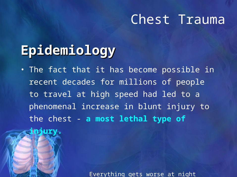
• The fact that it has become possible in recent
decades for millions of people to travel at high speed
had led to a phenomenal increase in blunt injury to
the chest - a most lethal type of injury.
EpidemiologyEpidemiology
Everything gets worse at night

Introduction
• Chest trauma is often sudden and dramatic
• Accounts for 25% of all trauma deaths• 2/3 of deaths occur after reaching hospital
• Major thoracic trauma is associated with multisystem injuries in 70% of cases. 16,000 deaths per year in the US
• 2/3 of victims of major blunt trauma suffer from thoracic injury.
• Second leading cause of trauma deaths after head injury (in USA)
•

Introduction to Thoracic Injury
• Vital Structures• Heart, Great Vessels, Esophagus,
Tracheobronchial Tree, & Lungs• 25% of MVC deaths are due to thoracic
trauma• 12,000 annually in US
• Abdominal injuries are common with chest trauma.
• Prevention Focus• Gun Control Legislation• Improved motor vehicle restraint
systems• Passive Restraint Systems• Airbags

Never operate on a patient who is getting rapidly better or rapidly worse
Thoracic Trauma
• Anatomical Injuries• Thoracic Cage (Skeletal)• Cardiovascular• Pleural and Pulmonary• Mediastinal• Diaphragmatic• Esophageal• Penetrating Cardiac
What structures
may be involved with
each injury?

Mechanism of Injury
• Blunt thoracic injuries• Forces distributed over large area
• Penetrating thoracic injuries• Forces distributed over small area• Organs injured usually those that lie
along path of penetrating object
Nothing spoils good results as much as follow-up

CAUSES OF CHEST INJURIES
• BLUNT TRAUMA• Motor vehicle
accidents• Auto vs.
pedestrian• Falls• Blast injuries
• PENETRATING TRAUMA• Gunshot
wounds• Stab wounds• Shrapnel
wounds

Pathophysiology of Thoracic Trauma
• Blunt Trauma• Results from kinetic energy forces• Subdivision Mechanisms
• Blast• Pressure wave causes tissue disruption• Tear blood vessels & disrupt alveolar tissue• Disruption of tracheobronchial tree• Traumatic diaphragm rupture
• Crush (Compression)• Body is compressed between an object and a hard surface• Direct injury of chest wall and internal structures
• Deceleration• Body in motion strikes a fixed object• Blunt trauma to chest wall• Internal structures continue in motion
• Ligamentum Arteriosum shears aorta
• Age Factors• Pediatric Thorax: More cartilage = Absorbs forces• Geriatric Thorax: Calcification & osteoporosis = More fractures

• Either: - direct blow (e.g. rib fracture) -
deceleration injury or - compression injury
• Rib fracture is the most common sign of blunt thoracic trauma
• Fracture of scapula, sternum, or first rib suggests massive force of injury
Blunt injuriesBlunt injuries

Mechanism of Injury
Penetrating injuries• E.g. stab wounds etc.• Primarily peripheral lung• Haemothorax• Pneumothorax• Cardiac, great vessel or oesophageal
injury

Pathophysiology of Thoracic Trauma
• Penetrating Trauma• Low Energy
• Arrows, knives, handguns• Injury caused by direct contact
and cavitation
• High Energy• Military, hunting rifles & high
powered hand guns• Extensive injury due to high
pressure cavitation
Trauma.org

Pathophysiology of Thoracic Trauma
• Penetrating Injuries (cont.)• Shotgun
• Injury severity based upon the distance between the victim and shotgun & caliber of shot
• Type I: >7 meters from the weapon
• Soft tissue injury• Type II: 3-7 meters from weapon
• Penetration into deep fascia and some internal organs
• Type III: <3 meters from weapon
• Massive tissue destruction

The first step is to make a rough estimate of
the status of the circulatory and respiratory
systems. This provides the first diagnostic clues and
often determines which therapeutic action is to be
taken. Specific questions are then posed pertaining to
individual injuries or their consequences.
Assessment of patient with Thoracic injury

• The treatment of polytraumatized patient must follow a certain protocol which includes. • Adequate oxygenation. • Fluid replacement. • Surgical intervention. • Treatment of septic complications. • Adequate caloric and substrate supplementation. • Prevention of stress bleeding. • Finally, be alert of possible complication (CNS,
ARDS, hepatic, renal, coagulation disorders, sepsis.
Management of patients with Thoracic Trauma

• The evaluation of thoracic injuries is only
one aspect of the total assessment of
severely injured patients.
• Both diagnosis and therapy go hand in
hand.
• The basic principle of elective surgery -
“First investigate and make the diagnosis,
then treat the illness” - is a dangerous
illusion.
Assessment of patient with Thoracic injury

Monitoring and evaluating the patient with Thoracic trauma
• Roentgenograms of the thorax (Chest wall i.e. ribs, sternum, vertebral, clavicles).
• Mediastinum (wide or normal) shifted or not.
• Lung parenchyma (Contusion).
• The heart (cardiac tamponade).
• Diaphragm.
• Pneumothorax, hemothorax.
ECGECG CVPCVP Arterial blood Arterial blood
gases. gases. Urine output. Urine output. Lab. Investigations.Lab. Investigations. Others. Others.

Immediately life-threatening; diagnosis
and therapy before taking
roentgenograms
TEN QUESTIONSTEN QUESTIONS to be asked in the initial to be asked in the initial assessment of severe blunt thoracic injuries assessment of severe blunt thoracic injuries
1. Hypovolemia?
2. Respiratory insufficiency?
3. Tension pneumothorax?
4. Cardiac tamponade

1. Multiple rib fractures? (Paradoxical respiration?)
2. Pneumothorax ? (subcutaneous emphysema? mediastinal emphysema?)
3. Hemothorax?
4. Diaphragmatic rupture?
5. Aortic rupture?
6. Cardiac contusion?
TEN QUESTIONSTEN QUESTIONS to be asked in the initial to be asked in the initial assessment of severe blunt thoracic injuries assessment of severe blunt thoracic injuries
If you are not sure, it isn’t

Common Injuries Develop After Blunt Chest Trauma
• Thoracic cage fractures• Lung contusion and tears• Myocardium contusion• Aortic rupture

Thoracic Trauma
• Initial exam directed toward life threatening:• Injuries
• Open pneumothorax• Flail chest• Tension pneumothorax• Massive hemothorax• Cardiac tamponade
• Conditions• Apnea• Respiratory Distress

TRAUMA DEATHS
EARLYEARLY
30%-35%
Within Hours (Golden Hour)
Thoracic Trauma
Liver/Spleen Injuries
Multiple Pelvic Fractures Others
Optimum Initial Care
IMMEDIATEIMMEDIATE
50%
Seconds or Minutes
Spinal Cord Injuries
Severe Brain Injuries
Lesions to Great Vessels
Prevention
Optimum Prehospital Care
LATE
15%-20%
2-3 Weeks
Sepsis
Multiple Organ Failure
Optimum Initial Care
(Future?)

Injuries Associated with Penetrating Thoracic Trauma
• Closed pneumothorax• Open pneumothorax (including
sucking chest wound)• Tension pneumothorax• Pneumomediastinum• Hemothorax• Hemopneumothorax• Laceration of vascular
structures
• Tracheobronchial tree lacerations
• Esophageal lacerations
• Penetrating cardiac injuries
• Pericardial tamponade• Spinal cord injuries• Diaphragm trauma• Intra-abdominal
penetration with associated organ injury

2003-3-31
Imaging Survey
• Chest x-rayChest x-ray : serve as a screening rather than a definite test repeat radiography should be ordered if suspicious
• Computed tomographyComputed tomography : highly sensitive in detecting injuries and superior to routine chest x-ray recommended in patients with multiple trauma and suspected chest trauma
• AngiogramAngiogram : for suspicious great vessel injuries
• Chest ultrasoundChest ultrasound : detect hemothorax, FAST

Massive HemothoraxMassive Hemothorax
• Need for thoracotomy Need for thoracotomy if: if:
• Immediate evacuation Immediate evacuation of 1500 mL of blood of 1500 mL of blood
• steady trend of more steady trend of more than 250 mL/hthan 250 mL/h

Indications for Thoracotomy in Indications for Thoracotomy in Chest TraumaChest Trauma• Cardiac tamponade Cardiac tamponade • Acute hemodynamic deterioration/cardiac arrest in the trauma Acute hemodynamic deterioration/cardiac arrest in the trauma
center center • Penetrating truncal trauma (resuscitative thoracotomy) Penetrating truncal trauma (resuscitative thoracotomy) • Vascular injury at the thoracic outlet Vascular injury at the thoracic outlet • Loss of chest wall substance (traumatic thoracotomy) Loss of chest wall substance (traumatic thoracotomy) • Massive air leak Massive air leak • Endoscopic or radiographic evidence of significant tracheal or Endoscopic or radiographic evidence of significant tracheal or
bronchial injury bronchial injury • Endoscopic or radiographic evidence of esophageal injury Endoscopic or radiographic evidence of esophageal injury • Radiographic evidence of great vessel injury Radiographic evidence of great vessel injury • Mediastinal passage of a penetrating object Mediastinal passage of a penetrating object • Significant missile embolism to the heart or pulmonary artery Significant missile embolism to the heart or pulmonary artery

Big Trouble
• Central lung injuries are deadly because they are difficult to control and repair.
• When confronted control the pulmonary hilum between thumb and forefinger or clamp it.
• Suture , lobectomy or pneumonectomy

Rib Fracture
• Fractures of 1st and 2nd second require high force• Frequently have injury to aorta or bronchi• Occur in 90% of patients with tracheo-
bronchial rupture• May injure subclavian artery/vein• May result in pneumothorax
• 30% will die

Rib Fracture
• Most common chest wall injury from direct trauma
• More common in adults than children
• Especially common in elderly
• Ribs form rings• Possibility of break in two places
• Most commonly 5th - 9th ribs• Poor protection

Pitfalls to Avoid• Elderly do not tolerate relatively minor
chest injuries• Anticipate progression to acute respiratory
insufficiency
• Children may sustain significant intrathoracic injury w/o evidence of thoracic skeletal trauma• Maintain a high index of suspicion

Tracheobronchial Rupture• Assessment Findings
• Respiratory Distress• Dyspnea• Tachypnea
• Obvious SQ emphysema• Hemoptysis
• Especially of bright red blood
• Signs of tension pneumothorax unresponsive to needle decompression
Diaphragmatic Penetration
• Suspect intra-abdominal trauma with any injury below 4th ICS
• Suspect intrathoracic trauma with any abdominal injury above umbilicus

Traumatic Aortic Dissection/Rupture
• Assessment Findings• Retrosternal or interscapular pain• Pain in lower back or one leg• Respiratory distress• Asymmetrical arm BPs• Upper extremity hypertension with
• Decreased femoral pulses, OR• Absent femoral pulses
• Dysphagia

Myocardial Contusion• Assessment Findings
• Cardiac arrhythmias following blunt chest trauma
• Angina-like pain unresponsive to nitroglycerin
• Precordial discomfort independent of respiratory movement
• Pericardial friction rub (late)

Pulmonary Contusion• Management
• Supportive therapy• Early use of positive pressure ventilation
reduces ventilator therapy duration• Avoid aggressive crystalloid infusion• Severe cases may require ventilator
therapy• Emergent Transport
• Hospital

Hemothorax• Assessment Findings
• Tachypnea or respiratory distress• Shock
• Rapid, weak pulse• Hypotension, narrow pulse pressure• Restlessness, anxiety• Cool, pale, clammy skin• Thirst
• Pleuritic chest pain• Decreased lung sounds• Collapsed neck veins• Dullness on percussion

Open Pneumothorax
• Assessment Findings• Opening in the chest wall• Sucking sound on inhalation• Tachycardia• Tachypnea• Respiratory distress• SQ Emphysema• Decreased lung sounds on affected side

CATEGORIES OF CHEST WOUNDS
• OPEN• Tension
pneumothorax• Sucking chest wound• Hemothorax• Impaled object
• CLOSED• Tension
pneumothorax• Hemothorax• Flail chest• Rib fractures

Simple Pneumothorax• Causes
• Commonly a fx rib lacerates lung• Paper bag effect• May occur spontaneously in tall, thin young males
following:
• Exertion• Coughing
Simple Pneumothorax• Incidence
• 10-30% in blunt chest trauma• almost 100% with penetrating chest trauma• Morbidity & Mortality dependent on
• extent of atelectasis• associated injuries
Flail Chest
• Mortality rates 20-40% due to associated injuries
• Mortality increased with• advanced age• seven or more rib fractures• three or more associated injuries• shock• head injuries

Flail Chest
• Usually secondary to blunt trauma• Most commonly in MVC• Also results from
• falls from heights• industrial accidents• assault• More common in older patients
Sternal Fracture
• Uncommon, 5-8% in blunt chest trauma
• Large traumatic force
• Direct blow to front of chest by• Deceleration
• steering wheel• dashboard
• Other object

Cardiac Tamponade
• Beck’s triad:
- hypotension, jugular venous distention, and muffled heart sounds
- causes decreased diastolic ventricular filling and resultant hypotension
- echocardiogram shows impaired diastolic filling of right atrium initially (1st sign)

Cardiac Tamponade

Hemopericardium with tamponade
Troublesome Injuries
• Blunt cardiac trauma - managementBlunt cardiac trauma - management
• Most cases do not require Tx; Symptomatic arrhythmia (2-5%) antiarrthythmics
• Abnormal ECG and cardiac enzymes almost return to normal within one week.
• Patients with abnormal cardiac echo finding or MUGA keep hospitalization till a repeat test show acceptable finding
• Cardiac rupture prompt surgical repair

Troublesome Injuries
• Lung contusionLung contusion• CxR finding may range from minimal
interstitial infiltrate to extensive lobar consolidation
• Chest CT is accurate diagnostic tool but not always mandatory
• Tx : same as flail chestsame as flail chest, but pay attention to avoid overhydration; use of steroid and prophylactic antibiotic are still controversial

Aortic Transection
• Signs:
- widened mediastinum, 1st rib fx, apical capping, left hemothorax, tracheal deviation to right
- widening from bridging veins and arteries, not aorta itself
- need aortic evaluation in pts with significant mechanism (deceleration injuries), usually tears at ligamentum
- 90% of patients die at the scene

Thoracic Aorta Injury
• 90% lethal before receiving emergency care• Usually a transverse laceration of part or all
of aortic circumference• 60% have adventia intact = pseudoaneurysm• Injury to root or ascending aorta is nearly
100% fatal• ~90% occur at aortic isthmus just distal to left
subclavian• 4-10% of cases have concomitant great
vessel injury

Airway Injury• Tracheobronchial tears are uncommon
• < 0.35-1.5% of BCT• bronchial > tracheal • 75% at R mainstem usually within 2.5 cm of carina
• Leads to persistent PTX• Specific Symptom: persistent PTX after chest
tube placement• Finding: “Fallen Lung Sign”,
pneumomediastinum, pneumopericardium, sub cut. Emphysema
• ET Tube balloon inflation >2.8cm implies tracheal rupture

Open Pneumothorax
• Opening in chest cavity that allows air to enter pleural cavity
• Causes the lung to collapse due to increased pressure in pleural cavity
• Can be life threatening and can deteriorate rapidly

TENSION PNEUMOTHORAX
• 33% of preventable combat deaths• Injured chest or lung acts as one-way valve• Air becomes trapped between the lung and
chest wall causing the lung to collapse• The heart is pushed to the other side causing
blood vessels to kink• Death will result if not quickly recognized and
treated with needle decompression• May occur in open and closed chest wounds

Tension Pneumothorax
Air collapses lung and pushes heart to other side
Blood return to heart restricted by kinked vessels, heart unable to pump
Air between lung and chest wall

OTHER SIGNS AND SYMPTOMS OF TENSION PNEUMOTHORAX
• Difficulty breathing • Chest pain• Unilateral decreased/absent breath sounds• Anxiety or agitation• Increased pulse• Tracheal deviation• Jugular venous distention (JVD)• Cyanosis

NEEDLE CHEST DECOMPRESSION

SUCKING CHEST WOUND (OPEN PNEUMOTHORAX)• Open chest wound allows air entry into chest and escape• Although lung is collapsed (pneumothorax), pressure is
relieved by air escape and tension pneumothorax is avoided
• Tension pneumothorax may develop later • Continually reassess the casualty for signs and symptoms
of tension pneumothorax

OPEN CHEST WOUND

Esophageal Injury
• <10% of esophageal rupture is caused by trauma
• < 1% of BCT’s• Findings:
• Pneumomediastinum• Left PTX• Left pleural effusion• Sub cut emphysema• Left lower lobe atelectasis

Traumatic Asphyxia

MANAGEMENT OF IMPALED OBJECT IN THE CHEST
• Immobilize the impaled object• Stabilize object with support dressings
• Use bulky dressings
• Construct protective structure using splint or sling
• Cover and dress open wounds


ConclusionChest injuries are directly responsible for more than
20% of all traumatic deaths (regardless of mechanism) and account for about 16,000 deaths per year in the United
States. The risk of death can be reduced by early recognition of serious signs and symptoms and rapid
treatment.


