
Therapeutics, Targets, and Chemical Biology
TLR Adaptor Protein MYD88 Mediates Sensitivityto HDAC Inhibitors via a Cytokine-DependentMechanismMaria New1, Semira Sheikh1, Mina Bekheet1, Heidi Olzscha1, Marie-Laetitia Thezenas2,Matthew A. Care3,4, Susan Fotheringham1, Reuben M. Tooze3, Benedikt Kessler2,and Nicholas B. La Thangue1
Abstract
Histone deacetylase (HDAC) inhibitors have proven usefultherapeutic agents for certain hematologic cancers. However,HDAC inhibition causes diverse cellular outcomes, and identifi-cation of cancer-relevant pathways within these outcomesremains unresolved. In this study, we utilized an unbiased loss-of-function screen and identified the Toll-like receptor (TLR)adaptor protein MYD88 as a key regulator of the antiproliferativeeffects of HDAC inhibition. High expression of MYD88 exhibitedincreased sensitivity to HDAC inhibitors; conversely, low expres-sion coincided with reduced sensitivity. MYD88-dependent TLRsignaling controlled cytokine levels, which then acted via anextracellular mechanism to maintain cell proliferation and
sensitize cells to HDAC inhibition. MYD88 activity was directlyregulated through lysine acetylation and was deacetylated byHDAC6. MYD88 was a component of a wider acetylationsignature in the ABC subgroup of diffuse large B-cell lympho-ma, and one of the most frequent mutations in MYD88, L265P,conferred increased cell sensitivity to HDAC inhibitors. Ourstudy defines acetylation of MYD88, which, by regulatingTLR-dependent signaling to cytokine genes, influences theantiproliferative effects of HDAC inhibitors. Our resultsprovide a possible explanation for the sensitivity of malignan-cies of hematologic origin to HDAC inhibitor–based therapy.Cancer Res; 76(23); 6975–87. �2016 AACR.
IntroductionAbnormal epigenetic control is a common feature of tumori-
genesis, and aberrant lysine acetylation is believed to take on animportant role in driving the malignant phenotype (1). Lysineacetylation is regulated by two groups of enzymes, with histoneacetyl-transferases (HAT) mediating the acetylation event, andhistone deacetylases (HDAC) providing the deacetylation event(2). This type of relationship, often referred to as "writer, reader,and eraser", plays an instrumental role in epigenetic control,where lysine acetylation provides a key regulatory event (1).Furthermore, HAT and HDAC, and bromodomain reader pro-teins, are families with multiple members, reflecting the diverse
biological roles that these enzymes and reader proteins undertake(2, 3). Lysine acetylation occurs on proteins with diverse biolog-ical functions, for example, many proteins involved with metab-olism and cell signaling (4, 5), and thus acetylation cannot beviewed as a modification that is restricted to the nucleus andepigenetic control.
There is increasing recognition that lysine acetylation is deregu-lated in cancer, either through mutation in HAT genes or con-versely deregulation of HDAC activity (6).
HDACs have been increasingly recognized as a relevant ther-apeutic target in neoplastic disease (7, 8). Small-molecule inhi-bition of HDAC activity has antiproliferative effects on cancercells, including cell-cycle arrest and apoptosis (9), and conse-quently there has been considerable interest in progressingHDACinhibitors into clinical studies (2, 10, 11). SomeHDAC inhibitorshave been approved for clinical use, for example SAHA/vorinostatfor treating cutaneous T-cell lymphoma (CTCL), and panobino-stat for multiple myeloma (10, 12, 13). It remains unclear howHDAC inhibitors should be optimally deployed for clinicalbenefit, reflecting the pleiotropic and diverse cellularmechanismsthat are controlled by acetylation, and lack of knowledge abouthow thesemechanisms are influenced byHDAC inhibitors and inmalignant disease.
To address this question, we have taken an unbiased loss-of-function screening approach to identify pathways thatinfluence the effect of HDAC inhibitors on cancer cells. Here,we report that the Toll-like receptor (TLR) adaptor proteinMYD88 regulates cell sensitivity to HDAC inhibitor treatment.MYD88 (myeloid differentiation primary response gene 88)augments TLR-dependent signaling, thereby affecting the level
1Department of Oncology, University of Oxford, Oxford, United Kingdom.2Target Discovery Institute, Nuffield Department of Medicine, University ofOxford, Oxford, United Kingdom. 3Section of Experimental Haematology, LeedsInstitute of Cancer and Pathology, University of Leeds, Leeds, United Kingdom.4Bioinformatics Group, School of Molecular and Cellular Biology, University ofLeeds, Leeds, United Kingdom.
Note: Supplementary data for this article are available at Cancer ResearchOnline (http://cancerres.aacrjournals.org/).
M. New, S. Sheikh, and M. Bekheet contributed equally to this article.
Corresponding Author: Nicholas B. La Thangue, University of Oxford, Old RoadCampus Research Building, Roosevelt Drive, Oxford OX3 7DQ, United Kingdom.Phone: 44-18-6561-7090; Fax: 44-18-6561-7092; E-mail:[email protected]
doi: 10.1158/0008-5472.CAN-16-0504
�2016 American Association for Cancer Research.
CancerResearch
www.aacrjournals.org 6975

of extracellular cytokines, which act in part through an auto-crine mechanism to sensitize cells to HDAC inhibition. Nota-bly, MYD88 is a direct target for lysine acetylation, and itsdeacetylation is mediated by HDAC6. Significantly, hypoace-tylation of MYD88 causes enhanced IL6 levels, which sensi-tizes cells to HDAC inhibition. MYD88 is expressed at highlevels and its gene is a target for somatic mutation in hema-tologic malignancy (14, 15), and cells expressing the mostfrequently observed mutation, L265P, exhibit enhanced sen-sitivity to HDAC inhibitors. Furthermore, MYD88 is a com-ponent of a wider acetylation signature in the ABC subgroupof diffuse large B-cell lymphoma (DLBCL). Thus, the role ofMYD88 in TLR-dependent signaling defines a novel mecha-nism that sensitizes tumor cells to HDAC inhibitors. Ourresults bear on the apparent sensitivity of malignancies ofhematologic origin to HDAC inhibitor–based therapy.
Materials and MethodsCell lines
U2OS cells were from ATCC. Cells were regularly tested with aLonzamycoplasma kit. Regular immunofluorescencemicroscopywith nuclear staining confirmed a negative mycoplasma result(last negative test November 2015). Cells were passaged twice aweek and used until 16 passages. The inducible U2OS-stable celllines were thawed before each experiment. RIVA and HBL-1 cellswere from Prof. Alison Banham (May 2013; Haemato-oncology,Nuffield Division of Clinical Laboratory Sciences, John RadcliffeHospital, Oxford, United Kingdom) at passage 8. MYD88 L265Pmutation status was confirmed in both cell lines (December2013) by DNA sequencing. Mycoplasma testing was carried outas above (last negative test May 2016). U2OS cells were main-tained inDMEM supplemented with 10%FCS and 1%penicillin/streptomycin (Invitrogen). RIVAandHBL-1 cellsweremaintainedin RPMI supplemented with the same. The inducible FLAG-MYD88 cells were created in parental U2OS cells using theClontech Tet-On gene expression system (Clontech). EctopicFLAG-protein expression was induced by doxycycline accordingtomanufacturer's instructions.HDAC6 shRNAknockdownU2OScell lines were created in U2OS cells using HDAC6 or controlshRNA lentiviral particles (Santa Cruz Biotechnology), and stableclones were selected for puromycin resistance as per the manu-facturer's protocol.
Plasmids, transfection, and screenCells were transfected usingGeneJuice (Merck) and harvested 24
to 72 hours after transfection. U2OS cells were transfected withsiRNAs against HDAC6 (Dharmacon, M-00349900), MYD88(Smart-Pool Dharmacon), and nontargeting control NT2 (Dhar-macon) using oligofectamine (Invitrogen) to a final concentrationof 50 nmol/L before harvesting. Lymphoma cells were transfectedwith MYD88 or control GFP siRNA mixed in OptiMEM andRNAiMaxaccording to themanufacturer's instructions.Themixturewas added to 2 � 106 cells in 1-mL medium (2 mL total) for 6hours, seeded into a 6-cm dish, and incubated for a further 4 days.Onedaybeforeharvesting, SAHAwas added to the transfected cells.
The PCMV-FLAG-WT MYD88 mutant derivatives L265P andK132R were created using oligonucleotides designed in accor-dance with Stratagene's QuikChange Site-Directed MutagenesisKit. MYD88 was identified by a loss-of-function screen asdescribed in ref. 16.
Antibodies and compoundsThe following antibodies were used: FLAG-M2 (Sigma), PARP
(Cell Signaling Technology), acetylated tubulin (Sigma), actin(Sigma), HDAC6 (Millipore), phospho-Chk1 S317 (Millipore),Chk1 (Cell Signaling Technology), cleaved caspase-3 (Cell Sig-naling Technology), MYD88 (Cell Signaling Technology forimmunoblotting and Abcam for IHC), TLR4 (Cell SignalingTechnology), p53 (Santa Cruz Biotechnology), IL6 (Abcam), IL8(Abcam), acetylated lysine (Cell Signaling Technology), andNFkB p65 (Santa Cruz Biotechnology). HDAC inhibitors wereobtained fromCaymanChemical, Sigma, andChemietek, and theMYD88 inhibitor T1617932 from AOBIOUS Inc.
Immunoblotting, immunoprecipitation, and preparation ofnuclear extracts
Immunoblotting, immunoprecipitation, and nuclear extractpreparation were carried out as described previously (8). Repre-sentative blots from at least three experiments are shown.
Colony formation assay and flow cytometryU2OS-Tet-FLAG-MYD88–inducible cells were seeded at 1 �
103 cells perwell in 6-well plates. FLAG-MYD88was induced for 4hours prior to 24 hours of drug treatment as indicated. At 9 daysafter drug treatment, cells were washed twice in PBS, fixed inmethanol for 10 minutes, and stained with crystal violet solutionfor 30 minutes before extensive washing with water. The colonieswere then counted. Flow cytometry was performed as describedpreviously (16).
Reporter assayCells were transfected with b-galactosidase (b-gal) and Bcl-xl
reporter plasmids and expressing vector as described previously(17). Triplicate samples were transferred to a 96-well plate andprocessed with aWinGlo 96Microplate luminometer (Promega),and relative activity calculated according to transfection efficiencyindicated by b-gal activity.
Cytokine level analysisThe levels of six cytokines, IL1b, IL12p, IL10, IL6, TNFa, and
IL8, were analyzed using a cytometric bead array as described bythe manufacturer (BD Biosciences).
Mass spectrometry and in-solution digestFLAG-MYD88–stable cell lines were prepared as described
previously (18, 19). Samples were digested with trypsin (0.2mg/mL) overnight at 37�C and processed for mass spectrometryas described.
IHC pathology examinationIHC on paraffin-embedded formalin-fixed tissue was per-
formed exactly as described previously (20).
Bioinformatics analysis and signature enrichmentA dataset of 14,816 gene signatures was created by merging
signatures downloaded from http://lymphochip.nih.gov/signa-turedb/ (SignatureDB), http://www.broadinstitute.org/gsea/msigdb/index.jsp MSigDB V4 (MSigDB C1–C7), http://comp-bio.dfci.harvard.edu/genesigdb/ Gene Signature Database V4(GeneSigDB), UniProt Keywords (UniProtKB; refs. 21–24). Geneontology (GO) and annotation files were downloaded from
New et al.
Cancer Res; 76(23) December 1, 2016 Cancer Research6976

http://www.geneontology.org (03/17/2015). The structure fromthe .obo and annotations from the .goa files were used to builda GO gene set. Enrichment of gene sets against signatures wasassessed using a hypergeometric test, where the draw is the geneset genes, the successes are the signature genes, and the pop-ulation is the number of genes present on the platform. The top500 most MYD88-correlated genes within ABC and GC DLBCLwere determined across 10 DLBCL datasets encompassing2,030 cases (Care and colleagues; manuscript submitted).These 500 genes were used for GO and gene signature enrich-ment analysis.
On the basis of the top 500 in ABC DLBCL, acetylation eventswere assigned to the 500 genes/proteins. Annotated acetylationand acetylation sites at lysine residues of proteins (experimentbased, N-terminal acetylation excluded) were obtained fromthe following databases: Compendium of Protein Lysine Mod-ifications (CPML), PhosphoSite Plus, Phosida, and UniProt. Onthe basis of the heatmaps and the signatures, the genes in theMYD88-related ABC subgroup DLBCL nonimmune clusters wereassigned to the respective clusters chr3p21/chr3p22, MCM com-plex/DNA unwinding, metabolic process (including pentose andorganophosphate pathways), macromolecule organization, and
020406080
100120140160180200
:SAHA (μmol/L)5 10 20
Col
ony
num
ber
0
– + – + – + – + :DOX
FLAG (MYD88)
Actin
(ii)– + – +
:SAHA
:DOX
** * *
(i)A
B
+ –
Actin
MYD88
:SAHA (μmol/L) 0 5 10 0 5 10
Casp
Sub-
G1
(%)
: SAHA (μmol/L)
+ – + – : MYD88 siRNA02 4
8
10
12
14
6
0 5 10
+ –
****
(i)
:MYD88 siRNA (ii)
PARP
Figure 1.
MYD88 expression regulates sensitivity to HDAC inhibitors. A, i, U2OS cells treated with MYD88 or control (NT) siRNA (50 nmol/L for 72 hours) in the presenceor absence of SAHA (5 or 10 mmol/L for 24 hours) and subsequently immunoblotted with anti-MYD88, anti-PARP (cleaved), anti-caspase-3 (cleaved), andanti-actin antibody. ii, U2OS cells (in triplicate) from i were analyzed by flow cytometry (blue bars, MYD88; red bars, NT siRNA). The sub-G1 phase (%) isshown. Statistical significance is indicated (�� , P � 0.01). B, U2OS cells stably expressing Tet-On inducible FLAG-tagged MYD88 were grown in triplicatein the absence (�) or presence (þ) of doxycycline (DOX) together with SAHA (5, 10, or 20 mmol/L) and, after 9 days, viable cell colonies were assessed bycrystal violet staining (and quantitated). The untreated control (no SAHA) cells are shown for comparison. i, Graphical representation with example colony stainsunderneath (in triplicate). Statistical significance is indicated; � , P� 0.05; �� , P� 0.01. ii, Immunoblot of ectopic FLAG-MYD88 in the same cells as used in i. Note thatMYD88 was one of the top seven hits in the loss-of-function screen, which ranked highly because the cells from the resistant colony contained a singleshRNA and the target gene was silenced in a stable fashion in SAHA-resistant colonies.
MYD88 Regulates Cell Sensitivity to HDAC Inhibitors
www.aacrjournals.org Cancer Res; 76(23) December 1, 2016 6977
FLAG (MYD88)
– MYD88(ii)
Rel
ativ
e ch
ange
+ DOX
(ii)
(iii) (iv)
Rel
ativ
e ch
ange
Rel
ativ
e ch
ange
Rel
ativ
e ch
ange
Rel
ativ
e ch
ange
Rel
ativ
e ch
ange
(v) (vi)
C
+ DOX
+ DOX + DOX
+ DOX + DOX
– – – + + + – – – + + + R
elat
ive
activ
ity (×
10–3
):SAHA
– MYD88
**
D
05
101520 25 30
– – + + – – + + – + – + – + – +
:SAHA
**
A (i)
B (i)
Sub
-G1
%
:Anti-IL6
:DOX
FLAG (MYD88)
– +
PARP
FLAG (MYD88)
:Anti-IL6 :SAHA
:DOX
– – + + – – + +
– +
– + – + – + – +
ActinActin
Actin
PARP
0
5
10
15
20
25
0
2
4
6
8 IL1β
0
2
4
6
8 IL10
0
2
4
6
8 IL12p70
0
2
4
6
8 TNFα
0
2
4
6
8 IL8
0
2
4
6
8 IL6
New et al.
Cancer Res; 76(23) December 1, 2016 Cancer Research6978
adhesion. Acetylation events of the genes in the different clusterswere assigned and a protein list of the occurring proteins in theclusters (nonoverlapping) was compiled. The percentage of acet-ylationwas calculated for the top 500 genes ABCDLBCL, the totalnumber of genes from ABC DLBCL nonimmune clusters, and theindividual clusters from ABC DLBCL nonimmune clusters.
ResultsMYD88 expression regulates cell sensitivity to HDAC inhibitortreatment
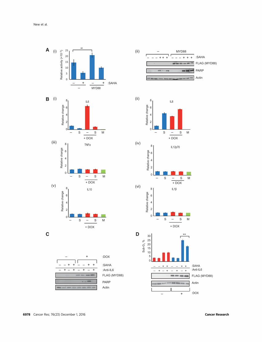
MYD88 was identified in a genome-wide loss-of-functionscreen for genes that regulate cell sensitivity to HDAC inhibitorsusing SAHA, as described previously (16). The effect of MYD88in regulating cell sensitivity to HDAC inhibitors was confirmedboth by short-term silencing and ectopic MYD88 expression;treating cells with MYD88 siRNA reduced sensitivity to apo-ptosis upon SAHA treatment and conversely expressing ectopicMYD88 enhanced sensitivity to SAHA (Fig. 1A and Supple-mentary Fig. S1A). Furthermore, a stable inducible cell lineexpressing MYD88 exhibited enhanced apoptosis to SAHA(Supplementary Fig. S1B). A similar effect was observed in cellcolony formation assays, where the inhibitory effect of SAHAwas enhanced upon stable expression of ectopic MYD88 rela-tive to uninduced cells (Fig. 1B). Moreover, MYD88 sensitizedcells to a variety of other HDAC inhibitors, such as panobino-stat, but not mechanistically unrelated agents (SupplementaryTable S1). These results establish that MYD88 regulates cellsensitivity to HDAC inhibitor treatment.
Extracellular IL6 sensitizes cells to HDAC inhibitionOne of the major effector arms of MYD88-dependent TLR
signaling regulates cytokine gene expression, via NFkB-depen-dent transcription (25). To explore the mechanistic role ofMYD88, we tested the impact of MYD88 on cytokine expressionand thereafter the influence of cytokines on HDAC inhibitor–treated cells. The increased level of ectopic MYD88 caused theactivation of NFkB and NFkB-responsive transcription (Fig. 2Aand Supplementary Fig. S1C). We then measured the extracel-lular level of different cytokines regulated by the NFkB path-way, including IL1b, IL6, IL8, IL10, IL12, and TNFa, andobserved that IL6 was elevated upon MYD88 expression andreduced by SAHA treatment (Fig. 2B). The level of IL8 was alsoenhanced by MYD88 but in contrast to IL6 remained high uponSAHA treatment. The effect of MYD88 expression was insignif-icant on the other cytokines measured under these assay con-ditions (Fig. 2B).
We addressed whether the increase in the level of extracellularIL6 or IL8 dependent upon MYD88 influenced cell sensitivity toHDAC inhibitors using neutralizing antibodies to inactivate IL6,which we added to the medium of cells expressing inducibleectopic MYD88 and then treated the cells with SAHA (Fig. 2C andD). We observed that the enhanced level of apoptosis (increasedPARP and sub-G1 cells) that occurred upon MYD88 expressionwith SAHA treatment was reduced by IL6 neutralization (Fig. 2Cand D). In contrast, neutralization of IL8, under the same exper-imental conditions of MYD88 expression, had a modest effect onSAHA-dependent apoptosis (Supplementary Fig. S1D), suggest-ing that IL6 rather than IL8 is the more relevant effector in theseconditions. The increased level of extracellular IL6 dependentupon MYD88 expression therefore augments cell sensitivity toHDAC inhibitors.
To explore the mechanism through which IL6 sensitizes cells toHDAC inhibitors, we considered an influence on cell-cycle pro-gression, an idea that is consistent with previous reports describingthe growth promoting effects of IL6 (26, 27). We comparedMYD88- induced to uninduced cells, and noticed impaired cell--cycle progression, reflecting an increase in the size of the G2 phaseand reduced S-phase population, upon treatment with anti-IL6antibody (25% � doxycycline to 37% þ doxycycline); the anti-IL6–treated MYD88-induced cells exhibited a profile typical ofcells stalled inG2 phase (Supplementary Fig. S1E, i and ii; comparedoxycycline þ/� with anti-IL6 treatment). Because the cell-cycleprofile was reminiscent of an active G2 checkpoint (3, 28), weinvestigated whether anti-IL6–treated cells had an active G2 check-point. Chk1 acts at the G2–M phase transition in response tocellular stress (28), and S317 phosphorylation is a marker foractive Chk1 kinase (3). Enhanced phosphorylation occurred atS317 upon IL6 neutralization (Supplementary Fig. S1E, iii), con-sistent with a role of Chk1 in the G2 checkpoint and the increasedG2 phase cells, which appeared upon IL6 neutralization. Anincrease in phosphorylation at S317 also occurred in uninducedcells, although the effect in MYD88-induced cells was far moreapparent (Supplementary Fig. S1E, iii). These results suggest thatan effect of IL6 is to allow cell-cycle progression.
MYD88 is acetylated and deacetylated by HDAC6Having established that MYD88 regulates cell sensitivity to
HDAC inhibition, we turned our attention to the control ofMYD88 activity. One possibility that we considered was a directinfluence of HDAC on MYD88; for example, if MYD88 was itselfacetylated. Significantly, we identified lysine acetylation on bothectopic and endogenous MYD88 protein (Fig. 3A and Supple-mentary Fig. S1F), and then by tandem mass spectrometry
Figure 2.MYD88 augments IL6 levels.A,U2OS cells were transfectedwith the indicated expression vectors (1 mg) for 48 hours and treatedwith SAHA (10 mmol/L;þ) for another24 hours. The NFkB-responsive Bcl-xL promoter luciferase activity is shown relative to cotransfected pCMV-bgal expression (i), together with the level ofectopic FLAG-MYD88 protein (ii), PARP (cleaved), and actin, performed in triplicate. Statistical significance is indicated (�� , P� 0.01). B, U2OS cells stably expressingTet-On inducible FLAG-tagged MYD88 were treated with SAHA (S; 10 mmol/L) for 24 hours under noninduced or induced doxycycline (þDOX) treatment conditions(1 mg/mL; induction for 72-hour treatment), and relative levels of the indicated cytokines (IL6, IL8, TNFa, IL12p70, IL10, and IL1b) in the tissue culture media weremeasured using BD Cytometric Bead Array Human Inflammatory Cytokine measurement kit. The reading for preculture medium (M) is indicated. Mean values fromtriplicate readings. C, U2OS cells stably expressing Tet-On inducible FLAG-tagged MYD88 were treated with SAHA (10 mmol/L; þ) and anti- IL6 neutralizingantibody (2 mg/mL) for 24 hours under noninduced (�) or induced (þ) doxycycline (DOX) treatment conditions (1 mg/mL; induction for 72 hours prior to treatment).Cells were immunoblotted with anti-FLAG (for ectopic MYD88) or PARP (cleaved) and actin antibodies as indicated. Quantification indicated that cleaved PARPdecreased by over 50% upon IL6 neutralization and less than 5% upon IL8 neutralization (Supplementary Fig. S1D) under conditions of MYD88 induction (þDOX) andSAHA treatment. D, U2OS cells stably expressing Tet-On inducible FLAG-tagged MYD88 were treated as in C and analyzed by flow cytometry for the level ofsub-G1 phase cells (as a percentage of the total population). Immunoblot shows the level of MYD88 (anti-FLAG) and actin. Statistical significance is indicated as�� , P � 0.01.
MYD88 Regulates Cell Sensitivity to HDAC Inhibitors
www.aacrjournals.org Cancer Res; 76(23) December 1, 2016 6979

C
0 1 2 4 8 24 0 1 2 4 8 24 :hr cyc
K132R WT :MYD88
p53
6
4
2
0
8
Rel
ativ
e ch
ange
in IL
6
WT K132R :MYD88
– + – + M :DOX
in IP
(ii)(i)
IDDD TIR
E
MYD88
131 142
1 296
F
– + – +
WT
– + – +
K132R
Sub
-G1
(%)
05
10
25
15
30 20
– – + + :DOX:SAHA
– – + +
**
200 300 400 500 600 700 800 900 1,000 1,100 1,200 1,300 1,400
m/z
0
5
10
15
20
25
30
35
40
45
50
55
60
65
70
75
80
85
90
95
100
ion
Rel
ativ
eab
unda
nce
y2(229.16)229.15
y5(557.56)557.33
y3(357.36)357.25
y4(486.37)486.29
b3(412.46)412.26
y6(686.23)686.37
y7(815.41)815.41
y+8
(926.51)926.45
b7(926.51)926.46
y10(1199.64)1199.59
b4(523.34)523.29
L K132 Q Q Q E E A EKPLy2y3y4y5y6y7y8y10
b3 b7b4Ac
A
:SAHA
FLAG (MYD88)
Ac-K (MYD88)
FLAG (MYD88)
in IP : Anti-FLAG
– WT K132R – WT K132R
4.5
3
1.5
0
Nor
mal
ized
PA
RP
cle
avag
e
:SAHA – + – + – + – +WT K132R :MYD88WT K132R
– + :DOX
M88 NS M88 NS
Ac-K (MYD88)
Actin
B
D
Figure 3.
MYD88 is acetylated. A, i, Cells stably expressing FLAG-MYD88 were treated with SAHA (þ; 10 mmol/L) for 16 hours and immunoprecipitated with FLAG (M88) ornonspecific (NS) antibody prior to immunoblottingwith MYD88 and acetyl-lysine antibody (Ac-K). Input (2%) level of MYD88 is shown. A 10% increase in the acetylationlevel of MYD88 occurred in SAHA-treated cells. ii, Domain organization of MYD88. DD, death domain; ID, intermediate domain; TIR, Toll-like receptor interactingdomain. MYD88 was immunopurified as described in A and analyzed by mass spectrometry. The MS/MS spectrum of the peptide 131LKQQQEEAEKPL142derived from MYD88 (SwissProt no Q99836) demonstrates acetylation at lysine (K) 132. B and y fragment ions are indicated. B, FLAG-MYD88 WT, FLAG-K132R, orcontrol (�) inducible U2OS cell lines were induced for 2 days with doxycycline (DOX). Cells were harvested and MYD88 or its mutant immunoprecipitated with anti-FLAG antibody and immunoblotted with anti-FLAG or anti-acetyl-lysine antibody. Input is indicated (inset). C, Stable U2OS cells expressing Tet-On inducibleWT or K132R FLAG-MYD88 were treated with cycloheximide (100 mmol/L) for the indicated time and immunoblotted with FLAG, p53, and actin antibody. Immunoblotquantitation of FLAG-MYD88 relative to actin performed using ImageJ software; WT and K132R had a half-life of 2 and 8 hours, respectively. D, Stable Tet-On inducibleU2OS cell lines expressing WT or K132R FLAG-MYD88 were treated with SAHA (10mmol/L) for 16 hours under noninduced (�) or induced (þ) doxycycline(DOX) treatment conditions (1 mg/mL; induction for 72 hours prior to treatment). Cells were immunoblotted for MYD88 (anti-FLAG), PARP (cleaved), and actinantibody. The graph represents quantitation of PARP cleavage. E, Level of sub-G1 in stable Tet-On inducible U2OS cell lines expressingWT or K132R FLAG-MYD88U2OSunder noninduced (�) or induced (þ) doxycycline (DOX) treatment conditions by flow cytometry, upon SAHA treatment (10 mmol/L) for 16 hours, as describedin D. Statistical significance is indicated as �� , P � 0.01. F, Relative level of IL6 in tissue culture medium was measured using BD Cytometric Bead Array HumanInflammatory Cytokine Kit. Mean values from triplicate readings are shown in U2OS cells stably expressing Tet-On inducible WT or K132R MYD88 under non-induced(-) or induced (þ) doxycycline (DOX) treatment conditions (1 mg/mL; induction for 72-hour treatment), as described in D.
New et al.
Cancer Res; 76(23) December 1, 2016 Cancer Research6980

identified a single acetylated lysine (K) residue (K132; Fig. 3A).Furthermore, the low but detectable acetylation level on MYD88contrasted with its mutant derivative, carrying a lysine to argininesubstitution (K132R), which showed reduced acetylation (Fig.3B), suggesting that K132R is an acetylation loss-of-function
mutant. Most importantly, K132R exhibited a longer half-liferelative to wild-typeMYD88 (Fig. 3C), suggesting that acetylationinfluenced the stability of MYD88. We then compared the prop-erties of wild-type MYD88 to K132R, beginning with HDACinhibitor sensitivity and observed that K132R expression
A
0 5 10 20 0 5 10 20 0 5 10 20 :SAHA
30
25
15
10
5
0 – + – + – + :SAHA
B (ii)(i)
C
Rel
ativ
e ch
ange
in IL
6
:SAHA 0 5 10 0 5 10
:shRNA HDAC6
HDAC6
MYD88
Actin
(i)
+ –
:SAHA
Rel
ativ
e le
vel o
f IL6
(ii)
:shRNA HDAC6 – +
10
8
6
4
2
0
Actin
Casp
FLAG (MYD88)
200
150
100
50
0Nor
mal
ized
cas
pase
-3 c
leav
age
D IP : Anti-FLAG in
WT− WTL265P L265P
FLAG (MYD88)
Ac-K (MYD88)
Actin
:MYD88WT
0 5 10 20 0 5 10 20 0 5 10 20
L265P –
:SAHA
−
Figure 4.
Properties of MYD88.A, i,Control (�) and HDAC6 knockdown (þ; stable shRNA knockdown) U2OS cells were treatedwith SAHA at the indicated concentrations for24 hours and analyzed by immunoblotting for HDAC6 and MYD88. Actin was the loading control. ii, Relative levels of IL6 in tissue culture medium weremeasured using BD Cytometric Bead Array Kit. Mean values from triplicate readings are shown in U2OS control (�) and HDAC6 knockdown (þ; stable shRNAknockdown) cells. B, i, U2OS cells were transfected with empty vector (�), FLAG-MYD88 (WT), or FLAG-L265P. SAHA (5, 10, or 20 mmol/L) or DMSO (0)was added, and 24 hours later, cells were immunoblotted with FLAG, caspase-3 (cleaved), or actin antibodies. The reduced level of L265P in SAHA-treated cellswas reproducibly observed in at least three independent experiments; the reasons for the reduction remain unclear. ii, Graph represents the quantitatedcaspase-3 (cleaved) from C (i), normalized to actin loading control using ImageJ software. C, Relative levels of IL6 in tissue culture medium (M) measured usingBD Cytometric Bead Array Kit. Mean values from triplicate readings are shown in U2OS expressing WT or L265P MYD88 in the presence or absence of SAHAtreatment (5 mmol/L). D, U2OS cells were transfected with empty vector, FLAG-MYD88 (WT), or FLAG-L265P. Cells were harvested and immunoprecipitatedwith anti-FLAG and immunoblotted with anti-FLAG or anti-acetyl-lysine (Ac-K) antibody. Input levels are indicated, together with acetylated MYD88.
MYD88 Regulates Cell Sensitivity to HDAC Inhibitors
www.aacrjournals.org Cancer Res; 76(23) December 1, 2016 6981

RIVA HBL-1
– + – + – – + +
A B
D RIVA
:SAHA MYD88
PARP Casp Actin
:SAHA :Anti-TLR4
MYD88
PARP Casp
Actin
MYD88
Actin
Casp
:SAHA – + – +
RIVA
E
C (i)
+ –
– + – + – + – + – – + + – – + +
RIVA HBL-1
:SAHA :Anti-IL6 MYD88
Casp
Actin
F G
:MYD88 siRNA
– + – +
Rel
ativ
e su
b-G
1
(ii)
0
1
2
3
4
5
6
7
8
9
:SAHA − + − + RIVA HBL-1
0
1
2
3
4
5
6
RIVA HBL-1 Medium
Rel
ativ
e cy
toki
ne le
vels
IL6IL8
1.0
0.8
0.6
0.4
0.2
0Nor
mal
ized
cas
pase
-3 c
leav
age
+ + :SAHA
− +
:Anti-IL6
0
1
2
3
4
5
6
7
8
9
RIVA
−− − −
−
−
HBL-1
Rel
ativ
e ch
ange
in s
ub-G
1
:SAHA
:MYD88 siRNA
Cancer Res; 76(23) December 1, 2016 Cancer Research6982
New et al.

sensitized cells to a higher level of apoptosis upon HDAC inhib-itor treatment compared with wild-type MYD88 (PARP cleavageand sub-G1 cells; Fig. 3D and E). Furthermore, the heightenedeffect of K132R coincided with an increased extracellular IL6 levelcomparedwithwild-typeMYD88 (Fig. 3F).MYD88 is thus a directtarget for acetylation, and acetylation impacts on its biochemicaland biological properties.
We considered HDAC6 as a possible deacetylase, given itscytoplasmic colocalization with MYD88 and role in regulatingcytoplasmic substrates (29), and assessed the effect of silencingHDAC6 on MYD88. We observed in HDAC6-depleted cells areduced level of MYD88 and concomitant reduction in IL6 (Fig.4A and Supplementary Fig. S2A–S2C). Furthermore, in cellstreated with tubastatin A, a small-molecule inhibitor of HDAC6(30), MYD88 levels were also reduced (Supplementary Fig. S2Band S2C); the acetylation of tubulin, a recognized substrate ofHDAC6 (31), increased under tubastatin A treatment (Supple-mentary Fig. S2C). Overall, the results highlight HDAC6 as a keyregulator of MYD88 and more generally suggest that acetylationcontrols the activity of MYD88.
Somatic mutation in MYD88 influences HDAC inhibitorsensitivity
MYD88 is expressed at high levels in the hematopoieticlineage and somatic mutation in MYD88, resulting in theL265P substitution observed in the ABC subtype of DLBCL andfrequently in Waldenstrom macroglobulinemia (14, 15). L265Pis believed to result in an oncogenic gain-of-function mutation,which augments signaling through NFkB (14). We reasonedtherefore that L265P may impact on MYD88 as a sensitizer toHDAC inhibitor treatment. When the effect of L265P was com-pared with wild-type MYD88, an enhanced level of apoptosiswas apparent upon HDAC inhibitor treatment and increased IL6production was evident (Fig. 4B and C). Furthermore, L265Pdisplayed a lower level of acetylation compared with its wild-type counterpart (Fig. 4D).
To establish the importance of the results for lymphoma cells,we used two pathologically relevant ABC-like DLBCL cell lines,RIVA and HBL-1, representing either wild-type MYD88 (RIVA) ora heterozygous somatic L265P mutation, respectively. We foundthat HBL-1 cells, expressing heterozygous MYD88 L265P, weremore sensitive than RIVA cells to apoptosis (Fig. 5A and B).Moreover, the apoptosis seen upon HDAC inhibitor treatmentrequired MYD88, as depleting MYD88 in RIVA cells reduced cellsensitivity to SAHA treatment (Fig. 5C) and TLR signaling, astreating cells with an anti-TLR4 antibody, which blocks TLR
signaling (32), reduced the level of apoptosis (Fig. 5D). We thenexamined the level of cytokine production by each cell line andobserved that extracellular IL6 was higher in HBL-1 comparedwith RIVA cells, in contrast to IL8 (Fig. 5E). Furthermore, neu-tralizing extracellular IL6 with anti-IL6 antibody caused a reduc-tion in HDAC inhibitor–induced apoptosis (caspase-3 cleavageand sub-G1 cells) in HBL-1 compared with RIVA cells (Fig. 5F andG), and conversely treatment with lipopolysaccharide (LPS) toaugment extracellular cytokine levels increased sensitivity toSAHA treatment (Supplementary Fig. S2D and S2E). Moreover,taking a chemical biology approach to inactivate MYD88 usingthe small-molecule MYD88 dimerization inhibitor T6167923(33) on cells treated with LPS caused a significant reduction inthe level of SAHA-induced apoptosis (Supplementary Fig. S2D).Altogether, these results suggest that MYD88, by virtue of itsability to act through TLR signaling and augment extracellularIL6 levels, regulates cell sensitivity to HDAC inhibition.
We also examined by IHC MYD88 expression in tissue micro-array biopsies from patients with DLBCL compared with biopsiestaken from patients with prostate and colorectal cancer, andnoncancerous tonsillar tissue biopsies; Waldenstrommacroglob-ulinemia sections stained for MYD88 served as a positive control(Supplementary Fig. S3). High levels of MYD88 were apparent inthe DLBCL sections, but not prostate and colorectal cancer (Sup-plementary Fig. S3A–S3E). The conclusions drawn from the cell-based studies relating to the role of MYD88 in lymphoma celllines could therefore be relevant to the clinical disease.
MYD88 expression coincides with an acetylation signatureAs MYD88 is believed to be important in the pathology of the
ABC subgroup of DLBCL (14) and because MYD88 confers cellsensitivity to HDAC inhibitors, we reasoned that there might be agroup of genes whose expression coincided with MYD88, thatwere functionally related with the role of MYD88 in HDACinhibition. We thus took a bioinformatics approach to determineat the genome-wide level whether a gene signature correlatedwithMYD88 expression by interrogating gene expression datasetsderived from DLBCL clinical biopsies. Accordingly, we rankedthe 500 most correlated genes with MYD88 expression andclustered them according to GO descriptors (Fig. 6A). There wassome overlap between the GO clusters within the ABC and GCsubgroups, mostly related to immunologic GO terms and there-fore likely to reflect a shared signature of host immune response(Fig. 6A and Supplementary Fig. S4). Notably, a number of ABCsubgroup–specific GO gene clusters were apparent (Fig. 6A,bottom right square); for example, the MCM complex/DNA
Figure 5.Sensitivity of DLBCL cell lines to HDAC inhibitors.A,RIVA andHBL-1 cells were treated with SAHA (5 mmol/L) for 24 hours prior to immunoblottingwith anti-MYD88,PARP (cleaved), caspase-3 (cleaved), and actin antibody. The IC50 of SAHA was 2.3 mmol/L for RIVA cells and 1.8 mmol/L for HBL-1 cells. B, RIVA and HBL-1cells were either untreated (�) or treated with SAHA (5 mmol/L;þ) for 24 hours and analyzed by flow cytometry. The level of sub-G1 cells is shown, normalized to thebasal level of sub-G1 in untreated cells. Mean with SEs based on three different experiments. C, i, RIVA cells were treated with MYD88 or control (GFP) siRNAfor 96 hours and either untreated (�) or treated with SAHA (5 mmol/L; þ) for 24 hours before immunoblotting with anti-MYD88, anti-caspase-3 (cleaved)and anti-actin antibody. ii, The graph represents the quantitated level of caspase-3 (cleaved) from C (i) upon SAHA treatment (þ) normalized to actin using ImageJsoftware.D,RIVA cellswere grown inmedium containing an anti-TLR4 antibody for 4 hours and then treatedwith SAHA (5 mmol/L) for 24 hours, and immunoblottedwith anti-MYD88, PARP (cleaved), caspase-3 (cleaved), and actin. E, Relative levels of IL6 (blue) and IL8 (red), normalized to the background level in mediumalone, detected in the tissue culture medium of RIVA and HBL-1 cells were measured using BD Cytometric Bead Array Human Inflammatory Cytokine measurementkit. Mean with SEs based on three different experiments is shown. The mean absolute cytokine readings were RIVA IL6, 13.1 pg/mL; IL8, 76.8 pg/mL; HBL-1 IL6,79.9 pg/mL; IL8, 38.8 pg/mL. F, RIVA and HBL-1 cells were treated with SAHA (5 mmol/L) and IL6-neutralizing antibody for 24 hours as indicated and thenimmunoblotted with anti-MYD88, caspase-3 (cleaved), and actin antibody. G, RIVA and HBL-1 cells were treated as described in F and analyzed by flow cytometry.The level of sub-G1 cells is shown, normalized to the basal level of sub-G1 in untreated cells. Mean with SEs based on three different experiments.
www.aacrjournals.org Cancer Res; 76(23) December 1, 2016 6983
MYD88 Regulates Cell Sensitivity to HDAC Inhibitors

Cancer Res; 76(23) December 1, 2016 Cancer Research6984
New et al.

unwinding andmetabolic process GO terms. Because some of thegenes in these GO clusters are known to encode lysine acetylationtargets, we examined in greater detail the acetylation content ofeach cluster. This indicated that each ABC subgroup–specificcluster had a high content of lysine acetylation targets, above theaverage lysine acetylation content of the top 500 genes (Fig. 6B).Thus, in the ABC subgroup, MYD88 expression is associated withan enriched set of genes, which, in addition to their GO term, arerelated through their high level of lysine acetylation.
DiscussionMYD88 is an essential cytosolic adaptor protein expressed at
high levels in the hematopoietic lineage, required for signalinginduced bymembers of the TLR and IL1R super family,which playan important role in innate and adaptive immunity by regulatingthe expression of diverse proinflammatory genes through theactivation of NFkB (25, 34). Somatic mutation in MYD88 result-ing in L265P has been described in a number of human hema-tologic malignancies, including the ABC subtype of DLBCL andWaldenstrom macroglobulinemia (14, 15). The L265P mutationis believed to provide a gain-of-function, which activatesMYD88-dependent TLR signaling, thereby driving NFkB activity andinflammatory gene expression (14).
We identified MYD88 through an unbiased loss-of-functionscreen aiming to define cellular pathways that are responsible forgrowth inhibition by HDAC inhibitors, as described previously(16). The screen identifies genes that, when expressed, sensitizecells to drug-induced cell death (8, 20). As MYD88 is a mediatorof inflammatory signaling, we assessed the role of cytokines, andfound that increased levels of extracellular IL6 are an importantdownstream effector of HDAC inhibitor cell sensitivity. The factthat neutralization of extracellular IL6 reduced the effect ofHDACinhibitors on cells provides a direct test for the autocrine action ofIL6 in mediating cell sensitivity to HDAC inhibitors.
Our results suggest that one role of the pleiotropic cytokine IL6(26, 27) is to enable cell-cycle progression by allowing transitionthrough the G2 phase checkpoint (28, 35), which is significantbecause, at a general level, HDAC inhibitors affect proliferatingcells more so than nonproliferating cells (9). Cells expressingMYD88 with an active TLR signaling pathway are therefore likelyto exhibit increased sensitivity to HDAC inhibitors (Fig. 6C).
We identified an acetylation site at residue K132. The K132R-mutant derivative exhibited increased protein stability andcaused higher levels of IL6 and apoptosis relative to wild-typeMYD88, establishing a mechanistic connection between MYD88acetylation and cytokine signaling. We further identified HDAC6
as a key upstream regulator of MYD88 acetylation, which in turnenables HDAC and HDAC inhibitors, which target HDAC6 toimpact on MYD88 activity. The reduced level of MYD88 inHDAC6-depleted cells is entirely consistent with the observedincrease in half-life of the hypoacetylated K132Rmutant, arguingthat deacetylation by HDAC6 endows MYD88 with enhancedsignaling capability (Fig. 6C). Furthermore, HDAC inhibitorsinfluence innate immunity and have immune-modulating func-tions (36–39). Consequently, our results detailing the acetyla-tion of MYD88 provide a possible mechanism that relates HDACinhibition to immune modulation.
It is of further interest thatMYD88undergoes somaticmutationin DLBCL and Waldenstrom macroglobulinemia (14, 15, 40),andour results suggest that L265P impacts on the sensitivity of themalignant cells to HDAC inhibitors. The increased sensitivity ofcells expressing L265P could reflect the ability of IL6 to promoteproliferation, as documented in the cell systems investigated inthis study, and is consistent with high level production of IL6 byDLBCL and Waldenstrom macroglobulinemia in clinical disease(14, 41). It will be interesting to assess how the ABC subgroup ingeneral, and specifically the L265P subgroup, responds to HDACinhibitor-based therapeutics. Furthermore, MYD88 has beenimplicated in other malignancies, for example, metastatic hepa-tocellular carcinoma (42), where it will be important to establishits role in mediating the effect of HDAC inhibitors in thepathology.
We uncovered a provocative relationship involving MYD88acetylation, where MYD88 expression in the ABC subgroup cor-related with a subgroup-specific cluster of genes, which exhibitdistinct ontological relationships relative to the GC subgroup.Most importantly, an analysis of each GO cluster indicated a highrepresentation of genes encoding lysine acetylation targets. ThusMYD88, which is itself subject to acetylation control, is a repre-sentative of a broader network of genes that share a propensity toencode proteins subject to acetylation control, perhaps suggestiveof a more general role for lysine acetylation in mechanismsrelevant to the ABC subgroup.
In conclusion, our studyhighlights a novelmechanism throughwhich MYD88 and its role as an adaptor protein in TLR signalingregulate cell sensitivity to HDAC inhibitors, in part, mediatedthrough the expression and autocrine action of cytokines like IL6(Fig. 6C). We suggest that the ability of IL6 to foster cellularproliferation thereby augments cell sensitivity to HDAC inhibitortreatment. Most importantly, the mechanism is likely to berelevant to the established sensitivity of hematologic malignan-cies to HDAC inhibitors (10) and argues in addition that
Figure 6.MYD88 is in an acetylation signature. A, The top gene signatures and GO terms enriched for the 500 most MYD88-correlated genes in ABC DLBCL (n ¼ 346signatures). The black box surrounds a subset of nonimmune–related signatures specific to the ABC subgroup (n¼ 127). The heatmap is clustered according to thecorrelation of the genes contributing to term enrichments. To the right of each heatmap, general categories corresponding to major correlation clusters areillustrated. A high resolution version of the heatmap is available in Supplementary Fig. S5. B, Graph representing the lysine acetylation content (as a percentage) ofeach nonimmune GO gene cluster reflecting the term enrichments in A, compared with the lysine acetylation content of total top 500 genes and total non immune–related clusters. Colored bars correspond to the nonimmune GO terms in A. C, Model depicting the role of MYD88 as a sensitizer to HDAC inhibitor treatment.It is envisaged, through its role as an adaptor protein in TLR-dependent signaling, that in untreated lymphoma cells (left-hand diagram), MYD88 augmentsNFkB activity, which thereby increases the expression of target genes, including cytokine genes like IL6. The autocrine action of IL6 retains cells in the proliferativestate and augments cell sensitivity to HDAC inhibitors. Acetylated MYD88 is deacetylated by HDAC6. Upon treating cells with HDAC inhibitor (right),increased acetylation of MYD88 occurs, resulting in reduced MYD88 and NFkB activity, lowering IL6 expression and thereby prompting growth inhibitionthrough reduced IL6 levels. It is suggested that altered MYD88 acetylation contributes to the antiproliferative effects of HDAC inhibitor treatment by preventing theautocrine growth-promoting action of cytokines like IL6.
www.aacrjournals.org Cancer Res; 76(23) December 1, 2016 6985
MYD88 Regulates Cell Sensitivity to HDAC Inhibitors

inflammatory diseasewhereMYD88 expression is highmight alsobe susceptible to HDAC inhibitor–based therapies. Our resultstherefore establish the importance of TLR signaling and innateimmunity, andmore generally the inflammatory properties of theextracellular tumor microenvironment, in mediating the effect ofHDAC inhibitors on malignant cells.
Disclosure of Potential Conflicts of InterestNo potential conflicts of interest were disclosed.
Authors' ContributionsConception and design: M. New, M. Bekheet, H. Olzscha, S. Fotheringham,N.B. La ThangueDevelopment of methodology: M. New, S. Sheikh, M. Bekheet, H. Olzscha,S. Fotheringham, N.B. La ThangueAcquisition of data (provided animals, acquired and managed patients,provided facilities, etc.): M. New, S. Sheikh, M. Bekheet, H. Olzscha,M.-L. Thezenas, B.M. Kessler, N.B. La ThangueAnalysis and interpretation of data (e.g., statistical analysis, biostatistics,computational analysis): M. New, S. Sheikh, M. Bekheet, H. Olzscha,M.-L. Thezenas, M.A. Care, R.M. Tooze, B.M. Kessler, N.B. La Thangue
Writing, review, and/or revision of the manuscript: M. New, S. Sheikh,M. Bekheet, H. Olzscha, R.M. Tooze, B.M. Kessler, N.B. La ThangueAdministrative, technical, or material support (i.e., reporting or organizingdata, constructing databases): M. New, H. Olzscha, N.B. La ThangueStudy supervision: M. New, N.B. La Thangue
AcknowledgmentsThe authors thank the MRC, CRUK, and Rosetrees Trust for supporting this
work.
Grant SupportThis work was supported by the MRC, CRUK (Programme grantC300/
A13058), and Rosetrees Trust. M. New was in receipt of a Gordon Pillarstudentship from LLR, S. Sheikh an Anya Sturdy Fellowship and a CRUKClinical Research Fellowship, M. Bekheet a Yousef Jameel Fellowship, and H.Olzscha a long-term EMBO Fellowship.
The costs of publication of this articlewere defrayed inpart by the payment ofpage charges. This article must therefore be hereby marked advertisement inaccordance with 18 U.S.C. Section 1734 solely to indicate this fact.
Received February 26, 2016; revised August 22, 2016; accepted September 20,2016; published OnlineFirst October 12, 2016.
References1. Dawson MA, Kouzarides T, Huntly BJ. Targeting epigenetic readers in
cancer. N Engl J Med 2012;367:647–57.2. Finley A, Copeland RA. Small molecule control of chromatin remodeling.
Chem Biol 2014;21:1196–210.3. Wilsker D, PetermannE,Helleday T, Bunz F. Essential function of Chk1 can
be uncoupled fromDNA damage checkpoint and replication control. ProcNatl Acad Sci U S A 2008;105:20752–57.
4. Choudhary C, Kumar C, Gnad F, NielsenML, RehmanM,Walther TC, et al.Lysine acetylation targets protein complexes and co-regulates major cel-lular functions. Science 2009;325:834–40.
5. Choudhary C, Weinert BT, Nishida Y, Verdin E, Mann M. The growinglandscape of lysine acetylation links metabolism and cell signalling. NatRev Mol Cell Biol 2014;15:536–50.
6. Stimson L, Wood V, Khan O, Fotheringham S, La Thangue NB. HDACinhibitor-based therapies and haematological malignancy. Ann Oncol2009;20:1293–302.
7. Dawson MA, Kouzarides T. Cancer epigenetics: from mechanism to ther-apy. Cell 2012;150:12–27.
8. New M, Olzscha H, La Thangue NB. HDAC inhibitor-based therapies: canwe interpret the code? Mol Oncol 2012;6:637–56.
9. Reichert N, ChoukrallahMA,Matthias P.Multiple roles of class I HDACs inproliferation, differentiation, and development. Cell Mol Life Sci 2012;69:2173–87.
10. Falkenberg KJ, Johnstone RW. Histone deacetylases and their inhibitors incancer, neurological diseases and immune disorders. Nat Rev Drug Discov2014;13:673–91.
11. Dokmanovic M, Clarke C, Marks PA. Histone deacetylase inhibitors:overview and perspectives. Mol Cancer Res 2007;5:981–9.
12. Marks PA. Discovery and development of SAHA as an anticancer agent.Oncogene 2007;26:1351–6.
13. San-Miguel JF, Hungria VT, Yoon SS, Beksac M, Dimopoulos MA,Elghandour A, et al. Panobinostat plus bortezomib and dexametha-sone versus placebo plus bortezomib and dexamethasone in patientswith relapsed or relapsed and refractory multiple myeloma: a multi-centre, randomised, double-blind phase 3 trial. Lancet Oncol 2014;15:1195–206.
14. Ngo VN, Young RM, Schmitz R, Jhavar S, Xiao W, Lim KH, et al. Onco-genically active MYD88 mutations in human lymphoma. Nature 2011;470:115–9.
15. Treon SP, Xu L, Yang G, Zhou Y, Liu X, Cao Y, et al. MYD88 L265P somaticmutation in Waldenstrom's macroglobulinemia. N Engl J Med 2012;367:826–33.
16. Fotheringham S, Epping MT, Stimson L, KhanO,Wood V, Pezzella F, et al.Genome-wide loss-of-function screen reveals an important role for the
proteasome in HDAC inhibitor-induced apoptosis. Cancer Cell 2009;15:57–66.
17. Zheng SS, Moehlenbrink J, Lu YC, Zalmas LP, Sagum CA, Carr S, et al.Arginine methylation-dependent reader-writer interplay governs growthcontrol by E2F-1. Mol Cell 2013;52:37–51.
18. Fischer R, Trudgian DC, Wright C, Thomas G, Bradbury LA, Brown MA,et al. Discovery of candidate serum proteomic and metabolomic biomar-kers in ankylosing spondylitis. Mol Cell Proteomics 2012;11:M111013904.
19. Cho EC, Zheng S, Munro S, Liu G, Carr SM, Moehlenbrink J, et al.Arginine methylation controls growth regulation by E2F-1. EMBO J2012;31:1785–97.
20. Khan O, Fotheringham S, Wood V, Stimson L, Zhang C, Pezzella F, et al.HR23B is a biomarker for tumor sensitivity to HDAC inhibitor-basedtherapy. Proc Natl Acad Sci U S A 2010;107:6532–7.
21. Culhane AC, Schwarzl T, Sultana R, Picard KC, Picard SC, Lu TH, et al.GeneSigDB-a curated database of gene expression signatures.Nucleic AcidsRes 2010;38:D716–D25.
22. Monti S, Savage KJ, Kutok JL, Feuerhake F, Kurtin P, Mihm M, et al.Molecular profiling of diffuse large B-cell lymphoma identifies robustsubtypes including one characterized by host inflammatory response.Blood 2005;105:1851–61.
23. Shaffer AL, Wright G, Yang LM, Powell J, Ngo V, Lamy L, et al. A library ofgene expression signatures to illuminate normal and pathological lym-phoid biology. Immunol Rev 2006;210:67–85.
24. SubramanianA, TamayoP,Mootha VK,Mukherjee S, Ebert BL,GilletteMA,et al. Gene set enrichment analysis: a knowledge-based approach forinterpreting genome-wide expression profiles. Proc Natl Acad Sci U S A2005;102:15545–50.
25. Takeda K, Akira S. TLR signaling pathways. Sem Immunol 2004;16:3–9.26. Hodge DR, Hurt EM, Farrar WL. The role of IL-6 and STAT3 in inflamma-
tion and cancer. Eur J Cancer 2005;41:2502–12.27. Chang Q, Daly L, Bromberg J. The IL-6 feed-forward loop: a driver of
tumorigenesis. Sem Immunol 2014;26:48–53.28. Liu QH, Guntuku S, Cui XS, Matsuoka S, Cortez D, Tamai K, et al. Chk1 is
an essential kinase that is regulated by Atr and required for the G(2)/MDNA damage checkpoint. Gene Dev 2000;14:1448–59.
29. Into T, Inomata M, Niida S, Murakami Y, Shibata K. Regulation ofMyD88 aggregation and the MyD88-dependent signaling pathway bysequestosome 1 and histone deacetylase 6. J Biol Chem 2010;285:35759–69.
30. Butler KV, Kalin J, Brochier C, Vistoli G, Langley B, Kozikowski AP. Rationaldesign and simple chemistry yield a superior, neuroprotective HDAC6inhibitor, tubastatin A. J Am Chem Soc 2010;132:10842–6.
Cancer Res; 76(23) December 1, 2016 Cancer Research6986
New et al.

31. Valenzuela-Fernandez A, Cabrero JR, Serrador JM, Sanchez-Madrid F.HDAC6: a key regulator of cytoskeleton, cell migration and cell-cellinteractions. Trends Cell Biol 2008;18:291–7.
32. Tomchuck SL, Zwezdaryk KJ, Coffelt SB, Waterman RS, Danka ES,Scandurro AB. Toll-like receptors on human mesenchymal stem cellsdrive their migration and immunomodulating responses. Stem Cells2008;26:99–107.
33. Olson MA, Lee MS, Kissner TL, Alam S, Waugh DS, Saikh KU. Discovery ofsmall molecule inhibitors of MyD88-dependent signaling pathways usinga computational screen. Sci Rep 2015;5:14246.
34. O'Neill LAJ, Golenbock D, Bowie AG. The history of Toll-like receptors -redefining innate immunity. Nat Rev Immunol 2013;13:453–60.
35. Medema RH, Macurek L. Checkpoint control and cancer. Oncogene 2012;31:2601–13.
36. Bode KA, Schroder K, Hume DA, Ravasi T, Heeg K, Sweet MJ, et al. Histonedeacetylase inhibitors decrease Toll-like receptor-mediated activation ofproinflammatory gene expression by impairing transcription factor recruit-ment. Immunology 2007;122:596–606.
37. Bode KA, Dalpke AH. HDAC inhibitors block innate immunity. Blood2011;117:1102–3.
38. Roger T, Lugrin J, Le Roy D, Goy G, Mombelli M, Koessler T, et al. Histonedeacetylase inhibitors impair innate immune responses to Toll-like recep-tor agonists and to infection. Blood 2011;117:1205–17.
39. Grabiec AM,Krausz S, de JagerW, Burakowski T,GrootD, SandersME, et al.Histone deacetylase inhibitors suppress inflammatory activation of rheu-matoid arthritis patient synovial macrophages and tissue. J Immunol2010;184:2718–28.
40. Sehn LH, GascoyneRD.Diffuse large B-cell lymphoma: optimizing outcomein the context of clinical and biologic heterogeneity. Blood 2015;125:22–32.
41. Chng WJ, Schop RF, Price-Troska T, Ghobrial I, Kay N, Jelinek DF, et al.Gene-expression profiling of Waldenstrom macroglobulinemia reveals aphenotype more similar to chronic lymphocytic leukemia than multiplemyeloma. Blood 2006;108:2755–63.
42. Liang BB, Chen R, Wang T, Cao L, Liu YY, Yin F, et al. Myeloid differen-tiation factor 88 promotes growth andmetastasis of human hepatocellularcarcinoma. Clin Cancer Res 2013;19:2905–16.
www.aacrjournals.org Cancer Res; 76(23) December 1, 2016 6987
MYD88 Regulates Cell Sensitivity to HDAC Inhibitors


