
Transgenic mouse analysis of Sry expression during the pre- and peri-
implantation stage David W. Silversides1*, Diana L. Raiwet1, Ouliana Souchkova2, Robert S. Viger3, Nicolas Pilon2* 1 Department of Veterinary Biomedicine, Centre de recherche en reproduction animale
(CRRA), Faculty of Veterinary Medicine, University of Montreal, St-Hyacinthe, QC, Canada, J2S 7C6.
2 Molecular Genetics of Development Laboratory, Department of Biological Sciences and BioMed Research Center, Faculty of Sciences, University of Quebec at Montreal (UQAM), 141 President-Kennedy Ave, Montreal, PQ, Canada, H2X 3Y7.
3 Reproduction, Perinatal and Child Health, Centre de Recherche du Centre Hospitalier Universitaire de Québec (CRCHUQ), Centre de Recherche en Biologie de la Reproduction (CRBR) and Department of Obstetrics and Gynecology, Faculty of Medicine, Laval University, Quebec City, QC, Canada, G1V4G2
*co-corresponding authors:
David W. Silversides email: [email protected] Nicolas Pilon email: [email protected]
BACKGROUND: The SRY/Sry gene is expressed in pre-Sertoli cells of the male genital ridge
and functions as the mammalian testis determining factor (TDF). In addition, expression of
SRY/Sry outside the genital ridge has been reported, including pre-implantation embryos,
although the functional significance of this is not well understood.
RESULTS: Using Cre-mediated lineage studies and transgenic reporter mouse models, we now
show that promoter sequences of human, pig and mouse SRY drive robust reporter gene
expression in epiblast cells of peri-implantation embryos between e4.5 and e6.5. Analysis of
endogenous Sry expression revealed that linear transcripts are produced via multiple
polyadenylation sites in e4.5 embryos. Within the epiblast, SRY reporter expression mimics the
expression seen using a Gata4 reporter model, but is dissimilar to that seen using an Oct4
reporter model. In addition, we report that overexpression of mouse Sry in ES cells leads to
downregulation of the core pluripotency markers Sox2 and Nanog.
CONCLUSION: We propose that SRY/Sry may function as a male specific maturation factor in
the peri-implantation mammalian embryo, providing a genetic mechanism to help explain the
observation that male embryos are developmentally more advanced compared to female
embryos, and suggesting a role for SRY beyond that of TDF.
Key words: SRY, promoter, blastocyst, epiblast, male specific maturation factor, pluripotency,
Sox2, Oct4, Nanog.
Page 1 of 31 Developmental Dynamics
John Wiley & Sons, Inc.

INTRODUCTION
The SOX family of DNA binding proteins are a developmentally important family of
vertebrate nuclear proteins involved in maintaining stem cell populations, making cell fate
decisions, and contributing to cell and tissue differentiation (Harley and Lefebvre, 2010). In
human and mouse, there are 20 SOX genes that can be organized by homology into eight
families, designated SOX A to SOX H (Bowles et al., 2000; Koopman et al., 2004). All SOX
proteins have a single 80 amino acid DNA binding motif known as a high-mobility-group
(HMG) box domain that confers low affinity binding to DNA at a rather low specificity
consensus sequence [(A/T)(A/T)CAA(A/T)G] (Harley and Lefebvre, 2010). Notably, binding
of the HMG-box domain is to the minor groove of DNA, forcing the DNA molecule to bend in
a characteristic fashion. The HMG box domain can also bind transcription factors such as POU
and PAX members (Yuan et al., 1995; Kamachi et al., 2001; Remenyi et al., 2003; Wissmuller
et al., 2006). For example, SOX2 can bind the POU factor OCT4 (POU5F1) in embryonic stem
(ES) cells (Yuan et al., 1995) or with PAX6 in lens cells (Kamachi et al., 2001). Outside of the
HMG box domain many SOX proteins have transcriptional activator domains, several have
transcriptional repressor domains, while SOX D and SOX E family members have homo-
dimerization domains (Kiefer, 2007). Cell specificity of SOX protein function is conferred at
several levels, including tissue specific expression as defined by promoter elements, the
presence (or absence) of competing (or collaborating) factors within the cell type in question,
and post translational modifications of the SOX protein itself. Since all SOX proteins bind to
the same DNA consensus sequences, if two or more SOX genes are expressed within the same
cell type, there can be competition and/or compensation between SOX proteins for binding the
same sites on target genes. For example, in oligodendrocytes, SOX D proteins (SOX5 and 6)
compete with SOX E family members (SOX9 and 10) for SOX binding sites in the promoters of
the MPZ and MBP genes (Stolt et al., 2006).
SRY (Sex determining region Y) was the first SOX gene identified (Sinclair et al., 1990), and
conferred its name to the family (SOX: SRY-related HMG bOX). SRY is found on the
mammalian Y chromosome and via homology is most closely related to SOX B1 members
(SOX1/2/3) (Bowles et al., 2000). SRY is believed to have evolved from SOX3, itself located
on the X chromosome (Waters et al., 2007), and SOX3 can substitute for SRY in transgenic
experiments (Sutton et al., 2011). SRY functions in mammalian sex determination as the testis
determination factor (TDF) (Koopman et al., 1991). It is expressed in male embryos within a
sub-population of somatic cells of the indifferent genital ridge (Hanley et al., 2000; Bullejos and
Koopman, 2001), thus defining the fate of these cells as pre-Sertoli cells and triggering the
differentiation of the bi-potential genital ridge in the direction of a testis. However, SRY is not a
typical SOX family member. Whereas most SOX members are represented broadly across
vertebrates and invertebrates, SRY is a young gene found only in therian mammals (Veyrunes et
al., 2008). Moreover, whereas most SOX orthologs have conserved sequences outside of the
HMG box domain, both between species and across phyla (Bowles et al., 2000), only the HMG-
Page 2 of 31Developmental Dynamics
John Wiley & Sons, Inc.

box sequences of SRY are conserved between mammalian species, with little to no structural
conservation found outside the HMG box. Virtually all documented functional mutations of
human SRY occur within the HMG box coding sequences (Cameron and Sinclair, 1997). In
addition, it has been reported that goat SRY, when introduced into XX mouse embryos as a
transgene, is able to induce testicular development (Pannetier et al., 2006); it is noteworthy that
there is no sequence conservation between mouse and goat SRY outside of the HMG box.
Taken together, these observations strongly suggest that SRY accomplishes its function as TDF
via its HMG box only.
Although no other functions beyond TDF are documented for the SRY gene, extra-genital
ridge expression of SRY has been reported in a variety of tissues and species. SRY expression is
reported in the adult testes of several species (Hacker et al., 1995; Salas-Cortes et al., 1999), in
both germ cells and Sertoli cells. SRY expression is also reported in the brain of human and
mouse (Clepet et al., 1993; Harry et al., 1995; Lahr et al., 1995; Mayer et al., 1998; Mayer et al.,
2000) and it was shown that SRY loss-of-function in brain tissues results in altered behavior in
rats (Dewing et al., 2006). In addition, SRY expression is reported in the rat adrenal gland
(Turner et al., 2007) and SRY gain-of-function in this tissue results in hypertension (Ely et al.,
2007). In both brain and adrenal gland, SRY is believed to act upstream of the tyrosine
hydroxylase gene and thus modulate the dopamine and norepinephrine biosynthesis pathway
(Dewing et al., 2006; Ely et al., 2007). SRY expression is also reported in the healthy and
neoplastic prostate gland (Tricoli et al., 1993; Lau and Zhang, 2000; Dasari et al., 2001).
Finally, SRY expression is reported in pre-implantation blastocysts of several species, including
human (Ao et al., 1994; Fiddler et al., 1995), mouse (Zwingman et al., 1993; Boyer and
Erickson, 1994; Cao et al., 1995) and bovine (Gutierrez-Adan et al., 1997). The functional
significance of these observations is currently not clear.
We have previously shown that human and pig SRY promoter sequences drive reporter gene
expression within the mouse genital ridge at the time of sex determination (Daneau et al., 2002;
Boyer et al., 2006). Furthermore, we reported that the human and pig SRY promoter sequences
contain information that can also direct reporter gene expression to migrating neural crest cells
(Boyer et al., 2006). We now report that human, pig as well as mouse SRY promoter sequences
can in addition direct reporter gene expression to cells of the epiblast of the peri-implantation
embryo. We confirm endogenous Sry expression in the e4.5 mouse embryo and identify novel
poly-adenylation sites for Sry mRNA. We also show that SRY reporter gene expression in the
epiplast is similar to what is seen for a Gata4 reporter transgene whereas it appears
complementary to the expression pattern of an Oct4 reporter transgene. Semi-quantitative RT-
PCR data further indicate that transient overexpression of Sry in ES cells results in reduced
expression of the core pluripotency markers Sox2 and Nanog. Consequently, we propose a
model whereby expression of SRY/Sry in the epiblast may influence, in a sex specific fashion,
the development of the pre- and peri-implantation mammalian embryo.
Page 3 of 31 Developmental Dynamics
John Wiley & Sons, Inc.

RESULTS
The SRY promoter can be active in epiblast cells of the pre- and peri-implantation embryo.
In order to generate a Cre recombinase mouse line useful for targeted gene excision within
pre-Sertoli cells of the bipotential genital ridge and given the specific activity of the pig SRY
[4.6kb] promoter in these cells (Daneau et al., 2002), we generated three transgenic mouse lines
using a Cre expression construct driven by the pig SRY [4.6kb] promoter. To test for Cre
recombinase activity, we then mated these pSRYp[4.6kb]-Cre lines to the Cre reporter mouse
line R26R-YFP (Srinivas et al., 2001). This reporter mouse line is particularly useful for cell
lineage studies because, in the R26R-YFP background, cells that express Cre recombinase as
well as their progeny will display fluorescence (Srinivas et al., 2001; Pilon et al., 2008).
At the time of birth, double transgenic animals (pSRYp[4.6kb]-Cre::R26R-YFP) showed
evidence for ubiquitous Cre recombinase expression. For example, the testes showed extensive
fluorescence but no distinct cell specificity (Fig. 1A, A’), and Cre activity could even be
demonstrated by observing fluorescence in the skin (Fig. 1B, B’). One line was selected for
more detailed analysis of the developing embryo. At e12.5, the genital ridges of both male and
female double transgenic embryos displayed extensive fluorescence (Fig. 1C, C’).
Fluorescence was additionally observed throughout the e9.5 embryo (Fig. 1D, D’).
The extensive tissue distribution of Cre activity in newborn pups and in the e12.5 and e9.5
embryos suggested that the 4.6 kb pig SRY promoter is active in an early embryonic cell
population. To identify this early embryonic cell population, pSRYp[4.6kb]-Cre::R26R-YFP
double transgenic embryos were collected at e6.5 and e5.5. At these stages, fluorescence was
seen in cells of the epiblast (Fig. 2). In double transgenic embryos at e4.5, faint fluorescence
was also inconsistently seen within cells of the inner cell mass. Given that our cell lineage
model needs the sequential activation of two proteins to detect SRY promoter activity, these
observations are in accordance with the previous description of SRY/Sry expression in pre-
implantation blastocysts (Zwingman et al., 1993; Ao et al., 1994; Boyer and Erickson, 1994;
Cao et al., 1995; Fiddler et al., 1995; Gutierrez-Adan et al., 1997). It is also interesting to note
that this pattern is similar to what we have previously observed with a Gata4Cre knockin allele
(Pilon et al., 2008).
Our cell lineage data suggested that the enhancer sequences necessary for SRY/Sry
expression in the pre-implantation blastocyst are contained within its proximal promoter. To
more directly assess this possibility, we then looked at early embryos from our human and pig
SRY reporter mouse lines (Daneau et al., 2002; Boyer et al., 2006). The human SRY promoter
(hSRYp[5 Kb]-YFP) transgene was observed to support YFP expression within the epiblast of
embryos from e4.5 to e6.5 (Fig. 3). At these stages, results were very similar to what was seen
using the Cre recombinase model (Fig. 2) with the exception that fluorescence was now more
consistently seen within the inner cell mass of e4.5 embryos. It was further noted that, in e4.5
Page 4 of 31Developmental Dynamics
John Wiley & Sons, Inc.

embryos, the inner cell mass showed mosaic cell expression of fluorescence: some cells express
fluorescence strongly while other cells express fluorescence moderately or not at all (Fig. 3B).
This pattern of mosaic cell expression was still evident at e5.5 (Fig. 3E) and at e6.5 (Fig. 3H).
Three additional mouse lines, containing pig and/or mouse SRY fluorescence reporter
transgenes, similarly supported reporter expression within epiblast cells of peri-implantation
embryos (Fig. 4). The same 4.6 kb of pig SRY promoter used to drive Cre recombinase, when
now used to drive GFP marker protein sequences directly (pSRYp[4.6kb]-GFP), resulted in
fluorescence in cells of the epiblast in a similar fashion to what was seen using the human SRY
promoter sequences (Fig.4A-C). A shorter pig SRY promoter (pSRYp[1.4kb]-YFP) gave a
similar pattern of fluorescence (Fig.4D-F). Finally, we also observed that a transgenic mouse
line containing a hybrid SRY promoter (-3 to -1 kb mouse, 1.4 kb pig) driving RFP
(SRYp[hybrid]-RFP) (Boyer et al., 2006) also exhibited fluorescence within epiblast cells of
e5.5 mouse embryos (Fig. 4G-I).
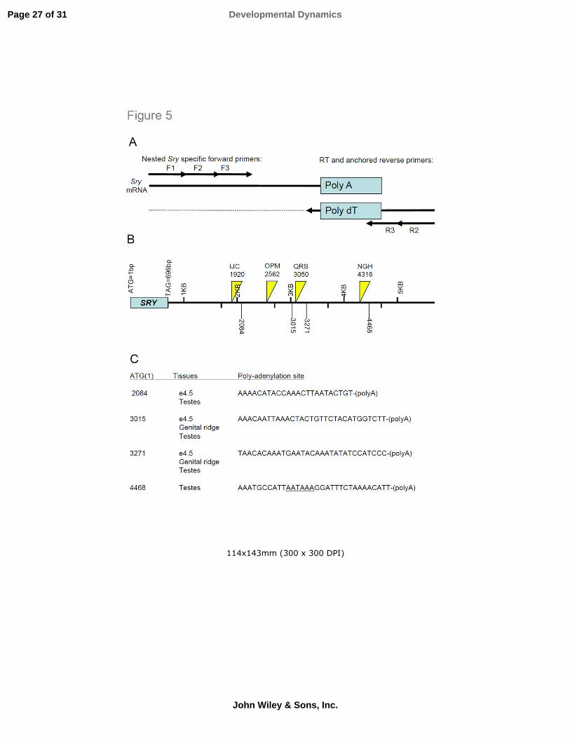
In e4.5 embryos, linear Sry transcripts are produced via usage of multiple polyadenylation
sites.
In order to support our observations described above and further characterize the previously
described Sry expression in the mouse blastocyst, we devised a RT-PCR approach for
determining which of the two known poly-adenylation sites is involved at this stage (Hacker et
al., 1995; Jeske et al., 1995). To this end, a single sided nested RT-PCR procedure was
designed to overcome difficulties with working with mouse Sry transcripts (Fig. 5A-B). Sry is
an intronless gene, so that genomic DNA and cDNA sequences are difficult to distinguish and
contaminating genomic DNA can result in false positive results. To avoid problems with
genomic DNA contamination, all RNA samples were treated with DNAse. The mouse Sry
locus is flanked by inverted genomic repeat elements, and a feature of mouse Sry RNA is a
circular, non-polyadenylated transcript that can be found in both adult and embryonic tissues
including the blastocyst (Capel et al., 1993; Cao et al., 1995). To prevent PCR amplification
from circular Sry transcripts, we performed the RT reaction using a poly-dT primer which
should only prime polyadenylated RNA sequences, and then performed subsequent PCR
reactions using non-specific reverse primers. As indicated in Fig. 5C, our analysis first revealed
that a subset of Sry transcripts from e4.5 blastocysts, as well as those found in e11.5 genital
ridges and adult testes, use the polyadenylation site previously reported by Jeske et al. (1995) at
3271 bp from the ATG start site. A novel polyadenylation site for these three tissues was also
observed at 3015 bp from the ATG site. Moreover, for e4.5 embryos and adult testes, an
additional novel polyadenylation site was observed at 2084 bp from the ATG start site. Of note,
the site reported by Hacker et al. (1995), at 4468 from the ATG start site, was only observed in
adult testes. Taken together, our data confirm that the Sry promoter is active in mouse
blastocysts and further indicate that different linear transcripts (compatible with translation) are
produced.
Page 5 of 31 Developmental Dynamics
John Wiley & Sons, Inc.

At the pro-amniotic cavity stage of the peri-implantation embryo, male embryos tend to be
developmentally more advanced compared to female embryos.
Interestingly, from observing numerous e5.5 day embryos from the SRYp[hybrid]-RFP line,
it became evident that within a given gestation, embryos could be slightly more (or less)
advanced developmentally (data not shown). For example, the pro-amniotic cavity could be
absent (indicating a younger embryo), present but not well developed, or present and well
defined (indicating a slightly older embryo). Furthermore, we found that fluorescence peaked
with the appearance of the pro-amniotic cavity and gradually became reduced with further
embryo development. These observations raise the intriguing possibility that SRY reporters can
be used as markers of maturation and/or early differentiation of the inner cell mass.
To verify this possibility, the SRYp[hybrid]-RFP reporter was used to study the well
documented observations that male pre-implantation embryos are developmentally more
advanced compared to female pre-implantation embryos (Tsunoda et al., 1985; Burgoyne,
1993; Cassar et al., 1994; Pergament et al., 1994; Cassar et al., 1995; Bernardi and Delouis,
1996). More precisely, the SRYp[hybrid]-RFP mouse line was used in a series of experiments
to demonstrate that, at e5.5, embryos that were more developmentally advanced tended to be
male (Fig. 6). In order to sex the embryos, we took advantage of a genetic marker consisting of
an X chromosome integrated GFP transgene driven by 5 kb of the rat Gata4 promoter
(Gata4p[5kb]-GFP(2)) (Cory et al., 2007; Mazaud Guittot et al., 2007). Briefly, when this
transgene is introduced from the paternal side, only XX embryos will receive it. Since this
transgene generates discernable fluorescence within the pre-implantation embryo (Pilon et al.,
2008), when this breeding strategy is followed, embryos at e5.5 that show GFP fluorescence are
female. Based on morphological criteria (size, presence of pro-amniotic cavity, organization of
the epiblast cell layer), embryos were ranked as being developmentally more or less advanced.
From 7 gestations and 58 embryos total (of which 58.6% were male and 41.4% were female),
22 embryos were ranked as developmentally advanced. Of these, 18 were male and 4 were
female. Interestingly, in early e5.5 gestations when developmentally advanced embryos are just
beginning to form the pro-amniotic cavity and concomitantly show peak expression of the RFP
fluorescence (due to the hybrid SRY promoter-RFP transgene), strong RFP fluorescence usually
indicates male embryos. These observations support the idea that SRY reporters can be used as
epiblast maturation/differentiation markers.
Within the epiblast of the peri-implantation embryo, SRY and GATA4 reporters mark the
same cell populations whereas SRY and OCT4 reporters mark different cell populations.
To further confirm that SRY reporters are markers of maturation and early differentiation
within the inner cell mass, mice homozygote for SRYp[hybrid]-RFP transgene were mated to
mice homozygote for an autosomal Gata4 reporter (Gata4p[5kb]-GFP(6a)), and embryos were
collected at e5.5 of gestation. GATA4 and GATA6 are considered key regulators of early
differentiation within the inner cell mass by triggering differentiation of the primitive endoderm
Page 6 of 31Developmental Dynamics
John Wiley & Sons, Inc.

(Soudais et al., 1995; Fujikura et al., 2002; Chazaud et al., 2006) and we have recently found
that the sequences necessary to drive expression of Gata4 in the primitive endoderm are
contained within its 5kb proximal promoter (Pilon et al., 2008). Interestingly, we have also
found that such Gata4 reporters additionally drive gene expression within epiblast cells not
necessary committed to the primitive endoderm lineage although the functional significance of
this observation is currently not known (Pilon et al., 2008). SRYp[hybrid]-RFP::Gata4p[5kb]-
GFP(6a) double transgenic embryos were examined at e5.5 via confocal microscopy and it was
observed that GFP and RFP emission patterns, when merged, mirrored each other in both
distribution and relative intensity (Fig. 7A-D). This experiment was then repeated using two
additional independent reporter mouse lines, to generate hSRYp[5 Kb]-YFP::Gata4p[5kb]-
RFP(2) double transgenic embryos. When these embryos were examined at e5.5 via confocal
microscopy, once again the GFP and RFP emission patterns mirrored each other (Fig. 7E-H).
Thus, these observations are in accordance with the possibility that both promoters (Gata4 and
Sry) are upregulated in differentiating cells of the maturating epiblast.
The same breeding scheme was then used to verify if a converse relationship can be seen
between the relative distribution of SRY promoter activity and a marker of pluripotency such as
Oct4 (Boyer et al., 2005). To this end, mice homozygote for the SRYp[hybrid]-RFP transgene
were mated to mice homozygote for an Oct4 promoter-GFP transgene (Anderson et al., 2000).
Embryos were collected at e5.5 of gestation and examined via confocal microscopy (Fig. 8). It
was now observed that GFP and RFP emission patterns in cells of the epiblast were mainly
discordant in both distribution and relative intensity: a population of cells was predominantly
green (Oct4 promoter transgene dominance), a second population of cells was predominantly
red (SRY promoter transgene dominance), while a third minority population of cells was yellow,
indicating overlapping GFP and RFP emission patterns. Taken together, these observations
suggest that, within the mammalian pre-implantation embryo, SRY and Gata4 promoter
activities (and by extension gene activities) share a developmental pathway which is divergent
from a developmental pathway involving Oct4 activity.
Overexpression of Sry in ES cells leads to downregulation of pluripotency genes.
The observations that the activity of the SRY/Sry promoter from multiple species correlates
with early differentiation of epiblast cells suggest that the SRY/Sry gene product may be
involved in this process by interfering with the activity of the closely related SOX2/Sox2.
Indeed, SOX2 is a well-known stemness factor that forms, together with OCT4 and NANOG
the core pluripotency network of ES cells (Boyer et al., 2005; Loh et al., 2006; Masui et al.,
2007). Interestingly, within this network, these transcription factors are also known to regulate
each other’s expression via particular composite OCT-SOX enhancers in proximity to NANOG
binding sites (Boyer et al., 2005; Masui et al., 2007; Chambers and Tomlinson, 2009). Thus, in
order to gain insights into the putative role of SRY/Sry in the epiblast, we transiently
overexpressed mouse Sry in undifferentiated R1 ES cells and assessed the expression levels of
the pluripotency genes Oct4, Sox2 and Nanog via semi-quantitative RT-PCR. To this end, we
Page 7 of 31 Developmental Dynamics
John Wiley & Sons, Inc.

also took advantage of a previously described bicistronic DomSry-IRES-GFP expression vector
allowing FACS-mediated recovery of transfected cells (Lau and Li, 2009). As shown in Fig. 9,
this analysis reveals that expression of Oct4 is unaffected in ES cells grown under
undifferentiating conditions but specifically overexpressing Sry. However, in the same
conditions, expression of Nanog and Sox2 is markedly reduced. These observations suggest that
the Oct-Sox enhancer of the Oct4 promoter may behave differently than the Oct-Sox enhancer
of both the Nanog and Sox2 promoter. Most importantly, they also strongly suggest that
SRY/Sry proteins might be involved in epiblast differentiation at least by promoting exit of
pluripotency.
DISCUSSION
In mammals, there are examples of developmental sexual dimorphisms that are evident
before gonadal sex determination and development (Erickson, 1997). Since these differences
occur before the production of dimorphic sex steroids by the gonads, there must be a genetic
rather than a hormonal explanation. An example of sexual dimorphism is the observation that
male pre-implantation embryos are developmentally more advanced compared to female pre-
implantation embryos. This was first reported in mice via classical re-implantation studies
which showed that developmentally more advanced pre-implantation embryos were enriched
for XY genotypes (Tsunoda et al., 1985; reviewed by Mittwoch, 1993). Follow up studies in
the mouse (Burgoyne, 1993) confirmed that the male pre-implantation embryo could be
developmentally more advanced and furthermore that this depended on the Y chromosome
haplotype. In vitro work with bovine embryos demonstrated more rapid development of male
pre-implantation embryos, including increased cell numbers and mitotic indices (Xu et al.,
1992). In vivo studies of pig embryos showed that male embryos were developmentally more
advanced compared to female embryos at days 5, 6 and 10 of pregnancy (Cassar et al., 1994;
Cassar et al., 1995). In studies of human pre-implantation embryos fertilized in vitro, it was
concluded that the presence of the Y chromosome causes accelerated growth rates immediately
after fertilization (Pergament et al., 1994). Similarly, in in vitro fertilized and matured ovine
embryos, fast developing embryos were found to be predominantly male (Bernardi and Delouis,
1996). At present, these observations are poorly understood at a mechanistic level. However,
recent advances in the field of embryonic stem cells may now provide the basis for a
mechanism to explain sex dimorphisms within the pre-implantation embryo.
Embryonic stem (ES) cells, whether of mouse or human origin, are pluripotent, non
immortalized cells that can be grown in tissue culture and may be equivalent to epiblast cells of
the mouse embryo, or primitive streak cells of the human embryo (Rossant, 2008).
Understanding how these cells maintain their pluripotency and how they undergo differentiation
has increased considerably in recent years. Indeed, this knowledge has been used to
successfully reprogram differentiated somatic cells into induced pluripotent stem (iPS) cells
displaying ES cell-like phenotypes (recently reviewed in Maury et al., 2012). The implication
Page 8 of 31Developmental Dynamics
John Wiley & Sons, Inc.

of this work for understanding in vivo embryo development as well as for providing potential
clinical applications has been noted (Jaenisch and Young, 2008; Rossant, 2008; Maury et al.,
2012). A core regulatory circuitry of transcription factors within ES cells is proposed to
maintain the pluripotent state, involving at a minimum the proteins OCT4, SOX2, and NANOG
(Boyer et al., 2005; Loh et al., 2006; Masui et al., 2007). These factors interact with their own
and each other’s promoters, forming a self perpetuating auto-regulatory loop (Boyer et al.,
2005; Chew et al., 2005; Chambers and Tomlinson, 2009). OCT4 and SOX2 proteins can
physically dimerize with each other via their respective DNA binding domains and this
interaction is thought to stabilize OCT4 binding on DNA (Remenyi et al., 2003; Williams et al.,
2004; Chambers and Tomlinson, 2009). This SOX2/OCT4 dimer can activate several gene
promoters, via binding of both proteins on a particular composite OCT-SOX element (Tomioka
et al., 2002; Chambers and Tomlinson, 2009). The vast majority of the promoters regulated by
this OCT-SOX element also contain a NANOG binding site in close proximity and these
include the promoter of OCT4, SOX2 and NANOG themselves (Boyer et al., 2005). The
autoregulatory loop formed by these three transcription factors is critically important for the
stability of the self-renewal circuitry and, as such, is maintained within tight limits that can be
subject to genetic or epigenetic perturbations. Slight increases or decreases in the expression of
OCT4, NANOG, or SOX2 can cause ES cells to exit the self-renewal circuit, thus triggering
differentiation pathways (Fong et al., 2008; Kopp et al., 2008). Thus, SOX2 is considered a
stemness master gene (Harley and Lefebvre, 2010), helping to maintain stemness in ES cells
and pluripotency in epiblast cells (Avilion et al., 2003). It is noteworthy that SOX2 can also
maintain stem cell status during neurogenesis, and that decreased SOX2 levels results in
increased nerve cell differentiation (Graham et al., 2003).
SRY/Sry expression has been reported in the pre-implantation embryo in several mammalian
species (Zwingman et al., 1993; Ao et al., 1994; Boyer and Erickson, 1994; Cao et al., 1995;
Fiddler et al., 1995; Gutierrez-Adan et al., 1997). We confirmed such observations for the
mouse blastocyst and also found that different linear Sry transcripts compatible with translation
are produced at this stage. Results from our in vivo cell lineage study using Cre recombinase
under the control of 4.6 kb of pig SRY promoter sequences, although precluding the utility of
this model for specific site directed excision in pre-Sertoli cells within the developing genital
ridge, now provide further evidence that the SRY promoter can be active at an early stage in
embryo development. Continued work with this model showed that, reminiscent of the pattern
we have reported for Gata4Cre-mediated lineage studies (Pilon et al., 2008), the SRY promoter is
active within epiblast cells of the blastocyst stage embryo. Using transgenic fluorescence
marker models, we confirmed that the human, pig and mouse SRY promoters contain sequence
information that can support transcriptional activity within the inner cell mass and epiblast of
the pre-implantation blastocyst, and that such transgene expression is possible in both male and
female embryos. We have previously shown that the SRY promoter is synergistically responsive
to GATA4 and WT1 levels (Miyamoto et al., 2008), and furthermore that Gata4 is expressed
within epiblast cells of the pre-implantation embryo (Pilon et al., 2008). GATA4/Gata4 and
Page 9 of 31 Developmental Dynamics
John Wiley & Sons, Inc.
WT1/Wt1 transcripts were also previously observed in undifferentiated human and mouse ES
cells (Brandenberger et al., 2004; Wei et al., 2005; Pilon et al., 2008). Collectively, these
observations suggest that transcription factors capable of driving SRY promoter activity are
present within cells of the inner cell mass of both the male and female blastocyst stage embryos.
Male specificity of SRY expression in the early embryo is obviously due to the fact that SRY is a
Y chromosome located gene and, as such, is not found in XX embryos.
In mammals, it has been long observed that the male (XY) embryo tends to be
developmentally more advanced in comparison to the female (XX) embryo (Mittwoch, 1993).
Differences include embryo size, weight, number of somites, body length in post implantation
embryos as well as cell number and stage of blastocyst maturity in pre-implantation embryos
(Burgoyne, 1993). Burgoyne (1993) demonstrated that there is a positive Y chromosome effect
on blastocyst cell numbers in mice, and furthermore that this effect can be strain and allele
dependent. In the same year, Zwingman et al. (1993) reported that Y chromosome genes,
including Sry and Zfy, are expressed within the male pre-implantation embryo as early as the 2
cell stage, and speculated that these factors may provide what has been called “growth factor
Y”. Further work by Burgoyne and coworkers (Thornhill and Burgoyne, 1993; Burgoyne et al.,
1995) demonstrated, in post-implantation mouse embryos (e10.5), that a growth inhibiting X
chromosome effect is present in XX embryos in addition to the growth accelerating effect of the
Y chromosome seen in XY embryos. Using genetic models, these investigators were able to
show that Sry is not responsible for the Y chromosome effect seen during this post-implantation
time period. Although not supported by genetic models, it was further suggested that Sry would
have little role to play in the Y chromosome effect observed during the pre-implantation of XY
embryo. As suggested by Mittwoch (1993), we rather believe that the accelerated growth rate
and increased maturity observed in male pre-implantation embryos is most likely due to a
combination of genetic mechanisms. As recently presented by Arnold (2012), the sex
chromosome complement can have differential effects on XX and XY embryos via several
genetic mechanisms, including: male exclusive genes, X inactivation and dosage effects, X
imprinting effects as well as X (or Y) heterochromatin sink effects (Arnold, 2012). Different
combinations of these genetic effects could well be responsible for the differential growth and
development seen in embryos of different ages, and it may not be prudent to equate post-
implantation embryos with pre-implantation embryos.
Our present findings suggest that the potential role of SRY within the pre-implantation XY
embryo should now be re-examined. In view of the importance of SOX2 levels for the
maintenance of stem cell potential in vitro and for making early lineage fate decisions in vivo
(Fong et al., 2008; Kopp et al., 2008), our data raise the possibility that the transcriptional
activity of the closely related SRY within cells of the inner cell mass of pre-implantation male
embryos may influence embryo development in a sexually dimorphic fashion. This possibility
is supported by our semi-quantitative RT-PCR data and may provide a molecular explanation
for the observations that pre-implantation male embryos in a number of mammalian species are
Page 10 of 31Developmental Dynamics
John Wiley & Sons, Inc.

developmentally more advanced compared to their female counterparts (Tsunoda et al., 1985;
Xu et al., 1992; Pergament et al., 1994; Cassar et al., 1995; Bernardi et al., 1996). All SOX
proteins including SRY recognize the same core DNA binding motif. Thus, the SRY protein
might compete with SOX2 via its HMG-box domain for the shared DNA binding sites of SOX
proteins found within the promoters of SOX2 target genes which include the promoters of
OCT4, NANOG and SOX2 itself (Boyer et al., 2005; Loh et al., 2006). In agreement with this
possibility, previous work with murine Sox2-null ES cells has shown that other Sox proteins
expressed in ES cells (Sox4, Sox11 and Sox15) can also fulfill an activating role in the
regulation of otherwise Sox2-regulated enhancers (Masui et al., 2007). Mechanistically, binding
of SRY on such SOX elements might thus interfere with the positive SOX2 (and other SOX)
activity either by passive competition or active repression (Lau and Li, 2009; Peng et al., 2009).
Additionally, given the high homology of the HMG box domain between SRY and SOX2, SRY
is also predicted to interact with OCT4 on composite OCT-SOX elements (Remenyi et al.,
2003). This possibility is supported by ChIP-chip experiments performed in Ntera2 testicular
embryonal carcinoma cells indicating that OCT4 and SRY colocalize on hundreds of target
promoters (Jin et al., 2007). Interestingly, precedents for both of these mechanisms are also
provided by the competitive inhibition of SOX E family proteins by SOX D family proteins in
oligodendrocytes (Stolt et al., 2006).
In conclusion, the possibility that SRY has a biological function beyond TDF and sex
determination, i.e. that of a genetic male specific maturation factor within cells of the inner cell
mass of the pre- and peri-implantation blastocyst, provides new insights and raises new
questions about this intriguing mammalian specific, male only molecule.
EXPERIMENTAL PROCEDURES
Mouse lines and trials
The production of mouse lines transgenic for the human SRY promoter [5 kb]-YFP, Hybrid SRY
promoter-RFP, rat Gata4 promoter [5 kb]-GFP with transgene integration on the X
chromosome (used for sexing embryos), and rat Gata4 promoter [5 kb]-RFP has been
previously described (Boyer et al., 2006; Mazaud Guittot et al., 2007; Pilon et al., 2008). All
mice used were of FVB/n genetic background. The Cre reporter R26R-YFP mouse line was
originally provided by Frank Costantini (Srinivas et al., 2001); we subsequently placed the
marker allele onto a FVB/n genetic background via backcrossing. Oct4-GFP marker mice
(Anderson et al., 2000) were a gift from Chris Wylie; the Oct4-GFP marker transgene was
introduced onto the FVB/n genetic background via backcrossing. The human SRY promoter [5
Kb]-YFP, SRY promoter [hybrid]-RFP, rat Gata4 promoter [5 Kb]-RFP, and Oct4-GFP lines
were all maintained as homozygous lines; thus appropriate matings for double marking trials
resulted in all embryos being doubly heterozygote for the appropriate transgenes. For staged
embryo collections, females were housed with fertile males, and noon on the day when a
Page 11 of 31 Developmental Dynamics
John Wiley & Sons, Inc.

seminal plug was observed was designated as e0.5. Embryos at e4.5 were collected by uterine
flushing using PBS; embryos at later stages were recovered via dissection.
Pig SRY promoter [4.6 kb]-Cre mouse
Using standard molecular biology construction techniques, a pig SRY promoter [4.6 kb]-GFP
genetic construction (Daneau et al., 2002) was modified such that the GFP coding sequence was
replaced by Cre recombinase coding sequences (Boyer et al., 2002). The resulting transgene,
designated pSRYp[4.6kb]-Cre, was microinjected into the male pronucleus of single celled
FVB/n embryos, and transgenic mice were generated using standard methodologies (Daneau et
al., 2002; Boyer et al., 2006). A tyrosinase minigene was co-injected to provide visual
identification of transgenic mice. Pigmented founder animals were bred to FVB/n non-
transgenic mice to insure single integration sites (Methot et al., 1995). Co-integration of Cre
sequences was verified by PCR analysis on genomic DNA (data not shown). Three lines were
retained and mated to the Cre reporter R26R-YFP mouse line; the activity of the Cre
recombinase was verified by observing newborn tissues of double transgenic animals with
fluorescence optics. All lines showed similar results; one line was retained for further studies.
Microscope studies and imaging
Newborn mice were photographed with a Leica MZFLIII stereomicroscope equipped with
fluorescence excitation and filters for GFP capture. Image acquisition was via a Nikon
DXM1200 digital camera, using Nikon Act-1 software. For visible light, fluorescence and
confocal imaging, a Nikon Eclipse E800 microscope was used that was equipped for
fluorescence excitation and with filters for GFP and RFP capture, and connected to a Nikon C1
confocal imaging system. The confocal system had an argon laser for excitation and filters for
GFP and YFP emission capture, as well as a green helium-neon laser for excitation and filters
for RFP emission capture.
Mapping of Sry polyadenylation sites in e4.5 mouse embryos.
For pre-implantation embryos, 232 day e4.5 embryos were collected by uterine flushing and
processed without attempting to sex the embryos. For collecting e11.5 male genital ridges,
matings were performed using a transgenic mouse line incorporating a Gata4 promoter-GFP
and tyrosinase transgenes incorporated on the X chromosome; sexing of embryos was
performed by the presence in females (and absence in males) of fluorescence and pigmentation
(Cory et al. 2007). Genital ridges from 34 male embryos at age e11.5 were dissected and
pooled. For adult testes, tissues were homogenized using a Polytron PT1300D apparatus with a
5mm probe. For all tissues, total RNA was then extracted using a RNeasy mini kit (Qiagen).
The samples were digested with DNAse enzyme, using RNase-free DNase Set (Qiagen),
according to the supplier’s instructions. Total RNA was then further processed to derive mRNA
using a Oligotex Direct mini kit (Qiagen), again according to the manufacturer’s instructions.
Reverse transcription was performed using Omniscript RT enzyme (Qiagen), incubating
samples at 37˚C for 1 hr, in the presence of a poly-dT primer (Reverse Primer 1, see Table1).
Page 12 of 31Developmental Dynamics
John Wiley & Sons, Inc.

An anchored, nested PCR procedure was devised, as presented in Fig. 5. Specific forward
nested primer triplets, and non-specific reverse primers, are presented in Table 1. All PCRs
consisted of cycles of denaturation at 95˚C for 30 sec, annealing at 60°C for 30 sec, and
elongation at 72˚C for 90 sec. PCR reactions were performed in a total reaction volume of
100µl. A first PCR reaction of 20 cycles was performed using a specific forward primer and
reverse primer 2. Five (5) µl of this completed reaction was used to seed the second reaction of
30 cycles using the first nested specific forward primer and reverse primer 3; 5µl of this
completed reaction was used to seed the third reaction of 30 cycles using the second nested
specific forward primer and reverse primer 3. PCR products were visualized on a 1% agarose
gel with ethidium bromide. PCR products were then subcloned into pDrive plasmid vector
(Qiagen), and plasmid miniprep DNA prepared and analyzed for insert size. Selected plasmids
were sequenced and insert sequences used to search mouse genomic sequences using the Blastn
function of Ensembl.org; positive hits for Sry sequences were recorded.
Semi-quantitative RT-PCR assays in ES cells
R1 ES cells were grown in standard ES cell medium containing LIF onto gelatin-coated dishes
as previously described (Pilon et al., 2008). Approximately 105 cells were transfected with 5µg
of either DomSry-IRES-GFP or empty IRES-GFP control vector. Transfections were performed
with Genejuice reagents (Novagen) in accordance with manufacturer’s instruction and
transfected cells were cultivated for 48h in complete ES cell medium. At the end of the
cultivation period, cells were harvested and GFP-positive cells recovered by FACS. For each
condition, mRNA was isolated from 3x10⁴ cells with the Oligotex Direct mRNA kit (Qiagen)
and reverse transcription was performed with an oligodT primer and Superscript II reagents
(Invitrogen). For PCR amplifications, with the sole exception of Sox2, primers were designed to
encompass an intron, allowing the detection of contaminating genomic DNA by the presence of
a larger band. Specific pairs of oligonucleotides and expected size of the amplicons are listed in
Table 2. PCR amplifications were performed with Platinum Taq (Invitrogen) and consisted of
20 to 35 cycles of 35 sec at 96°C, 30 sec at 61°C, and 45 sec at 68°C. PCR products were size
fractionated on a 1.5% agarose gel. PCR analyses were performed three times from three
independent reverse transcriptions.
ACKNOWLEDGEMENTS
The authors thank Denis Flipo (UQAM) for the FACS analyses, the DNA Diagnostic Lab
(Faculty of Veterinary Medicine, University of Montreal) for sequencing and Celine Forget for
her technical assistance in the production of the pig SRY promoter [4.6]-Cre mouse line. Chris
Lau is thanked for the DomSry-IRES-EGFP plasmid while Frank Costantini and Chris Wylie
are thanked for the R26R-EYFP and Oct4-GFP mouse lines, respectively. NP is a Fonds de la
Recherche en Santé du Québec (FRSQ) Jr1 scholar and RV holds a Canada Research Chair in
Reproduction and Sex Development.
Page 13 of 31 Developmental Dynamics
John Wiley & Sons, Inc.

REFERENCES
Anderson R, Copeland TK, Scholer H, Heasman J, Wylie C. 2000. The onset of germ cell migration in the mouse embryo. Mech Dev 91:61-68.
Ao A, Erickson RP, Winston RM, Handyside AH. 1994. Transcription of paternal Y-linked genes in the human zygote as early as the pronucleate stage. Zygote 2:281-287.
Arnold AP. 2012. The end of gonad-centric sex determination in mammals. Trends Genet 28:55-61.
Avilion AA, Nicolis SK, Pevny LH, Perez L, Vivian N, Lovell-Badge R. 2003. Multipotent cell lineages in early mouse development depend on SOX2 function. Genes Dev 17:126-140.
Bernardi ML, Delouis C. 1996. Sex-related differences in the developmental rate of in-vitro matured/in-vitro fertilized ovine embryos. Hum Reprod 11:621-626.
Bowles J, Schepers G, Koopman P. 2000. Phylogeny of the SOX family of developmental transcription factors based on sequence and structural indicators. Dev Biol 227:239-255.
Boyer A, Pilon N, Raiwet DL, Lussier JG, Silversides DW. 2006. Human and pig SRY 5' flanking sequences can direct reporter transgene expression to the genital ridge and to migrating neural crest cells. Dev Dyn 235:623-632.
Boyer LA, Lee TI, Cole MF, Johnstone SE, Levine SS, Zucker JP, Guenther MG, Kumar RM, Murray HL, Jenner RG, Gifford DK, Melton DA, Jaenisch R, Young RA. 2005. Core transcriptional regulatory circuitry in human embryonic stem cells. Cell 122:947-956.
Boyer TR, Erickson RP. 1994. Detection of circular and linear transcripts of Sry in pre-implantation mouse embryos: differences in requirement for reverse transcriptase. Biochem Biophys Res Commun 198:492-496.
Brandenberger R, Khrebtukova I, Thies RS, Miura T, Jingli C, Puri R, Vasicek T, Lebkowski J, Rao M. 2004. MPSS profiling of human embryonic stem cells. BMC Dev Biol 4:10.
Bullejos M, Koopman P. 2001. Spatially dynamic expression of Sry in mouse genital ridges. Dev Dyn 221:201-205.
Burgoyne PS. 1993. A Y-chromosomal effect on blastocyst cell number in mice. Development 117:341-345.
Burgoyne PS, Thornhill AR, Boudrean SK, Darling SM, Bishop CE, Evans EP. 1995. The genetic basis of XX-XY differences present before gonadal sex differentiation in the mouse. Philos Trans R Soc Lond B Biol Sci 350:253-260 discussion 260-251.
Cameron FJ, Sinclair AH. 1997. Mutations in SRY and SOX9: testis-determining genes. Hum Mutat 9:388-395.
Cao QP, Gaudette MF, Robinson DH, Crain WR. 1995. Expression of the mouse testis-determining gene Sry in male preimplantation embryos. Mol Reprod Dev 40:196-204.
Capel B, Swain A, Nicolis S, Hacker A, Walter M, Koopman P, Goodfellow P, Lovell-Badge R. 1993. Circular transcripts of the testis-determining gene Sry in adult mouse testis. Cell 73:1019-1030.
Cassar G, de la Fuente R, Yu Z, King GJ, King WA. 1995. Sex chromosome complement and developmental diversity in pre-and post-hatching porcine embryos. Theriogenology 44:879-884.
Cassar G, King WA, King GJ. 1994. Influence of sex on early growth of pig conceptuses. J Reprod Fertil 101:317-320.
Chambers I, Tomlinson SR. 2009. The transcriptional foundation of pluripotency. Development 136:2311-2322.
Page 14 of 31Developmental Dynamics
John Wiley & Sons, Inc.
Chazaud C, Yamanaka Y, Pawson T, Rossant J. 2006. Early lineage segregation between epiblast and primitive endoderm in mouse blastocysts through the Grb2-MAPK pathway. Dev Cell 10:615-624.
Chew JL, Loh YH, Zhang W, Chen X, Tam WL, Yeap LS, Li P, Ang YS, Lim B, Robson P, Ng HH. 2005. Reciprocal transcriptional regulation of Pou5f1 and Sox2 via the Oct4/Sox2 complex in embryonic stem cells. Mol Cell Biol 25:6031-6046.
Clepet C, Schafer AJ, Sinclair AH, Palmer MS, Lovell-Badge R, Goodfellow PN. 1993. The human SRY transcript. Hum Mol Genet 2:2007-2012.
Cory AT, Boyer A, Pilon N, Lussier JG, Silversides DW. 2007. Presumptive pre-sertoli cells express genes involved in cell proliferation and cell signalling during a critical window in early testis differentiation. Mol Reprod Dev 74:1491-1504.
Daneau I, Pilon N, Boyer A, Behdjani R, Overbeek PA, Viger R, Lussier J, Silversides DW. 2002. The porcine SRY promoter is transactivated within a male genital ridge environment. Genesis 33:170-180.
Dasari VK, Goharderakhshan RZ, Perinchery G, Li LC, Tanaka Y, Alonzo J, Dahiya R. 2001. Expression analysis of Y chromosome genes in human prostate cancer. J Urol 165:1335-1341.
Dewing P, Chiang CW, Sinchak K, Sim H, Fernagut PO, Kelly S, Chesselet MF, Micevych PE, Albrecht KH, Harley VR, Vilain E. 2006. Direct regulation of adult brain function by the male-specific factor SRY. Curr Biol 16:415-420.
Ely D, Milsted A, Bertram J, Ciotti M, Dunphy G, Turner ME. 2007. Sry delivery to the adrenal medulla increases blood pressure and adrenal medullary tyrosine hydroxylase of normotensive WKY rats. BMC Cardiovasc Disord 7:6.
Erickson RP. 1997. Does sex determination start at conception? Bioessays 19:1027-1032. Fiddler M, Abdel-Rahman B, Rappolee DA, Pergament E. 1995. Expression of SRY transcripts
in preimplantation human embryos. Am J Med Genet 55:80-84. Fong H, Hohenstein KA, Donovan PJ. 2008. Regulation of self-renewal and pluripotency by
Sox2 in human embryonic stem cells. Stem Cells 26:1931-1938. Fujikura J, Yamato E, Yonemura S, Hosoda K, Masui S, Nakao K, Miyazaki Ji J, Niwa H.
2002. Differentiation of embryonic stem cells is induced by GATA factors. Genes Dev 16:784-789.
Graham V, Khudyakov J, Ellis P, Pevny L. 2003. SOX2 functions to maintain neural progenitor identity. Neuron 39:749-765.
Gutierrez-Adan A, Behboodi E, Murray JD, Anderson GB. 1997. Early transcription of the SRY gene by bovine preimplantation embryos. Mol Reprod Dev 48:246-250.
Hacker A, Capel B, Goodfellow P, Lovell-Badge R. 1995. Expression of Sry, the mouse sex determining gene. Development 121:1603-1614.
Hanley NA, Hagan DM, Clement-Jones M, Ball SG, Strachan T, Salas-Cortes L, McElreavey K, Lindsay S, Robson S, Bullen P, Ostrer H, Wilson DI. 2000. SRY, SOX9, and DAX1 expression patterns during human sex determination and gonadal development. Mech Dev 91:403-407.
Harley V, Lefebvre V. 2010. Twenty Sox, twenty years. Int J Biochem Cell Biol 42:376-377. Harry JL, Koopman P, Brennan FE, Graves JA, Renfree MB. 1995. Widespread expression of
the testis-determining gene SRY in a marsupial. Nat Genet 11:347-349. Jaenisch R, Young R. 2008. Stem cells, the molecular circuitry of pluripotency and nuclear
reprogramming. Cell 132:567-582.
Page 15 of 31 Developmental Dynamics
John Wiley & Sons, Inc.

Jeske YW, Bowles J, Greenfield A, Koopman P. 1995. Expression of a linear Sry transcript in the mouse genital ridge. Nat Genet 10:480-482.
Jin VX, O'Geen H, Iyengar S, Green R, Farnham PJ. 2007. Identification of an OCT4 and SRY regulatory module using integrated computational and experimental genomics approaches. Genome Res 17:807-817.
Kamachi Y, Uchikawa M, Tanouchi A, Sekido R, Kondoh H. 2001. Pax6 and SOX2 form a co-DNA-binding partner complex that regulates initiation of lens development. Genes Dev 15:1272-1286.
Kiefer JC. 2007. Back to basics: Sox genes. Dev Dyn 236:2356-2366. Koopman P, Gubbay J, Vivian N, Goodfellow P, Lovell-Badge R. 1991. Male development of
chromosomally female mice transgenic for Sry. Nature 351:117-121. Koopman P, Schepers G, Brenner S, Venkatesh B. 2004. Origin and diversity of the SOX
transcription factor gene family: genome-wide analysis in Fugu rubripes. Gene 328:177-186.
Kopp JL, Ormsbee BD, Desler M, Rizzino A. 2008. Small increases in the level of Sox2 trigger the differentiation of mouse embryonic stem cells. Stem Cells 26:903-911.
Lahr G, Maxson SC, Mayer A, Just W, Pilgrim C, Reisert I. 1995. Transcription of the Y chromosomal gene, Sry, in adult mouse brain. Brain Res Mol Brain Res 33:179-182.
Lau YF, Li Y. 2009. The human and mouse sex-determining SRY genes repress the Rspol/beta-catenin signaling. J Genet Genomics 36:193-202.
Lau YF, Zhang J. 2000. Expression analysis of thirty one Y chromosome genes in human prostate cancer. Mol Carcinog 27:308-321.
Loh YH, Wu Q, Chew JL, Vega VB, Zhang W, Chen X, Bourque G, George J, Leong B, Liu J, Wong KY, Sung KW, Lee CW, Zhao XD, Chiu KP, Lipovich L, Kuznetsov VA, Robson P, Stanton LW, Wei CL, Ruan Y, Lim B, Ng HH. 2006. The Oct4 and Nanog transcription network regulates pluripotency in mouse embryonic stem cells. Nat Genet 38:431-440.
Masui S, Nakatake Y, Toyooka Y, Shimosato D, Yagi R, Takahashi K, Okochi H, Okuda A, Matoba R, Sharov AA, Ko MS, Niwa H. 2007. Pluripotency governed by Sox2 via regulation of Oct3/4 expression in mouse embryonic stem cells. Nat Cell Biol 9:625-635.
Maury Y, Gauthier M, Peschanski M, Martinat C. 2012. Human pluripotent stem cells for disease modelling and drug screening. Bioessays 34:61-71.
Mayer A, Lahr G, Swaab DF, Pilgrim C, Reisert I. 1998. The Y-chromosomal genes SRY and ZFY are transcribed in adult human brain. Neurogenetics 1:281-288.
Mayer A, Mosler G, Just W, Pilgrim C, Reisert I. 2000. Developmental profile of Sry transcripts in mouse brain. Neurogenetics 3:25-30.
Mazaud Guittot S, Tetu A, Legault E, Pilon N, Silversides DW, Viger RS. 2007. The proximal Gata4 promoter directs reporter gene expression to sertoli cells during mouse gonadal development. Biol Reprod 76:85-95.
Mittwoch U. 1993. Blastocysts prepare for the race to be male. Hum Reprod 8:1550-1555. Miyamoto Y, Taniguchi H, Hamel F, Silversides DW, Viger RS. 2008. A GATA4/WT1
cooperation regulates transcription of genes required for mammalian sex determination and differentiation. BMC Mol Biol 9:44.
Pannetier M, Tilly G, Kocer A, Hudrisier M, Renault L, Chesnais N, Costa J, Le Provost F, Vaiman D, Vilotte JL, Pailhoux E. 2006. Goat SRY induces testis development in XX transgenic mice. FEBS Lett 580:3715-3720.
Page 16 of 31Developmental Dynamics
John Wiley & Sons, Inc.

Peng H, Ivanov AV, Oh HJ, Lau YF, Rauscher FJ, 3rd. 2009. Epigenetic gene silencing by the SRY protein is mediated by a KRAB-O protein that recruits the KAP1 co-repressor machinery. J Biol Chem 284:35670-35680.
Pergament E, Fiddler M, Cho N, Johnson D, Holmgren WJ. 1994. Sexual differentiation and preimplantation cell growth. Hum Reprod 9:1730-1732.
Pilon N, Raiwet D, Viger RS, Silversides DW. 2008. Novel pre- and post-gastrulation expression of Gata4 within cells of the inner cell mass and migratory neural crest cells. Dev Dyn 237:1133-1143.
Remenyi A, Lins K, Nissen LJ, Reinbold R, Scholer HR, Wilmanns M. 2003. Crystal structure of a POU/HMG/DNA ternary complex suggests differential assembly of Oct4 and Sox2 on two enhancers. Genes Dev 17:2048-2059.
Rossant J. 2008. Stem cells and early lineage development. Cell 132:527-531. Salas-Cortes L, Jaubert F, Barbaux S, Nessmann C, Bono MR, Fellous M, McElreavey K,
Rosemblatt M. 1999. The human SRY protein is present in fetal and adult Sertoli cells and germ cells. Int J Dev Biol 43:135-140.
Sinclair AH, Berta P, Palmer MS, Hawkins JR, Griffiths BL, Smith MJ, Foster JW, Frischauf AM, Lovell-Badge R, Goodfellow PN. 1990. A gene from the human sex-determining region encodes a protein with homology to a conserved DNA-binding motif. Nature 346:240-244.
Soudais C, Bielinska M, Heikinheimo M, MacArthur CA, Narita N, Saffitz JE, Simon MC, Leiden JM, Wilson DB. 1995. Targeted mutagenesis of the transcription factor GATA-4 gene in mouse embryonic stem cells disrupts visceral endoderm differentiation in vitro. Development 121:3877-3888.
Srinivas S, Watanabe T, Lin CS, William CM, Tanabe Y, Jessell TM, Costantini F. 2001. Cre reporter strains produced by targeted insertion of EYFP and ECFP into the ROSA26 locus. BMC Dev Biol 1:4.
Stolt CC, Schlierf A, Lommes P, Hillgartner S, Werner T, Kosian T, Sock E, Kessaris N, Richardson WD, Lefebvre V, Wegner M. 2006. SoxD proteins influence multiple stages of oligodendrocyte development and modulate SoxE protein function. Dev Cell 11:697-709.
Sutton E, Hughes J, White S, Sekido R, Tan J, Arboleda V, Rogers N, Knower K, Rowley L, Eyre H, Rizzoti K, McAninch D, Goncalves J, Slee J, Turbitt E, Bruno D, Bengtsson H, Harley V, Vilain E, Sinclair A, Lovell-Badge R, Thomas P. 2011. Identification of SOX3 as an XX male sex reversal gene in mice and humans. J Clin Invest 121:328-341.
Thornhill AR, Burgoyne PS. 1993. A paternally imprinted X chromosome retards the development of the early mouse embryo. Development 118:171-174.
Tomioka M, Nishimoto M, Miyagi S, Katayanagi T, Fukui N, Niwa H, Muramatsu M, Okuda A. 2002. Identification of Sox-2 regulatory region which is under the control of Oct-3/4-Sox-2 complex. Nucleic Acids Res 30:3202-3213.
Tricoli JV, Yao JL, D'Souza SA, Bracken RB. 1993. Detection of sex-region Y (SRY) transcripts in human prostate adenocarcinoma and benign prostatic hypertrophy. Genes Chromosomes Cancer 8:28-33.
Tsunoda Y, Tokunaga T, Sugie T. 1985. Altered sex ratio of live young after transfer of fast- and slow-developing mouse embryos. Gamete Research 12:301-304.
Turner ME, Martin C, Martins AS, Dunmire J, Farkas J, Ely DL, Milsted A. 2007. Genomic and expression analysis of multiple Sry loci from a single Rattus norvegicus Y chromosome. BMC Genet 8:11.
Page 17 of 31 Developmental Dynamics
John Wiley & Sons, Inc.

Veyrunes F, Waters PD, Miethke P, Rens W, McMillan D, Alsop AE, Grutzner F, Deakin JE, Whittington CM, Schatzkamer K, Kremitzki CL, Graves T, Ferguson-Smith MA, Warren W, Marshall Graves JA. 2008. Bird-like sex chromosomes of platypus imply recent origin of mammal sex chromosomes. Genome Res 18:965-973.
Waters PD, Wallis MC, Marshall Graves JA. 2007. Mammalian sex--Origin and evolution of the Y chromosome and SRY. Semin Cell Dev Biol 18:389-400.
Wei CL, Miura T, Robson P, Lim SK, Xu XQ, Lee MY, Gupta S, Stanton L, Luo Y, Schmitt J, Thies S, Wang W, Khrebtukova I, Zhou D, Liu ET, Ruan YJ, Rao M, Lim B. 2005. Transcriptome profiling of human and murine ESCs identifies divergent paths required to maintain the stem cell state. Stem Cells 23:166-185.
Williams DC, Jr., Cai M, Clore GM. 2004. Molecular basis for synergistic transcriptional activation by Oct1 and Sox2 revealed from the solution structure of the 42-kDa Oct1.Sox2.Hoxb1-DNA ternary transcription factor complex. J Biol Chem 279:1449-1457.
Wissmuller S, Kosian T, Wolf M, Finzsch M, Wegner M. 2006. The high-mobility-group domain of Sox proteins interacts with DNA-binding domains of many transcription factors. Nucleic Acids Res 34:1735-1744.
Xu KP, Yadav BR, King WA, Betteridge KJ. 1992. Sex-related differences in developmental rates of bovine embryos produced and cultured in vitro. Mol Reprod Dev 31:249-252.
Yuan H, Corbi N, Basilico C, Dailey L. 1995. Developmental-specific activity of the FGF-4 enhancer requires the synergistic action of Sox2 and Oct-3. Genes Dev 9:2635-2645.
Zwingman T, Erickson RP, Boyer T, Ao A. 1993. Transcription of the sex-determining region genes Sry and Zfy in the mouse preimplantation embryo. Proc Natl Acad Sci U S A 90:814-817.
Page 18 of 31Developmental Dynamics
John Wiley & Sons, Inc.

FIGURE LEGENDS
Fig. 1. Pig SRY promoter sequences driving Cre recombinase results in indiscriminate
tissue marking in cell lineage studies. (A-D): visible light illumination; (A’-D’):
epifluorescence illumination with GFP filters. A: Newborn testes at 40X magnification. Wild
type (WT) on left, pSRYp[4.6kb]-Cre::R26R-YFP double transgenic on right. B: New born
pups, view of head. Wild type (WT) pup on top, pSRYp[4.6kb]-Cre::R26R-YFP double
transgenic pup on bottom. Image was taken at 8X magnification. C: Testes (T), ovaries (O)
from pSRYp[4.6kb]-Cre::R26R-YFP double transgenic embryos at e12.5 (100X magnification).
D: e9.5 embryo from pSRYp[4.6kb]-Cre::R26R-YFP cross. 40X magnification.
Fig. 2. Pig SRY promoter sequences drive Cre recombinase expression in an early
embryonic cell population. Embryos are double transgenic (pSRYp[4.6kb]-Cre::R26R-YFP).
(A-C): e6.5 gastrulation stage embryo, 200X magnification. (D-F): e5.5 epiblast stage embryo,
400X magnification. A,D: Visible light. B,E: GFP emission. C: Merge of A and B images. F:
Merge of D and E images.
Fig. 3. Human SRY promoter sequences drive reporter protein expression within cells of
the pre- and peri-implantation embryo. Transgene is hSRYp[5kb]-YFP. (A,D,G): visible
light. (B,E,H): YFP emission. (C,F,I): visible light, YFP emission merge. (A-C): e4.5 pre-
implantation embryo, 400X magnification. The outline of the inner cell mass is indicated by a
red dotted line in A. In B and C, it is evident that some inner cell mass cells display strong
fluorescence while others display no fluorescence. (D-F): e5.5 epiblast stage embryo, 400X.
The epiblast is now organized (indicated by a red dotted line in D), and the pro-amniotic cavity
has formed. Fluorescence displayed by epiblast cells is more uniform than seen in cells of the
inner cell mass, but still shows variation in intensity between cells. (G-I): e6.5 gastrulation
stage embryo, 200X. Fluorescence is quite variable between cells.
Fig. 4. At e5.5, pig and mouse SRY promoter sequences drive reporter transgene
expression in cells of the inner cell mass that resolve themselves into the epiblast. Confocal
optics at 400X magnification were used. (A,D,G): visible light. (B,E,H): fluorescence
emission. (C,F,I): merged images of visible light, fluorescence emission. (A-C):
pSRYp[4.6kb]-GFP transgene. The embryo depicted is at the early pro-amniotic cavity stage.
The promoter sequence used is the same length that was used in the Cre-recombinase study and,
at this stage, the two models give similar results. (D-F): pSRYp[1.4kb]-YFP transgene.
Embryos 1 and 2 are at e5.5, and are both slightly more advanced in development than the
embryo depicted in (A-C). Embryo 3 is at e4.5 and fluorescence is only just becoming evident.
(G-I): SRYp[hybrid]-RFP transgene consisting of 1.4kb of pig and 2 kb of mouse promoter
sequences. Fluorescence marking is similar to that seen in embryos with the hSRYp[5kb]-YFP,
pSRYp[4.6kb]-GFP, and pSRYp[1.4kb]-YFP reporter transgenes at all stages, and to
pSRYp[4.6kb]-Cre::R26R-YFP double transgenic embryos at e5.5.
Page 19 of 31 Developmental Dynamics
John Wiley & Sons, Inc.

Fig.5. Mapping of Sry polyadenylation sites in e4.5 embryos
A: Schematic presentation of the single sided, nested RT-PCR strategy for identifying
endogenous Sry mRNA from e4.5 embryos, e11.5 male genital ridges and adult testes. F1, F2
and F3 represent the nested triplet of forward primers; R1(poly-dT) was used for reverse
transcription, while R2 and R3 were used for anchored nested PCR amplifications
B: Polyadenylation site mapping of endogenous mouse Sry transcripts from non transgenic mice.
Map of FVB/n mouse Sry gene including open reading frame and 5 Kb of 3’ sequences
(numbered from ATG start = 1). Nested triplet forward primers for 3’sequences of mouse Sry
gene are marked (see Table 1), where numbering of primers refers to the 5’ end of the first nested
primer. Numbering below the horizontal line refer to sites where polyadenylation was observed
by the RT-PCR and sequencing strategy. C: Sites at which polyadenylation was observed for the
Sry transcript, correlated with tissues. The genomic sequences found just 5’ to the
polyadenylation sites are listed.
Fig. 6. At e5.5, developmentally advanced embryos are biased towards males. In this study,
male mice transgenic for Gata4 promoter-GFP(2) transgenes (with X-chromosome integration)
were mated to homozygote SRYp[hybrid]-RFP females. Resulting embryos were observed at
e5.5, with confocal optics at 200X magnification. Embryos from one gestation are shown. RFP
emission marks the epiblast cells of XX and XY embryos; GFP emission marks the epiblast of
XX embryos, and is used to sex embryos. Based on anatomic criteria (size of embryo, maturity
of pro-amniotic cavity, organization of epiblast) as well as sexing, more advanced embryos are
biased towards being male. A: Visible light. By physical size and maturity of the epiblast cell
layer, embryos labeled 1 and 2 are the most mature. For embryos 3, 4 and 5, the pro-amniotic
cavity is just becoming defined. B: RFP emission. Note that embryos 3, 4 and 5 show peak
RFP emission, and are overexposed to show expression of embryo 1. C: GFP emissions,
allowing sexing of embryos, since inner cell mass/epiblast cells of female embryos fluoresce.
Embryos 3, 4 and 5 are female. D: Merged GFP, RFP emissions and visible light. The more
developmentally advanced embryos (1, 2) are male.
Fig. 7. At e5.5, Gata4 and SRY reporter transgenes mark the same cell populations within
the epiblast. (A-D) Gata4 promoter-GFP(6a)::SRYp[hybrid]-RFP double transgenic embryo,
shown at a stage before the pro-amnionic cavity has formed. (E-H) hSRYp[5kb]-YFP::Gata4
promoter-RFP(2) double transgenic embryo, shown at the early pro-amniotic cavity stage. For
a given fluorescence marker, some cells of the epiblast are marked more strongly (or more
weakly) by fluorescence. Between markers, the same cells are marked more strongly (or more
weakly). Thus within the epiblast, the transgene marker pairs mark epiblast cells in a similar
fashion. Images are taken with confocal optics at 400X magnification. A,E: Embryo with
visible light. B,F: GFP emission. Note that all cells of the epiblast are green, but that the
intensity of green between cells can be variable. C,G: RFP emission. All cells of the epiblast
are red, although the intensity of red between cells can be variable. Note also that for the RFP
Page 20 of 31Developmental Dynamics
John Wiley & Sons, Inc.

channel, particularly in (C), trophectoderm cells give emission; this was commonly seen
irrespective of transgene. D,H: Merged GFP and RFP emission profiles. All cells of the
epiblast are yellow; the intensity (but not the hue) of yellow can be variable.
Fig. 8. At e5.5, Oct4 and SRY reporter transgenes mark different cell populations within
the epiblast. Shown are Oct4 promoter-GFP::SRYp[hybrid]-RFP transgenes in double
transgenic embryos at e5.5. Green fluorescence is seen in cells of the epiblast that express the
Oct4 promoter-GFP reporter transgene; red fluorescence is seen in cells that express the
SRYp[hybrid]-RFP reporter transgene. All images are at 400X magnification. (A-D): early
e5.5, before the formation of the pro-amniotic cavity and full organization of the epiblast. (E-
H): late e5.5, after formation of the pro-amniotic cavity and organization of the epiblast. (A,
E): Visible light. (B,F): GFP emission. (C,G): RFP emission. (D, H): Merged fluorescence
images show that there is a population of inner cell mass/epiblast cells that express
predominantly the Oct4 promoter transgene (green), cells that express both transgenes (yellow),
and cells that express predominantly the SRYp[hybrid]-RFP transgene (red), indicating two
populations of cells with some expression overlap.
Fig. 9. Transient overexpression of Sry in ES cells results in reduced expression of
pluripotency genes. A semi-quantitative RT-PCR approach was used to evaluate the effect of
Sry overexpression in ES cells on the expression levels of the core pluripotency markers Oct4,
Nanog and Sox2. Amplification of Gapdh was used for normalization. Prior to RT-PCR
analyses, murine R1 ES cells were transfected with DomSry-IRES-GFP (Sry) or empty IRES-
GFP (Ctl) vector and GFP-positive cells recovered by FACS. In the presence of Sry
overexpression, expression of Nanog and Sox2 is clearly reduced whereas expression of Oct4
appears unaffected. It is noteworthy that similar results were obtained from three independent
RT-PCR assays.
Data not shown: In e5.5 embryos, the SRYp[hybrid]-RFP transgene marks cells of the
epiblast, with variable intensity displayed between embryos. (A-C): Five embryos from a
single gestation are shown at 200x. A: visible light. B: RFP emission. C: merge of images in
A, B. Variable fluorescence signal intensity (stronger, weaker) is observed between embryos.
Through observations of e5.5 stage embryos from numerous gestations, it was observed that
intensity of fluorescence was maximal in embryos at the early pro-amniotic cavity stage, and
declined subsequently.
Page 21 of 31 Developmental Dynamics
John Wiley & Sons, Inc.

TABLE 1: Primers used for mapping of Sry polyadenylation sites:
Forward nested triplet primers I,J,C:
(F1):mSRY3’.i (1920 bp from ATG) 5’-GGTTGAGCATTCTAATGACTGGG
(F2):mSRY3’.j (1961 bp from ATG) 5’-GAGCAGGCAAAAACACAGTGCC
(F3):mSRY3’.C (2157 bp from ATG) 5’-GGGTTTCTCTCTAGCACACAAAAC
Forward nested triplet primers O,P,M:
(F1):mSRY3’.0 (2562 bp from ATG) 5’-GTGGGATGAATGAATGGCCATCTGC
(F2):mSRY3’.P (2619 bp from ATG) 5’-GGAATGGTTCAAAGTTACCCACTC
(F3):mSRY3’.M (2757 bp from ATG) 5’-TACCCAGACCTTCAGTTAGCCTG
Forward nested triplet primers Q,R,S:
(F1):mSRY3’.Q (3051 bp from ATG) 5’-GTATTGTGAACAGTAACTTACATGTATTG
(F2):mSRY3’.R (3101 bp from ATG) 5’-GACTGTTCTCTAGAATTCAATGGAAC
(F3):mSRY3’.S (3128 bp from ATG) 5’-GTTAGCTGGCACTACTGGACTTC
Forward nested triplet primers N,G,H:
(F1):mSRY3’.N (4317 bp from ATG) 5’-ATGCTAACTCCCTACTGACCAGGG
(F2):mSRY3’.G (4454 bp from ATG) 5’-CCATTAGCAAAAAGTATTCCCTGGG
(F3):mSRY3’.H (4478 bp from ATG) 5’-ATACTGTTCTTCTGGAAAGCTTAC
(R1):Reverse Primer 1 (poly-dT) 5’-GGATCCAAGCTTGAATTCTAATACGACTCACTATAGGG(T)17
(R2):Reverse Primer 2 5’-GGATCCAAGCTTGAATTCTAATACG
(R3):Reverse Primer 3 5’-GAATTCTAATACGACTCACTATAGGG
TABLE 2. Oligonucleotide Pairs Used for RT-PCR Analyses
Gene Forward primer Reverse primer Size (bp)
Oct4 5’-GATGGCATACTGTGGACCTCAGGTTG 5’-CTGATTGGCGATGTGAGTGATCTGC 560
Sox2 5’-TGTCGCACATGTGAGGGCTGGAC 5’-CGCCCTCAGGTTTTCTCTGTACAA 278
Nanog 5’-TCTCCTCCATTCTGAACCTGAGCT 5’-AAAGTCCTCCCCGAAGTTATGGAG 380
Gapdh 5’-TCCTGCACCACCAACTGCTTAGC 5’-AGGTCCACCACCCTGTTGCTGTA 530
Page 22 of 31Developmental Dynamics
John Wiley & Sons, Inc.

114x84mm (300 x 300 DPI)
Page 23 of 31 Developmental Dynamics
John Wiley & Sons, Inc.

114x110mm (300 x 300 DPI)
Page 24 of 31Developmental Dynamics
John Wiley & Sons, Inc.

114x152mm (300 x 300 DPI)
Page 25 of 31 Developmental Dynamics
John Wiley & Sons, Inc.

114x135mm (300 x 300 DPI)
Page 26 of 31Developmental Dynamics
John Wiley & Sons, Inc.
114x143mm (300 x 300 DPI)
Page 27 of 31 Developmental Dynamics
John Wiley & Sons, Inc.

114x50mm (300 x 300 DPI)
Page 28 of 31Developmental Dynamics
John Wiley & Sons, Inc.

Figure 7
114x87mm (300 x 300 DPI)
Page 29 of 31 Developmental Dynamics
John Wiley & Sons, Inc.

114x93mm (300 x 300 DPI)
Page 30 of 31Developmental Dynamics
John Wiley & Sons, Inc.

55x42mm (300 x 300 DPI)
Page 31 of 31 Developmental Dynamics
John Wiley & Sons, Inc.


![Transgenic Mouse Model - Cancer Research · [CANCER RESEARCH 53, 5690-5696, December 1, 1993] In Vivo Mutagenesis Induced by CC-1065 and Adozelesin DNAAlkylation in a Transgenic Mouse](https://cdn.vdocuments.net/doc/165x107/5f5f89d1830baf394a055e92/transgenic-mouse-model-cancer-research-cancer-research-53-5690-5696-december.jpg)


