By Mark L. Gordon, M.D.
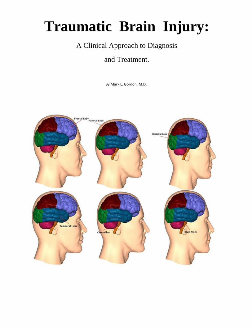
Traumatic Brain Injury: A Clinical Approach to Diagnosis
and Treatment.
Text Copyright ©2013 Millennium Health Centers, Inc. for Mark L. Gordon
All Rights Reserved
Other Books by the Author
The Clinical Application of Interventional Endocrinology (2007)
The Clinical Application of Interventional Endocrinology; the Handbook (2013)
The Laboratory of Interventional Endocrinology (2014)
Hormones and the Brain (2014)
Special Thanks*
It is greatly appreciated that Access Medical Laboratories of Jupiter, Florida have provided the
Millennium Health Centers, Inc. with a Grant in 2009 for the evaluation of 100 military veterans with
mTBI sustained while in combat. Mr. Ryan El-Hosseiny, CEO, Access Medical Laboratory.
Dedication
To Private Charles H. Kuhl, of L Company, U.S. 26th Infantry Regiment, who reported to an aid station
of C Company, 1st Medical Battalion, on 2 August 1943. At the time, Kuhl had been in the U.S. Army for
eight months, and attached to the 1st Infantry Division since th 2nd of June 1943. He was diagnosed with
"exhaustion," a diagnosis he had been given three times since the start of the campaign. Arriving at an aid
station he was subsequently evacuated to the 15th Evacuation Hospital near Nicosia for further evaluation
being started on treatment with sodium amytal. Notes in his medical chart indicated that he was
suffereing from "psychoneurosis anxiety state, moderately severe – this soldier has been twice before in
hospital within ten days. He can't take it at the front, evidently. He is repeatedly returned.”
Patton arrived at the hospital the same day, accompanied by a number of medical officers, as part of his
tour of the U.S. II Corps troops. He spoke to some patients in the hospital, commending the wounded. He
then approached Kuhl, who did not appear to be injured. Kuhl was sitting slouched on a stool midway
through a tent ward filled with injured soldiers. When Patton asked Kuhl where he was hurt, Kuhl
reportedly shrugged and replied that he was "nervous" rather than wounded, adding "I guess I can't take
it." Patton "immediately flared up," slapped Kuhl across the chin with his gloves, then grabbed him by the
collar and dragged him to the tent entrance. He shoved him out of the tent with a kick to his backside.
Yelling "Don't admit this son-of-a-bitch," Patton demanded that Kuhl immediately be sent back to the
front, adding, "You hear me, you gutless bastard? You're going back to the front."
Corpsmen picked up Kuhl and brought him to a ward tent, where it was discovered he had a temperature
of 102.2 °F (39.0 °C); he was later diagnosed with Malarial parasites. Speaking later of the incident, Kuhl
noted "at the time it happened, [Patton] was pretty well worn out ... I think he was suffering a little battle
fatigue himself." Kuhl wrote his parents about the incident, but asked them to "just forget about it." That
night, Patton recorded the incident in his diary: "[I met] the only errant coward I have ever seen in this
Army. Companies should deal with such men, and if they shirk their duty, they should be tried for
cowardice and shot."
I also dedicate this book to all the patients of the Millennium:
I am who I am because of all of you who have challenged me to find an alternative approach to wellness.
And, all of you who trusted the literature that I read finding answers that had already been discovered and
ignored for years before I dusted the cobwebs off. Not always an issue of selecting the right pill but more
so in listening to what you had to share. Not always about treating the numbers in a lab test but accepting
that we are not all made equally.
Pushing the envelop on interpretation of standard labs in a non-standard manner and treating to replenish
the chemicals, hormones, and minerals that were found to be less than optimal. Redefining the use of the
terms - deficiency and insufficiency – with healthy and non-healthy.
For all these reasons and more, I dedecate this book to you.

Forward by
Dr. Joseph Maroon
Table of Content
Traumatic brain injury (TBI) is nothing new having been developed in the rising of man and the
socialization of war. In the time of Australopithecus, as depicted by Stanley Kubrick’s 2001; A Space
Odyssey, a pre-homo sapien learns about weaponry by confronting a neighbor with a leg bone of some
large animal; wielding it like a club. Archeological records represented by skulls and not friable papyrus,
tell us the stories of traumatic brain injury before there were stories to tell.
It is unlikely that those skulls discovered with depression fractures of the frontal, temporal, or parietal
bones were the result of a motor vehicle accident, an improvised explosive device, or a slip-and-fall, and
all-of-which have now become the hallmark of socio-economic evolution, in the here and now.
In survey of countries that have weathered this socio-economical growth over the past two hundred years,
we see a significant rise in the occurrence of traumatic brain injury from a diversity of causes; those
individuals less than 20 years of age, motor vehicle accidents (mainly DUI related) make up the majority
of cases while those over 65 years of age, succumbing to over-medication, Frailty Syndrome, Vitamin D
deficiency all causing gait instability and falls as a close second.
The landscape between 20 and 65 years of age is filled with assaults, slip-and-falls, sports injuries, motor
vehicle accidents, blast trauma, occupational, and recreational injuries as causative factors.
Although not perceived literally as a trauma, any intracranial vascular event (hemorrhagic or ischemic) or
hypoxia, can lead to the same pathophysiological consequences as those produced by direct force trauma
to the skull. It appears that a diversity of causes can trigger the same pathways of destruction.
Taking things one step further, we can also add to this group of precipitating factors Alzheimer’s disease
since the critical process that leads to the loss of executive function and cognitive abilities are the same
chemical pathways that are initiated by the accumulation of Amyloid-β, stroke, ionizing radiation, and
traumatic brain injury.
The core of this book, chapter by chapter, is about the overwhelming number of individuals who have
sustained a mild traumatic brain injury (mTBI) and don’t even know that it has occurred. They are the
ones who develop progressive cognitive and psychosocial changes that appear as behavioral issues that
can manifest even after a delay of 30 years (1). These are the individuals that have forgotten the injury
and therefore, instead of looking beyond the superficial presentation of their apparent “illness”, are placed
on psychotropic medications to mask the underlying causes referred to as the “Stealth Syndrome”.
This book and the associated TBI Symposium will provide the fundamental knowledge for interested
physicians to learn how to recognize the presence of this condition and how to diagnose and treat the
individual.
This first chapter is a composite over-view of what the rest of the book has to offer you. It is in fact, the
beginning of my story to you…..
Epidemiology of Non-Combatant Traumatic Brain Injury
Traumatic brain injury (TBI) is a major cause of traumatic death and disability. In individuals younger
than 45 years of age, injury is the primary cause of death in the United States and other developing
nations (2). The general incidence of TBI in developed countries is approximately 200 per 100,000
population at risk. This estimate is skewed since the statistics are based upon those patients with TBI that
are hospitalized while the majority of those sustaining a TBI never seek medical attention (3).
Based upon a 2010 report looking at only the United States; a brain injury occurs every 7 seconds and
results in death every 5 minutes; representing about 4.5 million brain injuries and 53,000 deaths a year
(4). A prior CDC evaluation of TBI cases between 2002 and 2006, reported 1.7 million cases per year
with 80.7% emergency room visits, 16.3% hospitalizations, and 3.0% deaths (5). But again, this only took
into account those individuals who ended up in the medical system immediately after sustaining their TBI
while the majority were lost or delayed from inclusion.
Contrary to general believe, the majority of TBI cases are mild in nature and therefore, the bulk of the
injured never seek medical attention, that is, until the subtle behavioral and neuropsychological changes
start occurring forcing medical assessment. By that time, the association between the mild TBI(mTBI)
and symptomatology is lost. Unfortunately, this creates erroneous data by underestimating the frequency
of mild forms of TBI and overestimating the proportion of moderate to severe TBIs.
Approximately 50% of TBIs are the result of motor vehicle, bicycle or pedestrian–vehicle accidents. Falls
are the second-commonest cause of TBI (20–30% of all TBI), being more frequent among the elderly and
the very young population. Violence-related incidents account for approximately 20% of TBI, almost
equally divided into firearm and non-firearm assaults (6).
Several classifications for TBI severity are reported in the literature. The post-resuscitation Glasgow
Coma Scale (GCS) is the most widely used clinical classification of TBI severity. The GCS is based on
the patient’s response (eye opening, verbal and motor function) to various stimuli. A score of 13–15 is
considered mild, 9–12 moderate and ≤8 severe TBI (7). Clinical severity of TBI is also defined by
duration of loss of consciousness (LOC), loss of memory for events immediately before or after the
accident (post-traumatic amnesia) and identified intracranial lesion (8, 9). Radiological findings by
computed tomography (CT) may be helpful in TBI severity evaluation, and the most used radiological
scale is the Marshall’s CT classification of TBI (10). Functional outcome after TBI is usually evaluated
by the Glasgow Outcome Scale (GOS), a descriptive and easy to use scale, describing five outcome
categories (death, vegetative, severely disabled, moderately disabled, and good recovery) (11).
Given all these excellent resources to assess those with TBI, it still does not equate out to effective care
when you consider that almost 66% of those who need help fail to make it into the medical system. Those
that do, the system fails them in limiting diagnoses and treatment to antiquated superficial markers of
injury instead of looking deeper into the new science of Traumatic Brain Injury. But, its here now.
Epidemiology of Combatant Traumatic Brain Injury
Traumatic brain injury (TBI) has been described as the “signature wound” of the conflicts in Iraq and
Afghanistan. Improvised explosive devises (IED), mortars, tank cannons, hand grenades, and even
repetitive fire from an M-16 or M-4 Carbine, can produce the mechanical factors predisposing a soldier to
mTBI.
Of the total 178,876 TBI cases since 2000, 137,328 have been mild, 30,893 have been moderate, 1,891
have been severe, 3,175 have been penetrating, and 5,589 have not been classifiable but all are suffering
the sequela of brain trauma with psychological, physiological, and physical impairment (12).
In March 2009, USA TODAY’s caption read “360,000 Veterans May Have Traumatic Brain Injury.” The
story followed with; “WASHINGTON — Pentagon officials estimated for the first time Wednesday that
up to 360,000 Iraq and Afghanistan veterans may have suffered brain injuries. Among them are 45,000 to
90,000 veterans whose symptoms persist and warrant specialized care.”
Although these numbers are vast they could have been larger if not for the modern body armor worn by
soldiers that greatly enhanced chances of surviving mechanisms that are responsible for the mortality and
morbidity seen in combat. Blast injuries can cause brain damage with significant long-term and often
delayed neurological consequences.
Blast injury can be a complex clinical syndrome or “polytrauma” caused by the combination of four blast
effects; primary, secondary, tertiary and quaternary blast mechanisms. Air filled structures such as the
sinuses, ears, lungs, and intestines are most vulnerable to the primary blast wave. This can lead to
bleeding, alteration of ventilatory mechanisms with hypoxia, and tissue damage. The blast wave can
cause a sudden rotation of the brain leading to shearing forces that can sever cerebral capillary and long
axons responsible for intra-lobular communication.
Individuals exposed to a blast frequently manifest loss of memory for events before and after the
explosion; confusion, headache, impaired sense of reality, and reduced decision-making ability. Patients
with brain injuries acquired in explosions often develop sudden, unexpected brain swelling and cerebral
vasospasm despite continuous monitoring. However, the first symptoms of blast-induced neurotrauma
(BINT) may occur months or even years after the initial event, and are therefore categorized as secondary
brain injuries. The broad variety of symptoms includes weight loss, hormone imbalance, chronic fatigue,
headache, and problems in memory, speech and balance. These changes are often debilitating, interfering
with daily activities. Because BINT in blast victims is underestimated, valuable time is often lost for
preventive therapy and/or timely rehabilitation (13).
As is the case in the civilian population, laboratory assessment for hormonal disruption is infrequently
performed over the course of the individual’s rehabilitation for their physical trauma. An initial or
baseline level is rarely obtained thereby missing the 56% of those injured with TBI that do have one or
more hormonal deficiencies by 3 months. (14) Failure to recognize and correct the underlying hormonal
deficiencies (insufficiencies) may compound the physical and psychological complications of TBI and
interfere with rehabilitation (15).
The Military’s programs available are comprehensive but limited. They see the literature and
acknowledge that there is an association between mTBI and hormonal dysfunction but rarely pursue
testing as recommended by the AACE and others (XX). Since 2009, the Millennium Health Centers, Inc
has offered a free laboratory assessment for military personnel that have sustained a mTBI* with a
Glasgow Coma Score of 13-15.
Epidemiology of Sports-related Traumatic Brain Injury
Andre Waters was not the first, Dave Duerson was not the second, and nor will Junior Seau be the last to
succumb to the secondary psychological and physical consequences of hormonal insufficiency and
deficiencies precipitated by traumatic brain injury that led to their suicide.
Their suffering and ultimate sacrifice has shaken the very fiber of the sports world, not only in football
but in all contact sports, awakening us to the need for better protection, assessment, restrictions, and
treatment for those who have sustained a TBI on the field. Presently, a player can be taken out of a game
if their bench-side evaluation (after a suspected TBI) shows any sign of impaired brain functioning.
Injured players will no longer have their jerseys just brushed off and be sent back into the game, they will
be restricted from playing until their neurological evaluation has normalized.
Nonetheless, there will be many players from all sports that over time, sometimes up to a decade after
their last head injury, will start presenting to their physicians with altered personality, bouts of anger and
rage, cognitive, and functional impairments. Hopefully, at that time, they will have hormonal assessment
first as opposed to being placed on a psychotropic medication. Furthermore, if the athlete is found to be
hormone insufficient and not necessarily deficient they will be allowed to have the appropriate hormones
available for replenishment and on-going treatment instead of being denied this potentially lifesaving
treatment. (16, 17, 18)
The impact of sports related TBI has become a political bailiwick with congressional hearings and threats
of banning football and all contact sports. Head injuries suffered by the likes of Patrice Bergeron, Pierre-
Marc Bouchard and Marc Savard have raised awareness to new heights, so much so that the NHL
instituted in the 2010 season; Rule 48, banning blindside hits to the head. This was seen as a way to
reduce the public health risks and the consequential financial burden on insurance companies, families
and the team leagues.
Consequently, there are many NFL veterans who are still struggling post-career. A 1994 survey from the
players' association found that significant numbers were depressed, fat and managing chronic pain. A
2006 Scripps Howard report found that of 3,850 deceased players that NFL players are more than twice as
likely as Major League Baseball players to have died before their 50th birthday. Frequently, their much
heavier bodies failed in retirement.
What about our children? Football accounts for nearly two-thirds of the estimated 62,816 TBIs incurred in
high school sports each year, according to a 1999 survey published in the Journal of American Medical
Association.
Traumatic brain injury (TBI) has been associated with hypopituitarism in general and GH deficiency
(GHD) in particular; the consequences of this on growth and development are likely to be critical in
children and adolescents in the so-called "transition phase". At 3 months, hypopituitarism was present in
34.6%, and at 12 months, hypopituitarism was present in 30.3%. Growth hormone deficiency (GHD) and
secondary hypogonadism were the most common acquired pituitary deficits in this age group. These
results show the high risk of TBI-induced hypopituitarism also in the transition age. Thus it is
recommended that pediatric endocrinologists follow-up pituitary function of children and adolescents
after brain injuries. (19) I think there is little else to say here, more later.
Pain Alternatives, Solutions and Treatment Medical Team (P.A.S.T.)
How many medications are you on? “Eighteen (18)! Three are narcotics and then there is one that is a
stimulant to counter the side effects of the three narcotics so I can at least get up and go to the bathroom.
Otherwise, I’m so out of it I pee in my bed. Then there are the two muscle relaxers and the three for my
depression. Funny thing is, I’m still depressed. Then because of the stimulant I can’t sleep at night so they
have me take a couple of sleep pills(Trazodone and Ambien) that make me so groggy in the morning I
can’t wake up and take a stimulant medication.”
If you think that encounter was made up, consider spending some time at PAST headquarters in Clifton,
New Jersey and you will hear this conversation over and over again. When you walk away you will
become a true believer in how these notable professional sports retirees were led down a medication
pathway instead of addressing the underlying cause to their pain and altered mental status.
PAST is an organization and program developed by the compassionate Dr. William Focazio, a
gastroenterologist by training and a saint to boys who enter his sanctuary.
In explaining PAST, Dr. Focazio stated that, “.. many of the retired athletes that come to his organization
have expended their time-limited medical insurance policies graciously given to them upon retirement by
their relative sports team. After about 5 years when the progression of their underlying injures blossom
into a limiting disability many have no medical coverage left. Without coverage, their medical treatment
is more palliative then curative using an endless number of oral medications instead of corrective surgery
or indicated hormonal replenishment.”
“By the time they make it to PAST our first duty is to detoxify them from their personal pharmacopeia.
Our Neuropsychiatrist and Neurologist evaluates them and provide a program for detoxification.
Concurrently, the retiree is evaluated by one of our orthopedist to see if there is justification for surgical
intervention as a means of stopping their pain and retuning them to physical functioning and a healthy life
style with the least number of pills”, explains Dr. Facazio.
If they have a history of concussion(s) with or without loss of consciousness, they are evaluated by our
Interventional Endocrinologist for hormone dysfunction that plagues many players. It is the deficiency
(and insufficiency) of specific brain generated hormones that can cause outbursts of anger, depression,
fatigue, cognitive impairment, physical degradation, and suicidal tendencies.
Replenishment of those hormones found to be low (or low-normal) can return the individual to a higher
level of functioning and quality of life. Thousands of medical articles attest to the finding of hormonal
insufficiency and deficiency in TBI as well as documented beneficial affects when replacement therapy is
used to return levels to optimum and physiological.
PAST is a non-profit organization with a team of specialized physicians that provide services to those
who cannot afford it. If you are a physician with a desire to help and become a recognized member of the
PAST Medical Team; please contact them through their website.
Neuropathophysiology of Traumatic Brain Injury
Regardless of the type of non-penetrating trauma that is conveyed to and through the skull (blunt force,
blast waves, or ionizing radiation), there are two phases of damage that occur to the brain; an initial
mechanical injury (1°- primary) associated with contusion, hemorrhage, edema, ischemia, and diffuse
axonal injury (DAI), and a neuro- or biochemical assault (2°- secondary) associated with neuronal loss
resulting from the activation of one or more of the known terminal cascades - Necrosis, Apoptosis, and
Autophagy.
These events, mechanical and biochemical, can co-exist depending upon the magnitude of injury but
frequently, in mild traumatic brain injury(mTBI), the primary injury is short-lived while the subtle and
insidious secondary injury can linger on for years taking its toll piece by piece. It is the impact of these
biochemical processes that is most important for understanding the cause-and-effect relationships
between TBI and Hypothalamic-Pituitary Dysfunction (HPD).
The primary impact is frequently followed by the development of vasogenic and cytotoxic edema and
impairment of energy metabolism (ATP and mitochondrial loss). This sets in motion a cascade of
biochemical events both extra- and intracellular causing an indolent progression to cellular death. That is
indolent until either a critical mass of cells are lost or there is impact upon a region of brain that matters
(frontal lobe and executive functions).
Neurons that escaped the initial mechanical trauma are exposed to a secondary mechanism that few cells
can escape. At the immediate area of contusion and also remotely, neurons that succumbed to the initial
trauma release an array of physiological cellular components into the intracellular space that accumulates
to toxic levels. Nitric Oxide (NO), Reactive Oxygen Species (ROS), glutamate, cytokines, and
interleukins create an environment of collateral cellular damage as the oxidative stress (OS) increases.
The end result is a concentric ring of expanding cell death. It is by this process that over time, the
accruement of dying brain cells leads to the signature changes that have come to represent not only mild
traumatic brain injuries but all degrees of neurotrauma. (20)
Furthermore, capillaries that are torn leak red blood cells and plasma into the environment only to be
degradated to a more reactive hemoglobin/iron (OS). As damage includes blood vessels there is the loss
of the autoregulatory function of the cerebral vasculature with intravascular fluids leak into the white and
gray matter spaces causing edema. Owing to the edema, the intracranial pressure (ICP) increases and the
brain is crushed against the unyielding cranium increasing the risk for uncal herniation, as well as, further
diminution of vascular blood flow, cerebral spinal fluid circulation and death. The ensuing ischemia,
hypoxia, and further neuronal damage all cause acceleration of oxidative stress and expansion of the
involved area(s).
We look at these processes as having an overt negative net affect upon the brain but in fact, we are
learning that the ultimate goal of these neurochemical cascades is to help preserve and limit damage to the
brain and its neurons.
Necrosis may be the only process that does not stand out as a bad-good mechanism to help cells survive
like Apoptosis and Autophagy. In necrosis, which has classically been regarded as uncontrolled cell
death, recent research suggests that its occurrence and progression might be tightly controlled. This is
based upon finding signs of organized processes like mitochondrial dysfunction, ATP depletion,
increased Reactive Oxygen Species (ROS), cytoskeletal proteolysis by Calpain, and disruption of the cell
membrane. It is this loss of cell membrane integrity that precipitates irreversible cell death (21, 22).
Apoptosis, or programmed cell death, has always implied that the cell dies in an organized and
programmed manner, which is heralded by condensation of nuclear chromatin with DNA fragmentation,
cell shrinkage, and the formation of apoptosomes that are subsequently cleared by phagocytosis. In this
process, an organized removal of an irreversible damaged cell ensues in a surgical manner. The process of
Apoptosis is focused on the containment of intracellular components that can trigger inflammatory
responses that if allowed to float freely into contiguous cell spaces will precipitate collateral damage.
Contrary to Necrosis, in Apoptosis there is little collateral inflammation and no significant release of
chemotactic agents that would bring the immune defense system into play. This is more of a quiet process
that saves the surrounding cells and their morphology from a potential logarithmic process of destruction.
(23)
Finally, Autophagy also called the type 2, non-apoptotic cell death mechanism, differs from apoptosis in
that it doesn’t necessarily terminate in cell death. On the contrary, Autophagy is probably the most cell
protective mechanism against cell execution (24, 25, 26). In the event that damage occurs to a
cytoplasmic organelle, which brings with it the risk of leakage of its content into the cytoplasm,
autophagy would encapsulate it in an autophagosome. Once engulfed, the organelle would be processed
with lysozymes digesting it to its principle components for reutilization for other cell structures.
Cannibalization of this nature can also occur in cell starvation where the cell is forced to ingest its own
parts for survival. (27)
Traumatic brain injury in humans results in a pattern of neuronal loss that includes the cortex,
hippocampus, cerebellum and thalamus (hypothalamus). In the acute post-traumatic period, injured
neurons appear swollen then become shrunken and eosinophilic with condensation of chromatin.
Suggestive patterns of both necrotic and apoptotic processes. Under the electron microscope, these
degenerating neurons appear swollen, with swollen mitochondria, vacuolated cytoplasm and Pyknotic
nuclei, suggestive of necrosis (28, 29, 30).
In the delayed or chronic post-traumatic period, there is a progressive but indolent process of neuronal
degeneration that differs in the pattern seen in the acute phase, reflecting the apoptotic component of post-
traumatic brain injury (31).
Knowledge of the different biochemical cascades associated with each of the above processes has led
researchers to develop compounds that can interrupt the progression of neuronal destruction and cognitive
impairment (32). Though, it might take 3 to 5 years or longer before we see any of them in our local
pharmacies. Until that time, we can use prescriptive and alternative products that are available but are not
being utilized due to the lack of awareness and skills in how to use them.
Continued reading of this book might help to fill in many of those blank lines…
The Neuroradiology of Traumatic Brina Injury
It was November 2006; I had just given my classical lecture, Traumatic Brain Injury – The Hormone
Dysfunction Syndrome, to about 400 attendees of the ACAM symposium focusing on neurological
illnesses. As I walked off the stage and down the side aisle, a voice from behind called out “I got it, I got
it”. I turned around and asked in a demanding manner “What do you got?” The doctor repeated herself
and said, “I got it. I got it.” Again, “What do you have?”
She introduced herself as a Neuroradiologist who reviews the CTs and MRIs of patients who have
sustained anywhere from a mild to severe TBI. She explained that she rarely sees any structural
abnormalities in those patients with mTBI even months or years post-trauma even thought the patients are
being studied for progressive functional impairment. She added that almost all of the moderate to severe
TBI cases have recognizable contusions of the brain, diffuse axonal injury, and if not frank intracerebral
bleeding (subdural hematomas), punctuate areas of intracerebral bleeds.
This is the paradigm that is finally changing due to advancements in radiologic technology and the
unraveling of the biochemical mechanisms that activate both apoptosis and autophagy creating the telltale
micro-calcified scars. In the past, these microscopic remnants of injury were missed due to the smaller
Tesla coils (magnetic strength) but now with a 3.0 Tesla coil the resolution can identify these micro-
calcifications which are found within neuronal tissue representing scarring precipitated by axonal damage
(DAI), microhemorrhages, and necrosis (33).
The importance of this technology was clearly defined in the paper by Orson and Handson in 2009 when
they showed that 76% of non-combatant individuals who sustained a TBI had positive findings with only
14% involvement of their pituitary gland (77). This, in my mind, opened the door to looking beyond the
pituitary gland as the poor recipiant of all things bad and turn attention to other areas of the brain. In fact,
in this same paper, over 59% of findings involved the Hippocampus (memory and the ability to learn new
information) and 29% diffuse axonial injury (DIA). Together, these describe the foundation for the
majority of symptoms experienced by patients with mTBI and above.
Using newer software enhancements to this MRI technology (DTI-MRI) allows for images to be formed
that look at the directional flow of water within neurons from the bodies down the microtubules into the
axons. Diffusion Tension Imaging (DTI) or Tractography can delineate the course and continuity of long
axonal connections between one or more cerebral hemispheres. Traumatic or neurochemical (necrosis,
apoptosis or autophagy) damage to these neuronal conduits can be seen as though they were cut wires. It
is in these areas where there is loss of continuity and connectivity that produces the alterations in brain
functioning that we see as change in the personality and cognitive abilities of the individual (34).
The functional MRI (fMRI) is another neuroimaging technique that can display patterns of brain activity
based upon increases and decreases in regional blood flow (hemodynamic). After sustaining a TBI, there
can be a significant redistribution of the hemodynamics reflecting the changes in blood flow with
alterations in both glucose and oxygen utilization.(35)
There are a number of other technologies like SPECT, PET, and Fusion scans that will be discussed in
detail in Chapter 5.
Neuroendocrinology of Traumatic Brain Injury
Post-traumatic hypopituitarism (PTHP) was first recognized and described in 1918 by Cyran, but at that
time it was thought to be a rare occurrence (36). More recently, TBI has been recognized as a frequent
cause of hypothalamic–pituitary dysfunction and impairment, contributing to a delayed or diminished
capacity for recovery in many TBI patients (37-42).
Hypothalamic-pituitary dysfunction following a traumatic event can be divided into an acute phase;
during which there are transient alterations in the production of pituitary hormones such that they are
either increased or diminished and a delayed phase; when those alterations in hormones fail to return to
normal regulatory production as a result of permanent damage at the pituitary and/or hypothalamic levels.
The pituitary gland responds to acute traumatic events with two secretory patterns: adrenocorticotropin
(ACTH), prolactin (PRL) and growth hormone (GH) levels increase, while luteinizing hormone (LH),
follicle-stimulating hormone (FSH) and thyrotropin (TSH) levels may either decrease or remain
unchanged, associated with a decreased activity of their target organ. Changes in the circulating hormone
levels become apparent during the first hours or days after trauma, and may persist for the duration of the
acute critical illness. These alterations represent part of the acute adaptive response to the injury, and may
be influenced by the type of injury and pharmacological therapy used to treat the critical illness
(glucocorticoids, narcotic analgesics or dopaminergic agents) (43).
Accepting the premise that trauma to the brain leads to a progressive disruption of hypothalamic control
of hormonal production by both the anterior and posterior pituitary glands, would make it reasonable to
test for the adequacy of production of those central hormones directly regulated by the Hypothalamus.
This would include anterior pituitary hormone markers such as thyroid stimulating hormone (TSH),
growth hormone (GH), adrenocorticotropic hormone (ACTH), and posterior pituitary markers such as
prolactin, anti-diuretic hormone (ADH), luteinizing hormone (LH), and follicle stimulating hormone
(FSH). (44)
In addition to those hormones, we need to look at IGF-1, IGFBP-3, fT3, fT4, rT3, free testosterone, total
testosterone, dihydrotestosterone (DHT), DHEA-s, estradiol (E2), estrone (E1), progesterone,
pregnenolone, and cortisol. These are used to assess the patient for primary hormonal deficiencies
(peripheral failure) that could pre-date the impact of TBI. Additionally, all these hormones have
beneficial affects on the central nervous system as either Neurosteroids(NS) or Neuroactive
Steroids(NAS).
Our understanding of Neurosteroids is mind boggling. These steroid hormones were once thought to pass
into the brain throught the blood-brain-barrier(BBB) having been produced in the periphery. Today,
science shows us that these hormones are regionally manufactured de novo in the brain. Pregnenolone,
allopregnanolone(tetrahydroprogesterone), progesterone, alloprogesterone, and DHEA-s to name a few.
All with important functions to protect nerves from oxidative stress (ROS), reduce inflammatory
cytokines, interleukins, and to regenerate myelin.(45-55) An interesting finding in the frontal lobe of
patients with Alzheimer’s disease is a lack or deficiency of Allopregnanolone.(56).
Neuroactive steroids are those hormones produced in the periphery, which penetrate the BBB and enter
the cerebral circulation to participate in the same protective processes as the NS. Many of the NAS are the
same as the NS and are therefore, additive.
So, the issue becomes when is the best time to assess the potential of neurotrauma related hormone
dysfunction? The answer comes as no surprice in being as soon as possible after the head trauma.
Unfortunately, as indicated above, most individuals with head trauma fail to seek medical attention until
subtle or overt symptoms start to creep into their lives. The initial testing, if early, will represent the
baseline hormonal levels with follow-up hormone panels at 3 months, 6 months, and 12 months from the
date of the initial testing (injury). In a 2006 study by Schneider and Schneider, they followed new TBI
cases for a year taking blood at 6 months and 12 months finding a 56% and 36%, respectively of one or
multiple hormone deficiencies(57).
A more recent article in the Journal of Neurotrauma (2012), addressed the persistancy of Pituitary
Hormone Deficiency after Traumatic Brain Injury acknowledging that 69% of the patients had at least
one pituitary hormone deficiency, where GH deficiency was more prevalent (severe: 40.0%; partial:
23.6%) than corticotropin (27.3%) or thyrotropin (21.8%) deficiencies.(58)
Diagnosing TBI Hormone Dysfunction Syndrome (THDS) can be challenging for the majority of
clinicians, as well as, for the trained endocrinologists. This is because the general consensuses for what
constitutes a laboratory result as being insufficient or deficient is controversial as is the question of
whether-or-not to treat the insufficiency? Many of the hormonal test results have ranges that are broad,
representing two standard deviations so the numerical distance from the low-end to the high-end can be
immense as in the case of Total Testosterone; 270ng/ml to 1730ng/ml. So if a patient has a result of
280ng/ml is that deficient, insufficient, or normal?
The majority, if not all, of traditional physicians would call that “within the normal range” and not
address the myriad of patient complaints and elect not to treat. Fortunately, this is slowly changing and
physicians are treating patients with appropriate physiological doses to achieve a level that is the median
of the range (50th percentile for age).
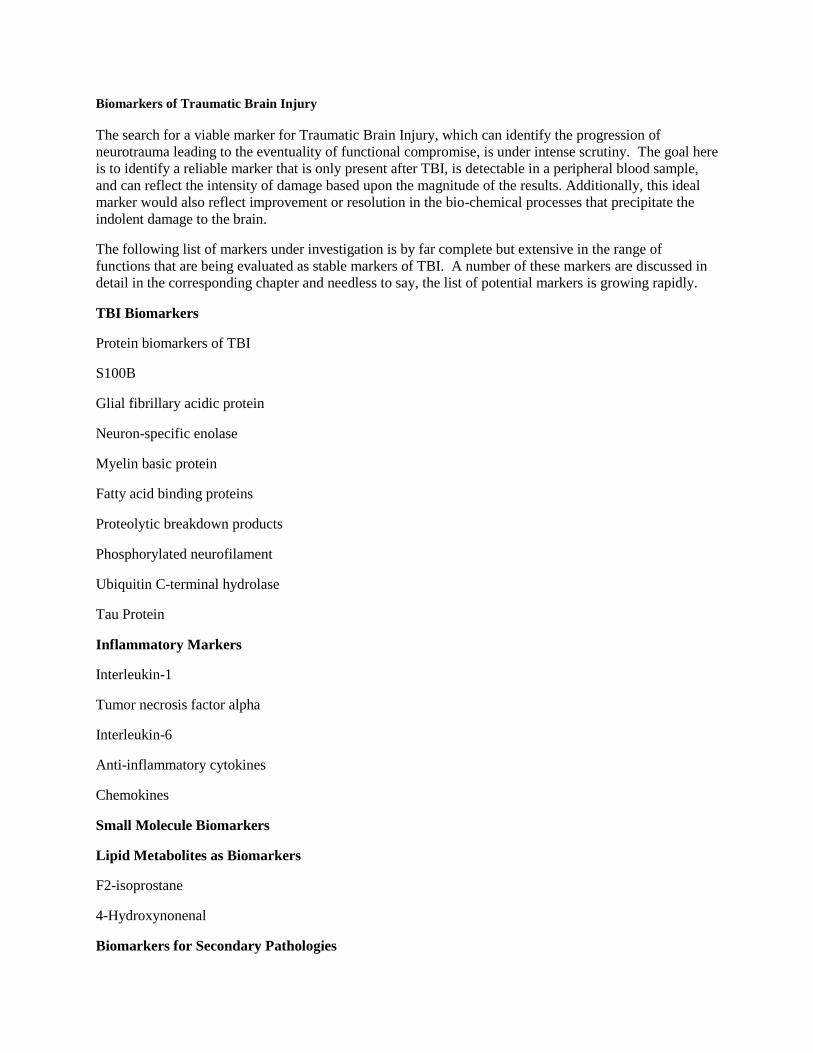
Biomarkers of Traumatic Brain Injury
The search for a viable marker for Traumatic Brain Injury, which can identify the progression of
neurotrauma leading to the eventuality of functional compromise, is under intense scrutiny. The goal here
is to identify a reliable marker that is only present after TBI, is detectable in a peripheral blood sample,
and can reflect the intensity of damage based upon the magnitude of the results. Additionally, this ideal
marker would also reflect improvement or resolution in the bio-chemical processes that precipitate the
indolent damage to the brain.
The following list of markers under investigation is by far complete but extensive in the range of
functions that are being evaluated as stable markers of TBI. A number of these markers are discussed in
detail in the corresponding chapter and needless to say, the list of potential markers is growing rapidly.
TBI Biomarkers
Protein biomarkers of TBI
S100B
Glial fibrillary acidic protein
Neuron-specific enolase
Myelin basic protein
Fatty acid binding proteins
Proteolytic breakdown products
Phosphorylated neurofilament
Ubiquitin C-terminal hydrolase
Tau Protein
Inflammatory Markers
Interleukin-1
Tumor necrosis factor alpha
Interleukin-6
Anti-inflammatory cytokines
Chemokines
Small Molecule Biomarkers
Lipid Metabolites as Biomarkers
F2-isoprostane
4-Hydroxynonenal
Biomarkers for Secondary Pathologies

Intracranial pressure
Blood brain barrier compromise
Secondary ischemia
Post-traumatic epilepsy
Biomarkers for Predicting Outcome
Neuropsychiatry and Traumatic Brain Injury
Patients who had suffered traumatic brain injury were evaluated to determine the occurrence of
psychiatric disorders during a 30-year follow-up; of the 60 patients, 29 (48.3%) had had an Axis I
disorder that began after traumatic brain injury, and 37 (61.7%) had an Axis I disorder during their
lifetimes. (60)
In this study, the most common disorders post-traumatic brain injury were major depression (26.7%),
alcohol abuse or dependence (11.7%), panic disorder (8.3%), specific phobia (8.3%), and psychotic
disorders (6.7%). Fourteen patients (23.3%) had at least one personality disorder. The most prevalent
individual disorders were avoidant (15.0%), paranoid (8.3%), and schizoid (6.7%) personality disorders.
Nine patients (15.0%) had DSM-III-R organic personality syndrome. The results suggest that traumatic
brain injury may cause decades-lasting vulnerability to psychiatric illness in some individuals. (60)
Traumatic brain injury seems to make patients particularly susceptible to depressive episodes, delusional
disorder, and personality disturbances. The high rate of psychiatric disorders found in this study
emphasizes the importance of psychiatric follow-up after traumatic brain injury. (60)
This study was published in 2002 and at that time the correlation between hormonal insufficiencies and
causative factors for cognitive and emotional changes were still not appreciated let alone treated.
Fortunately, at the writing of this book there seems to be an increased appreciation for the association
between hormonal dysfunction and TBI although, it is still a rare occurrence that a treatment protocol will
be started to address it. (60)
This brings me to a case of a 41 year old male who we’ll call Bill. Bill played rugby between the ages of
18 and 22 during which time he sustained 5 significant head traumas with loss of consciousness on three
separate occasions and one which precipitated hospitalization. At 36 years of age, and after a slow
progression of depression, self-medicated with alcohol for ten plus years, Bill was interred for evaluation
and treatment for severe depression. Once he was detoxified from the alcohol addiction, he was started on
a number of SSRIs which failed one after another to control his depression. The psychiatrist eventually
placed him on three times the usual dose of one medication acknowledging the persistent mild depression
as acceptable.
At age 41, and after being on psychotropic medication for 5 years, Bill read about our approach to TBI
with assessment and replenishment of hormones. The article on TBI causing a hormonal dysfunction
syndrome contained many of the elements that he was experiencing. He flew in from Boston to start his
evaluation. After lengthy documentation of his history, a TBI Blood Panel was drawn, and he was given a
Testosterone provocative challenge test while waiting for the lab confirmation of his deficiency.
Bill was sent home with instruction to call in one week to review his response to the provocative testing
and to review his laboratory results. At the time Bill informed me of an appointment he had scheduled
with an Orthopedist to discuss his chronic right shoulder pain and “turf toe” (frozen left 1st toe). His
appointment was two days after our initial appointment.
On that Thursday, Bill called the office and when we spoke he asked me to remind him why he went to
the Orthopedist. When I jokingly reminded him that it was for his shoulder pain and frozen toe, he said
that he awoke that morning and the shoulder pain was gone and the toe was moving. He also, stated that
he slept the night of his injection better than he had in years. He also noted a rise in his libido and a
calming of his depression.
Making this long story short, after 2 months on hormone replenishment treatment (self administering a
shot of testosterone once a week), Bill started working with his psychiatrist to taper him off of the anti-
depressants. At 6 months, not only did Bill’s depression abate but he was off his SSRIs, had mental
clarity, slept very well, emotionally was stable, had a new girlfriend, had a return of his libido, and was
working out harder in the gym with improved recovery.
I can go on with these annecdotal cases for months. The impact of hormonal replenishment in patients
with and without TBI who have neuropsychiatic, cognitive, and physical issues frequently subside or
abate within a few weeks of starting treatment.
A recently published book by Springer (2011), entitled; Acquired Brain Injury - an Integrative Neuro-
Rehabilitation Approach, provides a comprehensive dissertation on Neuropsychiatry and Traumatic Brain
Injury. The chapter brilliantly written by Dr. Angela Scicutella discusses the clinical manifestations,
assessment, and treatment for an array of psychological complexes (depression, mania, anxiety,
psychosis, agitation, arousal and attention, dementia, and sexual dysfunction) that are acknowledged as
being associated with traumatic brain injury. Unfortunately, each section contains an educational case
where the patient is inevitable placed on psychotropic medications. (61)
Of significant concern while reading this modern text book of psychiatric intervention was, not a single
word was offered discussing the impact of TBI on hypothalamic-pituitary dysfunction. This is in light of
thousands of articles published over the past 30 years addressing initially, the presence of hypopituitarism
in survivors of TBI, and then the association of hormonal deficiencies and their impact on psychological
functioning ( both emotional and cognitive).
For whatever reason, we seem to be focusing in on only the superficial aspects of psychological maladies
without going after the cause (biochemical). A patient presents with depression put them on an
antidepressant and increase the dosing until the symptoms resolve. I was unaware that humans can have
an antidepressant deficiency and therefore, replacing them is all that is needed. Medicine has become
shortsighted in the quest for health and wellness.
Treatment for Traumatic Brain Injury
I wish there was something cryptic I could say about this section (Chapter 8), which deals with treatment
of mTBI with laboratory confirmed hormonal insufficiencies, but there isn’t. Insufficiencies are generally
looked at as being within the normal reference ranges and therefore, they are never considered for being
brough up to the median physiologic level; and there lies the major issue. We need to reasses our thinking
about what is healthy in each individual patient and not what numbers look most acceptable on paper.
Treatment must be tailored for each patient with the goal of optimizing all hormone levels. This should
include the lessor hormones as well as the major ones.
We know from a 2002 review article about our insufficiencies in ; “Outcome Measures for Clinical Trials
in Neuro-Trauma” that dozens of studies were stopped prematurely due to their failure to improve the
condition of patients with mild to severe TBI. The medications and procedures that were being evaluated
did not work at best and many worsened the patients’ condition.(62)
More recently, (2009) a review of the benefits of “cooling” in TBI concluded, “… our present knowledge
on the use of active cooling in TBI patients has not been shown to be effective for outcome after
TBI…”(63)
In light of these and hundreds of other articles addressing the lack of beneficial affects, these treatments
continue to be used while the literature promoting the positive outcomes from the use of Growth
hormone, IGF-1, Testoserone, DHEA-s and DHT on neuropsychiatic, cognitive, and physical sequalea
are ignored. (64,65,66,67,68,69,70,71)
A major reason why the hormonal literature is being ignored is because the studies that have been
performed, over the past twenty years were looking at the benefits of replacement of a single hormone
such as testosterone; growth hormone, estrogen, progesterone, DHEA, Vitamin D, and Thyroid, have not
yielded significant results. In fact the results at times were contrary to the expectations or there were no
results at all. Why would that happen? A good guess would be that there is a level of hormonal synergy
that is being omitted. (72,73,74,75)
In 1993, Drs. Marc Blackman and Mitchell Harman looked at this issue of synergy in both genders. In the
males replenished with just testosterone or growth hormone alone, had lower beneficial effects then when
testosterone and growth hormone were used concurrently. The same happened in the females when they
were replenished with only growth hormone or only estrogen and progesterone (Progestin). When they
were given all replenishment hormones concurrently, the responses confirmed the synergy.(76)
Additionally, in order to prescribe medication such as growth hormone, an Insulin Challenge Test must be
performed. Furthermore, to avoid dissenting criticism by your state boards, the patient’s laboratory levels
must fit into the standards of care in order to provide legitimate treatment protocols. What this means is
that we can’t practice medicine to the fullest.
Nonetheless, when treating cases of mTBI we must replenish all hormones to at least the median
physiologic levels. Then, add secondary hormones that can be lost, such as Pregnanolone and DHEA,
when using testosteone. This will maintain optimal levels of NAS and NS and provide for added
protection against oxidative stress and promote remylenization of damaged neurons. The addition of
specific antioxidant regimens by oral or IV routes is encouraged along with a number of potential
Nootropin products (L-Dyprenyl, ammantadine, and neurotransmitter precursor amino acids (NPAA)).
Knowing the mechanisms behind the subtle progression of nerve damage can only help us to provide a
means of diminishing their long-term affect on brain functioning so that we may maintain who we are for
a longer period of uninterrupted quality of life.
Medical-Legal Issues relative to Traumatic Brain Injury
This topic is really a composition of a number of legal issues that arise in the treatment of patients with
traumatic brain injury.
Just as important as the “who to blame” for the traumatic brain injury, is the understanding of the legal
ramifications if providing treatment. As of the date of this writing, there is no approved diagnostic code
for using growth hormone in patients with TBI. Of all the thousands of medications listed in the
physician’s desk reference, only recombinant human growth hormone (rhGH) has the restriction of “no
off label” use! Therefore, we must prove that there is a state of Adult Growth Hormone Deficiency with
an approved challenge test. The safest and apparently most reliable one is the Glucagon Challenge test.
This can be preformed in the office within 3 hours and the results will justify your use of growth
hormone. Otherwise, you are breaking federal guidelines and can be subject to both DEA and State
Medical Board challenge; another kind of challenge testing that you mostly fail!
The benefit of GH replenishment is in its ability to up-regulates the serum level of IGF-I that has been
shown to help in the repair of neurons, dendrites, axons, microtubules, and stabilization of tau proteins.
As an alternative, the use of a clinically proven growth hormone secretagogue – Secretropin, can initially
be used and monitored for its ability to raise GH/IGF-I production in the patient. A three month trial is
suggested before a Glucagon Challenge test.
Some years ago, I called and spoke to an attorney that specialized in cases of Traumatic Brain Injury. I
shared with him what I was doing in regards to testing and treating with hormones. At some point in the
one sided conversation he interrupted me and said, “I’m afraid of you!” When I asked him why he said
that he replied, “you mean that if you find that the patient is hormanlly out of balance and replace the
hormones they can get better?” I replied, “Yes”. He said, “.. that is then a problem since my remuneration
is based upon the level of persistent damage that the patient has and if you improve the level of injury,
payout for the damages decreases.” He went on to say, “how about waiting until the case is closed and
then treat the patient.” I had nothing nice to say in response so I just hung up.
In cases that are litigated or anticipated to be litigated, we as physicians must learn how to best protect the
patients’ case by providing some additional testing along with the standards of clinical medical care. A
comprehensive past medical history, a specific history of the presenting injury, as well as, prior injuries, a
comprehensive physical examination, appropriate standard laboratory testing with a TBI blood panel, and
a Cognitive-Emotional questionnaire. If there are issues of neuropsychobehavior a referral to a
neuropsychologist to perform appropriate testing is suggested. These tests can also be used as rulers of
improvement when the patient undergoes hormonal replenishment.
ImPACT to Assess mild Traumatic Brain Injures (mTBI)
ImPACT (Immediate Post-Concussion Assessment and Cognitive Testing) is the first, most-widely used,
and most scientifically validated computerized concussion evaluation system established for Traumatic
Brain injury.
Developed in the early 1990's by Drs. Mark Lovell and Joseph Maroon, ImPACT is a 20-minute test that
has become a standard tool used in comprehensive clinical management of concussions for athletes of all
ages. ImPACT Applications, Inc. was co-founded by Mark Lovell, PhD, Joseph Maroon, MD, and
Michael Collins, PhD.
Given the inherent difficulties in concussion management, it is important to manage concussions on an
individualized basis and to implement baseline testing and/or post-injury neurocognitive testing. This type
of concussion assessment can help to objectively evaluate the concussed athlete's post-injury condition
and track recovery for safe return to play, thus preventing the cumulative effects of concussion. In fact,
neurocognitive testing has recently been called the "cornerstone" of proper concussion management by an
international panel of sports medicine experts.
ImPACT can be administered by an athletic trainer, school nurse, athletic director, team doctor or
psychologist, provided that they have completed training in the administration of the test. Post-concussion
care and the management of concussion should only be administered by a trained medical professional.
ImPACT assists doctors in making return-to-play decisions and should never be used as a stand-alone
tool.
ImPACT is the most widely used computer-based testing program in the world and is implemented
effectively across high school, collegiate, and professional levels of sport participation.
The test battery consists of a near infinite number of alternate forms by randomly varying the stimulus
array for each administration. This feature was built in to the program to minimize the "practice effects"
that have limited the usefulness of more traditional neurocognitive tests.
ImPACT takes approximately 20 minutes to complete. The program measures multiple aspects of
cognitive functioning in athletes, including; attention span, working memory, sustained and selective
attention time, response variability, non-verbal problem solving and reaction time.
Conclusion
Regarless of the etiology precipitating a traumatic brain injury – motor vehicle accident while under the
influence of alcohol, improvised explosive device, or any given Sunday, the biochemical processes or the
secondary phase of neurotrauma that are initiated at the moment of a TBI all cascade to the same end-
points; necrosis, apoptosis, and autophagia. The major difference is the extent of physical destruction of
brain matter ascribed to the primary insult thereby, creating the conditional status of the TBI from
minimal to severe.
As you delve into the chapters of this book you will learn about clinical neurology and how to use the
TBI Locatortm
to define the areas of the brain that are involved in the neurotrauma associated with mTBI.
Understanding where the injuries might be by evaluation of neuro-cognitive deficits by the bedside, can
help improve your understanding of the pathophysiology that is underlying any alteration in the
personality and cognitive abilities of the injured.
The results of the TBI Locatortm
may also assist in the prediction of the out come of neuroradiologic
procedures that are used to further define the area(s) of damage. The use of post-acute phase
neuroradiographic assessment has been simplified so that we can assess glucose up-take with a PET Scan
showing areas of hypo- or hyper-metabolism, regional blood flow with a fMRI looking at the iron in the
hemaglobin molecule, or following the course of long axons with Tractography produced by the DTI-
MRI looking at water in the axons. These are but a few radiologic techniques that can help deliniate the
areas and extent of neurotrama.
The secondary insult of TBI, the biochemistry that triggers Caspase 3 – the messenger of cell death, BAX,
and BCl2 products all lead to death of cells in the region of the trauma as well as in remote areas of the
brain. There appears to be areas of the brain that are overly sensitive to ROS and by-products of necrosis
and increasing levels of glutamate. These areas of preferential neuronal loss are found in the Cortex,
Hippocampus, Cerebellum, and Thalamus (Hypothalamus).(78)
Involvement of the Hippocampus alters acute and long term memory and the ability to learn and store
new information but, it is the involvement of the Hypothalamus that interests us the most. Damage to the
Hypothalamus not only alters thermal control of the body but affects the ability to regulate all of our
hormones. Within the hypothalmus are cell-sensors for the levels of circulating hormones. Damage to this
mechanism can lead to intial hormone insufficiencies that can progress to frank deficiencies. Failure of
the Hypothalamic-Pituitary Axis is at the core of this books and the foundation for our approach to the
diagnosis and treatment of TBI with Hormone Dysfunction syndrome.
Without adequate levels of hormones in the brain, whether neurosteroids produced regionally or
neuroactive steroids passing throught the blood brain barrier from the periphery, central hormonal
deficiencies can be causative for the progression of the secondary phase of neurotrauma and the failure of
the brain to heal itself. We have seen the literature attesting to the regeneration of a diverse population of
nerve cells under the influence of IGF-I and II, and DHEA-s along with the protection from oxidative
stress from pregnanolone, progesterone, and estrogens. (79-DD)
The perinatal histories of 46 children with idiopathic hypopituitarism were assessed in order to define the
relationship between perinatal insult and the development of hypopituitarism. Compared to normal
control siblings, the pregnancies resulting in hypopituitary children were complicated by a significantly
higher incidence of vaginal bleeding at various times during gestation (13 pregnancies). Twenty-four
percent of the hypopituitary children were delivered by breech, three times the incidence of control
siblings and seven times the incidence of breech delivery in the general population. Prolonged or
unusually short labors were more common in the hypopituitary children (13 patients), as were signs of
intrapartum distress and asphyxia (10 patients). At the time of the study, 19 hypopituitary children had
neurologic abnormalities; of these, 15 had histories of significant perinatal insult. The findings in this
study suggest that, in many cases, perinatal insults may cause hypopituitarism. (last)
*Access Medical Laboratory Grant 2009 for TBI; for the evaluation of 100 non-duty, discharged, male
individuals with mTBI sustained while in combat. Jupiter, Florida, Mr. Ryan El-Hosseiny, CEO, Access
Medical Laboratory.
References Chapter 1: Introduction
1. Axis I and II psychiatric disorders after traumatic brain injury a 30 year follow up study. Am J Psychiatry. 2002;
159(8):1315-21, Koponen S; et. al., Department of Psychiatry, Turku University Central Hospital, Finland. 2002
2. Head Injury associated deaths in the United States from 1979 to 1986. JAMA 1989;262:2251-5. Sosin Dm, et. Al.
3. The Epidemiology of Traumatic Brain Injury: A Review. Epilepsia, Vol 44, Suppl. 10, 2003. Burns and Hauser, Columbia
University. 2003
4. Traumatic Brain Injury, Handbook of Neurocritical Care; second edition, Chapter 18, G.S.F. Ling, S.A. Marshall,
Uniformed Services. 2010
5. Traumatic Brain Injury in the United States: Emergency Room Visits, Hospitalizations, and Deaths 2002-2006, Centers for
Disease Control and Prevention, National Center for Injury and Prevention, Atlanta Georgia, 2010
6. Consensus Conference. Rehabilitation of persons with traumatic brain injury. NIH Consensus Development Panel on
Rehabilitation of Persons with Traumatic Brain Injury. JAMA 1999 282 974–983.
7. Assessment of coma and impaired consciousness. A practical scale. Lancet 1974 2 81–84. Teasdale G & Jennett B.
8. The epidemiology of traumatic brain injury: a review. Epilepsia 2003 44 (Suppl 10) 2–10. Bruns J Jr & Hauser WA.
9. Congenital and acquired brain injury. 1. Brain injury: epidemiology and pathophysiology. Archives of Physical Medicine
and Rehabilitation 2003 84 S3–S7. Greenwald BD, Burnett DM & Miller MA.
10. The diagnosis of head injury requires a classification based on computed axial tomography. Journal of Neurotrauma 1992 9
S287–S292. Marshall LF, Marshall SB, Klauber MR, Van Berkum Clark M, Eisenberg H, Jane JA, Luerssen TG, Marmarou
A & Foulkes MA.
11. Assessment of outcome after severe brain damage. Lancet 1975 1 480–484.Jennett B & Bond M.
12. U.S. Military Casualty Statistics: Operation New Dawn, Operation Iraqi Freedom, and Operation Enduring Freedom Hannah
Fischer Information Research Specialist September 28, 2010 Congressional Research Service RS22452
13. Traumatic brain injury: An overview of pathobiology with emphasis on military populations. J Cerebral Blood Flow Metab
30(2):255-266 2010. Cernak, I., and L. J. Noble-Haeusslein.
14. Prevalence of anterior pituitary insufficiency 3 and 12 months after traumatic brain injury. Europe J Endocrinology. 2006;
154(2):259-65. Schneider HJ; et al. GK. Max Planck Institute of Psychiatry, Clinical Neuroendocrinology Group
Kraepelinstr. 10, 80804 Munich, Germany.
15. Prevalence of neuroendocrine dysfunction in patients recovering from traumatic brain injury. J Clin Endocrinology Metab.
2001; 86(6):2752-6 . Lieberman SA; et al. Transition Learning Community (B.E.M.) and Departments of Internal Medicine
and Neurology, University of Texas Medical Branch, Galveston, Texas 77555.
16. Anterior pituitary dysfunction following traumatic brain injury (TBI). Clinical Endocrinology 2006 64 481–488. Agha A & Thompson CJ.
17. Hypothalamic lesions following closed head injury. Brain 1971 94 165–172. Crompton MR.
18. Analysis of abnormalities in pituitary gland in non-missile head injury: study of 100 consecutive cases. Journal of Clinical Pathology 1986
39 769–773. Harper CG, Doyle D, Hume Adams J & Graham DI.
19. Hypopituitarism induced by traumatic brain injury in the transition phase. J Endocrinology Invest. 2005; 28(11):984-9. Aimaretti G;
Division of Endocrinology and Metabolism, Department of Internal Medicine, University of Turin, Turin, Italy.
20. The Role of Reactive Oxygen Species in the Pathogenesis of Traumatic Brain Injury, Esther Shohami and Ron Kohen
21. Cell death by necrosis: towards a molecular definition. TRENDS in Biochemical Sciences. 2006 Vol.32 No.1, Pierre Golstein et a;, Centre
d’Immunologie de Marseille-Luminy, Universite´ de laMe´ diterrane´e, Case 906, Marseille Cedex 9, France
22. Classification of cell death: recommendations of the nomenclature committee on cell death. Cell Death Differ. 12, 1463–1467. Kroemer, G.
et al. (2005)
23. Bench-to-bedside review: Apoptosis/programmed cell death triggered by traumatic brain injury. Critical Care 2005, 9:66-75 Xiaopeng
Zhang , Yaming Chen, Larry W Jenkins, Patrick M Kochanek and Robert SB Clark. 1Assistant Professor, Department of Critical Care
Medicine, University of Pittsburgh School of Medicine, Safar Center for Resuscitation Research, Pittsburgh, PA, USA
24. Autophagy: from phenomenology to molecular understanding in less than a decade. Nat Rev Mol Cell Biol. 2007 Nov;8(11):931-7.
Klionsky DJ. Life Sciences Institute, University of Michigan, 210 Washtenaw Avenue, Ann Arbor, Michigan 48109-2216, USA.
25. Autophagy fights disease through cellular self-digestion Nature. 2008 February 28; 451(7182): 1069–1075 Noboru Mizushima1, Beth
Levine2, Ana Maria Cuervo3, and Daniel J. Klionsky Life Sciences Institute and Departments of Molecular, Cellular and Developmental
Biology and Biological Chemistry, University of Michigan, Ann Arbor 48109, USA
26. Apoptosis and autophagy: regulatory connections between two supposedly different processes Apoptosis (2008) 13:1–9 Andrew Thorburn
Department of Pharmacology, University of Colorado at Denver and Health Sciences Center, Aurora, CO 80045, USA e-mail:
27. Protein turnover via autophagy: implications for metabolism. Annual Rev Nutrit, 2007; 27:19-40, Mizushima N et al.
28. Dysfunction of hypothalamic hypophysial axis after traumatic brain injury in adults. Journal of Neurosurgery September 2010 Volume 113,
Number 3 David Krahulik, M.D.1, Jirina Zapletalova, M.D., Ph.D.2, Zdenek Frysak, M.D.3, and Miroslav Vaverka, M.D.1 1Neurosurgical Clinic Faculty Hospital; 2Department of Paediatrics, Faculty Hospital; and 3IIIrd Internal Clinic Faculty Hospital, Olomouc, Czech Republic
29. Does the type and severity of brain injury predict hypothalamo–pituitary dysfunction? Does post-traumatic hypopituitarism predict worse
outcome? Pituitary (2008) 11:255–261 M. Klose and Feldt-Rasmussen. Department of Medical Endocrinology, PE2131, Copenhagen University Hospital, Rigshospitalet, Blegdamsvej 9, 2100 Copenhagen, Denmark
30. Cell death by necrosis: towards a molecular definition. TRENDS in Biochemical Sciences Vol.32 No.1 Pierre Golstein1,2,3,4 and Guido
Kroemer
31. Cell death mechanisms following traumatic brain injury. Brain Pathol. 2004; 14(2):215-22. Raghupathi R Drexel University College of
Medicine, 2900 Queen Ln, Philadelphia, PA, USA. [email protected]
32. X
33. Traumatic brain injury: a review and high-field MRI findings in 100 unarmed combatants using a literature-based checklist approach.
Journal of Neurotrauma. 2009; 26(5):689-701 Orrison WW; Hanson EH; Alamo T; Watson D; Sharma M; Perkins TG; Tandy RD. Nevada
Imaging Centers, Las Vegas, Nevada, USA. 2009
34. Diffusion Tensor Imaging Detects Clinically Important Axonal Damage after Mild Traumatic Brain Injury: A Pilot Study. Journal of
Neurotrauma. 24:1447–1459 2007.Bazarian JJ. et al.
35. A longitudinal fMRI study of working memory in severe TBI patients with diffuse axonal injury. Neuroimaging 43 (2008) 421–429. Rocio
Sanchez-Carrion, Davinia Fernandez-Espejo, et al, Dept of Neuropsychology, Institute Universitari de Neuro rehabilitation Guttmann,
Badalona, Spain
36. Hypophysenschadigung durch Schadelbasisfraktur. Deutsche Medizinische Wochenschrift 1918 44 1261. Cyran E.
37. Occurrence of pituitary dysfunction following traumatic brain injury. Journal of Neurotrauma 2004 21 685–696. Bondanelli M, de Marinis
L, Ambrosio MR, Monesi M, Valle D, Zatelli MC, Fusco A, Bianchi A, Farneti M & degli Uberti EC. 2004
38. Traumatic brain injury and subarachnoid haemorrage are conditions at high risk for hypopituitarism: screening study at 3 months after brain
injury. Clinical Endocrinology 2004 61 320–326. Aimaretti G, Ambrosio MR, Di Somma C, Fusco A, Cannavo` S, Gasperi M, Scaroni C,
De Marinis L, Benvenga S, degli Uberti EC, Lombardi G, Mantero F, Martino E, Giordano G & Ghigo E. 2004
39. Prevalence of neuroendocrine dysfunction in patients recovering from traumatic brain injury. Journal of Clinical Endocrinology and
Metabolism 2001 86 2752–2756. Lieberman SA, Oberoi AL, Gilkinson CR, Masel BE & Urban RJ. 2001
40. Rehabilitation and hypopituitarism after traumatic brain injury. GH and IGF Research 2004 14. (Suppl A) S108–S113. Masel BE.
41. Anterior pituitary dysfunction in survivors of traumatic brain injury. Journal of Clinical Endocrinology and Metabolism 2004 89 4929–
4936. Agha A, Rogers B, Sherlock M, O’Kelly P, Tormey W, Phillips J & Thompson CJ. 2004
42. Anterior pituitary dysfunction after traumatic brain injury. Part I. Journal of Head Trauma Rehabilitation 2003 18 541–543. Elovic EP.
43. Hormonal responses to trauma. Critical Care Medicine 1992 20 216–226. Woolf PD. 2003
44. Traumatic infarction of the anterior lobe of the pituitary gland. Lancet 1959 2 927–930. Daniel PM, Prichard MML & Treip CS.
45. Hypopituitarism following traumetic brain injury. Growth Hormone and IGF Research 2005 15 177–184. Popovic V, Aimaretti G,
Casanueva FF & Ghigo E.
46. Hypopituitarism and growth hormone deficiency (GHD) after traumatic brain injury (TBI). Growth Hormone and IGF Research 2004 14
S114–S117. Aimaretti G, Ambroso MR, Benvenga S, Borretta G, De Marinis L, De Menis E, Di Somma C, Faustini-Fustini M, Grottoli S,
Gasco V, Gasperi M, Logoluso F, Scaroni C, Giordano G & Ghigo E.
47. Neurosteroids in the Hippocampus: Neuronal Plasticity and Memory Schumacher M. Stress 1997 Oct;2(1):65-78
48. Mechanisms of progesterone-induced Neuroprotection. Ann N Y Acad Sci 2005 Jun;1052:145-51. Singh M. Department of Pharmacology
and Neuroscience, University of North Texas Health Science Center, 3500 Camp Bowie Blvd., Fort Worth, TX 76107.
49. Vitamin D deficiency reduces the benefits of progesterone treatment after brain injury in aged rats Neurobiology of Aging 32 (2011) 864–
874 Milos Cekic, et al. Department of Emergency Medicine, Emory University School of Medicine, Atlanta, GA,USA 2011
50. DHEA Stimulates Growth of Brain Cells. UCSF Researchers Conclude , Proceedings of the National Academy of Sciences, Jeffrey Norris
April 1998
51. Analysis of Neurosteroid levels in attention deficit hyperactivity disorder. International Journal of Neuropsychopharmacol 2001
Sep;4(3):259-64. Strous RD et al.
52. Neurosteroids in the Hippocampus: Neuronal Plasticity and Memory.. Stress 1997 Oct;2(1):65-78 Schumacher M, et al.
53. Pregnenolone Sulfate and Aging of Cognitive Functions: Behavioral, Neurochemical and Morphological Investigations. Hormones and
Behavior 40, 215-217 (2001), Mayo M et al.
54. Progesterone and Allopregnanolone reduce inflammatory cytokines after traumatic brain injury. Exp Neurol 2004 Oct;189(2):404-12. He J;
et al. Department of Psychology, Emory University, Atlanta, GA 30322, USA
55. Regeneration in a degenerating brain: potential of Allopregnanolone as a neuroregenerative agent. Curr Alzheimer Res. 2007; 4(5):510-7.
Wang JM; Irwin RW; Liu L; Chen S; Brinton RD. Department of Pharmacology and Pharmaceutical Sciences and Program in
Neuroscience, University of Southern California, School of Pharmacy, 1985 Zonal Avenue, Los Angeles, CA 90089, USA.
56. The neurosteroid Allopregnanolone is reduced in prefrontal cortex in Alzheimer's disease. Biol Psychiatry. 2006; 60(12):1287-94.
Marx CE; Trost WT; Shampine LJ; et. Al. Duke University Medical Center, Department of Psychiatry and Behavioral Sciences, Durham,
North Carolina 27705, USA
57. Prevalence of anterior pituitary insufficiency 3 and 12 months after traumatic brain injury. Eur J Endocrinol. 2006; 154(2):259-
65.Schneider HJ; Schneider M; Saller B; Petersenn S; Uhr M; Husemann B; von Rosen F; Stalla GK. Max Planck Institute of Psychiatry,
Clinical Neuroendocrinology Group Kraepelinstr. 10, 80804 Munich, Germany.
58. Lasting Pituitary Hormone Deficiency after Traumatic Brain Injury Odile Kozlowski Moreau, Edwige Yollin, Emilie Merlen, Walter
Daveluy, and Marc Rousseaux. Journal of Neurotrauma. January 1, 2012, 29(1):
59. biomarkers
60. Predictors of New-Onset Depression after Mild Traumatic Brain Injury J Neuropsychiatry Clin Neurosci. 2010 ; 22(1): 100–104. Vani Rao,
MD, Division of Neuropsychiatry & Geriatric Psychiatry; Dept. of Psychiatry, Johns Hopkins University; 5300 Alpha Commons Drive; 4th
Floor; # 444, Baltimore, MD 21224,
61. Axis I and II psychiatric disorders after traumatic brain injury: a 30-year follow-up study. Am J Psychiatry. 2002; 159(8):1315-21.
Koponen S; Taiminen T; Portin R; Himanen L; Isoniemi H; Heinonen H; Hinkka S; Tenovuo O Department of Psychiatry, Turku
University Central Hospital, Finland.
62. Outcome Measures for Clinical Trials in Neuro-Trauma M. Ross Bullock, M.D. Ph.D., Randall E. Merchant, Ph.D., Sung C. Choi, Ph.D.,
Charlotte B. Gilman, B.S.N., Jeffrey S. Kreutzer, Ph.D., Anthony Marmarou, Ph.D., And Graham M. Teasdale, F.R.C.P., F.R.C.S.
Neurosurg Focus 13(1), 2002.
63. Active cooling in traumatic brain-injured patients: a questionable therapy? Acta Anaesthesiol Scand. 2009 Nov;53(10):1233-8. Epub 2009
Aug 13. Grände PO Reinstrup P Romner B Department of Anaesthesia and Intensive Care, Lund University Hospital, Lund, Sweden.
64. Low testosterone levels predict incident depressive illness in older men: effects of age and medical morbidity. J Clin Psychiatry 2005
Jan;66(1):7-14 Shores MM; et al. Veterans Affairs Puget Sound Health Care System, Seattle , WA 98108, USA.
65. Testosterone therapy in late-life major depression in males. J Clin Psychiatry 2002 Dec;63(12):1096-101 Perry PJ; et al. Division of
Clinical Pharmacy, College of Pharmacy, University of Iowa, Iowa City 52242, USA
66. Replacement of DHEA in aging men and women. Potential remedial effects. Ann NY Academy of Science 1995;774:128-142. Yen SS,
Morales AJ, Khorrram O.
67. Depression in aging men: the role of testosterone. Drugs Aging 2004;21(6):361-76. Carnahan RM; et al. Clinical and Administrative
Pharmacy, College of Pharmacy, University of Iowa, Iowa City, Iowa 52242, USA
68. The role of the somatotropic system in cognition and other cerebral functions. Semin Vasc Med 2004 May;4(2):167-72 . Creyghton WM; et
al. Department of Clinical Endocrinology, University Medical Centre, Utrecht, The Netherlands.
69. Insulin-like growth factor 1 is essential for normal dendritic growth. J Neurosci Res 2003 Jul 1;73(1):1-9 Cheng CM; et al. Developmental
Endocrinology Branch, National Institute of Child Health and Human Development, National Institutes of Health, Bethesda, Maryland.
70. Growth hormone in the brain: characteristics of specific brain targets for the hormone and their functional significance. Front
Neuroendocrinol 2000 Oct;21(4):330-48. Nyberg F Department of Pharmaceutical Biosciences, Uppsala University, Uppsala, Sweden
71. Prevalence of Neuroendocrine dysfunction in patients recovering from traumatic brain injury. J Clin Endocrinol Metab. 2001; 86(6):2752-
6 . Lieberman SA; et al. Transition Learning Community (B.E.M.) and Departments of Internal Medicine and Neurology, University of
Texas Medical Branch, Galveston, Texas 77555.
72. X
73. X
74. X
75. X
76. Effects of GH and/or Sex Steroid Administration on Abdominal Subcutaneous and Visceral Fat in Healthy Aged Women and MenT
Munzer, M Harman, P Hees, E Shapiro, C Christmas, M Bellantoni, T Stevens, K O'Connor, K Pabst, C ST. Clair, J. Sorkin, M. Blackman.
Endocrine and Metabolism Sections, Laboratory of Clinical Investigation, Intramural Research Program, National Institute on Aging,
National Institutes of Health, and the Divisions of Geriatric Medicine and Gerontology, Cardiology and Endocrinology and Metabolism
, Departments of Medicine, Johns Hopkins Bay view Medical Center and Johns Hopkins University School of Medicine, Baltimore,
Maryland 21224. (2001)
77. Traumatic brain injury: a review and high-field MRI findings in 100 unarmed combatants using a literature-based checklist approach.
Journal of Neurotrauma. 2009; 26(5):689-701 Orrison WW; Hanson EH; Alamo T; Watson D; Sharma M; Perkins TG; Tandy RD. Nevada
Imaging Centers, Las Vegas, Nevada, USA.
78. Cell Death Mechanisms Following Traumatic Brain Injury. Brain Pathology 2004;14:215-222. Ramesh Ragupathi, PhD. 2004
79. X
80. x
(last)High incidence of perinatal insult in children with idiopathic hypopituitarism, The Journal of Pediatrics, Volume 96, Issue 3, Pages 397-402.
W. Craft, L. Underwood, J. Van Wyk
CRS Report to Congress, Former NFL Players: Disabilities, Benefits, and Related Issues, April 8, 2008, L. Elaine Halchin, Analyst in American
National Government, Government and Finance Division


