User Success Story
TABLE
Modality description (University Hospital of Geneva)
MR 1 Philips Eclipse 1.5 T (Marconi)MR 1 Philips Proview 0.23T (Marconi)MR, OT 1 Twinstar Workstation Philips (Marconi)MR, OT 1 Vistar Workstation Philips (Marconi)
CT 2 Philips MX8000 Quad (Marconi)CT, MR, OT 3 Philips Workstation MXView (Marconi)CT 1 GE Highspeed CT/ICT, MR, OT 2 GE Workstation Advantage-Windows release 2.0OT 1 Vitrea 2 Workstation Vital Images release 3.01CT 1 Philips PQ2000s (Marconi)CT, OT 3 Philips Workstations Voxel Q
CR 2 ADC AGFACR 2 Thoravision Philips
XA 1 Philips IntegrisOT 1 Workstation Philips 3DRAXA, OT 1 Workstation Philips EasyVision release 5.1
RF 3 Philips DSIRF 1 Workstation Philips EasyVision release 3.0
US 1 Aloka Prosound SSD-5000US 1 ATL Philips HDI-5000US 2 Acuson Sequoia 512
OT 1 Hologic Delphi Bone DensitometryOT 1 GE Prodigy Bone Densitometry
NM 1 Philips AXISNM 1 Philips IRIX
OT 8 Paxport AGFA
30 Image providers14 workstationsSite is mostly using the AGFA Drystar 3000
Hôpitaux Universitaires de GenèveConsistent Presentation of Images—A Case Study and Success StoryHôpitaux Universitaires de Genève (HUG) of Switzerland is a hospital with 2187 beds and a staff of7865. They provide consultation services for a wide geographical region, seeing upwards of550,000 people a year. Staffed by 200 employees and 70 radiologists, the Radiology departmentperforms 150,000 procedures annually. Radiology is deployed in seven different geographical loca-tions throughout the region.
Hôpitaux Universitaires de Genève’s department workflow utilizes Cedara Software Corp.’s PACSsolutions. Images are acquired from one of HUG’s 30 modalities See table (including XA, CT, MRI,PET, SPECT, US, CR, DF, MG, and Bone Densitometry), most of them with digital capabilities. TheCerner Image Manager stores these acquired images and then automatically routes them to theCedara I-SoftView 5.2 Image Creator/Display workstation.
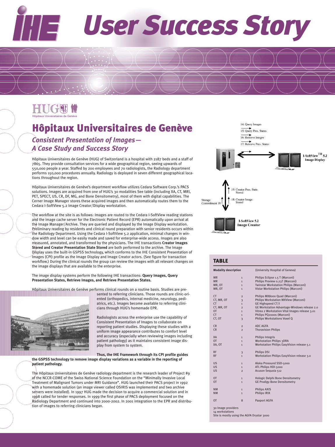
The workflow at the site is as follows: Images are routed to the Cedara I-SoftView reading stationsand the image cache server for the Electronic Patient Record (EPR) automatically upon arrival atthe Image Manager/Archive. They are queried and displayed by the Image Display workstation.Preliminary reading by residents and clinical round preparation with senior residents occurs withinthe Radiology Department. Using the Cedara I-SoftView 5.2 application, minimal changes in win-dow width and level can be easily made and saved for enterprise-wide access. Images are alsomeasured, annotated, and transformed by the physicians. The IHE transactions Creator ImagesStored and Creator Presentation State Stored are both performed to the archive. The ImageDisplay uses the built-in GSPSS technology, which conforms to the IHE Consistent Presentation ofImages (CPI) profile as the Image Display and Image Creator actors. (See figure for transactionworkflow.) During the clinical rounds the group can review the images with all relevant changes onthe image displays that are available to the enterprise.
The image display systems perform the following IHE transactions: Query Images, QueryPresentation States, Retrieve Images, and Retrieve Presentation States.
Hôpitaux Universitaires de Genève performs clinical rounds on a routine basis. Studies are pre-sented to referring clinicians. Those rounds are clinic-ori-ented (orthopedics, internal medicine, neurology, pedi-atrics, etc.). Images become available to referring clini-cians through HUG’s homemade EPR.
Radiologists across the enterprise use the capability ofConsistent Presentation of Images to collaborate onreporting patient studies. Displaying these studies with auniform image appearance contributes to comfort leveland accuracy (especially when reviewing images includingpatient pathology) as it maintains consistent image dis-play from system to system.
Thus, the IHE Framework through its CPI profile guidesthe GSPSS technology to remove image display variations as a variable in the reporting ofpatient pathology.
The Hôpitaux Universitaires de Genève radiology department is the research leader of Project #9of the NCCR-COME of the Swiss National Science Foundation on the “Minimally Invasive LocalTreatment of Malignant Tumors under MRI Guidance”. HUG launched their PACS project in 1992with a homemade solution (an image viewer called OSIRIS was implemented and two archiveservers were installed). In 1997 HUG made the decision to acquire a commercial solution and in1998 called for tender responses. In 1999 the first phase of PACS deployment focused on theRadiology Department and continued into 2000-2002. In 2001 integration to the EPR and distribu-tion of images to referring clinicians began.


