downloaded from on june 27, 2017 by guest · tu herpes simplex virus and, as reported here,...
TRANSCRIPT

1
Blocking ESCRT-mediated envelopment inhibits microtubule-dependent 1 trafficking of alphaherpesviruses in vitro. 2 Himanshu Kharkwal, Caitlin G. Smith, and Duncan W. Wilson# 3 4 Department of Developmental and Molecular Biology, 5 Albert Einstein College of Medicine, Bronx, New York 10461, USA 6 E-mails: [email protected], [email protected], 7 Running Title: Alphaherpesvirus envelopment and microtubule-motility 8 9 Key Words: Herpes Simplex Virus, Pseudorabies Virus, Microtubule-trafficking in vitro, 10 Viral Envelopment, Vps4 11 12 Manuscript JVI02777-14 Revised 09/30/14 13 14 # Corresponding author. Mailing address: Department of Developmental and 15 Molecular Biology, Albert Einstein College of Medicine, 1300 Morris Park Avenue, 16 Bronx, New York 10461, USA 17 Tel: (718) 430-2305. Fax: (718) 430-8567. E-mail: [email protected] 18 19 Abstract Word Count: 194 Text Word Count: 5,349 20 21
JVI Accepts, published online ahead of print on 8 October 2014J. Virol. doi:10.1128/JVI.02777-14Copyright © 2014, American Society for Microbiology. All Rights Reserved.
on June 27, 2017 by guesthttp://jvi.asm
.org/D
ownloaded from

2
Abstract 22 Herpes simplex virus and, as reported here, Pseudorabies virus utilize the 23
ESCRT-apparatus to drive cytoplasmic envelopment of their capsids. Here we 24 demonstrate that blocking ESCRT-mediated envelopment using the dominant negative 25 inhibitor Vps4A-EQ reduced the ability of HSV and PRV particles to subsequently traffic 26 along microtubules in vitro. HSV and PRV capsid-associated particles with bound GFP-27 Vps4A-EQ were readily detected by fluorescence microscopy in cytoplasmic extracts of 28 infected cells. These Vps4A-EQ-associated capsid-containing particles bound to 29 microtubules in vitro but were unable to traffic along them. Using a PRV strain 30 expressing a fluorescent capsid and a fluorescently tagged form of the envelope protein 31 gD, we found that similar numbers of gD-positive and gD-negative capsid-associated 32 particles accumulated in cytoplasmic extracts under our conditions. Both classes of 33 PRV particle bound to microtubules in vitro with comparable efficiency, and similar 34 results were obtained for HSV using anti-gD immunostaining. The gD-positive and gD-35 negative PRV capsids were both capable of trafficking along microtubules in vitro, 36 however motile gD-positive particles were less numerous and their trafficking was more 37 sensitive to the inhibitory effects of Vps4A-EQ. We discuss our data in the context of 38 microtubule-mediated trafficking of naked and enveloped alphaherpesvirus capsids. 39
40 Importance 41
The alphaherpesviruses include several important human pathogens. These 42 viruses utilize microtubule-mediated transport to travel through the cell cytoplasm, 43 however the molecular mechanisms of trafficking are not well understood. In this study 44
on June 27, 2017 by guesthttp://jvi.asm
.org/D
ownloaded from

3
we have used a cell-free system to examine the requirements for microtubule-45 trafficking, and have attempted to distinguish between the movement of “naked” and 46 membrane-associated cytoplasmic alphaherpesvirus capsids. 47
48 INTRODUCTION 49
Members of the Alphaherpesvirinae subfamily include Herpes simplex virus 50 (HSV) type 1 and 2, Varicella zoster virus and Pseudorabies virus (PRV). Like all 51 Herpesviruses, members of this subfamily replicate their genomes and assemble DNA-52 packaged capsids in the cell nucleus. It is then generally accepted that capsids bud into 53 the inner nuclear membrane to generate perinuclear virions that subsequently fuse with 54 the outer nuclear membrane to release mature nucleocapsids (also termed “naked” 55 capsids) into the cytoplasm. Naked cytoplasmic herpes capsids subsequently undergo 56 secondary envelopment at a post-nuclear organelle to assemble the mature, infectious 57 virion (1-4). 58
By performing subcellular fractionation during a single synchronized wave of 59 HSV egress we showed that HSV capsids bypass the cis, medial and trans 60 compartments of the Golgi apparatus (5) and accumulate in a buoyant membrane 61 fraction with the biochemical and antigenic properties of the Trans Golgi network (TGN) 62 and endosomes (5, 6). Studies by other laboratories have similarly concluded that 63 cytoplasmic HSV capsids acquire their envelopes at the TGN (4) or early endosomes 64 (7, 8). For HSV (9, 10) envelopment is known to utilize components of the cellular 65 ESCRT (Endosomal-Sorting Complex required for Transport) pathway (11-13) in 66 common with many other families of viruses (14, 15). In both normal and infected cells 67
on June 27, 2017 by guesthttp://jvi.asm
.org/D
ownloaded from

4
the last step in the ESCRT pathway is ATP hydrolysis by the AAA ATPase Vps4 at the 68 bud neck; this drives ESCRT-III release and membrane scission (16-19). Substitution 69 of a Glutamate by a Glutamine in the Vps4 ATPase active site generates Vps4-EQ, an 70 ATP-locked dominant negative form (20, 21) capable of blocking the final step and thus 71 arresting viral envelopment (9, 14, 18, 22). Although to our knowledge the ESCRT 72 pathway has not yet been shown to be required for PRV envelopment, Vps4 is found 73 associated with extracellular PRV (23) as it is for HSV (10). 74
Quantitative microscopic studies in non-polarized cells suggest that naked HSV 75 (24) and PRV (25) capsids utilize microtubules to reach the site of secondary 76 cytoplasmic envelopment, and non-enveloped HSV capsids are clearly capable of 77 recruiting multiple plus and minus-end directed motors (26). Following envelopment, 78 newly formed egress organelles, with their cargo of lumenal virions, probably also use 79 microtubules to reach the cell surface, however this step and the structure of the 80 exocytosing compartment are not well understood. It remains unclear to what degree 81 naked and enveloped viral capsids are capable of microtubule-association and 82 movement, and the relative importance of microtubules for the trafficking of each type of 83 particle. The situation is particularly complex in neurons, where it remains contentious 84 whether naked capsids (27), enveloped capsids or both (28-35) are capable of 85 trafficking out of the cell body and along axons. Moreover, opinions differ for the closely 86 related alphaherpesviruses HSV and PRV, as has recently been discussed (28, 36-38). 87
We previously demonstrated that exocytosing HSV particles present in a buoyant 88 subcellular membrane fraction (5) are capable of ATP-dependent trafficking along 89 fluorescently-labeled microtubules in a microscopic imaging chamber (6). These GFP-90
on June 27, 2017 by guesthttp://jvi.asm
.org/D
ownloaded from

5
tagged HSV capsids (39) demonstrated Kinesin and Dynein-mediated motility with a 91 velocity and processivity similar to that seen in vivo (6). Further, in vitro motility was 92 dependent (40) upon the large virally-encoded tegument protein UL36p (VP1/2) that is 93 known to recruit motors and to be required for motility and invasiveness in vivo (24-26, 94 41-46). In this study we modified our in vitro trafficking system to permit study of HSV 95 and PRV particle motility in bulk cytosol, containing both naked and enveloped viral 96 capsids. We next used the well-defined properties of the dominant negative Vps4A-EQ 97 protein to arrest HSV and PRV envelopment, blocking the formation of egress 98 organelles and their cargo of assembled viral particles. We found that the arrested 99 capsid-associated structures became decorated with bound Vps4A-EQ, and remained 100 able to bind microtubules but lost the ability to traffic along them. Loss of motility by this 101 population abolished approximately half of the alphaherpesvirus trafficking that we 102 observed in vitro. Studies of a PRV strain expressing a fluorescently labeled capsid and 103 a fluorescent gD envelope protein revealed that both gD-positive and gD-negative 104 capsids were able to traffic in vitro, but that the majority of trafficking particles were gD-105 negative. Of these trafficking virions, Vps4A-EQ was a more potent inhibitor of gD-106 positive than gD-negative particles. 107
108 109
on June 27, 2017 by guesthttp://jvi.asm
.org/D
ownloaded from
6
MATERIALS AND METHODS 110 Cells and viruses. Vero cells were maintained in Dulbecco Modified Eagle’s 111
Medium (DMEM) supplemented with 10% newborn calf serum and 1% penicillin-112 streptomycin (Gibco Laboratories). Cell lines expressing tetracycline-inducible GFP-113 tagged forms of Vps4A and Vps4A-EQ are derived from T-REx HEK293 cells 114 (Invitrogen) and have been previously described (22). These cells were cultured in 115 DMEM supplemented with 10% tetracycline-free fetal calf serum (FCS) (Clontech), 1% 116 penicillin-streptomycin and 100μg/ml Zeocin (Gibco Laboratories) at 37oC. The HSV-1 117 strain 2822 and PRV strain 847 carry in-frame fusions of mRFP1 to their UL35 gene, 118 and have been previously described (47, 48). PRV strain 4853 carries both the 119 mRFP1-UL35 fusion and an in-frame fusion of TagBFP (49) to US6 (encoding 120 glycoprotein D), similar to strains previously described (50). All viruses were 121 propagated and titered on Vero cells as previously described (51). 122
Isolation of post-nuclear supernatants. Vps4A or Vps4A-EQ-expressing cells 123 were induced with 1μg/ml tetracycline for 16 hours before infection, or at the time of 124 infection, with HSV or PRV at a multiplicity of infection (m.o.i.) of 20 for 1 hour at 37oC. 125 At the end of the infection cells were washed, then overlaid with, fresh pre-warmed 126 tetracycline-containing medium. At 16 hours post-infection, cells were washed with ice-127 cold MEPS buffer (5mM MgSO4, 5mM EGTA, 0.25M Sucrose, 35mM PIPES 128 [Piperazine-N,N’-bis (2-ethanesulfonic acid)], pH 7.1) (6, 52), collected by scraping and 129 resuspended in ice cold MEPS buffer containing 2mM Phenylmethylsulfonyl Fluoride, 130 5% (vol/vol) mammalian cell Protease Inhibitor Cocktail (Sigma, catalog number P8340) 131 and 4mM Dithiothreitol (DTT). Cells were then broken by repeated passage through a 132
on June 27, 2017 by guesthttp://jvi.asm
.org/D
ownloaded from

7
25-gauge needle, centrifuged at 2,000g for 5 minutes to remove nuclei and unbroken 133 cells, and a Post-Nuclear Supernatant (PNS) containing exocytosing viral particles 134 collected (6, 40). The PNS was supplemented by an additional 5% (vol/vol) of 135 mammalian protease inhibitor cocktail, and small aliquots flash frozen in liquid nitrogen 136 prior to storage at -80°C. 137
Preparation of fluorescent microtubules. Porcine brain tubulin, unlabeled or 138 labeled with HiLyte FluorTM 488 were purchased from Cytoskeleton, Inc. and used to 139 prepare microtubules in vitro as previously described (6, 40, 53). Labeled and 140 unlabeled tubulin were mixed at a molar ratio of 10:1 (total concentration of 6.5μg/μl) in 141 BRB80/G buffer (1mM EGTA, 1mM MgCl2, 1mM GTP, 3% Glycerol, 80mM PIPES pH 142 7.1). Polymerization was allowed to occur at 37°C for 25 minutes, then microtubules 143 stabilized by addition of pre-warmed BRB80/G buffer containing Taxol, to a final 144 concentration of 20μM. Stabilized microtubules were pelleted by room-temperature 145 centrifugation at 15,000g in an Eppendorf 5415R centrifuge, then resuspended in fresh 146 BRB80/G buffer containing 20μM Taxol and stored in darkness at room temperature 147 until needed. 148
Optical micro-chambers and microtubule binding/motility assays. Optical 149 micro-chambers were prepared as previously described (6, 40). 24x40mm coverslips 150 (Corning) were coated using 10μg/ml of DEAE-Dextran (Pharmacia) then 2 parallel 151 strips of double-sided tape (Scotch 3M) put into place and covered with a piece of cut 152 glass, creating a micro-chamber of 6x4 mm area able to hold a volume of 3-5μl. 153
For motility and binding assays, microtubules were diluted to the required 154 concentration in freshly prepared PMEE buffer (35mM PIPES pH 7.4, 5mM MgSO4, 155
on June 27, 2017 by guesthttp://jvi.asm
.org/D
ownloaded from

8
1mM EGTA, 0.5mM EDTA) supplemented with 20μM Taxol then flowed into DEAE 156 dextran-coated micro-chambers. Microtubules were allowed to attach for 4 minutes at 157 room temperature, and unbound microtubules removed by three washes with Blocking 158 Buffer (BB: PMEE supplemented with 20μM Taxol, 2mg/ml Bovine Serum Albumin, 159 4mM DTT, 2mg/ml Ascorbic acid, 5mg/ml Casein). PNS samples prepared from HSV 160 or PRV-infected cells were thawed, diluted as necessary into Assay Buffer (AB: 161 Identical to Blocking Buffer but lacking Casein) and a 5μl volume added to the micro-162 chambers. To allow binding of viral particles to microtubules, chambers were incubated 163 at room temperature for 10 minutes in a moist environment to minimize dehydration, 164 and then chambers washed with AB and transferred to a microscope stage preheated to 165 37oC. For motility assays, microtubule-dependent motion was initiated by exchanging 166 the micro-chamber buffer for AB containing either 50μM ATP or an ATP regenerating 167 system as previously described (6, 40). For motor inhibitor studies, 1 mM AMP-PNP or 168 5 μM Na3VO4 in AB was added into the chamber during imaging as previously 169 described (6). 170
Immunocytochemistry, membrane-staining and Western blotting. For 171 immunostaining, optical micro-chambers were prepared exactly as usual except that 172 they were precoated using 10 μg/ml Poly-L-Lysine (Sigma) rather than DEAE-Dextran. 173 After binding of microtubules and viral particles the chambers were fixed with 4% 174 paraformaldehyde in Assay Buffer (AB) lacking BSA for 30 minutes at room 175 temperature, then washed with Assay Buffer (AB). Membranes were permeabilized 176 with 0.1% Triton X-100 in PBS for 15 minutes, washed and blocked with 10% FCS in 177 PBS, and incubated with or without the mouse anti-HSV gD monoclonal IgG H170 178
on June 27, 2017 by guesthttp://jvi.asm
.org/D
ownloaded from
9
(Santa Cruz Biotechnology). After washing the chambers were incubated with Alexa 179 Fluor 350-labeled donkey anti-mouse IgG (Life Technologies), washed and finally 180 mounted for imaging using ProLong Gold antifade (Life Technologies). 181
To perform fluorescent staining of membranes, PNS preparations were attached 182 to coverslips using 100 μg/ml Poly-L-Lysine (Sigma), fixed with 4% paraformaldehyde in 183 Assay Buffer (AB) lacking BSA for 15 minutes at room temperature, then washed with 184 Assay Buffer (AB). They were then incubated with or without 1 μM 1,1'-Dioctadecyl-185 3,3,3',3'-Tetramethylindodicarbocyanine Perchlorate (DiD) (Life Technologies) in AB. 186 After 30 minutes at room temperature unbound dye was washed away with AB and 187 coverslips mounted for imaging exactly as in our immunocytochemistry studies. 188
For Western blotting, PNS samples were prepared as usual, subjected to SDS 189 PAGE electrophoresis on 12% gels then transferred to a polyvinylidene difluoride 190 membrane (Bio-Rad), blocked in a 5% milk/PBS solution and incubated with the 191 appropriate primary and secondary antibodies. Primary antibodies were as follows. 192 Anti-HSV VP5; mouse monoclonal antibody HA018-100 (Virusys). PRV structural 193 proteins were detected using the polyclonal antisera PA1-081 raised against purified 194 PRV Bartha particles (Thermo Scientific). mRFP1 was detected using a rabbit 195 polyclonal antiserum generously provided by Dr. Erik Snapp (Albert Einstein College of 196 Medicine). 197
Image Recording and Analysis. Imaging was performed in the Analytical 198 Imaging Facility (AIF) of the Albert Einstein College of Medicine. Time-lapse movies 199 were recorded using a cooled Charged-Coupled-Device camera mounted on a Zeiss 200 Axio Observer CLEM inverted microscope with a 63X oil-immersion, 1.4-numerical 201
on June 27, 2017 by guesthttp://jvi.asm
.org/D
ownloaded from

10
aperture objective. The maximum shutter speed available for each exposure time was 202 used to take sequential images of microtubules and capsid-associated vesicles using 203 appropriate filters. 204
All Images were saved in Zeiss AxioVision ZVI (Zeiss Vision Image) format using 205 AxioVision Rel. 4.8.2 Image Acquisition and Management software. Volocity 6.2.2 206 software (Perkin Elmer) was used for image analysis; numbers of vesicles/field, 207 microtubule-bound vesicles/field and moving microtubule-bound vesicles/field were 208 counted using Point Tool. 209
210 RESULTS 211
Naked and enveloped cytoplasmic herpesvirus capsids should be differentially 212 sensitive to the effects of a Vps4-EQ dominant-negative mutant, as summarized in Fig. 213 1. Motile, enveloped viral particles form when cytoplasmic capsids engage the ESCRT 214 machinery to bud into the lumen of an organelle, forming a structure that is 215 subsequently able to traffic along microtubules (Fig. 1, steps 1-3). In this case Vps4-216 EQ-arrested envelopment occurs before the generation of motile particles, and cells 217 expressing Vps4-EQ should contain reduced numbers of motile virions in their 218 cytoplasm. Conversely, naked cytoplasmic capsids (Fig. 1, steps a-c) must first travel 219 along microtubules in order to reach the site at which they undergo envelopment. In 220 this case inhibition by Vps4-EQ occurs after trafficking along microtubules, and Vps4-221 EQ expression should have no effect on motility. We performed our studies in parallel 222 using both HSV and PRV. 223
on June 27, 2017 by guesthttp://jvi.asm
.org/D
ownloaded from

11
Effect of a dominant-negative Vps4 mutant upon HSV and PRV replication 224 and accumulation of cytoplasmic virions. In order to test the effects of Vps4-EQ 225 expression on motility we first needed a source of cytoplasmic virions that had been 226 challenged with Vps4-EQ protein during their assembly. To do this we made use of 227 stable cell lines expressing tetracycline inducible-GFP-tagged versions of the wild type 228 and dominant negative alleles of the A isoform of Vps4 (22). Fig. 2A demonstrates 229 Vps4A-EQ-mediated inhibition of HSV replication in these cell lines similar to that 230 described by Crump and colleagues (9). The same result was observed for PRV (Fig. 231 2B), which to our knowledge has not been previously demonstrated. Induction of 232 Vps4A/Vps4A-EQ expression either at the time of infection (Pre Tet-, open symbols) or 233 from 16 hours prior to infection (Pre Tet+, solid symbols) had similar effects on overall 234 titer. Western blotting with an antiserum raised against purified PRV particles, which 235 reacts with an ~75 kDa PRV structural protein, or for mRFP1 (fused to the VP26 capsid 236 subunit of the PRV and HSV strains in this study) or for HSV VP5 showed similar levels 237 of expression of these viral proteins in Vps4A and Vps4A-EQ expressing cells when the 238 Vps4A proteins were induced at the time of infection (Fig. 2C and 2D). When Vps4A or 239 Vps4A-EQ expression was induced 16 hours prior to infection, levels of HSV structural 240 protein expression were also comparable in the two cell lines (Fig. 2C) but in the case of 241 PRV were slightly reduced in Vps4A-EQ cells compared to the Vps4A control (Fig. 2D). 242 We next prepared PRV and HSV Post Nuclear Supernatants (PNS) from these 243 cells as we previously described (6), flowed them into our motility imaging chamber and 244 used capsid-bound mRFP1-VP26 to visualize them. Fig. 3 (A to H) shows 245 representative fluorescence microscopy images for HSV and PRV capsid-associated 246
on June 27, 2017 by guesthttp://jvi.asm
.org/D
ownloaded from

12
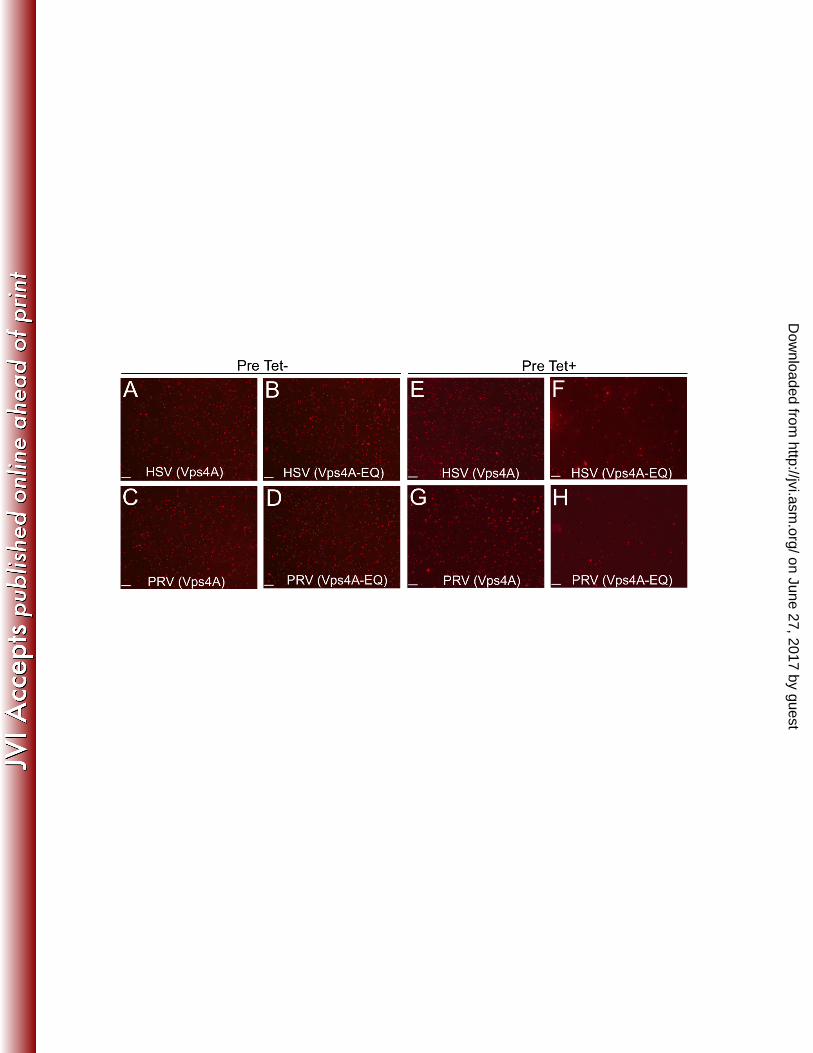
particles from Vps4A and Vps4A-EQ expressing cells under each of our tetracycline 247 induction conditions. HSV and PRV particles from Vps4A (Fig. 3A, C, E, G) or Vps4A-248 EQ-expressing cells (Figs. 3B, D, F, H) appeared comparable in apparent size and 249 brightness. Quantitation revealed that Vps4A/Vps4A-EQ expression from the time of 250 infection resulted in similar numbers of HSV and PRV particles in each cell type (Fig. 3I 251 and 3J, Pre Tet-). In contrast, Vps4A-EQ induction 16 hours prior to infection (Fig. 3I 252 and 3J, Pre Tet+) resulted in a 4.6 fold (HSV) and 2.2 fold (PRV) reduction in the 253 numbers of cytoplasmic particles when Vps4A-EQ was expressed, compared to Vps4A. 254 Because of this, we were careful in all subsequent experiments to test normalized 255 numbers of viral particles in our imaging chambers. Moreover, whether Vps4A/Vps4A-256 EQ expression was induced at the time of infection, or 16 hours earlier, the cytoplasmic 257 viral particles were found to exhibit identical trafficking properties (see below). 258 259
The dominant-negative form of Vps4A accumulates on cytoplasmic virions. 260 Whereas the wild type Vps4A protein cycles on and off membranes to complete 261 ESCRT-mediated envelopment, the Vps4A-EQ allele should become locked onto 262 enveloping virions during inhibition of their assembly. PNS samples from infected cells 263 were flowed into micro-chambers containing HiLyte FluorTM 488-labeled microtubules, 264 and fluorescent images collected in the red and green channels. Fig. 4 (A to D) shows 265 that GFP-tagged Vps4A-EQ could be readily visualized colocalizing with PRV capsid-266 containing cytoplasmic particles, and similar observations were made for HSV (data not 267 shown). Fig. 4E & 4F summarize the degree of colocalization of the Vps4A and Vps4A-268 EQ proteins with HSV and PRV cytoplasmic particles respectively. Relatively few 269
on June 27, 2017 by guesthttp://jvi.asm
.org/D
ownloaded from
13
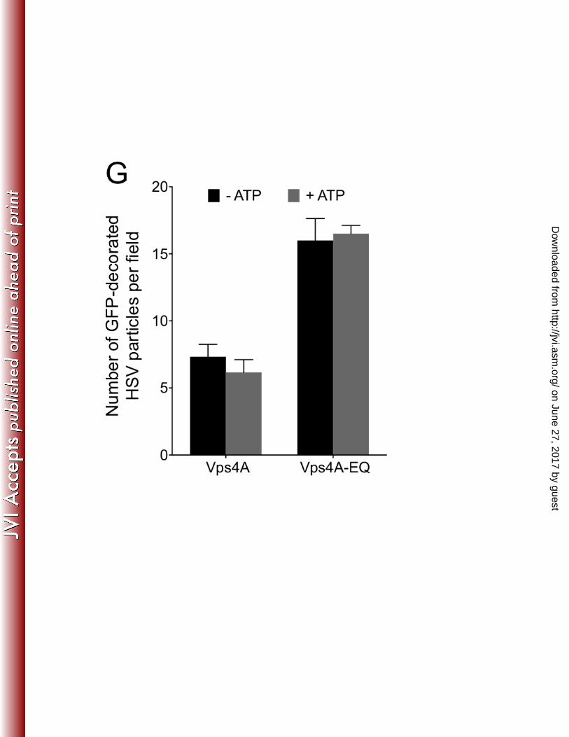
particles colocalized with wild type Vps4A (~2% and ~4% for HSV and PRV 270 respectively), reflecting its steady-state distribution during cycling. In contrast, Vps4A-271 EQ was found to decorate capsid-associated particles at a 3 to 5 fold higher level, 272 consistent with it locking on to enveloping structures in an irreversible fashion. Fig. 4G 273 shows that the presence or absence of ATP had no effect on the numbers of HSV 274 particles that were found to be decorated by Vps4A or Vps4A-EQ, suggesting that ATP-275 dependent release of Vps4A from organelles is not occurring in vitro under our 276 conditions. 277
278 Microtubule-dependent motility, but not microtubule-binding, is inhibited 279
when ESCRT-dependent envelopment is blocked. We next tested the microtubule-280 binding and motility properties of these cytoplasmic viral particles. Our earlier studies 281 used sucrose gradient density centrifugation to prepare buoyant, membrane-associated 282 HSV trafficking intermediates from the cytoplasm of infected cells (6, 40). However, to 283 ensure that all cytoplasmic virions including naked capsids, membrane-associated 284 enveloped virions, and any other assembly/trafficking intermediates were present, we 285 performed in vitro microtubule-binding and trafficking studies using total cytoplasmic 286 PNS samples. 287
HSV and PRV capsid-associated structures in the PNS bound to microtubules 288 with similar efficiencies whether they had been isolated from cells expressing Vps4A or 289 Vps4A-EQ and under both induction conditions (Figs. 5A and 5B and data not shown 290 Supplemental Figure S2). Upon addition of ATP to the microtubule-bound virions, the 291 HSV and PRV particles became motile, in both cases with a range of velocity and 292
on June 27, 2017 by guesthttp://jvi.asm
.org/D
ownloaded from
14
processivity similar to that in our earlier studies (6), and the presence of either Vps4A 293 allele did not affect the velocity or processivity of motile virions (data not shown). 294 However, the overall numbers of trafficking HSV and PRV particles were reduced by 295 approximately 50% as a result of Vps4A-EQ expression, compared to control (Figs. 5C 296 and 5D, and data not shown). We next examined that specific subpopulation of viral 297 particles demonstrating bound Vps4A or Vps4A-EQ, and therefore representing 298 envelopment intermediates. HSV and PRV virions that colocalized with Vps4A and 299 Vps4A-EQ were able to bind microtubules with an efficiency similar to the overall virion 300 population (Figs. 5E and 5F). However, fewer than 4% of HSV and PRV particles with 301 bound Vps4A or Vps4A-EQ moved along microtubules (Figs. 5G and 5H, and data not 302 shown). Table 1 summarizes the mean numbers of capsid-associated HSV and PRV 303 particles present in our imaging chambers (mRFP1+), that colocalized with Vps4A or 304 Vps4A-EQ (mRFP1+GFP+) and that became motile upon addition of ATP. 305
We previously showed that, for membrane-associated HSV particles, most of the 306 motility in our in vitro system is sensitive to AMP-PNP, which inhibits Kinesin but not 307 Dynein-mediated traffic under our conditions (6). Similarly, we found that the motility of 308 HSV in the PNS was more sensitive to AMP-PNP than to the Dynein inhibitor Sodium 309 Orthovanadate (Fig. 6). Those HSV particles which remained motile in the presence of 310 Vps4A-EQ were still sensitive to AMP-PNP but not Orthovanadate (Fig. 6), suggesting 311 this population utilizes Kinesin motors but not Dynein. 312
313 Quantitation of the relative numbers and in vitro properties of gD-314
associated and gD-negative cytoplasmic capsids. Vps4A and Vps4A-EQ 315
on June 27, 2017 by guesthttp://jvi.asm
.org/D
ownloaded from

15
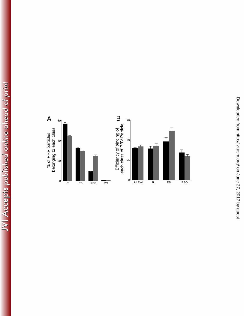
expressing cells were infected with HSV or a PRV strain expressing mRFP1-VP26 and 316 gD fused to the Blue Fluorescent Protein TagBFP (49). This fluorescent gD fusion is 317 known to be incorporated normally into the PRV envelope, as shown for a similar strain 318 expressing VP26-mRFP1 and gD-GFP (50). In our cytoplasmic particle preparations 319 gD-TagBFP is expected to be present in the envelopes of correctly assembled mature 320 PRV particles and in the bounding membranes of the organelles that contain PRV in 321 their lumen. PNS was prepared from Vps4A/Vps4A-EQ–expressing cell lines and viral 322 particles allowed to attach to microtubules, then capsids, gD and Vps4A/Vps4A-EQ 323 visualized by direct mRFP1, BFP and GFP fluorescence, respectively (Fig. 7, A-C). In 324 parallel, we performed similar experiments on microtubule-bound fixed and 325 permeabilized HSV particles (Fig. 8A-D), detecting gD by indirect immunocytochemistry. 326 In each case, alphaherpesvirus particles fluorescing in the Red (R), Red/Blue (RB) or 327 Red/Blue/Green (RBG) channels were quantitated and the data is presented for HSV in 328 Fig. 8E and PRV in Fig. 9A. 329
We next estimated the efficiency with which each class of PRV particle was able 330 to bind to microtubules, and found that gD-containing (Red/Blue) and gD-negative (Red 331 alone) capsids both bound efficiently (Fig. 9B). Upon addition of ATP (Fig. 9C) Red 332 and Red/Blue particles trafficked along the microtubules, though more Red than 333 Red/Blue particles became motile, despite the two populations being present in 334 comparable numbers (Fig. 9A) and able to bind with comparable efficiencies (Fig. 9B). 335 As we observed earlier (Fig. 5) the effect of Vps4A-EQ expression was to inhibit the 336 overall trafficking of all capsid-associated particles (termed “All Red” in Fig. 9C), 337 whether they colocalized with gD-TagBFP or not, by half. When considering the 338
on June 27, 2017 by guesthttp://jvi.asm
.org/D
ownloaded from

16
populations of capsid-associated particles separately, we observed that those with 339 detectable levels of gD-TagBFP fluorescence were more sensitive to the trafficking-340 inhibitory effects of Vps4A-EQ than were those lacking gD-TagBFP. Vps4A-EQ 341 expression reduced the trafficking of Red particles by about one quarter (R, Fig. 9C), 342 but that of Red/Blue particles by two thirds (RB, Fig. 9C). 343
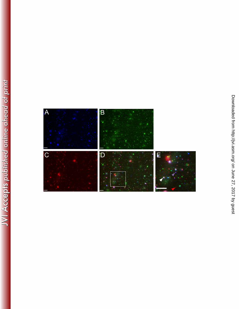
We reasoned that some of the Red particles lacking gD-TagBFP fluorescence 344 might be sensitive to the effects of Vps4A-EQ because they were membrane-345 associated, but lacked sufficient gD-TagBFP fluorescence for detection under our 346 conditions. To test whether the PNS contained such particles we stained PNS samples 347 identical to those examined in Fig. 9 (A-C) with the far-red fluorescent, lipophilic dye 348 1,1'-Dioctadecyl-3,3,3',3'-Tetramethylindodicarbocyanine Perchlorate (DiD). Far-red 349 DiD staining was imaged in the CY5 channel and is shown pseudo-colored Yellow in 350 Figs. 9D and 9F, but is absent from the minus-DiD control sample in Fig. 9E. Fig. 9F 351 shows a gallery of fluorescent particles collected from fields similar to that in Fig. 9D. 352 White arrowheads indicate capsids associated with both membranes and gD 353 fluorescence (Red/Yellow/Blue particles), and such virions are expected to exist. More 354 importantly, it is clear that the population of Red-alone particles (gD-TagBFP negative) 355 includes capsids that do not stain with DiD (Red alone, indicated by Red arrowheads) 356 and also those that do stain with DiD (Red/Yellow, indicated by Green arrowheads). 357 This is consistent with the notion that some of these gD-negative PRV capsids are truly 358 naked, and some are associated with membranes. 359
360 361
on June 27, 2017 by guesthttp://jvi.asm
.org/D
ownloaded from

17
DISCUSSION 362 We have used our previously established in vitro assay to test the consequences 363
of inhibition of ESCRT-mediated envelopment for microtubule-dependent motility of the 364 alphaherpesviruses HSV and PRV. We first tested whether PRV replication is inhibited 365 by expression of the dominant negative Vps4A-EQ allele. We compared the effects of 366 two different sets of tetracycline-induction conditions in most of our studies, initiating 367 Vps4A/Vps4A-EQ expression either 16 hours prior to the time of viral infection (Pre 368 Tet+) or at the time of infection (Pre Tet-). In both cases tetracycline-mediated induction 369 was then continued throughout the entire time course of viral replication. We found that, 370 as previously reported for HSV (9), expression of Vps4A-EQ resulted in a dramatic 371 effect on overall viral titer, reducing it by three to four logs for both HSV and PRV (Fig. 372 2A and 2B). Both Pre Tet- and Pre Tet+ conditions gave similar results. The principal 373 effect of Vps4A-EQ was upon viral assembly, since induction of Vps4A-EQ expression, 374 whether at the time of infection or 16 hours prior, had little effect on levels of expression 375 of the HSV structural proteins VP5 and VP26 (the latter detected by virtue of its fusion 376 to mRFP1) compared to the Vps4A control (Fig. 2C). Similarly, for PRV expression of 377 Vps4A-EQ from the time of infection (Pre Tet-) had no effect on expression of VP26-378 mRFP1 and a ~75kDa structural protein (Fig. 2D), though some reduction in their levels 379 was apparent in the Pre Tet+ conditions (Fig. 2D). 380
When Post Nuclear Supernatants (PNS) were prepared from infected cells and 381 capsid-bound VP26-mRFP1 examined by fluorescence microscopy, viral capsids or 382 capsid-associated particles were readily apparent (Fig. 3, A-H). Similar numbers of 383 particles were present in Vps4A and Vps4A-EQ expressing cells when expression was 384
on June 27, 2017 by guesthttp://jvi.asm
.org/D
ownloaded from

18
induced at the time of infection (Pre Tet-) Fig. 3 (I-J). However particles numbers were 385 reduced approximately five fold for HSV (Fig. 3I) and two fold for PRV (Fig. 3J) 386 (compared to wild type Vps4A) when expression was induced 16 hours prior to 387 infection. This correlates with some reduction in the levels of structural proteins under 388 these induction conditions (Fig. 2), and the observation that long term expression of 389 Vps4A-EQ can be toxic to cells (9). In subsequent studies we were careful to normalize 390 numbers of particles examined when comparing the effects of Vps4A and Vps4A-EQ 391 expression from Pre Tet+ cells. 392
The accumulation of GFP-tagged forms of Vps4A and Vps4A-EQ on cytoplasmic 393 virions was readily detectable (Fig. 4, A-D). Since wild type Vps4A binds transiently to 394 complete the process of ESCRT-driven envelopment, Vps4A-labeled structures likely 395 represent the steady state levels of HSV and PRV capsids that were undergoing 396 envelopment at the time cell extracts were prepared. In contrast, the larger numbers of 397 Vps4A-EQ-labeled particles (Fig. 4E and 4F) would be expected to represent a 398 population that is accumulating due to inhibition of the ATPase cycle, locking Vps4A-EQ 399 irreversibly onto the surface of the membrane. Nevertheless, both Vps4A and Vps4A-400 EQ decorated-structures would be expected to represent partially enveloped capsids 401 and to have similar biological properties; indeed, both bind efficiently to microtubules 402 (Figs. 5E and 5F) but show very little motility (Figs. 5G and 5H). Since fewer capsid-403 associated particles became decorated by wild-type Vps4A than by Vps4A-EQ (Fig 4E 404 and 4F), motile Vps4A-bound virions were barely detectable (Figs. 5G and 5H). 405
Arresting envelopment with Vps4A-EQ renders Vps4A-EQ-associated capsids 406 unable to traffic: what are the consequences of this for the total population of 407
on June 27, 2017 by guesthttp://jvi.asm
.org/D
ownloaded from

19
cytoplasmic virions? There was no effect of Vps4A-EQ upon the overall ability of 408 cytoplasmic HSV and PRV capsids to bind microtubules (Figs. 5A and 5B), which is to 409 be expected since even Vps4A/Vps4A-EQ-associated capsids bind microtubules 410 efficiently (Figs. 5E and 5F). However, the expression of Vps4A-EQ reduced the overall 411 numbers of trafficking HSV and PRV particles by approximately 50% compared to wild-412 type controls (Figs. 5C and 5D). By considering absolute numbers of trafficking 413 particles in a field, it is apparent that the numbers of Vps4A-EQ-bound, non-motile 414 capsids that accumulated in Vps4A-EQ-expressing cells are more than sufficient to 415 account for all of the 50% loss in overall motility (Table 1). 416
The naked HSV capsid is able to bind multiple molecular motors for both 417 anterograde and retrograde traffic (26) and HSV and PRV are believed to move 418 bidirectionally during egress (33, 54-56). To further characterize the trafficking 419 properties of viral particles in the presence and absence of Vps4A/Vps4A-EQ we 420 examined their sensitivity to AMP-PNP and sodium orthovanadate, inhibitors of Kinesin 421 and Dynein, respectively. As shown in Fig. 6, most trafficking of HSV particles in control 422 cells expressing Vps4A was sensitive to AMP-PNP, and to a lesser extent inhibited by 423 sodium orthovanadate, as we previously described (6). That population of viral particles 424 remaining motile in the presence of Vps4A-EQ could not be further inhibited by 425 orthovanadate but were still sensitive to AMP-PNP, suggesting they had retained 426 primarily Kinesin-mediated motility. 427
Vps4A-EQ should manifest its inhibitory effects predominantly upon enveloped 428 and membrane-associated capsids. To test this we made use of a dual-labeled PRV 429 strain, expressing mRFP1-tagged VP26 and the envelope protein gD fused to TagBFP. 430
on June 27, 2017 by guesthttp://jvi.asm
.org/D
ownloaded from

20
Imaging (Fig. 7) and quantitation of particle numbers (Fig. 9) enabled us to measure the 431 microtubule-binding and motility of gD-labeled (membrane-associated) capsids and gD-432 negative (naked capsids or membrane-associated capsids lacking detectable levels of 433 gD) in the same microscopic field in the same imaging chamber. We also performed 434 microtubule-binding studies of gD-associated and gD-negative HSV capsids using anti-435 gD immunostaining in fixed samples (Fig. 8A-E). As shown in Fig. 9A, approximately 436 half of the PRV particles in our preparations fluoresced only in the Red channel (termed 437 R in Fig. 9A), and lacked detectable levels of gD-TagBFP. The other half of the 438 mRFP1-labeled capsids colocalized with Blue fluorescence (termed RB or RBG in Fig. 439 9A), implying close proximity to gD-TagBFP-associated membranes. Approximately 440 one fifth of all gD-positive capsids also colocalized with Vps4A, and almost half became 441 decorated by the irreversible binding of Vps4A-EQ (Vps4A/Vps4A-EQ-labeled particles 442 are termed RBG in Fig. 9A). Only small numbers of PRV Red/Green particles were 443 observed (RG in Fig. 9A). Similar results were obtained by anti-gD immunostaining of 444 HSV, except that larger numbers of RG particles were apparent (Fig. 8E). This could be 445 due to greater numbers of enveloped HSV capsids lacking detectable gD in their 446 membrane, or might reflect inefficient immunostaining by the anti-gD antibody. 447
For the dual fluorescence-labeled PRV strain, the efficiency with which each 448 population of particles bound to microtubules was largely the same (Fig. 9B) whether 449 Vps4A or Vps4A-EQ was being expressed, consistent with our earlier data for HSV and 450 PRV in Fig. 5. However we did see some increase in the efficiency of Red/Blue (RB) 451 particle binding (from about 50% to about 60%) in the presence of Vps4A-EQ (Fig. 9B). 452 We are at present unsure of the reasons for this. 453
on June 27, 2017 by guesthttp://jvi.asm
.org/D
ownloaded from
21
Addition of ATP enabled us to test the relative motility of gD-positive and gD-454 negative capsids in vitro (Fig. 9C). In Vps4A-expressing (control) cells the majority of 455 motile particles were Red (R in Fig. 9C), lacking any other fluorescence; approximately 456 25% of these particles trafficked along microtubules. In contrast, about 4% of the Red 457 particles that colocalized with gD-TagBFP fluorescence (RB in Fig. 9C) were motile 458 when prepared from Vps4A cells. This could be a consequence of only small numbers 459 of PRV capsids completing a productive envelopment/egress pathway, generating an 460 enveloped, membrane-bounded particle that contains gD (and other envelope proteins) 461 and is competent for trafficking and export from the cell. Alternatively, the biochemical 462 conditions of our in vitro system may favor the trafficking of the Red population of 463 particles over those that are Red/Blue. 464
Although we do not know how many of the Red particles are truly naked capsids, 465 and how many are membrane-associated but lack detectable levels of gD-TagBFP, it is 466 reasonable to presume that the Red/Blue particles are predominantly enveloped or 467 membrane-associated PRV capsids, and that the Red particles contain at least a 468 mixture of naked capsids and enveloped virions. Consistent with this, the populations 469 were differentially sensitive to the effects of Vps4A-EQ; expression of Vps4A-EQ 470 reduced the motility of Red particles by one quarter, and Red/Blue particles by two 471 thirds (Fig. 9C). Furthermore, staining with the lipophilic dye DiD revealed that Red 472 particles included capsids that were, and were not, membrane-associated, despite 473 lacking detectable levels of gD-TagBFP (Fig. 9, D-F). At the moment we have only 474 been successful in staining fixed particles with such membrane-fluorescent dyes, but 475 are attempting to develop labeling conditions that are compatible with our in vitro assay. 476
on June 27, 2017 by guesthttp://jvi.asm
.org/D
ownloaded from

22
This will enable us to continue to examine the structure of the various populations of 477 trafficking virion. 478
The incomplete nature of Vps4A-EQ-inhibition on enveloped PRV particles is 479 likely a consequence of the partial effect of the Vps4A-EQ dominant negative allele, 480 which has previously been observed for Vps4-dependent intracellular protein sorting 481 events (57, 58). The extent of dominant-negative inhibition is dependent upon the 482 levels of Vps4A-EQ that become incorporated into the Vps4 dodecamer, and also 483 whether Vps4A-EQ subunits are among those that stochastically engage with ESCRT-484 III during each cycle of scission (18, 19). 485
For both HSV and PRV we consistently found that viral particles with bound 486 Vps4A/Vps4A-EQ were able to bind to microtubules but could not traffic along them. 487 This suggests that molecular motors are present on the surface of these particles and 488 able to mediate microtubule-attachment, but are not active in transport. Elegant studies 489 by the Enquist and Johnson laboratories have demonstrated roles for US9p and the 490 gE/gI heterodimer in recruiting Kinesin to HSV and PRV particles (28, 30, 35, 36). 491 Organelles into which HSV and PRV bud to acquire their envelopes are therefore 492 expected to contain US9p and/or gE/gI in their bounding membranes to recruit Kinesin 493 for subsequent anterograde traffic. On the basis of our findings we speculate that 494 US9p/gE/gI-bound motors are not active until after viral capsids have completed Vps4-495 dependent scission. Entry of a sealed, completely enveloped capsid into the lumen of 496 the bounding organelle would then signal completion of assembly, via the US9p/gE/gI 497 complex or other factors, activating bound Kinesin on the cytoplasmic surface of the 498 particle. 499
on June 27, 2017 by guesthttp://jvi.asm
.org/D
ownloaded from

23
500 ACKNOWLEDGMENTS 501
This work was supported by National Institutes of Health Grant AI083285 (to D.W.W). 502 We thank Lily Huang for excellent technical assistance and Drs. Margaret Kielian, John 503 Murray, Xin Tao and Allan Wolkoff for helpful discussions. We thank Dr. Gregory Smith 504 (Northwestern University School of Medicine, Chicago, IL.) for providing the HSV and 505 PRV strains used in this study. The Vps4A-inducible cell lines were a kind gift from Dr. 506 Margaret Kielian, and the anti-mRFP1 antibody was generously provided by Dr. Erik 507 Snapp. 508 509 510 511
on June 27, 2017 by guesthttp://jvi.asm
.org/D
ownloaded from

24
REFERENCES 512 1. Browne H, Bell S, Minson T, Wilson DW. 1996. An endoplasmic reticulum-513
retained herpes simplex virus glycoprotein H is absent from secreted virions: 514 evidence for reenvelopment during egress. Journal of virology 70:4311-4316. 515
2. Skepper JN, Whiteley A, Browne H, Minson A. 2001. Herpes simplex virus 516 nucleocapsids mature to progeny virions by an envelopment --> deenvelopment --517 > reenvelopment pathway. Journal of virology 75:5697-5702. 518
3. Mettenleiter TC, Klupp BG, Granzow H. 2006. Herpesvirus assembly: a tale of 519 two membranes. Current opinion in microbiology 9:423-429. 520
4. Turcotte S, Letellier J, Lippe R. 2005. Herpes simplex virus type 1 capsids 521 transit by the trans-Golgi network, where viral glycoproteins accumulate 522 independently of capsid egress. Journal of virology 79:8847-8860. 523
5. Harley CA, Dasgupta A, Wilson DW. 2001. Characterization of herpes simplex 524 virus-containing organelles by subcellular fractionation: role for organelle 525 acidification in assembly of infectious particles. Journal of virology 75:1236-1251. 526
6. Lee GE, Murray JW, Wolkoff AW, Wilson DW. 2006. Reconstitution of herpes 527 simplex virus microtubule-dependent trafficking in vitro. Journal of virology 528 80:4264-4275. 529
7. Hollinshead M, Johns HL, Sayers CL, Gonzalez-Lopez C, Smith GL, Elliott G. 530 2012. Endocytic tubules regulated by Rab GTPases 5 and 11 are used for 531 envelopment of herpes simplex virus. The EMBO journal 31:4204-4220. 532
on June 27, 2017 by guesthttp://jvi.asm
.org/D
ownloaded from
25
8. Johns HL, Gonzalez-Lopez C, Sayers CL, Hollinshead M, Elliott G. 2014. 533 Rab6 dependent post-Golgi trafficking of HSV1 envelope proteins to sites of virus 534 envelopment. Traffic 15:157-178. 535
9. Crump CM, Yates C, Minson T. 2007. Herpes simplex virus type 1 cytoplasmic 536 envelopment requires functional Vps4. Journal of virology 81:7380-7387. 537
10. Pawliczek T, Crump CM. 2009. Herpes simplex virus type 1 production requires 538 a functional ESCRT-III complex but is independent of TSG101 and ALIX 539 expression. Journal of virology 83:11254-11264. 540
11. Alam SL, Sundquist WI. 2007. Structural biology: ESCRT service. Nature 541 447:921-922. 542
12. McCullough J, Colf LA, Sundquist WI. 2013. Membrane fission reactions of the 543 mammalian ESCRT pathway. Annual review of biochemistry 82:663-692. 544
13. Stuchell-Brereton MD, Skalicky JJ, Kieffer C, Karren MA, Ghaffarian S, 545 Sundquist WI. 2007. ESCRT-III recognition by VPS4 ATPases. Nature 449:740-546 744. 547
14. Votteler J, Sundquist WI. 2013. Virus budding and the ESCRT pathway. Cell 548 host & microbe 14:232-241. 549
15. Morita E, Sandrin V, McCullough J, Katsuyama A, Baci Hamilton I, 550 Sundquist WI. 2011. ESCRT-III protein requirements for HIV-1 budding. Cell host 551 & microbe 9:235-242. 552
16. Babst M, Wendland B, Estepa EJ, Emr SD. 1998. The Vps4p AAA ATPase 553 regulates membrane association of a Vps protein complex required for normal 554 endosome function. The EMBO journal 17:2982-2993. 555
on June 27, 2017 by guesthttp://jvi.asm
.org/D
ownloaded from

26
17. Babst M, Sato TK, Banta LM, Emr SD. 1997. Endosomal transport function in 556 yeast requires a novel AAA-type ATPase, Vps4p. The EMBO journal 16:1820-557 1831. 558
18. Shestakova A, Hanono A, Drosner S, Curtiss M, Davies BA, Katzmann DJ, 559 Babst M. 2010. Assembly of the AAA ATPase Vps4 on ESCRT-III. Molecular 560 biology of the cell 21:1059-1071. 561
19. Davies BA, Azmi IF, Payne J, Shestakova A, Horazdovsky BF, Babst M, 562 Katzmann DJ. 2010. Coordination of substrate binding and ATP hydrolysis in 563 Vps4-mediated ESCRT-III disassembly. Molecular biology of the cell 21:3396-564 3408. 565
20. Bishop N, Woodman P. 2000. ATPase-defective mammalian VPS4 localizes to 566 aberrant endosomes and impairs cholesterol trafficking. Molecular biology of the 567 cell 11:227-239. 568
21. Yoshimori T, Yamagata F, Yamamoto A, Mizushima N, Kabeya Y, Nara A, 569 Miwako I, Ohashi M, Ohsumi M, Ohsumi Y. 2000. The mouse SKD1, a 570 homologue of yeast Vps4p, is required for normal endosomal trafficking and 571 morphology in mammalian cells. Molecular biology of the cell 11:747-763. 572
22. Taylor GM, Hanson PI, Kielian M. 2007. Ubiquitin depletion and dominant-573 negative VPS4 inhibit rhabdovirus budding without affecting alphavirus budding. 574 Journal of virology 81:13631-13639. 575
23. Kramer T, Greco TM, Enquist LW, Cristea IM. 2011. Proteomic characterization 576 of pseudorabies virus extracellular virions. Journal of virology 85:6427-6441. 577
on June 27, 2017 by guesthttp://jvi.asm
.org/D
ownloaded from

27
24. Sandbaumhuter M, Dohner K, Schipke J, Binz A, Pohlmann A, Sodeik B, 578 Bauerfeind R. 2013. Cytosolic herpes simplex virus capsids not only require 579 binding inner tegument protein pUL36 but also pUL37 for active transport prior to 580 secondary envelopment. Cellular microbiology 15:248-269. 581
25. Luxton GW, Lee JI, Haverlock-Moyns S, Schober JM, Smith GA. 2006. The 582 pseudorabies virus VP1/2 tegument protein is required for intracellular capsid 583 transport. Journal of virology 80:201-209. 584
26. Radtke K, Kieneke D, Wolfstein A, Michael K, Steffen W, Scholz T, Karger A, 585 Sodeik B. 2010. Plus- and minus-end directed microtubule motors bind 586 simultaneously to herpes simplex virus capsids using different inner tegument 587 structures. PLoS pathogens 6:e1000991. 588
27. Penfold ME, Armati P, Cunningham AL. 1994. Axonal transport of herpes 589 simplex virions to epidermal cells: evidence for a specialized mode of virus 590 transport and assembly. Proceedings of the National Academy of Sciences of the 591 United States of America 91:6529-6533. 592
28. Kratchmarov R, Taylor MP, Enquist LW. 2012. Making the case: married versus 593 separate models of alphaherpes virus anterograde transport in axons. Reviews in 594 medical virology 22:378-391. 595
29. Wisner TW, Sugimoto K, Howard PW, Kawaguchi Y, Johnson DC. 2011. 596 Anterograde transport of herpes simplex virus capsids in neurons by both 597 separate and married mechanisms. Journal of virology 85:5919-5928. 598
on June 27, 2017 by guesthttp://jvi.asm
.org/D
ownloaded from

28
30. Snyder A, Polcicova K, Johnson DC. 2008. Herpes simplex virus gE/gI and 599 US9 proteins promote transport of both capsids and virion glycoproteins in 600 neuronal axons. Journal of virology 82:10613-10624. 601
31. Howard PW, Howard TL, Johnson DC. 2013. Herpes simplex virus membrane 602 proteins gE/gI and US9 act cooperatively to promote transport of capsids and 603 glycoproteins from neuron cell bodies into initial axon segments. Journal of 604 virology 87:403-414. 605
32. Ibiricu I, Huiskonen JT, Dohner K, Bradke F, Sodeik B, Grunewald K. 2011. 606 Cryo electron tomography of herpes simplex virus during axonal transport and 607 secondary envelopment in primary neurons. PLoS pathogens 7:e1002406. 608
33. Antinone SE, Zaichick SV, Smith GA. 2010. Resolving the assembly state of 609 herpes simplex virus during axon transport by live-cell imaging. Journal of virology 610 84:13019-13030. 611
34. Negatsch A, Granzow H, Maresch C, Klupp BG, Fuchs W, Teifke JP, 612 Mettenleiter TC. 2010. Ultrastructural analysis of virion formation and intraaxonal 613 transport of herpes simplex virus type 1 in primary rat neurons. Journal of virology 614 84:13031-13035. 615
35. Kratchmarov R, Kramer T, Greco TM, Taylor MP, Ch'ng TH, Cristea IM, 616 Enquist LW. 2013. Glycoproteins gE and gI are required for efficient KIF1A-617 dependent anterograde axonal transport of alphaherpesvirus particles in neurons. 618 Journal of virology 87:9431-9440. 619
on June 27, 2017 by guesthttp://jvi.asm
.org/D
ownloaded from
29
36. Cunningham A, Miranda-Saksena M, Diefenbach R, Johnson D. 2013. Letter 620 in response to: Making the case: Married versus Separate models of alphaherpes 621 virus anterograde transport in axons. Reviews in medical virology 23:414-418. 622
37. Johnson DC, Baines JD. 2011. Herpesviruses remodel host membranes for 623 virus egress. Nature reviews. Microbiology 9:382-394. 624
38. Kramer T, Enquist LW. 2013. Directional spread of alphaherpesviruses in the 625 nervous system. Viruses 5:678-707. 626
39. Desai P, Person S. 1998. Incorporation of the green fluorescent protein into the 627 herpes simplex virus type 1 capsid. Journal of virology 72:7563-7568. 628
40. Shanda SK, Wilson DW. 2008. UL36p is required for efficient transport of 629 membrane-associated herpes simplex virus type 1 along microtubules. Journal of 630 virology 82:7388-7394. 631
41. Zaichick SV, Bohannon KP, Hughes A, Sollars PJ, Pickard GE, Smith GA. 632 2013. The herpesvirus VP1/2 protein is an effector of dynein-mediated capsid 633 transport and neuroinvasion. Cell host & microbe 13:193-203. 634
42. Radtke K, Dohner K, Sodeik B. 2006. Viral interactions with the cytoskeleton: a 635 hitchhiker's guide to the cell. Cellular microbiology 8:387-400. 636
43. Dodding MP, Way M. 2011. Coupling viruses to dynein and kinesin-1. The EMBO 637 journal 30:3527-3539. 638
44. Desai PJ. 2000. A null mutation in the UL36 gene of herpes simplex virus type 1 639 results in accumulation of unenveloped DNA-filled capsids in the cytoplasm of 640 infected cells. Journal of virology 74:11608-11618. 641
on June 27, 2017 by guesthttp://jvi.asm
.org/D
ownloaded from

30
45. Bottcher S, Granzow H, Maresch C, Mohl B, Klupp BG, Mettenleiter TC. 2007. 642 Identification of functional domains within the essential large tegument protein 643 pUL36 of pseudorabies virus. Journal of virology 81:13403-13411. 644
46. Bottcher S, Maresch C, Granzow H, Klupp BG, Teifke JP, Mettenleiter TC. 645 2008. Mutagenesis of the active-site cysteine in the ubiquitin-specific protease 646 contained in large tegument protein pUL36 of pseudorabies virus impairs viral 647 replication in vitro and neuroinvasion in vivo. Journal of virology 82:6009-6016. 648
47. Smith GA, Pomeranz L, Gross SP, Enquist LW. 2004. Local modulation of plus-649 end transport targets herpesvirus entry and egress in sensory axons. Proceedings 650 of the National Academy of Sciences of the United States of America 101:16034-651 16039. 652
48. Antinone SE, Smith GA. 2010. Retrograde axon transport of herpes simplex 653 virus and pseudorabies virus: a live-cell comparative analysis. Journal of virology 654 84:1504-1512. 655
49. Subach OM, Gundorov IS, Yoshimura M, Subach FV, Zhang J, Gruenwald D, 656 Souslova EA, Chudakov DM, Verkhusha VV. 2008. Conversion of red 657 fluorescent protein into a bright blue probe. Chemistry & biology 15:1116-1124. 658
50. Bohannon KP, Jun Y, Gross SP, Smith GA. 2013. Differential protein 659 partitioning within the herpesvirus tegument and envelope underlies a complex 660 and variable virion architecture. Proceedings of the National Academy of Sciences 661 of the United States of America 110:E1613-1620. 662
51. Church GA, Wilson DW. 1997. Study of herpes simplex virus maturation during a 663 synchronous wave of assembly. Journal of virology 71:3603-3612. 664
on June 27, 2017 by guesthttp://jvi.asm
.org/D
ownloaded from

31
52. Murray JW, Bananis E, Wolkoff AW. 2000. Reconstitution of ATP-dependent 665 movement of endocytic vesicles along microtubules in vitro: an oscillatory 666 bidirectional process. Molecular biology of the cell 11:419-433. 667
53. Howard J, Hyman AA. 1993. Preparation of marked microtubules for the assay 668 of the polarity of microtubule-based motors by fluorescence microscopy. Methods 669 in cell biology 39:105-113. 670
54. Smith GA, Gross SP, Enquist LW. 2001. Herpesviruses use bidirectional fast-671 axonal transport to spread in sensory neurons. Proceedings of the National 672 Academy of Sciences of the United States of America 98:3466-3470. 673
55. Luxton GW, Haverlock S, Coller KE, Antinone SE, Pincetic A, Smith GA. 674 2005. Targeting of herpesvirus capsid transport in axons is coupled to association 675 with specific sets of tegument proteins. Proceedings of the National Academy of 676 Sciences of the United States of America 102:5832-5837. 677
56. Antinone SE, Smith GA. 2006. Two modes of herpesvirus trafficking in neurons: 678 membrane acquisition directs motion. Journal of virology 80:11235-11240. 679
57. Fujita H, Yamanaka M, Imamura K, Tanaka Y, Nara A, Yoshimori T, Yokota S, 680 Himeno M. 2003. A dominant negative form of the AAA ATPase SKD1/VPS4 681 impairs membrane trafficking out of endosomal/lysosomal compartments: class E 682 vps phenotype in mammalian cells. Journal of cell science 116:401-414. 683
58. Hislop JN, Marley A, Von Zastrow M. 2004. Role of mammalian vacuolar 684 protein-sorting proteins in endocytic trafficking of a non-ubiquitinated G protein-685 coupled receptor to lysosomes. The Journal of biological chemistry 279:22522-686 22531. 687
on June 27, 2017 by guesthttp://jvi.asm
.org/D
ownloaded from
32
688 FIGURE LEGENDS 689
FIG. 1. Vps4-EQ is expected to differentially perturb the microtubule-dependent 690 trafficking of naked and enveloped alphaherpesvirus capsids. The assembly of 691 enveloped trafficking particles (Steps 1-3) requires that cytoplasmic capsids (black 692 discs) engage organelles (Step 1) to undergo ESCRT-mediated envelopment (Step 2). 693 Vps4 (grey-rings)-driven scission (Step 3) then generates enveloped, organelle 694 bounded-virions that subsequently traffic along microtubules. In this model, Vps4-EQ-695 arrested envelopment (Step 3) occurs before the generation of motile particles. 696 Alternatively, for naked capsid trafficking prior to envelopment (Steps a-c), cytoplasmic 697 capsids first travel along microtubules (Step a) to reach the site at which they undergo 698 ESCRT-mediated budding (Step b) and scission (Step c). In this case, Vps4-EQ blocks 699 envelopment (Step c) after microtubule-dependent motility has occurred. 700 701 on June 27, 2017 by guest
http://jvi.asm.org/
Dow
nloaded from

33
FIG. 2. Cell lines expressing Vps4A-EQ inhibit production of HSV and PRV infectious 702 particles. Cells expressing tetracycline-inducible Vps4A (circles) or Vps4A-EQ 703 (squares) were incubated with tetracycline from either 16 hours prior to infection (Pre 704 Tet+, solid symbols) or at the time of infection (Pre Tet-, open symbols). They were 705 then infected with HSV (A) or PRV (B) at a multiplicity of 10 for 1 hour at 37oC, acid 706 washed to inactivate unpenetrated virus and incubated in the continued presence of 707 tetracycline for the times shown, harvested and titer determined. Plotted values are 708 mean and standard deviation from the mean for duplicate (A) and triplicate (B) dishes 709 each titrated in duplicate. (C, D). Cells were grown and infected by HSV (C) or PRV (D) 710 similarly to A and B except that PNS was prepared at 16 hours post infection, subjected 711 to SDS PAGE and Western blotted for HSV-VP5, mRFP1 or a 75kDa PRV structural 712 protein as indicated. For panels C and D, each pair of lanes corresponds to Vps4A and 713 Vps4A-EQ cells respectively, under the following conditions. Lanes 1, 2: Infected, no 714 Tet preinduction (Pre Tet-). Lanes 3, 4: Uninfected, with Tet preinduction (Pre Tet+). 715 Lanes 5, 6: Infected, with Tet preinduction (Pre Tet+). 716 717
on June 27, 2017 by guesthttp://jvi.asm
.org/D
ownloaded from

34
FIG. 3. Visualization of HSV and PRV-associated particles prepared from the 718 cytoplasm of Vps4A and Vps4A-EQ expressing cells. Cells were preinduced (PreTet+) 719 or not preinduced (Pre Tet-) with tetracycline for 16 hours, then infected with HSV (A, B, 720 E, F) or PRV (C, D, G, H) in the presence of tetracycline, to express Vps4A or Vps4A-721 EQ as indicated. After 16 hours PNS samples were prepared and mRFP1-decorated 722 capsids imaged by fluorescence microscopy in the red channel. Representative fields 723 are shown. Scale bars represent 10μm. (I, J) Quantitation of particle numbers from 724 fields similar to those shown in panels A to H. For both HSV and PRV, particle fields 725 from five independent imaging chambers were counted for each cell-type and condition. 726 Plotted values represent mean particle numbers per field, and standard deviation from 727 the mean. Total numbers of particles counted (n=) for each virus, cell line and induction 728 condition are indicated under the bars. 729 730 on June 27, 2017 by guest
http://jvi.asm.org/
Dow
nloaded from

35
FIG. 4. Cytoplasmic viral particles bound by GFP-tagged Vps4A and Vps4A-EQ. 731 PNS samples prepared from HSV or PRV-infected Vps4A or Vps4A-EQ-expressing 732 cells were flowed into an imaging chamber precoated with green fluorescent 733 microtubules. (A-D) Representative field of mRFP1-PRV particles prepared from 734 Vps4A-EQ-expressing cells and viewed in the green (A) or red (B) channels or merged 735 (C). (D) 4.5x magnification of the region boxed in C. White arrowheads indicate two 736 PRV capsid-associated particles decorated by Vps4A-EQ. Scale bar in all panels 737 represents 10μm. (E, F) Quantitation of colocalization of mRFP1-tagged HSV and PRV 738 capsids, respectively, with GFP-tagged Vps4A or Vps4A-EQ. Vps4A (black bars) or 739 Vps4A-EQ (grey bars) expression was induced at the time of infection (Pre Tet-) or from 740 16 hours prior to infection (Pre Tet+), as indicated. Plotted values show the mean and 741 standard deviation from the mean. Fields counted, and numbers of particles counted in 742 each case, were identical to those in Fig. 3I and 3J. (G) ATP addition does not change 743 the numbers of Vps4A and Vps4A-EQ labeled HSV particles in vitro. PNS samples were 744 prepared from HSV-infected Vps4A and Vps4A-EQ expressing cells then flowed into an 745 imaging chamber precoated with microtubules, The chambers were incubated in the 746 absence (black bars) or presence (grey bars) of ATP and images of microscopic fields 747 recorded in the red (capsid) and green (Vps4A/Vps4A-EQ) channels. Plotted values 748 represent mean and standard deviation from the mean. Total numbers of particles 749 examined, in the absence and presence of ATP respectively, were as follows: 344 and 750 255 (Vps4A cells), 151 and 173 (Vps4A-EQ cells). 751 752 753 754
on June 27, 2017 by guesthttp://jvi.asm
.org/D
ownloaded from

36
FIG. 5. Vps4A-EQ binding inhibits the motility of HSV and PRV particles. Cells were 755 induced with tetracycline to express Vps4A or Vps4A-EQ at the time of infection by PRV 756 and HSV (Pre Tet-) or were induced 16 hours prior to HSV infection (Pre Tet+) as in Fig. 757 3. Induction was continued for the following 16 hours then a PNS prepared and viral 758 particles tested for their ability to bind and traffic along microtubules. Percent binding 759 was calculated by comparing numbers of capsid-associated particles added to the 760 imaging chamber with the number bound to microtubules after washing. Motility 761 efficiency was calculated by comparing numbers of capsid-associated particles bound 762 to microtubules to those that became motile following addition of ATP. (A, B) Efficiency 763 of microtubule binding of all HSV or PRV particles. (C, D) Efficiency of motility of all 764 bound HSV or PRV particles. (E, F) Efficiency of microtubule binding by that 765 subpopulation of HSV and PRV viral particles that were associated with Vps4A or 766 Vps4A-EQ. (G, H) Efficiency of motility of microtubule-bound Vps4A or Vps4A-EQ-767 decorated HSV and PRV particles. For all panels, plotted values represent the mean 768 and standard deviation from the mean. Total numbers of particles counted (n=) for each 769 virus, cell line and induction condition are indicated under the bars in panels A and B. 770 771 772
on June 27, 2017 by guesthttp://jvi.asm
.org/D
ownloaded from

37
FIG. 6. Effects of Kinesin and Dynein inhibitors. HSV particles were prepared from 773 the cytoplasm of Vps4A and Vps4A-EQ expressing cells and motility measured in the 774 presence of ATP supplemented with: buffer (no inhibitor-black bars), 1 mM AMP-PNP 775 (light gray bars), 5 μM Sodium Orthovanadate (dark gray bars) or a mixture of the 776 inhibitors together (white bars). Plotted values represent the mean and standard 777 deviation from the mean. Numbers of HSV particles examined under each condition, for 778 Vps4A and Vps4A-EQ samples respectively, were as follows: No inhibitor (1149, 942). 779 AMP-PNP (1069, 1099). Sodium Orthovanadate (778, 954). AMP-PNP+Sodium 780 Orthovanadate (2073, 745). 781 782 783 784 FIG. 7. Representative field of dual-labeled PRV particles from the cytoplasm of 785 Vps4A-EQ expressing cells. PNS samples prepared from PRV-infected Vps4A-EQ-786 expressing cells (Pre Tet- conditions) were flowed into a micro-chamber precoated with 787 green fluorescent microtubules. (A-D) Representative field of PRV particles viewed in 788 the Blue (A), Green (B), Red (C) channels or merged (D). (E) 2.7x magnification of the 789 region boxed in D. Red arrowhead indicates PRV capsid fluorescing only in the Red 790 channel, purple arrowhead indicates gD-TagBFP positive Red capsid and white 791 arrowhead indicates Red capsid colocalizing with gD-TagBFP and Vps4A-EQ-GFP. 792 Scale bar in all panels represents 10μm. 793 794
on June 27, 2017 by guesthttp://jvi.asm
.org/D
ownloaded from

38
FIG. 8. PNS was prepared from HSV-infected cells and flowed into a micro-chamber 795 precoated with green fluorescent microtubules, fixed and probed with an anti-gD mouse 796 monoclonal antibody (panels A and C) or control lacking primary antibody (panels B and 797 D) then with an Alexa Fluor 350 labeled secondary antibody. Images were collected in 798 the red (mRFP1-VP26 capsids), green (Vps4A/Vps4A-EQ-GFP and microtubules) and 799 blue (anti-gD) channels then merged. (A, B) PNS from Vps4A expressing cells. (C, D) 800 PNS from Vps4A-EQ expressing cells. Scale bars represent 10μm. (E) Quantitation of 801 microtubule-binding by each population of HSV particles in PNS of cells expressing 802 Vps4A (black bars) or Vps4A-EQ (grey bars). Data was obtained by counting 803 microscopic fields similar to those in panels A and C. Particle populations are labeled 804 below the bars as follows. R: HSV labeled only with mRFP1. RB: HSV particles 805 exhibiting mRFP1 and anti-gD fluorescence. RBG: HSV particles exhibiting mRFP1, 806 anti-gD and GFP fluorescence. RG: HSV particles exhibiting mRFP1 and GFP 807 fluorescence. Plotted values represent mean and standard deviation from the mean. 808 Total numbers of particles examined were 1546 (from Vps4A cells) and 1276 (from 809 Vps4A-EQ cells). 810 811 812 813 814 815 816 817
on June 27, 2017 by guesthttp://jvi.asm
.org/D
ownloaded from

39
FIG. 9. Binding and motility properties of a dual fluorescently labeled PRV strain. (A) 818 Distribution of PRV particle types in the PNS. PNS samples were prepared from Vps4A 819 (black bars) and Vps4A-EQ (grey bars) expressing cells and examined in each 820 fluorescence channel. Vertical axis indicates each class of PRV particle as a 821 percentage of the total PRV particles present. (B) Efficiency of binding of each class of 822 PRV particle to microtubules calculated as in Fig. 5. (C) Motility of each population of 823 PRV particle following addition of ATP. For Panels A-C particle populations are labeled 824 below the bars as follows. All Red: any PRV particle exhibiting at least mRFP1 825 fluorescence. R: PRV labeled only with mRFP1. RB: PRV particles exhibiting mRFP1 826 and TagBFP fluorescence. RBG: PRV particles exhibiting mRFP1, TagBFP and GFP 827 fluorescence. Plotted values represent mean and standard deviation from the mean. 828 Total numbers of particles examined were 2025 (from Vps4A cells) and 1593 (from 829 Vps4A-EQ cells). (D) Field of PNS-derived PRV particles from a Vps4A-expressing cell 830 stained with the lipophilic dye DiD. Image is a merge of Red (capsid), Blue (gD-831 TagBFP) and CY5 (DiD, pseudo-colored Yellow) channels. (E) Similar field to panel D 832 but DiD was omitted during staining. Scale bar in panels D and E represents 10μm. (F) 833 4x magnification of fields similar to those in Panel D. Indicated are Red/Yellow/Blue 834 particles (White arrowheads), Red/Yellow particles (Green arrowheads) and particles 835 that are Red alone (Red arrowheads). Scale bar in panel F represents 2.5μm. 836 837 838 839 840
on June 27, 2017 by guesthttp://jvi.asm
.org/D
ownloaded from

40
TABLE 1. Quantitation of particle numbers per microscopic field for PNS samples 841 prepared from HSV and PRV-infected cellsa 842 843 Virus/Vps4 allele
mRFP1+ mRFP1+ GFP+ Particlesb
Motile mRFP1+ Particlesb Particlesb
Pre Tet- conditions HSV/Vps4A 365 ± 18 29 ± 4 20 ± 2 HSV/Vps4A-EQ
319 ± 25
72 ± 6
7 ± 1
PRV/Vps4A
204 ± 42 24 ± 5 13 ± 2
PRV/Vps4A-EQ
189 ± 21 67 ± 7 7 ± 1
Pre Tet+ conditions HSV/Vps4A 291 ± 49 16 ± 4 17 ± 4 HSV/Vps4A-EQ
180 ± 39
42 ± 15
4 ± 1
844 a All values shown are Mean ± Standard Deviation from the Mean. 845 846 b Microscopic fields from eight HSV (Pre Tet-), eight HSV (Pre Tet+) and twelve 847 PRV (Pre Tet-) independently-prepared chambers were counted to determine 848 mean particle number per field. 849
on June 27, 2017 by guesthttp://jvi.asm
.org/D
ownloaded from