downloaded from on october 1, 2018 by guest · suz zär ázär v áxäx tät trásär tät tr...
TRANSCRIPT

1
Genes related to H2 and formate metabolism, flagellum and biofilm formation are necessary 1 for syntrophic growth of Desulfovibrio alaskensis. 2 Lee R. Krumholz1,2, Peter Bradstock1, Cody S. Sheik3, Yiwei Diao1, Ozcan Gazioglu1, Yuri 3
Gorby4 and Michael J. McInerney1 4 Department of Botany and Microbiology1 and Institute for Energy and the Environment2, 5 The University of Oklahoma, Norman, OK 73019 and 6 Department of Earth and Environmental Science, University of Michigan, Ann Arbor, MI, 7 481093 8 Dept of Civil and Environmental Engineering, Renssellaer Polytechnic Institute, Troy, NY 9 121804 10 11 Running title: 12 Desulfovibrio alaskensis Syntrophy Genes 13 Word count: 14 Tables: 3 Figures: 5 15 *Corresponding author: 16 Department of Botany and Microbiology, The University of Oklahoma, 17 770 Van Vleet Oval, Norman, OK 73019, USA 18 Tel: 405-325-0437 Fax: 405-325-7619 Email: [email protected] 19
Contents category: Microbial metabolism 20 21 22 23
AEM Accepted Manuscript Posted Online 23 January 2015Appl. Environ. Microbiol. doi:10.1128/AEM.03358-14Copyright © 2015, American Society for Microbiology. All Rights Reserved.
on February 14, 2019 by guest
http://aem.asm
.org/D
ownloaded from

2
Abstract 24 In anaerobic environments, mutually beneficial metabolic interactions between 25 microorganisms (syntrophy) are essential for oxidation of organic matter to carbon dioxide 26 and methane. Syntrophic interactions typically involve a microorganism degrading an 27 organic compound to primary fermentation byproducts and sources of electrons (i.e. 28 formate, hydrogen, or nanowires) and a partner producing methane or respiring the 29 electrons via alternative electron accepting processes. Using a transposon gene mutant 30 library of the sulfate-reducing Desulfovibrio alaskensis G20, we screened for mutants 31 incapable of serving as the electron-accepting partner of the butyrate-oxidizing bacterium, 32 Syntrophomonas wolfei. A total of 17 gene mutants of D. alaskensis were identified as 33 incapable of serving as the electron-accepting partner. The genes identified predominately 34 fell into three categories; membrane surface assembly, flagellum-pilus synthesis and energy 35 metabolism. Among these genes required to serve as the electron accepting partner, the 36 glycosyl transferase, pilus assembly protein (tadC) and flagellar biosynthesis protein 37 showed reduced biofilm formation, suggesting that each of these components is involved in 38 cell-to-cell interactions. Energy metabolism genes encoded proteins primarily involved in H2 39 uptake and electron cycling, including a rhodanese-containing complex that is 40 phylogenetically conserved among sulfate-reducing Deltaproteobacteria. Utilizing an mRNA 41 sequencing approach, analysis of transcript abundance in wild-type axenic and co-cultures 42 confirmed that genes identified as important for serving as the electron-accepting partner 43 were more highly expressed under syntrophic conditions. Results imply that sulfate-44 reducing microorganisms require flagellar and outer membrane components to effectively 45
on February 14, 2019 by guest
http://aem.asm
.org/D
ownloaded from

3
couple to their syntrophic partners; furthermore, H2 metabolism is essential for syntrophic 46 growth of D. alaskensis G20. 47 Introduction 48 Anaerobic oxidation of organic compounds, such as alcohols and fatty acids, is 49 thermodynamically unfavorable when protons or carbon dioxide are used as the electron 50 acceptor, unless the H2 partial pressure or formate concentration, respectively, can be 51 maintained at extremely low concentrations (1). Thus, complete degradation of organic 52 matter in methanogenic and some other anaerobic systems requires a microbial 53 consortium composed of two or more microbial species to oxidize the carbon and 54 subsequently remove hydrogen (2-5). This synergistic interaction between different 55 microorganisms is defined as syntrophy. The term was first coined in anaerobic, sulfur-56 oxidizing phototrophic co-cultures (6) but was experimentally verified when the 57 methanogenic, archaean Methanobacterium bryantii MOH was separated from an ethanol-58 oxidizing partner both of which were present in a culture called Methanobacillus 59 omelianskii, which at the time was believed to be a pure culture (7). The above work 60 established the general syntrophic model of two microorganisms mutualistically 61 cooperating by transferring electrons from one species to the other through H2 and/or 62 formate (8). However, direct electron transfer via nanowires has also been described (9). 63 Regardless of the electron transfer mechanism, the keystone of syntrophy requires that the 64 metabolic interactions be mutually beneficial (10). Such is the case in many anaerobic 65 environments where the degradation of alcohols, fatty and alicyclic acids, and aromatic 66 compounds is thermodynamically possible only when the electron-accepting partner 67 maintains very low H2 or formate concentrations (11). Because these compounds are only 68
on February 14, 2019 by guest
http://aem.asm
.org/D
ownloaded from

4
degraded by syntrophic consortia under methanogenic conditions, the net free energy 69 available from the overall reaction, which is typically small, must be shared between both 70 partners (9, 12-15). 71 To date, syntrophic organisms have been shown to be either strictly fermentative (e.g. 72 Syntrophus aciditrophicus and Syntrophomonas wolfeii) or those capable of syntrophy in the 73 absence of a terminal electron-accepting process (e.g. Geobacter and Desulfovibrio)(9, 16). 74 The electron-accepting organisms are often methanogens (3), but sulfate-reducing (16) 75 and fumarate-reducing organisms (9) have been shown to function efficiently with H2 76 and/or formate-producing syntrophic metabolizers. Sulfate-reducing bacteria acting as 77 syntrophic electron acceptors are found in bioreactors treating waste streams containing 78 sulfate (17-20) and are likely also present in many other anaerobic environments in which 79 both methanogenesis and sulfate reduction occur together. 80 As syntrophic growth is energetically restrictive, the syntrophic partners must adapt 81 to take advantage of this energy-limited, ecological niche. Several studies detected 82 significant alterations in global gene expression of Desulfovibrio species when comparing 83 lifestyles of a sulfate reducer (with lactate and sulfate) to a syntrophic electron donor 84 (lactate) with a methanogen as the electron-accepting partner (16, 21). Microarray and 85 real-time PCR analyses have revealed a functionally unknown, yet transcriptionally active 86 gene set in D. vulgaris Hildenborough, consisting of three co-located genes, which may be 87 involved in the lifestyle change of D. vulgaris from sulfate respiration to syntrophic 88 metabolism (22). Two of these protein-coding genes contain an iron-sulfur cluster-89 binding/ATPase domain while the third encodes an MTH1175-like hypothetical protein. 90 Furthermore, under syntrophic conditions D. vulgaris mutants significantly elevate 91
on February 14, 2019 by guest
http://aem.asm
.org/D
ownloaded from

5
transcription for genes coding a number of hydrogenases and membrane associated 92 electron transfer functions including Coo, Hmc, Hyd, and Hyn (21). Mutations in the genes 93 encoding the latter four enzymes impaired or severely limited the ability of D. vulgaris to 94 grow as the syntrophic electron-donating partner, but had little effect on growth in axenic 95 cultures. Similarly, 20 mutants in D. alaskensis were identified which were attenuated in 96 the ability to grow as electron-donating partner (16, 23). Genes involved in electron 97 transfer functions including hyd (Fe-only hydrogenase), qrc (quinone reactive complex), 98 cyc (cytochrome C3) and hyp (NiFe hydrogenase maturation) were shown to be important 99 as well as a number of other functional and regulatory genes. Many of these mutants were 100 deficient in motility as well as syntrophic growth. While the above studies highlight the 101 genes necessary for D. vulgaris to function in the role of syntrophic electron producer, little 102 is known of genes necessary for syntrophic growth where Desulfovibrio accepts electrons 103 and respires sulfate. 104 In order to most efficiently transfer electrons in the form of H2, small interspecies 105 distances are desirable and are facilitated by the formation of cell aggregates (12). In 106 anaerobic wastewater treatment systems, the formation of aggregates is common (13). 107 Syntrophic cultures also can transfer electrons directly between the two species using 108 filamentous structures (9). Aggregates of pure cultures of D. vulgaris occurring in biofilms 109 show a flagellum-like structure, which appears to connect cells together (12,14). The 110 composition of the latter filaments have not been identified nor has a role for flagella been 111 proven in the syntrophic interaction. 112 In a previous study, we described a transposon mutant library of 5760 mutants in 113 Desulfovibrio alaskensis G20 created using a mini-Tn10 transposon-bearing plasmid (24). 114
on February 14, 2019 by guest
http://aem.asm
.org/D
ownloaded from

6
Here, we use the library to identify genes that are important for syntrophic growth of D. 115 alaskensis as the electron acceptor with S. wolfei as the electron donor. (17-20) The mutant 116 screen and subsequent gene expression studies allowed us to identify genes necessary for 117 syntrophic interactions. A focus was on the importance of flagella and the role of H2 and 118 formate using enzymes. 119 Materials and Methods 120 Medium components and culture growth. A lactate-sulfate (LS) medium was used to grow D. 121 alaskensis G20 and its mutants axenically. Each liter of medium contained 2.0 mL vitamin 122 solution, 12.5 mL metals solution, 25 mL of 1 M HEPES, 1.0 g yeast extract, 16.1 g 123 NaSO4•10H20, 5.79 g sodium lactate, 1.97 g MgSO4•7H20, 0.088 g CaCl2•2H2O, 0.383 g 124 K2PO4, 1.07 g NH4Cl, 0.62 mL 0.1% resazurin and the pH was adjusted to 7.2. The vitamin 125 solution contained (per liter) 2 mg biotin, 2 mg folic acid, 10 mg pyridoxine HCl, 5 mg 126 thiamine HCl, 5 mg riboflavin, 5 mg nicotinic acid, 5 mg calcium D-(+)- pantothenate, 5 mg 127 cyanocobalamin, 5 mg p-aminobenzoic acid, 5 mg thioctic acid and 10 mg 128 mercaptoethanesulfonic acid. Metals solution contained (per liter) 12.8 g nitrilotriacetic 129 acid,; 0.42 g FeSO4•7H2O, 0.1 g MnCl2•4H2O, 0.17 g CoCl2•6H2O, 0.02 g CuCl2•2H2O, 0.21 g 130 ZnSO4•7H2O and 0.01 g Na2MoO4•2H2O. Media were prepared using strict anaerobic 131 procedures (25). Anoxic media (10 ml) were dispensed into serum tubes under N2 and 0.1 132 ml of 7% sodium bicarbonate and 0.15 mL of 2.5% cysteine•HCl were added post autoclave 133 sterilization. D. alaskensis transposon mutants were grown axenically in LS media with the 134 addition of 1050 µg/mL kanamycin sulfate. 135 A crotonate minimal medium was used to grow S. wolfei and S. aciditrophicus 136 axenically (26). Each liter of media contained 50 mL Pfennig I (10.0 g/L K2HPO4,), 50 mL 137
on February 14, 2019 by guest
http://aem.asm
.org/D
ownloaded from

7
Pfennig II (8.0 g/L NaCl, 8.0 g/L NH4Cl, 6.6 g/L MgCl2.2H20, 1.0 g/L CaCl2.2H20), 5.0 mL 138 metals solution, 10.0 mL vitamins solution, 1mL of 0.1% resazurin, and 0.4 g crotonic acid. 139 Metal and vitamin solutions are described above. Serum bottles with 100 mL of crotonate 140 medium were prepared with a N2/CO2 (80/20, vol/vol) headspace and reduced with 1.0 141 mL/L of 2.5% cysteine•HCl/2.5% sodium sulfide solution. A butyrate-sulfate minimal 142 medium was used to grow cocultures of G20 and S. wolfeii and a similar benzoate-sulfate 143 medium was used to grow G20-S. aciditrophicus cocultures. Media were prepared as for 144 crotonate-minimal medium except 6.6 g/L NaSO4•10H20, and 2.2 g/L sodium butyrate or 145 1.7 g/L of sodium benzoate were substituted for crotonate. When co-cultures were grown 146 for RNA seq analysis, 0.01% yeast extract was added to provide higher growth yields. 147 Cultures of D. alaskensis G20, S. aciditrophicus and S. wolfei were all grown at their optimal 148 temperature of 37 °C. Growth was monitored weekly using a Spec21 spectrophotometer to 149 determine optical density at 600nm (OD600). 150 Screening of G20 mutants in coculture with S. wolfei. A mutant library of 5,760 D. alaskensis 151 G20 mutants providing 1.5-fold coverage of total genes was prepared with a mini-tn10 152 transposon-bearing plasmid as described in previous work (24). The transposon was 153 shown to insert randomly. To screen for syntrophic growth deficiency, butyrate medium 154 was inoculated with 0.5 mL of washed cells of each mutant of D. alaskensis (to remove the 155 antibiotic) and 1 mL of a late log to early-stationary phase culture of S. wolfei grown on 156 crotonate. Growth was monitored weekly and individual mutants with normal syntrophic 157 growth formed co-cultures, which increased in OD600 by more than 0.1, which was 158 approximately one doubling during a 2-3 week growth period. Mutants that exhibited an 159 increase of less than 0.05 (final OD600) in coculture with S. wolfei were considered to be 160
on February 14, 2019 by guest
http://aem.asm
.org/D
ownloaded from

8
growth deficient and chosen for further study. Mutants identified during the initial 161 screening were retested in coculture to verify that they were truly deficient in coculture 162 growth. Mutants were then grown in LS medium with and without yeast extract to 163 distinguish mutations related to syntrophy versus those with nutritional or other 164 physiological defects. Mutants that grew poorly in pure culture were not chosen for further 165 study. 166 Identifying the transposon insertion location 167 Total DNA from an overnight culture of each D. alaskensis syntrophy mutant was 168 extracted with a Qiagen DNeasy kit and a two-round arbitrary PCR was performed (24). 169 The PCR products were sequenced and compared to the D. alaskensis genome (accession 170 no. NC_007519) using BlastN. 171 Morphological analysis of flagella mutants. The parent strain G20 and the flagella (flhA) 172 mutant were grown on the semisolid 0.1% LS agar inoculated with a drop of culture to 173 grossly compare relative motility. Plates were incubated at 370C in an anaerobic growth 174 chamber with an N2:H2 (95:5) atmosphere. Phase-contrast microscopy was also used to 175 determine whether the parent strain and flagella mutants were motile by observing 176 motility in several fields. To determine whether flagella mutants exhibited a structural 177 phenotype, cultures were prepared for scanning electron microscopy by allowing the 178 culture to grow in the presence of glass slides for 30 h. Slides were rinsed with phosphate 179 buffered saline (PBS, pH 7.2) and immersed in 4% paraformaldehyde in 50mM Na 180 cacodylate buffer (pH 7.0) overnight. Slides were then rinsed in water, dehydrated through 181 an ethanol series and dried using a critical point dryer (Tousimis Autosamdri 814). 182
on February 14, 2019 by guest
http://aem.asm
.org/D
ownloaded from

9
Samples were then sputter coated with silver and examined on a Zeiss Neon dual Beam 183 field emission scanning electron microscope. Three fields were photographed. 184 Quantification of biofilm production. LS media (15 ml with 1050 ug/mL kanamycin added 185 to the mutant culture tubes) were prepared and dispensed into serum tubes, which 186 contained one half of a microscope slide. Individual tubes were then inoculated with the 187 parent strain of G20, flhA, fliF, and tadC mutants and grown to early stationary phase (20 188 hours). Slides were then harvested, rinsed briefly with PBS buffer and then submerged in a 189 0.1% crystal violet solution for 10 min. Excess crystal violet was removed from slides with 190 distilled water and then slides were placed into a 20 ml ethanol:acetone (4:1) solution 191 overnight to leach crystal violet adhered to cells and biofilm attached to the slide. Crystal 192 violet was quantified spectrophotometrically at 580 nm. The relative quantity of biofilm 193 formation was expressed as OD(580nm destain solution)/OD(600nm of the culture at 20 hrs). Significant 194 differences in biofilm formation between mutants and the parent strain were determined 195 using the Students’ t-test. 196 Operon analysis of flagellar genes 197 Operon analysis was done to determine whether transposon insertions interrupted 198 transcription. Total RNA was extracted from the parent strain, and flhA, and fliF mutants 199 using the Qiagen RNeasy kit. Pure cultures grown for 12 h were chilled in a dry ice-ethanol 200 bath, harvested by centrifugation at 4°C, and stabilized with Qiagen RNAprotect reagent. 201 The RNAprotect Protocol 1 was followed with enzymatic lysis of cells using lysozyme. RNA 202 was treated with the Ambion Turbo DNAfree kit and a PCR control reaction was performed 203 to ensure there was no DNA contamination. Purity and adequate yield was confirmed 204 spectrophotometrically. First-strand cDNA synthesis was performed using the Fermentas 205
on February 14, 2019 by guest
http://aem.asm
.org/D
ownloaded from

10
Revertaid kit with gene-specific primers covering the entirety of the flhA and fliF mutants’ 206 operons (Table 1). Primers were designed to span the gaps between each gene in the 207 operon. PCR was performed with the parent strain RNA and each primer set to determine 208 first whether all genes were expressed on each operon and whether the genes were on the 209 same operon. PCR was then performed on the gaps for flagellar mutants flhA and fliF’s 210 respective operons to determine whether the transposon insertion affected the expression 211 of other genes in the operon. 212 Transcriptional analysis of parent and mutant cultures with RNAseq 213 Pure cultures of D. alaskensis, S. wolfei and S. aciditrophicus and cocultures of S. 214 wolfei with D. alaskensis and S. aciditrophicus with D. alaskensis were grown as described 215 above to mid log phase in 10 ml tubes. The different syntrophic electron producers were 216 used because they have been shown capable of shuttling electrons differentially (26). After 217 2 to 3 weeks initial growth, 2 ml of each coculture was transferred to a tube with 10 ml 218 medium. The 2 to 3 week incubation and transfer was repeated twice, several tubes were 219 pooled and 20 ml of the pooled cocultures was used to inoculate a serum bottle containing 220 100 ml of either butyrate-sulfate or benzoate-sulfate media for S. wolfei-D. alaskensis and S. 221 aciditrophicus-D. alaskensis respectively. The transfers continued every 2-3 weeks with 222 growth determined visually. At each of four subsequent transfers, 80ml was recovered for 223 RNA extraction and 20ml used for the next transfer. Frozen cells from each coculture were 224 pooled and RNA was extracted with the Qiagen RNeasy kit, DNA was removed with the 225 Ambion Turbo DNAFree kit, and the MicrobeExpress kit was used to deplete rRNA. RNA 226 was ethanol precipitated into TE buffer prior to rRNA removal. Purified RNA concentration 227 was quantified by Nanodrop (Thermo Scientific). RNA was shipped on dry ice to the 228
on February 14, 2019 by guest
http://aem.asm
.org/D
ownloaded from

11
University of Georgia Genomics facility for 100 base pair single-end sequencing with 229 Illumina HiSeq. 230 Processing Illumina reads and genome mapping. The transcriptome of D. alaskensis G20 and 231 mutant cultures were passed through a previously described pipeline (27). Briefly, each 232 growth condition was represented by a single RNA sample consisting of single samples of 233 pure cultures and pooled cocultures, which were processed independently whereby cDNA 234 reads generated were screened for quality and trimmed using Sickle 235 (https://github.com/ucdavis-bioinformatics/sickle.git). Single-end reads were then 236 mapped with BWA v.06.0 (28) (29) using default settings to described genes from the 237 parent strain D. alaskensis G20 genome (IMG object ID 637000095). Gene abundance was 238 extracted from BAM files with SAMtools v. 0.1.18 (29). Individual gene abundance was 239 normalized to the number reads recruited to protein coding genes. 240 Results 241 Identification and characterization of syntrophy mutants. Seventeen genes in D. alaskensis 242 G20 were identified as necessary for syntrophic growth as the electron-accepting partner 243 with S. wolfei as the electron-producing partner (Table 2). These genes were not necessary 244 for growth of G20 in pure culture on lactate and sulfate. Coculture growth curves of 245 mutants and parent strain G20 show the relative growth deficiencies observed for some of 246 the mutants identified (Fig. 1). The lack of coculture growth for the mutants is 247 reproducible. Some of the mutants shown appeared to be growing much slower than the 248 parent strain, while others had little detectable growth. We found that some mutants, 249 which were not able to grow with S. wolfei, grew well with S. aciditrophicus (Fig. 1B; e.g. 250 flhA and tadC) indicating that some of the genes will not have a important phenotypic effect 251
on February 14, 2019 by guest
http://aem.asm
.org/D
ownloaded from

12
with all electron-donating syntrophs. However, not all mutants were tested with S. 252 aciditrophicus. 253 Strain G20 genes necessary for syntrophic growth with S. wolfei had annotated 254 functions that fell into five functional categories; glycosyl transferases, flagella and pilus 255 synthesis, energy metabolism, cell wall/membrane synthesis, transport, and gene 256 regulation (serine phosphatase). Mutants involved with flagellum and pilus synthesis and 257 biofilm formation (glycosyl transferase genes) were tested further for potential phenotype 258 changes. Microscopic visualization showed that the flhA and fliF mutants had little-to-no 259 motility. When grown without shaking on H2 and sulfate, the parent strain formed a turbid 260 layer on the surface, which was not observed with the flhA mutant indicating that the 261 mutant is unable to move towards the source of H2. Scanning electron microscopy (SEM) 262 confirmed the absence of flagella in flhA and fliF mutants (See Figure 2 for flhA mutant), 263 whereas flagella were clearly visible on cells of the parent strain in pure culture (Fig. 2) and 264 in coculture with S. wolfei (Fig. 3). To determine whether the transposon eliminated 265 downstream expression of genes in the flh and fli operons, we performed gap analysis, 266 amplifying non-protein coding regions of RNA transcripts. The flhA gene mutant is in the 267 third gene of an operon with the first 4 genes involved in flagellar biosynthesis. The fliF 268 mutation is in the fourth gene in a seven-gene flagellar protein operon. The first 3 gaps in 269 the flh operon and the 6 gaps in the fli operon were PCR amplifiable from cDNA indicating 270 that mutants were capable of generating full transcripts despite the transposon insertion. 271 Thus, all genes in these two operons are expressed although an insertion in one of the 272 genes (flhA or fliF, respectively) disrupted the cell’s ability to assemble a functional 273 flagellum. Significantly decreased biofilm production in flhA, tadC and glycosyl transferase 274
on February 14, 2019 by guest
http://aem.asm
.org/D
ownloaded from

13
mutants relative to the parent strain was also observed (Fig. 4). Similarly, we observed by 275 scanning electron microscopy a dearth of extracellular polymeric substance (EPS) in the 276 flhA mutant when compared to parent strain biofilms (Fig. 2). 277 Energy metabolism mutants identified under syntrophic growth conditions fell into 278 two primary categories based on whether or not the mutant grew on H2-sulfate in pure 279 culture (Table 2). While several of these mutants have been described previously (Iron-280 only hydrogenase and Qrc-quinone reactive complex (16, 23), we also identified a putative 281 multi-protein complex needed for growth of D. alaskensis G20 as the electron-accepting 282 partner. Mutation to the gene Dde_0681, annotated as an 4Fe-4S cluster hydrogenase 283 component was incapable of syntrophic coupling. Pure culture growth experiments 284 showed that the mutant grew at a slower growth rate on H2 or lactate with sulfate than the 285 wild-type strain, but its growth rate was similar to that of the wild-type when thiosulfate or 286 sulfite were the electron acceptors (Fig. 5). Upon further inspection, this gene is within a 287 putative operon (Dde_0678-83) containing genes annotated as a molybdopterin and 288 selenocysteine containing, 4Fe-4S cytochrome protein, a formate dehydrogenase gamma 289 subunit (membrane spanning), and two non-duplicated rhodanese-like proteins. 290 Homologous operons are found in many other sequenced Desulfovibrio species listed on the 291 IMG website (Figure S1). The molybdopterin, cytochrome and rhodaneses are likely 292 translocated to the periplasm and linked to the membrane, as signal peptides were 293 identified in all three genes and membrane spanning helices were detected in the 294 cytochrome and rhodanese protein sequences (Dde_0679). The first rhodanese gene 295 (Dde_0679) contains three rhodanese pfams in its deduced amino acid sequence, 296 suggesting a true rhodanese protein. In contrast, the second rhodanese gene (Dde_0678) 297
on February 14, 2019 by guest
http://aem.asm
.org/D
ownloaded from

14
does not contain a transmembrane helix and only contains a single rhodanese domain and 298 a single thioredoxin-like superfamily domain in its deduced amino acid sequence. The 299 formate dehydrogenase gamma subunit does not contain a signal peptide but has four 300 predicted transmembrane helices. The putative Fe-S hydrogenase subunit did not contain 301 either membrane components or signal peptides. 302 In addition to this putative complex, the syntrophic mutant screen also identified 303 several previously studied energy usage genes including the Fe-only hydrogenase mutant, 304 the Quinone reactive complex (qrc) mutant (16, 23) and the high molecular weight 305 cytochrome c (HMC) complex mutant (30). Fe-Hydrogenase, hmc and qrc mutants, in 306 addition to being syntrophically deficient, had little to no growth with H2/SO4- in this study 307 although the parent strain is cable of growing on H2 or formate with sulfate (Table 2). 308 Previously, Fe-hydrogenase (hyd), and qrc mutants were also shown to be deficient in 309 growth on formate plus sulfate (16). We also identified two mutants in the rnf operon. Rnf 310 is an electron transfer protein that has been shown in several bacteria to couple the 311 oxidation of reduced ferredoxin to the reduction of NAD+ (31, 32). This process has been 312 shown to generate a Na+ ion gradient in Acetobacterium woodii and a proton gradient in 313 Clostridium ljungdahlii (33). The need for Rnf in Desulfovibrio coupled to the inability of the 314 rnf mutant to grow on H2, suggests that Rnf may be involved in a pathway for H2 315 metabolism. 316 Expression of genes identified in parent strain G20 under syntrophic conditions. Expression 317 profiling using RNA-seq analysis was done to confirm whether the genes identified as 318 important for syntrophy as the electron-accepting partner were transcriptionally active 319 and abundant under syntrophic growth in the parent strain G20. Among the 17 genes 320
on February 14, 2019 by guest
http://aem.asm
.org/D
ownloaded from

15
identified during the syntrophy screen, six were induced more than two fold during 321 syntrophic growth, in most cases under both syntrophic coculture conditions (Table 2). 322 These included one of the glycosyl transferase genes (Dde_0439), flagellar protein genes 323 (Dde_0353, Dde_0380), rnfD (Dde_0582), the hydrogenase component gene (Dde_0681) 324 and the serine phosphatase gene (Dde_3047) (Table 2). Due to limitations in sequencing of 325 multiple replicates, a traditional statistical analysis could not be performed. To identify 326 potentially significantly, upregulated genes we calculated z-scores for all genes in each 327 condition. All six of these genes were greater than two sigma units from the mean z-scores, 328 indicating these genes are most likely significantly more expressed during syntrophy and 329 are among the most highly expressed genes under syntrophic conditions. 330 Based on the results from the screening studies, we investigated transcription of 331 several putative operons, specifically those involved in flagellar proteins, chemotaxis, 332 biofilm and cell surface, energy metabolism, hydrogen and formate consumption. This 333 allowed us to explore the role of genes related to those identified during the mutant screen. 334 Results are presented for the expression of the putative operons to simplify the 335 presentation of the results (Table 3). The most dramatically upregulated group of genes 336 were the flagella structural and biosynthesis genes, suggesting an important role for 337 flagella during syntrophy. As well, a variety of chemotaxis related genes are about two-fold 338 upregulated indicating an involvement of chemotaxis during syntrophic growth. Several 339 biofilm and cell surface genes were also upregulated. This includes several glycosyl 340 transferase operons as well as a highly expressed phage shock protein gene, perhaps 341 involved in dealing with stress (34). 342
on February 14, 2019 by guest
http://aem.asm
.org/D
ownloaded from

16
The most highly differentially expressed region of the genome during syntrophic 343 growth was the region from Dde_3523-Dde_3530. This region encodes an alcohol 344 dehydrogenase, heterodisulfide reductase complex, and a putative FAD, selenocysteine-345 containing NAD+ binding protein complex. The alcohol dehydrogenase has previously been 346 shown necessary for ethanol dependent growth in D. vulgaris (35). This gene is situated 347 next to a large heterodisulfide reductase operon that is also highly expressed (45-105 fold). 348 Without additional information, it is difficult to say why these genes are highly upregulated. 349 It is possible that the physiological change associated with slow growth or with the 350 decreased level of yeast extract could have induced these changes, or that ethanol is an 351 interspecies intermediate during syntrophic growth. 352 Hydrogen and formate are important electron transport metabolites for S. wolfeii 353 and S. aciditrophicus (26). However, under syntrophic conditions, the D. alaskensis Fe-only 354 hydrogenase was downregulated (Table 3) but this operon was highly expressed in axenic 355 H2-SO4 growth. The Ni-Fe-Se hydrogenase was the most transcriptionally abundant 356 periplasmic hydrogenase during axenic growth on H2. However, of the three periplasmic 357 hydrogenases only the two Ni-Fe hydrogenases (Dde_2137-38, Dde_3754-56) were 358 significantly upregulated when cells were grown syntrophically. No significant 359 upregulation of the hmc operon was detected, but it was transcriptionally active under all 360 growth conditions, suggesting this cytochrome is important in metabolism of strain G20. 361 When grown syntrophically the formate dehydrogenases are likely involved in formate 362 oxidation, as this is a primary electron shuttle for both S. wolfeii and S. aciditrophicus. One 363 of four predicted fdh operons in strain G20 was upregulated during syntrophic growth 364
on February 14, 2019 by guest
http://aem.asm
.org/D
ownloaded from

17
(Dde_0715-18) as well as a nearby formate dehydrogenase formation protein (Dde_0706-365 09) indicating the importance of formate oxidation during syntrophy. 366 Discussion 367 Syntrophic growth typically involves electron transfer, typically mediated by H2 and 368 formate (36-39). Because most organisms growing syntrophically can use or produce both 369 H2 and formate, it has been difficult to determine which compound is preferred for electron 370 transfer. H2 has traditionally been thought to be the electron transfer currency. However, 371 the use of formate facilitates electron transfer at much higher rates, due to its much higher 372 diffusivity in aqueous solutions (39). Several studies have provided either chemical 373 evidence (38), proteomic (40) or transcriptional support (26, 41) for a contribution of 374 formate uptake along with H2 uptake during methanogenic syntrophic interactions. 375 Several of the aforementioned studies used M. hungatei in coculture with the S. wolfei or S. 376 fumaroxidans. Results suggest that M. hungatei can adjust for different relative levels of 377 formate and H2 produced. Here, we present evidence that the same is true for D. alaskensis 378 during syntrophic growth with both S. wolfei and S. aciditrophicus. 379 Our results showed that all hydrogenases were transcriptionally active under both 380 axenic and syntrophy growth experiments (Table 3). The two Ni-Fe (Dde_2137-38, 381 Dde_3754-56) and the Ni-Fe-Se (Dde_2134-35) hydrogenases were more abundant than 382 the iron-only hydrogenase during syntrophic growth. However, the two Ni-Fe 383 hydrogenases were markedly more abundant under syntrophic conditions than in axenic 384 cultures, where the Ni-Fe-Se hydrogenase was most abundant. Pure culture studies with D. 385 vulgaris showed that expression of the Ni-Fe hydrogenase did not vary with different H2 386 levels in the headspace and when one of the two Ni-Fe hydrogenases was functionally 387
on February 14, 2019 by guest
http://aem.asm
.org/D
ownloaded from

18
disrupted, there was little influence on growth (42) providing evidence that the Ni-Fe and 388 Ni-Fe-Se hydrogenase may be functionally equivalent in pure cultures. Studies in D. 389 vulgaris determining protein levels as well as activity have shown that the Ni-Fe-Se 390 hydrogenase can be the most abundant periplasmic hydrogenase in the cell under a variety 391 of growth conditions (43). We also observed high levels of the Ni-Fe-Se hydrogenase in 392 pure and cocultures (Table 3). However we observed a greater than 40-fold increase in 393 expression levels of one of the Ni-Fe hydrogenases during syntrophic growth increasing its 394 mRNA concentration above that of the Ni-Fe-Se hydrogenase. Since the major periplasmic 395 hydrogenases all are capable of reducing the same cadre of electron carriers and the Ni-Fe 396 and Ni-Fe-Se behave similarly in terms of forward and backwards activity (44), the 397 increased abundance of Ni-Fe hydrogenase in syntrophy cultures suggests that it may have 398 a unique function during syntrophic growth. While we cannot fully explain the expression 399 behavior of the Ni-Fe hydrogenase genes, we speculate binding affinities for hydrogen by 400 their proteins may be higher than for the Ni-Fe-Se hydrogenases. Some Ni-Fe 401 hydrogenases have been shown to have very high H2 binding affinities (Km= 30-130 nM) 402 (45). Thus under syntrophic conditions where the fermentative partner is inhibited 403 quickly by H2 accumulation (46), a high affinity hydrogenase would maintain sufficiently 404 low hydrogen partial pressures, and thereby keep its partner’s metabolism uninhibited by 405 thermodynamic constraints. 406 While no specific formate dehydrogenases were identified as essential to syntrophic 407 coupling, we did identify a novel operon (Dde_0683-Dde_0678) containing a fdh gamma 408 subunit that was needed for syntrophic growth and was highly upregulated under 409 syntrophic conditions. Interestingly the operon structure is highly conserved among 410
on February 14, 2019 by guest
http://aem.asm
.org/D
ownloaded from

19
sulfate-reducing Deltaproteobacteria, but is not universally present amongst genome 411 sequenced members of this group (Fig. S1). Dde_0682/0683 is predicted to be a 412 selenocysteine-containing molybdopterin oxidoreductase. Dde_0683 has the signal peptide 413 and also a domain for cytochrome cbb3. Dde_0682 has the molybdopterin-binding 414 domains and FeS clusters as do many molybdopterin oxidoreductases. Interestingly this 415 operon contains two, putative rhodanese genes (Dde_0678/0679), which could function as 416 thiosulfate reductases. The presence of the two genes, one of which contains a single 417 sulfane-binding domain (Dde_0678), suggests a potential role as sulfane transferases. 418 Dde_0681 is the FeS protein that likely links the catalytic unit (Dde_0682/0683 gene 419 products) to the membrane protein (Dde_0680 gene product, cytochrome b). Almost all of 420 the homologous membrane subunits are annotated as formate dehydrogenase gamma 421 subunit, as the transmembrane-helix sequences are conserved and were first described in 422 the Fdh. The physiological data suggest that this complex may be involved in the reduction 423 of sulfate under both heterotrophic and autotrophic conditions. However, more work is 424 necessary to fully elucidate this novel, operon’s function. 425 Previous work has shown cellular aggregation is commonplace in methanogenic 426 systems (21) and, in our parent strain syntrophy cocultures, we observed similar small, 427 visible aggregates. However, flagella mutants did not produce a similar phenotype 428 suggesting that flagella are integral components of syntrophic cell aggregates (Fig. 2)(12). 429 Expression experiments show that expression of flagellin gene encoding the main protein 430 in flagella increased by 3- to 20-fold (Tables 1, 2) during syntrophic growth relative to pure 431 culture growth. While gene expression does not directly equate to proportionate amounts 432 of protein, this marked increase in expression of flagellin and related proteins by the 433
on February 14, 2019 by guest
http://aem.asm
.org/D
ownloaded from

20
“hydrogen user” suggests a specific role for flagella under syntrophic conditions. Given the 434 increased abundance of chemotaxis genes under syntrophy conditions (Table 3), increased 435 production of flagella would facilitate chemotaxis to the syntrophic partner. Once a partner 436 cell is located, the increased flagella production could be used to maintain specific 437 distances for efficient interspecies electron transfer, via H2 or formate, and sustained 438 production(30) would help stabilize the subsequent biofilm. While our data do not 439 specifically address cell-to-cell communication, strain G20 could initiate syntrophic 440 coupling via the flagella, as previous work has shown that syntrophic systems respond 441 transcriptionally to the presence of flagella (47). However, more work is needed to 442 elucidate whether these communication mechanisms are present in G20. 443 Acknowledgements 444 This work was supported by a grant from the Physical Biosciences program, DOE Office of 445 Basic Energy Sciences, Chemical Sciences, Geosciences and Biosciences Division grant 446 number DE-FG02-06ER15769. 447 448 449 450 References 451 1. McInerney MJ, Bryant MP, Hespell RB, Costerton JW. 1981. Syntrophomonas 452
wolfei gen. nov. sp. nov., an anaerobic, syntrophic, fatty acid-oxidizing bacterium. 453 Appl. Environ. Microbiol. 41:1029-1039. 454 2. McInerney MJ, Rohlin L, Mouttaki H, Kim U, Krupp RS, Rios-Hernandez L, 455 Sieber J, Struchtemeyer CG, Bhattacharyya A, Campbell JW, Gunsalus RP. 2007. 456 The genome of Syntrophus aciditrophicus: Life at the thermodynamic limit of 457 microbial growth. Proc. Natl. Acad. Sci. U. S. A. 104:7600-7605. 458 3. McInerney MJ, Struchtemeyer CG, Sieber J, Mouttaki H, Stams AJM, Schink B, 459 Rohlin L, Gunsalus RP. 2008. Physiology, ecology, phylogeny, and genomics of 460 microorganisms capable of syntrophic metabolism. Ann. N. Y. Acad. Sci. 1125:58-72. 461 4. Schink B, Friedrich M. 1994. Energetics of Syntrophic Fatty-Acid Oxidation. FEMS 462 Microbiol. Rev. 15:85-94. 463
on February 14, 2019 by guest
http://aem.asm
.org/D
ownloaded from

21
5. Stams AJM. 1994. Metabolic interactions between anaerobic bacteria in 464 methanogenic environments. Antonie van Leeuwenhoek 66:271-294. 465 6. Biebl H, Pfennig N. 1978. Growth Yields of Green Sulfur Bacteria in Mixed Cultures 466 with Sulfur and Sulfate Reducing Bacteria. Arch. Microbiol. 117:9-16. 467 7. Bryant MP, Wolin EA, Wolin MJ, Wolfe RS. 1967. Methanobacillus omelianskii, a 468 symbiotic association of two species of bacteria. Arch. Microbiol. 59:20-31. 469 8. Schink B. 1992. Syntrophism among prokaryotes, p. 276-299. In Balows A, Truper 470 HG, Dworkin M, Harder W, Schleifer KH (ed.), The prokaryotes. Springer-Verlag, 471 New York, N.Y. 472 9. Summers ZM, Fogarty HE, Leang C, Franks AE, Malvankar NS, Lovley DR. 2010. 473 Direct Exchange of Electrons Within Aggregates of an Evolved Syntrophic Coculture 474 of Anaerobic Bacteria. Science 330:1413-1415. 475 10. Sieber JR, McInerney MJ, Gunsalus RP. 2012. Genomic insights into syntrophy: 476 The paradigm for anaerobic metabolic cooperation. Ann Rev Microbiol 66:429-452. 477 11. Schink B, Stams AJM. 2003. Syntrophism among procaryotes. In Dworkin M (ed.), 478 The Procaryotes: An Evolving Electronic Resource for the Microbial Community. 479 release 3.14, July, 31, 2003. Springer-Verlag, New York. 480 12. Ishii S, Kosaka T, Hori K, Hotta Y, Watanabe K. 2005. Coaggregation facilitates 481 interspecies hydrogen transfer between Pelotomaculum thermopropionicum and 482 Methanothermobacter thermautotrophicus. Appl. Environ. Microbiol. 71:7838-7845. 483 13. Gonzalez-Gil G, Lens PN, Van Aelst A, Van As H, Versprille AI, Lettinga G. 2001. 484 Cluster structure of anaerobic aggregates of an expanded granular sludge bed 485 reactor. Appl Environ Microbiol 67:3683-3692. 486 14. Clark ME, Edelmann RE, Duley ML, Wall JD, Fields MW. 2007. Biofilm formation 487 in Desulfovibrio vulgaris Hildenborough is dependent upon protein filaments. 488 Environ. Microbiol. 9:2844-2854. 489 15. Jackson BE, McInerney MJ. 2002. Anaerobic microbial metabolism can proceed 490 close to thermodynamic limits. Nature 415:454-456. 491 16. Li XZ, McInerney MJ, Stahl DA, Krumholz LR. 2011. Metabolism of H2 by 492 Desulfovibrio alaskensis G20 during syntrophic growth on lactate. Microbiology-SGM 493 157:2912-2921. 494 17. Colleran E, Finnegan S, Lens P. 1995. Anaerobic treatment of sulfate-containing 495 waste streams. Anton Leeuw Int J G 67:29-46. 496 18. Elferink S, Boschker HTS, Stams AJM. 1998. Identification of sulfate reducers and 497 Syntrophobacter sp. in anaerobic granular sludge by fatty-acid biomarkers and 16S 498 rRNA probing. Geomicrobiol. J. 15:3-17. 499 19. Elferink S, Vorstman WJC, Sopjes A, Stams AJM. 1998. Characterization of the 500 sulfate-reducing and syntrophic population in granular sludge from a full-scale 501 anaerobic reactor treating papermill wastewater. FEMS Microbiol. Ecol. 27:185-194. 502 20. Elferink SJWH, Visser A, Pol LWH, Stams AJM. 1994. Sulfate Reduction in 503 Methanogenic Bioreactors. FEMS Microbiol. Rev. 15:119-136. 504 21. Walker CB, He ZL, Yang ZK, Ringbauer JA, He Q, Zhou JH, Voordouw G, Wall JD, 505 Arkin AP, Hazen TC, Stolyar S, Stahl DA. 2009. The electron transfer system of 506 syntrophically grown Desulfovibrio vulgaris. J. Bacteriol. 191:5793-5801. 507 22. Scholten JC, Culley DE, Brockman FJ, Wu G, Zhang WW. 2007. Evolution of the 508 syntrophic interaction between Desulfovibrio vulgaris and Methanosarcina barkeri: 509
on February 14, 2019 by guest
http://aem.asm
.org/D
ownloaded from

22
Involvement of an ancient horizontal gene transfer. Biochem. Biophys. Res. Comm. 510 352:48-54. 511 23. Li XZ, Luo QW, Wofford NQ, Keller KL, McInerney MM, Wall J, Krumholz LR. 512 2009. A molybdopterin oxidoreductase is involved in H2 oxidation in Desulfovibrio 513 desulfuricans G20. J. Bacteriol. 191:2675-2682. 514 24. Groh JL, Luo Q, Ballard JD, Krumholz LR. 2005. A method adapting microarray 515 technology for signature-tagged mutagenesis of Desulfovibrio desulfuricans G20 and 516 Shewanella oneidensis MR-1 in anaerobic sediment survival experiments. Appl. 517 Environ. Microbiol. 71 7064-7074. 518 25. Balch WE, Wolfe RS. 1976. New approach to cultivation of methanogenic bacteria - 519 2-mercaptoethanesulfonic acid (Hs-Com)-dependent growth of Methanobacterium-520 ruminantium in a pressurized atmosphere. Appl. Environ. Microbiol. 32:781-791. 521 26. Sieber JR, Le HM, McInerney MJ. 2014. The importance of hydrogen and formate 522 transfer for syntrophic fatty, aromatic and alicyclic metabolism. Environ. Microbiol. 523 16:177-188. 524 27. Baker BJ, Sheik CS, Taylor CA, Jain S, Bhasi A, Cavalcoli JD, Dick GJ. 2013. 525 Community transcriptomic assembly reveals microbes that contribute to deep-sea 526 carbon and nitrogen cycling. Isme Journal 7:1962-1973. 527 28. Li H, Durbin R. 2009. Fast and accurate short read alignment with Burrows-528 Wheeler transform. Bioinformatics 25:1754-1760. 529 29. Li H, Handsaker B, Wysoker A, Fennell T, Ruan J, Homer N, Marth G, Abecasis G, 530 Durbin R, 1000 Genome Project Data Processing S. 2009. The sequence 531 alignment/map format and SAMtools. Bioinformatics 25:2078-2079. 532 30. Rossi M, Pollock WBR, Reij MW, Keon RG, Fu R, Voordouw G. 1993. The hmc 533 operon of Desulfovibrio vulgaris subsp. vulgaris Hildenborough encodes a potential 534 transmembrane redox protein complex. J. Bacteriol. 175:4699-4711. 535 31. Boiangiu CD, Jayamani E, Brügel D, Herrmann G, Kim J, Forzi L, Hedderich R, 536 Vgenopoulou I, Pierik AJ, Steuber J, Buckel W. 2005. Sodium ion pumps and 537 hydrogen production in glutamate fermenting anaerobic bacteria. J. Mol. Microbiol. 538 Biotechnol. 10:105-119. 539 32. Biegel E, Muller V. 2010. Bacterial Na+-translocating ferredoxin: NAD(+) 540 oxidoreductase. Proc. Natl. Acad. Sci. USA 107:18138-18142. 541 33. Tremblay P-L, Zhang T, Dar SA, Leang C, Lovley DR. 2012. The Rnf complex of 542 Clostridium ljungdahlii is a proton-translocating ferredoxin:NAD+ oxidoreductase 543 essential for autotrophic growth. Mbio 4. 544 34. Brissette JL, Russel M, Weiner L, Model P. 1990. Phage shock protein, a stress 545 protein of Escherichia coli. Proc. Natl. Acad. Sci. U S A 87:862-866. 546 35. Haveman SA, Brunelle V, Voordouw JK, Voordouw G, Heidelberg JF, Rabus R. 547 2003. Gene expression analysis of energy metabolism mutants of Desulfovibrio 548 vulgaris Hildenborough indicates an important role for alcohol dehydrogenase. J. 549 Bacteriol. 185:4345-4353. 550 36. Stams AJ, Plugge CM. 2009. Electron transfer in syntrophic communities of 551 anaerobic bacteria and archaea. Nature reviews. Microbiology 7:568-577. 552 37. Stams AJ, de Bok FA, Plugge CM, van Eekert MH, Dolfing J, Schraa G. 2006. 553 Exocellular electron transfer in anaerobic microbial communities. Environ. 554 Microbiol. 8:371-382. 555
on February 14, 2019 by guest
http://aem.asm
.org/D
ownloaded from

23
38. Thiele JH, Zeikus JG. 1988. Control of interspecies electron flow during anaerobic 556 digestion: significance of formate transfer versus hydrogen transfer during 557 syntrophic methanogenesis in flocs. Appl. Environ. Microbiol. 54:20-29. 558 39. Boone DR, Johnson RL, Liu Y. 1989. Diffusion of the interspecies electron carriers 559 H2 and formate in methanogenic ecosystems and its implications in the 560 measurement of Km for H2 or formate uptake. Appl. Environ. Microbiol. 55:1735-561 1741. 562 40. Schmidt A, Muller N, Schink B, Schleheck D. 2013. A proteomic view at the 563 biochemistry of syntrophic butyrate oxidation in Syntrophomonas wolfei. PloS one 564 8:e56905. 565 41. de Bok FAM, Luijten MLGC, Stams AJM. 2002. Biochemical evidence for formate 566 transfer in syntrophic propionate-oxidizing cocultures of Syntrophobacter 567 fumaroxidans and Methanospirillum hungatei. Appl. Environ. Microbiol. 68:4247-568 4252. 569 42. Caffrey SA, Park HS, Voordouw JK, He Z, Zhou J, Voordouw G. 2007. Function of 570 periplasmic hydrogenases in the sulfate-reducing bacterium Desulfovibrio vulgaris 571 Hildenborough. J. Bacteriol. 189:6159-6167. 572 43. Valente FAA, Almeida CC, Pacheco I, Carita J, Saraiva LM, Pereira IAC. 2006. 573 Selenium is involved in regulation of periplasmic hydrogenase gene expression in 574 Desulfovibrio vulgaris Hildenborough. J. Bacteriol. 188:3228-3235. 575 44. Valente FM, Oliveira AS, Gnadt N, Pacheco I, Coelho AV, Xavier AV, Teixeira M, 576 Soares CM, Pereira IA. 2005. Hydrogenases in Desulfovibrio vulgaris 577 Hildenborough: structural and physiologic characterisation of the membrane-bound 578 [NiFeSe] hydrogenase. Journal of biological inorganic chemistry : JBIC : a publication 579 of the Society of Biological Inorganic Chemistry 10:667-682. 580 45. Greening C, Berney M, Hards K, Cook GM, Conrad R. 2014. A soil actinobacterium 581 scavenges atmospheric H2 using two membrane-associated, oxygen-dependent 582 [NiFe] hydrogenases. Proc. Natl. Acad. Sci. U S A 111:4257-4261. 583 46. McInerney MJ, Bryant MP. 1981. Basic principles of bioconversions in anaerobic 584 digestion and methanogenesis, p. 277-296. In Sofer SS, Zaborsky OR (ed.), Biomass 585 conversion processes for energy and fuels. Plenum, New York, N.Y. 586 47. Shimoyama T, Kato S, Ishii S, Watanabe K. 2009. Flagellum mediates symbiosis. 587 Science 323:1574-1574. 588 589 590
on February 14, 2019 by guest
http://aem.asm
.org/D
ownloaded from

24
Table 1. Primers used for gap analysis to determine whether transcription of all of the 591 genes in the operon occurred. 592 Gaps Forward Primers Reverse Primers Gap BC (flgB/flgC) TCGTCCACGGCGAAGATCAGGT TATGGCCCGCCGCCTATGGT Gap CE (fligC/fliE) GACCTCGCAGCGCGGGTATC TCCACTCTCGGTTCCGGCGT Gap EF (fliE/fliF) AGTGATGGAAGCGTACCGCGAAC CGACCACAGTCAGGCCGACG Gap FG (fliF/fliG) CACCGGCGAAGAGCGTCTGG GGCACGGTGTCCAGTTCGACCGap GH (fliG/fliH) GCGCGGCCACCATGATCAGA GGGCTTCTTCACGCAGCCGT Gap HI (fliH/fliI) ACACCCCCGCGGTGGAGGATA GGCGCCCAGAGGCGCTTTTA Gap RB (fliR/flhB) GCAGATGAACCTGCTGATGA GCCATGGTCACAAAGGTTTT Gap AB (flhB/flhA) GCCTTGTATAGGCAGGTGGA GGTAACCAGCAGAAGCGAAG Gap AF (flhA/flhF) CAGCTGCTCCAGAGGTTTCT GCCATGATGTGCCCTTTTAT 593 594 595 596 597 598 599 600 601 602 603 604 605 606 607 608 609 610 611 612 613 614 615 616 617 618 619 620 621 622 623
on February 14, 2019 by guest
http://aem.asm
.org/D
ownloaded from

25
Table 2: D. alaskensis G20 genes identified as important for syntrophic growth as the 624 electron-accepting partner. Genes are grouped into their functional categories, Whether 625 each mutant was able to grow on H2- sulfate was indicated by the relative amount of 626 growth relative to the parent strain with a (-) being no detectable growth and (+++) being 627 equivalent to the parent strain. Relative expression of those individual genes with S. wolfei 628 (SW), S. aciditrophicus (SB) or in pure culture on H2-sulfate is given relative to growth in 629 pure culture on lactate-sulfate. 630
aRNA expression was measured in G20 parent strain under axenic or syntrophically with S. wolfei (SW) or S. 631 aciditrophicus (SB). 632 bIndicates a value calculated with both normalized read values below 1. 633 Fold regulation values calculated by: log10(normalized reads for SB, SW, or hydrogen-grown G20/normalized 634 reads for G20 lactate-sulfate). 635 *Indicates gene abundance that was one sigma unit from the mean. 636 **Indicates gene abundance that was two or more sigma units from the mean, putatively significant. 637 638 639 640 641 642
Locus Tag Gene Annotation H2-SO42-
Growth
Expression fold change in G20a
Coculture with SB
Coculture with SW
Pure Culture H2-SO42-
Surface assembly genes Dde_0215 Glycosyl transferase, group 2 family protein ++ 0.659 0.193b -0.28b Dde_0439 Glycosyl or glycerophosphate transferase +++ 3.06** 1.85* 0.04 Dde_0590 Cardiolipin synthase +++ 0.393b 0.11b 0.068b Flagella and pilus assembly genes Dde_0353 FliF flagellar ring protein +++ 3.88** 2.02** 0.323 Dde_0380 flhA flagellar biosynthesis protein ++ 2.79** 1.80* -0.19 Dde_2365 TadC, flp pilus assembly protein +++ ND ND 1.22 b* Energy metabolism genes Dde_0582 Electron transport complex rnfD + 0.494 2.02** 1.21* Dde_0585 Electron transport complex rnfA - 0.413 1.91* 1.37* Dde_0604 Pyruvate phosphate dikinase +++ -0.51 0.36b 0.0004 b Dde_0653 High molecular weight cytochrome c - -1.34* -0.67 0.82 Dde_0681 Fe-S-cluster hydrogenase component +++ 4.52** 3.51b* 2.29b** Dde_2281 Iron-only hydrogenase + -0.079 -1.99b 2.87** Dde_2933 Qrc-quinone reactive complex - -0.893 -0.27 0.966* Miscellaneous genes Dde_0304 Glyoxylase family protein +++ 0.032b 1.20* 0.321b Dde_2611 Putative lipoprotein +++ -0.983 0.51 -0.038 Dde_3047 Serine phosphatase +++ 3.75** 2.88* 0.041b Dde_3305 Choline glycine betaine transporter +++ -0.035 -0.08 -0.249
on February 14, 2019 by guest
http://aem.asm
.org/D
ownloaded from

26
Table 3. Relative expression of D. alaskensis G20 operons involved in specific cellular 643 functions that were highly expressed during syntrophy. Relative expression for multiple 644 gene operons was determined as the average change for all of the genes in the operon 645 when grown with S. wolfei (SW), S. aciditrophicus (SB) or in pure culture on H2-sulfate 646 relative to growth in pure culture on lactate-sulfate. 647 648 649 Expression Fold change vs. Axenic G20
Avg. Normalized expression
Operon range Operon Annotation
Coculture with SB
Coculturewith SW
On H2-
SO42-
SB Coculture
Lactate SO42-
Flagella-related genes Dde_0350-56 Fli flagellar components 18.74 5.76 1.19 31.7 1.69 Dde_0378–86 Flh flagellar biosynthesis proteins 2.32 1.35 0.82 8.65 3.72 Dde_1119–20 Fli flagellar components 5.33 2.05 1.09 9.98 1.87 Dde_1501 Flagellin 4.53 1.63 0.69 7.03 1.55 Dde_1502 Flagellin 3.08 1.9 1.27 57.5 18.6 Dde_1570 Flagellin 21.7 8.6 4.24 28.8 1.32 Dde_1709 Flagellin 10.14 3.91 1.06 62.8 6.2 Dde_2705–08 Fli flagellar protein 3.7 1.06 0.9 5.85 1.58 Dde_3149–59 Flg flagellar components operon 11.0 3.54 1.09 22.2 2.02 Dde_3582–90 Fli flagellar biosynthesis proteins 6.35 3.06 1.04 8.21 1.29 Chemotaxis-related genes Dde_0281 Chemotaxis protein 2.29 0.68 0.70 7.63 3.33 Dde_0369 Methyl-accepting chemotaxis protein (MACP) 2.37 0.23 0.89* 1.40 0.59 Dde_1322 MACP 1.69 1.27* 1* 1.05 0.62 Dde_1665 MACP 2.07 1.57 1.68 3.25 1.57 Dde_2411 Chemoreceptor protein A 1.82 1.87 1.23 1.89 1.04 Dde_2814 MACP 1.56 1.63 1.22 1.32 0.85 Dde_2857 Chemoreceptor protein A 2.19 2.06 1.32 27.1 12.3 Dde_2968 MACP 2.03 2.22 0.93* 1.34 0.66 Biofilm, pilus and cell surface-related genes Dde_0215–17 Glycosyl transferase 2.22 1.09 0.84 3.11 1.40 Dde_0337 Glycosyl transferase, group 1 family 2.01 0.53 0.68* 2.04 1.01 Dde_0425–28 Glycosyl transferase operon with a GDP-fucose synthetase 3.97 1.45 0.72 4.65 1.17 Dde_0438–39 Glycosyl or glycerophosphate transferase 13.97 6.03 1.14 24.3 1.74 Dde_0590 Cardiolipin synthase 1.32* 1.08* 1.05* 0.98 0.75 Dde_3224–26 Phage shock protein 25.7 33.4 1.03 23.1 0.90 Dde_2358–70 Tad pilus operon 2.6* 1.7* 0.95* 0.30 0.12 Alcohol Dehydrogenase- Heterodisulfide reductase Dde_3523 Alcohol dehydrogenase, iron- 260.3 60.81 11.60 118.7 0.46
on February 14, 2019 by guest
http://aem.asm
.org/D
ownloaded from
27
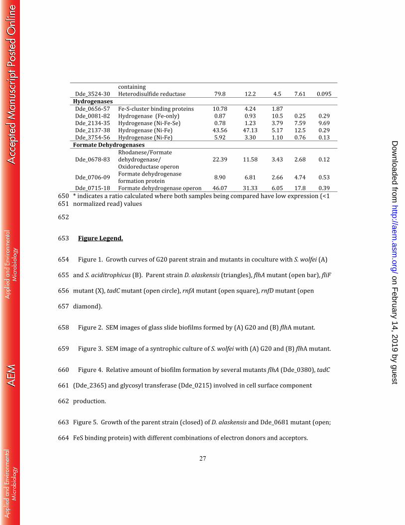
containing Dde_3524-30 Heterodisulfide reductase 79.8 12.2 4.5 7.61 0.095 Hydrogenases Dde_0656-57 Fe-S-cluster binding proteins 10.78 4.24 1.87 Dde_0081-82 Hydrogenase (Fe-only) 0.87 0.93 10.5 0.25 0.29 Dde_2134-35 Hydrogenase (Ni-Fe-Se) 0.78 1.23 3.79 7.59 9.69 Dde_2137-38 Hydrogenase (Ni-Fe) 43.56 47.13 5.17 12.5 0.29 Dde_3754-56 Hydrogenase (Ni-Fe) 5.92 3.30 1.10 0.76 0.13 Formate Dehydrogenases Dde_0678-83 Rhodanese/Formate dehydrogenase/ Oxidoreductase operon 22.39 11.58 3.43 2.68 0.12 Dde_0706-09 Formate dehydrogenase formation protein 8.90 6.81 2.66 4.74 0.53 Dde_0715-18 Formate dehydrogenase operon 46.07 31.33 6.05 17.8 0.39 * indicates a ratio calculated where both samples being compared have low expression (<1 650 normalized read) values 651 652
Figure Legend. 653 Figure 1. Growth curves of G20 parent strain and mutants in coculture with S. wolfei (A) 654 and S. aciditrophicus (B). Parent strain D. alaskensis (triangles), flhA mutant (open bar), fliF 655 mutant (X), tadC mutant (open circle), rnfA mutant (open square), rnfD mutant (open 656 diamond). 657 Figure 2. SEM images of glass slide biofilms formed by (A) G20 and (B) flhA mutant. 658 Figure 3. SEM image of a syntrophic culture of S. wolfei with (A) G20 and (B) flhA mutant. 659 Figure 4. Relative amount of biofilm formation by several mutants flhA (Dde_0380), tadC 660 (Dde_2365) and glycosyl transferase (Dde_0215) involved in cell surface component 661 production. 662
Figure 5. Growth of the parent strain (closed) of D. alaskensis and Dde_0681 mutant (open; 663 FeS binding protein) with different combinations of electron donors and acceptors. 664
on February 14, 2019 by guest
http://aem.asm
.org/D
ownloaded from

Fig. 1
0.02
0.06
0.10
0.14
0 2 14 17 23 34
Time (days)
Gro
wth
(O
D6
00)
0.07
0.12
0.17
0.22
0.27
0 4 7 10 13 18 21 26
Time (days)
A B
on February 14, 2019 by guest
http://aem.asm
.org/D
ownloaded from


Fig. 3 D. alaskensis
S. wolfei
on February 14, 2019 by guest
http://aem.asm
.org/D
ownloaded from

Figure 4
0
0.5
1
1.5
2
2.5
Wild Type GlycosylTransferase
TadC FlhA
Re
lati
ve B
iofi
lm f
orm
atio
n
(A5
80
/A6
00
) on February 14, 2019 by guest
http://aem.asm
.org/D
ownloaded from

0.005
0.05
0.5
H2-Sulfate Lactate-Sulfate
0.005
0.05
0.5
Lactate-Thiosulfate
H2-Thiosulfate
0.005
0.05
0.5
0 10 20
Lactate-Sulfite
0 5 10 15 20 25
H2-Sulfite
Figure 5
Time (hours)
Gro
wth
(O
D6
00)
on February 14, 2019 by guest
http://aem.asm
.org/D
ownloaded from