downregulation of type i collagen expression in the
TRANSCRIPT

RESEARCH ARTICLE Open Access
Downregulation of type I collagenexpression in the Achilles tendon bydexamethasone: a controlled laboratorystudyZilu Ge, Hong Tang, Wan Chen* , Yunjiao Wang, Chengsong Yuan, Xu Tao, Binghua Zhou and Kanglai Tang
Abstract
Background: Spontaneous Achilles tendon rupture associated with long-term dexamethasone (Dex) use has beenreported. However, few studies have investigated the potential mechanism. The aim of this study was to evaluatethe effects of oral Dex on type I collagen in humans and rats and its association with tendon rupture.
Methods: First, six Achilles tendons from patients who received long-term Dex treatment, and another six normaltendons were harvested for histological evaluation. Secondly, 8-week-old rats (n = 72) were randomly assigned to aDex group or a control group. Type I collagen was studied at the mechanical, histological, and molecular levelsafter 3 and 5 weeks. Tenocytes isolated from normal human and rat tendon were used to investigate the effect ofDex on cellular scale.
Results: Histological analysis of human and rat tendon tissue revealed an irregular, disordered arrangement of typeI collagen in the Dex group compared with the control group. In addition, In the Dex+ group, type I collagenexpression decreased in comparison with the Dex− group in both human and rat tenocytes. The mechanicalstrength of tendons was significantly reduced in the Dex group (68.87 ± 11.07 N) in comparison with the controlgroup (81.46 ± 7.62 N, P = 0.013) after 5 weeks. Tendons in the Dex group were shorter with smaller cross-sectionalareas (10.71 ± 0.34mm2, 1.44 ± 0.22mm2, respectively) after 5 weeks than those in the control group (11.13 ± 0.50mm2,P = 0.050, 2.74 ± 0.34mm2, P < 0.001, respectively).
Conclusions: This finding suggests long-term use of Dex that decreases the expression of type I collagen at molecularand tissue levels both in human and rat Achilles tendons. Furthermore, Dex decreases the mechanical strength of thetendon, thereby increasing the risk of Achilles tendon rupture.
Keywords: Achilles tendon, Dexamethasone, Human, Rats, Type I collagen
IntroductionSince the first use of glucocorticoids (GC) in September1948, at the Mayo Clinic, to treat a patient with rheuma-toid arthritis (RA) [1], they have been extensively usedto treat osteoarthritis, tendinopathy, inflammatoryarthritis, and degenerative spine disease. In addition torest, oral nonsteroidal anti-inflammatory drugs, specific
stretching, and strengthening exercises, GC treatment iscommonly prescribed for Achilles tendon diseases [2].Although it is generally accepted that local injections ofGCs temporarily alleviate local inflammation and pain,the adverse effects and long-term functional conse-quences have limited their use [3]. The local administra-tion of GCs has significant negative effects on tendons,increasing the risk of tendon rupture, impairing tendonhealing, and leading to poorer long-term outcomes [4].However, only a few studies have reported a link be-tween oral GCs and the risk of Achilles tendon rupture[5, 6]. Considering occasional case reports of patients
© The Author(s). 2020 Open Access This article is distributed under the terms of the Creative Commons Attribution 4.0International License (http://creativecommons.org/licenses/by/4.0/), which permits unrestricted use, distribution, andreproduction in any medium, provided you give appropriate credit to the original author(s) and the source, provide a link tothe Creative Commons license, and indicate if changes were made. The Creative Commons Public Domain Dedication waiver(http://creativecommons.org/publicdomain/zero/1.0/) applies to the data made available in this article, unless otherwise stated.
* Correspondence: [email protected] of Orthopedics/Sports Medicine Center, State Key Laboratory ofTrauma, Burn and Combined Injury, Southwest Hospital, Third MilitaryMedical University (Army Medical University), Gaotanyan Street. 30,Shapingba District, Chongqing 400038, China
Ge et al. Journal of Orthopaedic Surgery and Research (2020) 15:70 https://doi.org/10.1186/s13018-020-01602-z

receiving long-term GC treatment due to systematic dis-eases or autoimmune disorders, who experienced spon-taneous Achilles tendon rupture [7–9], it is important todetermine the exact mechanism by which the Achillestendon is weakened by GC.The strength and stability of the Achilles tendon are
significantly affected by collagen, which is the maincomponent of the tendon. The amount of collagen andits spatial arrangement are important determinants ofthe mechanical properties of tendons [10]. In particular,type I collagen accounts for approximately 85–95% ofthe dry weight of an Achilles tendon, making it the fore-most factor in the stabilization of the tendon [11]. TypeI collagen, as well as elastin fibers and many other extra-cellular matrix components (cytokines, enzymes, andglycosaminoglycans), is produced by tenocytes [12]. Anyfactor contributing to collagen denaturation, decrease, orchange in arrangement may lead to the weakening orrupture of the Achilles tendon.Researchers have suspected that spontaneous and low-
impact ruptures of the Achilles tendon are associatedwith GC treatment. This can be explained by a disrup-tion in the structure of type I collagen by GC. A seriesof animal experiments support this hypothesis. Underdifferent doses of GCs, the level of type I collagen de-creases, to a certain extent, in the rotator cuff tendon ofrats [13]. An in vitro analysis of tendon cells has shownthat GCs reduce cell viability, cell proliferation, and col-lagen synthesis [14]. However, few studies have directlydemonstrated the effect of oral GCs on type I collagenin humans. We hypothesized that long-term use of dexa-methasone (Dex) could decreased the expression of typeI collagen in the Achilles tendon. Therefore, we usedsamples harvested from patients who underwent long-term Dex treatment to evaluate its effect on type I colla-gen in human Achilles tendons. To avoid any interfer-ence from the clinic, we further confirmed the resultsusing mechanical, histological, and molecular analysis ofrat tendons.
MethodsAchilles tendon samplesAll experiments and study protocols, including sampleacquisition and analysis, were approved by the institu-tional review board of the Human Research Ethics Com-mittee of Army Military Medical University (KY201838).Human Achilles tendon specimens were obtained frompatients in the Department of Orthopaedic Sports Medi-cine Center, Southwest Hospital, Army Military MedicalUniversity, Chongqing, China, after obtaining their writ-ten informed consent. Patients who met the inclusionand exclusion criteria were involved in this study. Ingeneral, the inclusion criteria for Dex group included pa-tients who suffered Achilles tendon rupture and had a
history of long-term Dex treatment for RA, and the cu-mulative duration of Dex treatment was more than 3months. The inclusion criteria for control group in-cluded patients who suffered Achilles tendon rupturedue to acute trauma but had no treatment history of anyglucocorticoids. For both groups, patients aged below 18or above 50 years, diagnosis of Achilles tendinopathy,type 1 or 2 diabetes, hypertension and hyperlipidemia,Achilles tendon infection, smoke and alcohol addiction,previous injury or surgery on Achilles tendon, and over-use of the Achilles tendon would lead to exclusion. Afterensuring they met the inclusion and exclusion criteria,six patients were included in the Dex group, and six pa-tients were served as the control group. Patients’ demo-graphics, including age (Dex group, 37.3 ± 1.6; controlgroup, 35.3 ± 2.0; P = 0.451), body mass index (Dexgroup, 22.8 ± 0.8; control group, 23.5 ± 0.9; P = 0.586),and gender (Dex group, 50%; control group, 50%),showed no significant differences between the Dex andcontrol groups.All samples were collected during tendon repair sur-
gery. After removing the peritendinous tissue, 2 × 2-mmsample near the edge of torn Achilles tendon was har-vested from each patient in the Dex and control group.The tissue was immediately placed in formaldehydefixation fluid to prepare it for histological staining. An-other 2 × 2-mm tissue sample was harvested from eachpatient in the control group and was placed in sterilephosphate-buffered saline (PBS) for cellular experiment.For the animal experiment, 72 male, 8-week-old Spra-
gue–Dawley rats, weighing approximately 200 g, werestratified by body weight and then randomized into fourgroups (18 animals per group, 9 for the mechanical test,3 for western blotting, 3 for PCR, and 3 for immuno-staining (Fig. 1). Owing to the poor solubility of Dex, 3%dimethyl sulfoxide (DMSO) was added [15]. The animalsthat were randomly selected to receive Dex and DMSO(Dex group) were treated by intraperitoneal injection
Fig. 1 Flowchart of animal experiment. MT, mechanical testing; HIS,histology; WB, western blotting; qRT-PCR, quantitative real-timepolymerase chain reaction
Ge et al. Journal of Orthopaedic Surgery and Research (2020) 15:70 Page 2 of 10

into the abdomen with 10 mg/mL/kg [16] once daily for3 and 5 weeks. Animals in the control group wereinjected with saline and DMSO at the same dose. At theendpoint, animals were euthanized under inhalationanesthesia with isoflurane, and the left tendons wereused for all experiments. Cells isolated from tendons ofthe control group were used to evaluate the impact ofDex on rat tenocytes. All animals were kept in individualcages under the same conditions of feeding and environ-ment and without activity restriction.
Isolation and culture of tenocytesIsolation and culture of human and rat tenocytes wereperformed as per our previous description [17, 18].Briefly, after collecting the mid-portion of the Achillestendon, the peritendinous connective tissue was carefullyremoved. The remaining tissue was then placed in PBSand finely diced before being digested in type I collage-nase (3 mg/mL Sigma–Aldrich, St. Louis, MO, USA) for2.5 h at 37 °C on a rocking bed set at 200 rpm. A single-cell suspension was yielded using a 70-μm cell strainer(Becton Dickinson, Franklin Lakes, NJ, USA). Afterwashing in PBS, the released cells were centrifuged at1500 rpm for 5 min and were then resuspended in Dul-becco’s modified Eagle’s medium (DMEM) (Gibco,Carlsbad, CA, USA) containing 100 U/mL penicillin,100 mg/mL streptomycin, 10% fetal bovine serum, and 2mmol/L L-glutamine (all from Invitrogen, Carlsbad, CA,USA). The isolated cells were plated at 37 °C in 5% CO2
for 2 days to form colonies and were then washed twicein PBS to remove nonadherent cells. On day 7 of cul-ture, cells were trypsinized with trypsin-EDTA solution(Sigma-Aldrich), combined together, and were seededonto plates as passage 0 (P0) cells. All subsequent exper-iments were conducted using cells at P3. Tenocytes wereseeded onto 6-well plates with a density of 6 × 10⁴ cells/well and 10-cm-diameter petri dishes for qRT-PCR andprotein extraction, respectively. During the culture period,DMEM used for culturing cells in the Dex+ group wassupplemented with 1 μM Dex (Sigma–Aldrich) and dis-solved in DMSO, and the Dex− group were cultured inDMEM supplemented with DMSO only. All media werechanged every 3 days.
Tenocyte identificationHuman and rat tenocyte identification was carried outbased on the immunoreactivity of the cells for collagen I[19] and vimentin [20] and negative immunoreactivityfor collagen III [21]. The primary cultured tenocyteswere used for immunostaining. Briefly, tenocytes werecultured on coverslips in a 12-well plate at a density of1–2 × 10⁵ cells/ml for 24 h. The cells were then fixed in4% formaldehyde for 30 min at room temperature. Afterwashing, normal goat serum (5%) was added to the
coverslips and incubated for 30min at room temperatureto block non-specific binding sites. Cells were incubatedwith diluted primary antibody in 5% PBS Tween 20(PBST) overnight at 4 °C. The cells were then incubatedwith secondary antibody (fluorescein-isothiocyanate-la-beled goat anti-mouse, Santa Cruz, Dallas, USA) at roomtemperature in the dark with 5% BSA for 1 h. After incu-bating with 0.5 μg/mL DAPI (Beyotime, Shanghai, China)for 5 min, the coverslips were mounted using mountingmedium. Five areas were randomly selected and observedunder × 200 magnification using a confocal laser scanningmicroscope (TCS2NT; Leica, Wetzlar, Germany).The primary antibodies used in the human tenocytes
identification were as follows: monoclonal mouse anti-human collagen I (Sigma-Aldrich, Munich, Germany),mouse anti-human collagen III (Sigma-Aldrich), andmouse anti-human vimentin (Sigma-Aldrich). The primaryantibodies used in the rat tenocyte identification were asfollows: rabbit anti-rat collagen I, rabbit anti-rat collagenIII, and rabbit anti-rat vimentin (all from Abcam).
Immunohistochemical stainingSerial sagittal paraffin sections were prepared from theAchilles tendons as previously described [22]. Briefly, allsamples (including rat and human) for histology werefixed in 4% buffered formalin. After embedding in paraf-fin, the tissue was cut into 4-μm sections. The sectionswere blocked with H2O2 and methanol for 15 min underdark light after deparaffinization. Sections were then in-cubated with pepcase and tryptase for 30 min at 37 °Cfor antigen retrieval. After incubating with goat serum for30min, sections were incubated with anti-collagen type I(1:500) (Abcam) in 5% BSA in PBST overnight at 4 °C.Sections were then incubated with enzyme-conjugatedsecondary antibody for 2 h at room temperature. After be-ing dehydrated and mounted, five areas obtained fromeach sample were randomly selected, and an OLYMPUSEX-51 light microscope (Tokyo, Japan) was used to ob-serve samples at × 40 and × 200 magnification. The wholeareas were evaluated by the Bonar score system [23]. Thescore of the intact group was defined as 12 points, and theaverage optimal density (AOD) of type I collagen wascalculated by ImageJ.
qRT-PCRQuantitative real-time polymerase chain reaction (qRT-PCR) was used to evaluate type I collagen mRNA levels.After extraction of RNA from the cells or tissues byTRIzol (TaKaRa, Kusatsu, Japan), and synthesizingcDNA (TaKaRa), qRT-PCR was performed using a SYBRGreen RT-PCR Kit (TaKaRa) and ABI Prism 7900 Se-quence Detection System (PE Applied Biosystems, FosterCity, CA, USA). The expression levels were calculatedrelative to the expression of glyceraldehyde 3-phosphate
Ge et al. Journal of Orthopaedic Surgery and Research (2020) 15:70 Page 3 of 10

dehydrogenase (GAPDH). Primer sequences are shownin Table 1.
Protein extraction and western blottingAfter tissue was cut into pieces and cells were scraped,they were homogenized in lysis buffer (50 mmol/L Tris-HCl, pH 8.0, 1 mmol/L EDTA, 1% Triton X-100, 0.5%sodium deoxycholate, 0.1% sodium dodecyl sulphate(SDS), 150 mmol/L NaCl) containing proteinase inhibi-tors (Thermo Fisher Scientific Inc., Rockford, IL, USA).A BCA protein assay kit (Thermo Fisher Scientific Inc.,Rockford, IL, USA) was used for protein concentrationmeasurement. Protein samples (30 μg/lane) were re-solved by SDS-polyacrylamide gel electrophoresis andthen transferred onto polyvinylidene difluoride mem-branes. The membranes were blocked with 0.1% TBS-Tween containing 5% non-fat milk for 1 h at roomtemperature, then incubated sequentially with primaryantibody (anti-collagen I (Abcam)) and secondary anti-body (goat anti-rabbit IgG (H&L)-HRP conjugated (Pro-teintech, 1:3000)). The results were visualized, andimages were captured using a LiCoR Odyssey Imager(LI-COR Biosciences, Lincoln, NE, USA).
Mechanical testingA high-precision caliper was used to record the length,width, and thickness of the tendon. Subsequently, theAchilles tendon, with half of the muscle, was fixed in amechanical testing machine to determine the load tofailure (N) and the elastic modulus (MPa) [24, 25]. Afterthe gastrocnemius was frozen in liquid nitrogen [26], itwas fastened to the machine (Regerl, China). Tendonswere tested one by one under a constant displacementrate of 50 mm/min. Ringer’s solution was used to mois-ten the tendon during the test. Data on load to failure(N) were obtained from the load-elongation graph. Elas-tic modulus (MPa) was calculated by the linear portionof the graph and was correlated with strain and cross-sectional area.
Statistical analysisA 5% significance level and an 80% power were set basedon previous studies [16]. The sample size was 10 ratsper group in the mechanical test. Similar experiments inprevious studies have used 7–9 animals in mechanicaltests and 3–10 animals for histological outcomes. There-fore, we believe that this sample size was reasonable.
Comparisons of mechanical properties among groupswere performed using independent sample t tests afternormality testing and homogeneity testing of variance.Differences with P < 0.05 were considered statisticallysignificant. All analyses were performed using SPSS ver-sion 22.0 (IBM Corp., Armonk, NY, USA).
ResultsDex downregulates type I collagen expression in humanAchilles tendonsIn a comparison between tissue harvested from the rup-tured Achilles tendon of patients who had a history oflong-term Dex use and from patients who suffered fromacute trauma, we observed a distinct difference in type Icollagen (Fig. 2a). The quality and density of type I colla-gen in Achilles tendons that ruptured by acute traumawere basically normal. Collagen was arranged regularlyand was thicker than the collagen in the Dex group. Theruptured human Achilles tendon induced by Dex treat-ment showed collagen attenuation, with a highly irregulararrangement and a disordered and curled appearance inthe whole field of vision. The histological score of tissueand AOD of type I collagen was showed in Fig. 2b and c.To evaluate our hypothesis regarding the role of type I
collagen in tendon rupture at the cellular level, we iso-lated human tenocytes from tissues damaged by traumaand cultured them in DMEM with and without Dex.The human tendon cells were fusiform-shaped, as shownin Fig. 3a, and qRT-PCR analysis showed that there wereno significant changes in type I collagen expression aftertreatment with Dex for 1 day. The expression level grad-ually increased in the Dex− group. However, unlike theupward trend observed in the Dex− group, the expres-sion of type I collagen decreased gradually after 3 and 5days and increased slightly at 7 days in the Dex treat-ment group. Expression levels at all time points weresignificantly lower than those in the Dex− group, andthe gap increased over time (Fig. 3b). The western blot-ting results showed the same trend (Fig. 3c).
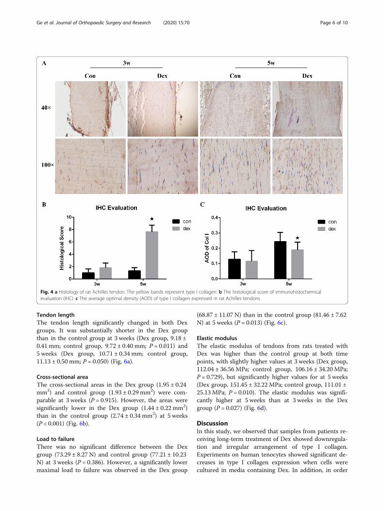
Dex downregulates type I collagen expression in ratAchilles tendonsTo identify the effect of Dex on rat tendons, we observedchanges in type I collagen expression at 3 and 5 weeks inDex and control groups (Fig. 4a). The general pattern inthe Dex group was the same as that for cells harvestedfrom patients. Histological examination of tissue samples
Table 1 Sequences of primers used for qRT-PCR
Gene Species Forward primer (5′→3′) Reverse primer (3′→5′)
Col I Human TGGTGAGACGTGGAAACCTG CTTGGGTCCCTCGACTCCTA
Col I Rats TGGCAAAGAAGGCGGCAAAGG AGGAGCACCAGCAGGACCATC
GAPDH TGACTTCAACAGCAACTC TGTAGCCATATTCATTGTCA
Ge et al. Journal of Orthopaedic Surgery and Research (2020) 15:70 Page 4 of 10

revealed that type I collagen of the Dex group was ar-ranged irregularly and was curled and disordered com-pared with that of the control group. The overall collagenstaining intensity in the field of view was also lower thanthat of the control group. The arrangement became sub-stantially worse at 5 weeks. The histological score of tissueand AOD of type I collagen was showed in Fig. 4b and c.
Tissue samples were harvested at 3 and 5 weeks, andtenocytes were collected at day 3, day 5, and day 7 of cul-ture. Results from qRT-PCR and western blotting areshown in Fig. 5. As the duration of Dex use increased, theexpression level of type I collagen decreased, consistentwith the previously described results. The identification ofrat tenocytes is shown in Supplementary Figure 1.
Fig. 2 a Histology of human Achilles tendons. The yellow fasciculate bands represent type I collagen. The Dex group, receiving long-term Dextreatment, have irregular and curled collagen type I. b The histological score of immunohistochemical evaluation (IHC). c The average optimaldensity (AOD) of type I collagen expressed in human Achilles tendons
Fig. 3 a Identification of human tenocytes. Collagen type I and vimentin were positively expressed, and Collagen type III was negativelyexpressed. b mRNA expression of type I collagen in human Achilles tenocytes. The black and gray bars represent the Dex− and Dex+ groups,respectively. The asterisk represents a significant change between the two groups. c Protein expression of type I collagen in human Achillestenocytes. Relative expression levels were normalized to GAPDH
Ge et al. Journal of Orthopaedic Surgery and Research (2020) 15:70 Page 5 of 10
Tendon lengthThe tendon length significantly changed in both Dexgroups. It was substantially shorter in the Dex groupthan in the control group at 3 weeks (Dex group, 9.18 ±0.41 mm; control group, 9.72 ± 0.40 mm; P = 0.011) and5 weeks (Dex group, 10.71 ± 0.34 mm; control group,11.13 ± 0.50 mm; P = 0.050) (Fig. 6a).
Cross-sectional areaThe cross-sectional areas in the Dex group (1.95 ± 0.24mm2) and control group (1.93 ± 0.29 mm2) were com-parable at 3 weeks (P = 0.915). However, the areas weresignificantly lower in the Dex group (1.44 ± 0.22 mm2)than in the control group (2.74 ± 0.34 mm2) at 5 weeks(P < 0.001) (Fig. 6b).
Load to failureThere was no significant difference between the Dexgroup (73.29 ± 8.27 N) and control group (77.21 ± 10.23N) at 3 weeks (P = 0.386). However, a significantly lowermaximal load to failure was observed in the Dex group
(68.87 ± 11.07 N) than in the control group (81.46 ± 7.62N) at 5 weeks (P = 0.013) (Fig. 6c).
Elastic modulusThe elastic modulus of tendons from rats treated withDex was higher than the control group at both timepoints, with slightly higher values at 3 weeks (Dex group,112.04 ± 36.56MPa; control group, 106.16 ± 34.20MPa;P = 0.729), but significantly higher values for at 5 weeks(Dex group, 151.45 ± 32.22MPa; control group, 111.01 ±25.13MPa; P = 0.010). The elastic modulus was signifi-cantly higher at 5 weeks than at 3 weeks in the Dexgroup (P = 0.027) (Fig. 6d).
DiscussionIn this study, we observed that samples from patients re-ceiving long-term treatment of Dex showed downregula-tion and irregular arrangement of type I collagen.Experiments on human tenocytes showed significant de-creases in type I collagen expression when cells werecultured in media containing Dex. In addition, in order
Fig. 4 a Histology of rat Achilles tendon. The yellow bands represent type I collagen. b The histological score of immunohistochemicalevaluation (IHC). c The average optimal density (AOD) of type I collagen expressed in rat Achilles tendons
Ge et al. Journal of Orthopaedic Surgery and Research (2020) 15:70 Page 6 of 10

to exclude interference from the clinic, we explored thishypothesis in rats on the cellular and tissue level. Theresults showed that Dex had a significant influence onthe expression of type I collagen, both after 3 weeks and5 weeks, but had a minimal effect on histology after 3weeks. From the above findings, we concluded thatlong-term treatment with Dex downregulated type I col-lagen expression.According to the results of the mechanical testing, the
load to failure and elastic modulus of rat Achilles ten-dons were altered by Dex treatment. In addition, thelength and cross-sectional area of the tendons were re-duced by Dex treatment. However, aside from the ten-don length, other parameters were not significantlychanged after 3 weeks of Dex treatment in rats, indicat-ing that the short-term use of Dex has little effect ontendon mechanical properties. By contrast, after 5 weeksof Dex treatment, all measured parameters changed sig-nificantly. The mean values for length, cross-sectionalarea, and load to failure decreased, and the elastic modu-lus increased. The difference in tendon length and cross-sectional area may be explained by the effect of Dex ontype I collagen expression. According to the results ofqRT-PCR and western blot, Dex treatment downregu-lated collagen type I on both the RNA and protein level.This likely contributed to the abnormal synthesis of
collagen type I, leading to narrower and shorter Achillestendons. Moreover, the mechanical properties of ten-dons are related to the fibril diameter, and the size ofcollagen fibrils determines functional differences. Fibrilswith larger diameters exhibit an increased density ofintramolecular cross-links, and those with smaller diam-eters are more elastic and resistant to creep [27]. Silveret al. reported that the elastic modulus and ultimate ten-sile strength are more dependent on fibril length thanon diameter [28]. Additionally, type III collagen is moreflexible than type I collagen [29]. Taguchi et al. reportedthat systemic administration of GC lead to smaller colla-gen fibers [16]. Considering Taguchi’s findings, thehistological and mechanical changes noted in our studymay be explained by the change of type I collagen.Long-term treatment with Dex resulted in irregular ar-rangement of type I collagen and a reduction in content,leading to a substantial effect on the load to failure andelastic modulus. With an increased elastic modulus anddecreased maximal load to failure, the Achilles tendonhad an increased risk of rupture.Similar studies on the influence of corticosteroid injec-
tions on tendon mechanical properties have yielded dif-ferent outcomes. Some studies have shown nosignificant change after local injection [30, 31], whileothers have reported the opposite. Haraldsson et al.
Fig. 5 mRNA and protein expression of type I collagen in rat Achilles tendons tissues after 3 and 5 weeks (a) and (c). mRNA and proteinexpression of type I collagen in rat tenocytes at 3, 5, and 7 days (b) and (d). Relative expression levels were normalized to GAPDH. The asteriskrepresents a significant change between the two groups
Ge et al. Journal of Orthopaedic Surgery and Research (2020) 15:70 Page 7 of 10

proved that corticosteroid injection reduces the tensilestrength of isolated collagen fascicles [32]. Mikolyzket al. used a rat model and found that a single cortico-steroid dose has significant short-term effects on thebiomechanical properties of both injured and uninjuredrotator cuff tendons [33]. In our study, significantchanges in the molecular, histological, and mechanicalcharacteristics of Achilles tendon tissue were identifiedafter 5 weeks of Dex treatment, while only slight changesin some mechanical parameters and histological findingswere observed by 3 weeks. This suggests that Dex prob-ably had an impact on type I collagen since the treat-ment began; however, the impact was not sufficient tosignificantly alter the phenotype. As the treatment
duration increased, significant histological and mechan-ical changes were identified, which will have increasedthe risk of Achilles tendon rupture.These results suggest that type I collagen in the Achil-
les tendon can be decreased by Dex, but the underlyingmechanism is not yet fully elucidated. Previous researchprovides some insight. Tomoyuki observed an increasedexpression of matrix metalloproteinase (MMP)-3 in re-sponse to corticosteroids [34]. MMP-3 is a potentproteoglycan-degrading enzyme that plays a vital role inthe degeneration of collagen and other structural com-ponents [35]. MMP-3 expression levels are unchangedin the synovial tissues of osteoarthritic joints after theintra-articular administration of steroids, indicating its
Fig. 6 Mechanical testing of rat Achilles tendons. Control group (gray boxes); Dex group (white boxes); x-axis, 3 and 5 weeks. The line in theboxes represents the median, with the lower and upper ends of the boxes representing the 1st and 3rd quartiles, respectively. The asteriskrepresents a significant difference between the two groups
Ge et al. Journal of Orthopaedic Surgery and Research (2020) 15:70 Page 8 of 10

unique characteristic in tendons [36]. Other latent MMPscan degrade the extracellular matrix indirectly. In hu-man supraspinatus tendon cells, migration is inhib-ited, and the levels of MMP2, MMP8, MMP9, andMMP13 are reduced, while tissue inhibitor of metallo-proteinases 1, which is an MMP inhibitor, is upregu-lated [14]. In addition to MMPs, scleraxis is aprobable gene involved in the alteration of type I col-lagen [37]. Therefore, a complex regulatory mechan-ism may determine the ultimate changes. Furtherresearch should focus on determining the detailedmechanism.Some patients receive GC to treat the pain associated
with Achilles tendinopathy. Recent research has shownthat chronic inflammation is a feature of Achilles tendi-nopathy [38, 39]. Some doctors insist on using Dex to al-leviate symptoms, as GCs inhibit the progression ofinflammation. Although the specific pathophysiologicalmechanisms underlying tendinopathy remain unknown,four main drug classes have been implicated in itsexacerbation, including long-term GCs. The main tar-gets of GC toxicity are the lower limb tendons, includingthe Achilles tendon [40]. Considering the histologicalchanges resulting from tendinopathy, including signifi-cant fiber disorientation [41], it is possible that GCsaccelerate the progress of tendinopathy by leading to thedecrease in type I collagen. Moreover, inflammationplays a vital role in tendon healing, but excessive or per-sistence inflammation can be deleterious [42]. As GCcan inhibit angiogenesis, worsen the inflammation ofmicrangium, and trigger coagulation [43], it may impaircollagen synthesis and tendon healing due to microdamage.However, our study has a few limitations. Firstly, we
obtained samples from patients who suffered from RA.The disease, as well as other factors, may cause struc-tural damage to tendons, but the underlying mechanismsare not clear [44]. We performed an animal experimentto exclude these effects. However, we cannot conclu-sively exclude the effects of the disease. Secondly, thein vitro model of cultured human tendon cells onlyshows the short-term effect but not the long-term effectof Dex treatment. The difference in species and dosemay also influence the findings. Although we usedDMSO to help dissolve Dex to form a solution, the pre-cipitation of Dex may still have occurred during in vivoand in vitro experiments, potentially affecting the con-clusions regarding clinical applications. Moreover, themechanism by which GC decreases type I collagen inthe Achilles tendon remains to be fully elucidated.
ConclusionIn conclusion, Dex downregulates type I collagenexpression in Achilles tendons, resulting in tendon
dysfunction and an increased risk of rupture. Dex,whether administered orally or by injection, requiresan evaluation of the balance between the temporarypositive responses and potential long-term side ef-fects. Further research is needed to determine theprecise mechanisms underlying the observed effects.
Supplementary informationSupplementary information accompanies this paper at https://doi.org/10.1186/s13018-020-01602-z.
Additional file 1: Figure S1. The identification of rat tenocytes.Collagen type I and vimentin were positively expressed, and Collagentype III was negatively expressed.
AbbreviationsAOD: Average optimal density; Dex: Dexamethasone; DMEM: Dulbecco’smodified Eagle’s medium; DMSO: Dimethyl sulfoxide; GC: Glucocorticoids;MMP: Matrix metalloproteinase; RA: Rheumatoid arthritis
AcknowledgementsNot applicable.
Authors’ contributionsGZL and TH contributed to research design, development of animal models,and analysis and drafting of the manuscript. WYJ contributed to thedevelopment of animal models. YCS, TX, and ZBH contributed in samplecollection and revising the manuscript. CW and TKL contributed in researchdesign and revising the manuscript. All authors read and approved the finalmanuscript.
FundingThis Project was supported by the National Science Foundation for YoungScientists of China (Grant No.81601943).
Availability of data and materialsNot applicable.
Ethics approval and consent to participateThis study was performed at southwest hospital and was approved by theinstitutional review board of the Human Research Ethics Committee of ArmyMilitary Medical University (KY201838).
Consent for publicationNot applicable.
Competing interestsThe authors declare that they have no competing interests.
Received: 19 October 2019 Accepted: 17 February 2020
References1. Glyn J. The discovery and early use of cortisone. J R Soc Med. 1998;91(10):
513–7.2. Alfredson H, Lorentzon R. Chronic Achilles tendinosis: recommendations for
treatment and prevention. Sports Med. 2000;29(2):135–46.3. Metcalfe DJ, Costa ML. Glucocorticoid injections in lesions of the Achilles
tendon. Foot Ankle Int. 2009;30(7):661–5.4. Coombes BK, Bisset L, Brooks P, Khan A, Vicenzino B. Effect of corticosteroid
injection, physiotherapy, or both on clinical outcomes in patients withunilateral lateral epicondylalgia: a randomized controlled trial. JAMA. 2013;309(5):461–9.
5. van der Linden PD, Sturkenboom MC, Herings RM, Leufkens HM, RowlandsS, Stricker BH. Increased risk of Achilles tendon rupture with quinoloneantibacterial use, especially in elderly patients taking oral corticosteroids.Arch Intern Med. 2003;163(15):1801–7.
Ge et al. Journal of Orthopaedic Surgery and Research (2020) 15:70 Page 9 of 10

6. Spoendlin J, Meier C, Jick SS, Meier CR. Oral and inhaled glucocorticoid useand risk of Achilles or biceps tendon rupture: a population-based case-control study. Ann Med. 2015;47(6):492–8.
7. Hersh BL, Heath NS. Achilles tendon rupture as a result of oral steroidtherapy. J Am Podiatr Med Assoc. 2002;92(6):355–8.
8. Rao SK, Navadgi BC, Vasdev A. Bilateral spontaneous rupture of Achillestendons: a case report. J Orthop Surg (Hong Kong). 2005;13(2):178–80.
9. Baruah DR. Bilateral spontaneous rupture of the Achilles tendons in apatient on long-term systemic steroid therapy. Br J Sports Med. 1984;18(2):128–9.
10. Saito M, Marumo K. Collagen cross-links as a determinant of bone quality: apossible explanation for bone fragility in aging, osteoporosis, and diabetesmellitus. Osteoporos Int. 2010;21(2):195–214.
11. Bernard-Beaubois K, Hecquet C, Houcine O, Hayem G, Adolphe M. Cultureand characterization of juvenile rabbit tenocytes. Cell Biol Toxicol. 1997;13(2):103–13.
12. Kannus P, Józsa L, A. N. Effects of training, immobilization andremobilization on tendons. Scand J Med Sci Sports 2010;7(2):67–71.
13. Wei AS, Callaci JJ, Juknelis D, Marra G, Tonino P, Freedman KB, et al. Theeffect of corticosteroid on collagen expression in injured rotator cufftendon. J Bone Joint Surg Am. 2006;88(6):1331–8.
14. Tempfer H, Gehwolf R, Lehner C, Wagner A, Mtsariashvili M, Bauer HC, et al.Effects of crystalline glucocorticoid triamcinolone acetonide on culteredhuman supraspinatus tendon cells. Acta Orthop. 2009;80(3):357–62.
15. Mitchell A, Rivas KA, Smith R 3rd, Watts AE. Cryopreservation of equinemesenchymal stem cells in 95% autologous serum and 5% DMSO does notalter post-thaw growth or morphology in vitro compared to fetal bovineserum or allogeneic serum at 20 or 95% and DMSO at 10 or 5. Stem CellRes Ther. 2015;6:231.
16. Taguchi T, Kubota M, Saito M, Hattori H, Kimura T, Marumo K. Quantitativeand qualitative change of collagen of Achilles tendons in rats with systemicadministration of glucocorticoids. Foot Ankle Int. 2016;37(3):327–33.
17. Chen W, Deng Y, Zhang J, Tang K. Uniaxial repetitive mechanicaloverloading induces influx of extracellular calcium and cytoskeletondisruption in human tenocytes. Cell Tissue Res. 2015;359(2):577–87.
18. Liu J, Chen L, Tao X, Tang K. Phosphoinositide 3-kinase/Akt signaling isessential for prostaglandin E2-induced osteogenic differentiation of rattendon stem cells. Biochem Biophys Res Commun. 2013;435(4):514–9.
19. Schulze-Tanzil G, Mobasheri A, Clegg PD, Sendzik J, John T, Shakibaei M.Cultivation of human tenocytes in high-density culture. Histochem Cell Biol.2004;122(3):219–28.
20. Bjur D, Danielson P, Alfredson H, Forsgren S. Presence of a non-neuronalcholinergic system and occurrence of up- and down-regulation inexpression of M2 muscarinic acetylcholine receptors: new aspects ofimportance regarding Achilles tendon tendinosis (tendinopathy). Cell TissueRes. 2008;331(2):385–400.
21. Pauly S, Klatte F, Strobel C, Schmidmaier G, Greiner S, Scheibel M, et al.Characterization of tendon cell cultures of the human rotator cuff. Eur CellsMaterials. 2010;20(7):84.
22. Docheva D, Muller SA, Majewski M, Evans CH. Biologics for tendon repair.Adv Drug Deliv Rev. 2015;84:222–39.
23. Maffulli N, Longo UG, Franceschi F, Rabitti C, Denaro V. Movin and Bonarscores assess the same characteristics of tendon histology. Clin Orthop RelatRes. 2008;466(7):1605–11.
24. Muller SA, Durselen L, Heisterbach P, Evans C, Majewski M. Effect of a simplecollagen type I sponge for Achilles tendon repair in a rat model. Am JSports Med. 2016;44(8):1998–2004.
25. Muller SA, Quirk NP, Muller-Lebschi JA, Heisterbach PE, Durselen L, MajewskiM, et al. Response of the injured tendon to growth factors in the presenceor absence of the paratenon. Am J Sports Med. 2019;47(2):462–7.
26. Wieloch P, Buchmann G, Roth W, Rickert M. A cryo-jaw designed for in vitrotensile testing of the healing Achilles tendons in rats. J Biomech. 2004;37(11):1719–22.
27. Ottani V, Raspanti M, Ruggeri A. Collagen structure and functionalimplications. Micron. 2001;32(3):251–60.
28. Silver FH, Christiansen DL, Snowhill PB, Chen Y. Role of storage on changesin the mechanical properties of tendon and self-assembled collagen fibers.Connect Tissue Res. 2000;41(2):155–64.
29. Silver FH, Horvath I, Foran DJ. Mechanical implications of the domainstructure of fiber-forming collagens: comparison of the molecular and
fibrillar flexibilities of the α1-chains found in types I–III collagen. J Theor Biol.2002;216(2):243–54.
30. Wiggins ME, Fadale PD, Ehrlich MG, Walsh WR. Effects of local injection ofcorticosteroids on the healing of ligaments. a follow-up report. J Bone JointSurg Am. 1995;77(11):1682–91.
31. Hugate R, Pennypacker J, Saunders M, Juliano P. The effects ofintratendinous and retrocalcaneal intrabursal injections of corticosteroid onthe biomechanical properties of rabbit Achilles tendons. J Bone Joint SurgAm. 2004;86(4):794–801.
32. Bjarki Thor H, Henning L, Per A, Anne-Marie Z, Benno VE, Jeroen D, et al.Corticosteroids reduce the tensile strength of isolated collagen fascicles. AmJ Sports Med. 2006;34(12):1992.
33. Mikolyzk DK, Wei AS, Tonino P, Marra G, Williams DA, Himes RD, et al. Effectof corticosteroids on the biomechanical strength of rat rotator cuff tendon.J Bone Joint Surg Am. 2009;91(5):1172–80.
34. Muto T, Kokubu T, Mifune Y, Inui A, Harada Y, Yoshifumi, et al. Temporaryinductions of matrix metalloprotease-3 (MMP-3) expression and cellapoptosis are associated with tendon degeneration or rupture aftercorticosteroid injection. J Orthop Res. 2014;32(10):1297–304.
35. Matrisian LM, Hogan BL. Growth factor-regulated proteases and extracellularmatrix remodeling during mammalian development. Curr Top Dev Biol.1990;24:219–59.
36. Young L, Katrib A, Cuello C, Vollmer-Conna U, Bertouch JV, Roberts-Thomson PJ, et al. Effects of intraarticular glucocorticoids on macrophageinfiltration and mediators of joint damage in osteoarthritis synovialmembranes: findings in a double-blind, placebo-controlled study. ArthritisRheum. 2001;44(2):343–50.
37. Chen W, Tang H, Zhou M, Hu C, Zhang J, Tang K. Dexamethasone inhibitsthe differentiation of rat tendon stem cells into tenocytes by targeting thescleraxis gene. J Steroid Biochem Mol Biol. 2015;152:16–24.
38. Dakin S, Newton J, Martinez F, Hedley R, Gwilym S, Jones N, et al. Chronicinflammation is a feature of Achilles tendinopathy and rupture. Br J SportsMed. 2018;52(6):359–67.
39. Millar NL, Murrell GA, McInnes IB. Inflammatory mechanisms intendinopathy - towards translation. Nat Rev Rheumatol. 2017;13(2):110–22.
40. Kirchgesner T, Larbi A, Omoumi P, Malghem J, Zamali N, Manelfe J, et al.Drug-induced tendinopathy: from physiology to clinical applications. JointBone Spine. 2014;81(6):485–92.
41. Cipollaro L, Sahemey R, Oliva F, Maffulli N. Immunohistochemical features ofrotator cuff tendinopathy. Br Med Bull. 2019;130(1):105–23.
42. Thomopoulos S, Parks WC, Rifkin DB, Derwin KA. Mechanisms of tendoninjury and repair. J Orthop Res. 2015;33(6):832–9.
43. Coutinho AE, Chapman KE. The anti-inflammatory and immunosuppressiveeffects of glucocorticoids, recent developments and mechanistic insights.Mol Cell Endocrinol. 2011;335(1):2–13.
44. Janta I, Stanciu D, Hinojosa M, Nieto-González JC, Valor L, Bello N, et al.Structural damage in rheumatoid arthritis: comparison between tendondamage evaluated by ultrasound and radiographic damage. Rheumatology(Oxford). 2016;55(6):1042–6.
Publisher’s NoteSpringer Nature remains neutral with regard to jurisdictional claims inpublished maps and institutional affiliations.
Ge et al. Journal of Orthopaedic Surgery and Research (2020) 15:70 Page 10 of 10