dr abukakr h mossa anatomy instructor mbbs, 22-25/9/2011 histology lab connective tissue
TRANSCRIPT

Dr Abukakr H MossaAnatomy instructor
MBBS, 22-25/9/2011
Histology lab
Connective tissue

Outlines…• Connective tissue (CT) features • CT components:
– Cells – Extracellular matrix
• Fibers: collagen, elastic and reticular fibers• Ground substance
• Classification of CT:– Embryonic:
• Mesenchymal: givers rise to all types of adult connective tissue• Warton’s jelly: in the umbilical cord
– CT proper • Loose CT• Dense CT (regular & irregular)
– Specialized CT: bone, cartilage, blood, adipose tissue, hemopoietic tissue and lymphatic tissue.


CT features
• Unlike the highly packed cells of the epithelium, the CT consists of variety of cells which are widely spaced in the intercellular matrix.
• It forms a vast and continuous compartment throughout the body, between the epithelia and muscular or nervous tissues.
• CT has many functions:– Supportive – Storage – Protection – ……..
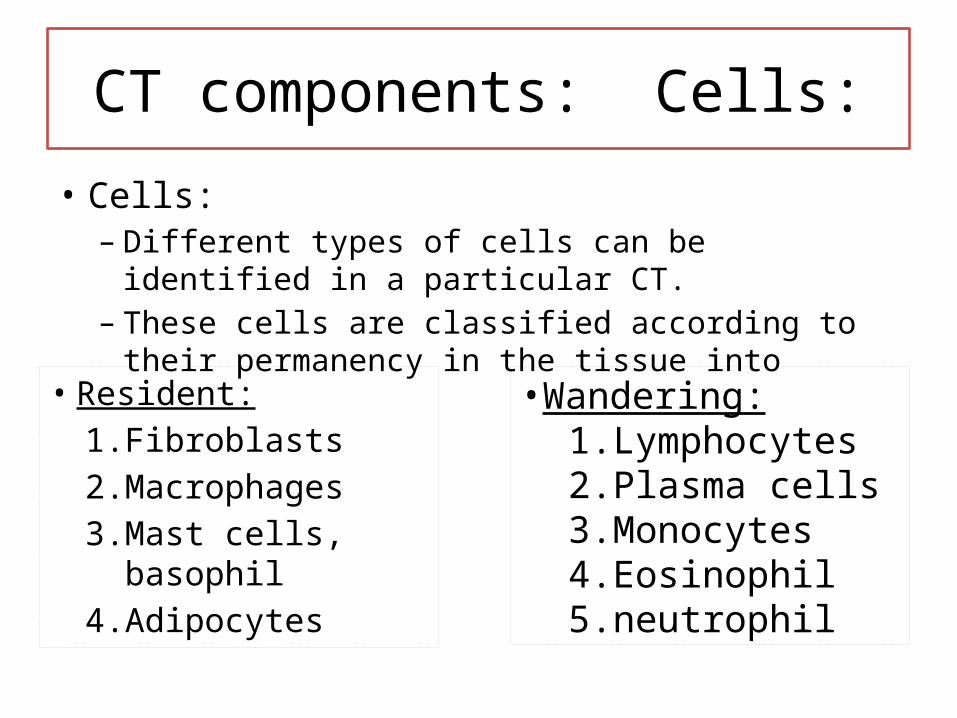
CT components: Cells:
• Cells:– Different types of cells can be identified in a particular CT.– These cells are classified according to their permanency
in the tissue into
• Resident:1. Fibroblasts2. Macrophages3. Mast cells, basophil4. Adipocytes
•Wandering: 1. Lymphocytes2. Plasma cells3. Monocytes 4. Eosinophil5. neutrophil

Resident CT cells:
• It is the principal cell of the CT• Function: production of CT
fibers & ground substance.• In H & E: – the nucleus appears as flat disc
with a thin pale surrounding of cytoplasm
1. FIBROBLAST (FB):

2. MACROPHAGES:• Also called histiocytes
– In conventional studies: Difficult to identify unless showing evidence of phagocytosis “visible ingested material in cytoplasm”
Resident CT cells:
Functions: 1. Phagocytosis: 2. Secretion of substances needed
for inflammation:3. Immune reaction

3. Mast cells:• Description:
– large, ovoid with spherical nucleus, metochromatic cytoplasmic granules.
• Function: release of histamine and other inflammatory substances.
Resident CT cells:
• Location: – in the connective tissue surrounding the blood vessels of the previously
mentioned sites– CNS:
• Only in the meninges• Not found in brain & spinal cord?
– Lymphoid tissue except spleen4. BASOPHILS: are similar to mast cells in function but they
are smaller and has shorter life span
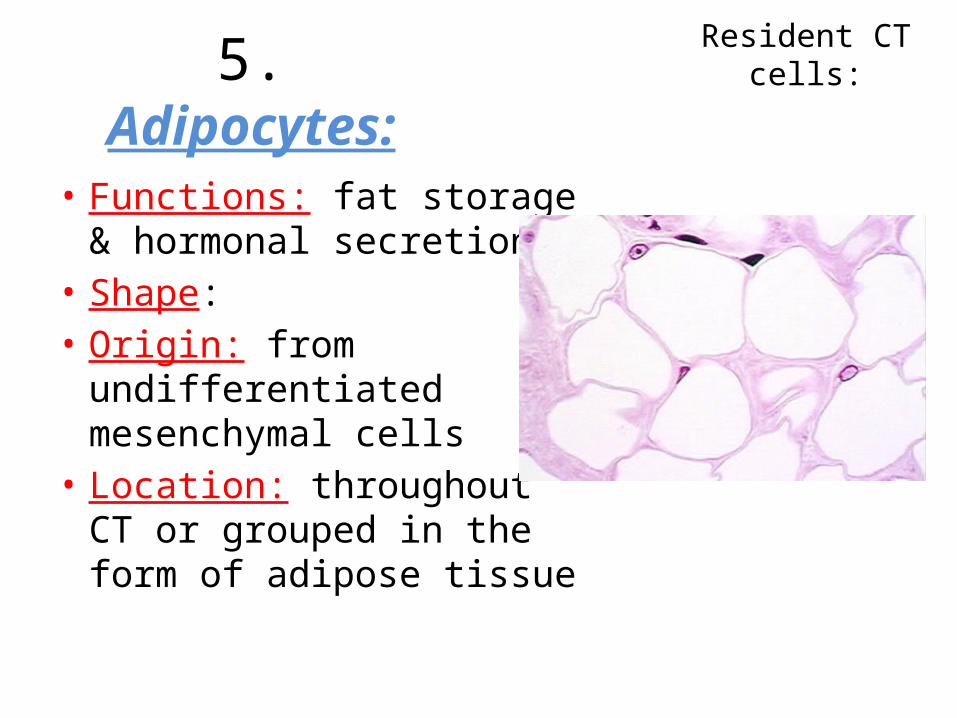
5. Adipocytes:
• Functions: fat storage & hormonal secretion
• Shape: • Origin: from
undifferentiated mesenchymal cells
• Location: throughout CT or grouped in the form of adipose tissue
Resident CT cells:
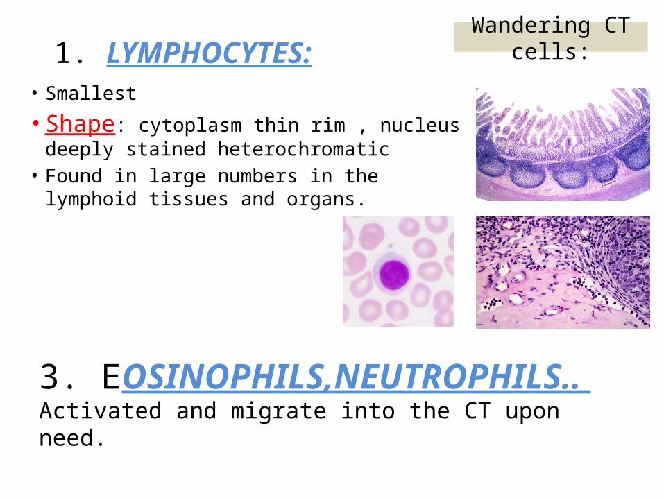
1. LYMPHOCYTES:• Smallest
• Shape: cytoplasm thin rim , nucleus deeply stained heterochromatic
• Found in large numbers in the lymphoid tissues and organs.
Wandering CT cells:
3. EOSINOPHILS,NEUTROPHILS.. Activated and migrate into the CT upon need.
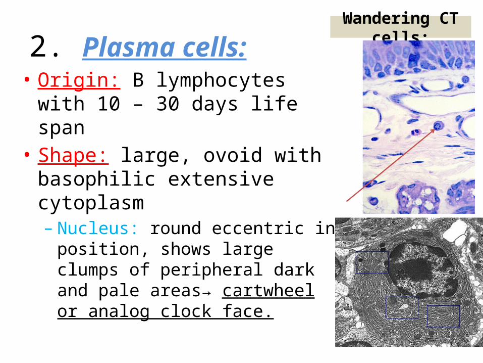
2. Plasma cells:• Origin: B lymphocytes with 10 –
30 days life span• Shape: large, ovoid with
basophilic extensive cytoplasm– Nucleus: round eccentric in
position, shows large clumps of peripheral dark and pale areas→ cartwheel or analog clock face.
Wandering CT cells:
• EM is a complex structural network that surrounds and supports the CT cells.
• Composed of fibers and ground substance.
CT components: Extracellular matrix

• Ground substance:– A viscous, clear substance with a slippery feel and
high water content.– Mainly formed by: proteoglycans, multiadhesive
glycoprotiens and glycosaminoglycans. – The ground substance plays important role in cell-
to-cell communication and support.
CT components: Extracellular matrix

Fibers: • Three main types:
1. Collagen fibers:• Most abundant • Different (many) types with different structure, function
and location.• Can be synthesized by different cells of the CT and
epithelial cells (where can you find CT in the epithelium??)• Vitamin C is needed for their synthesis. (what is Scurvy?
What happens in it?)• Histologically stained with eosin and other acidic dyes as
wavy structures.
CT components: Extracellular matrix
Collagen fiber
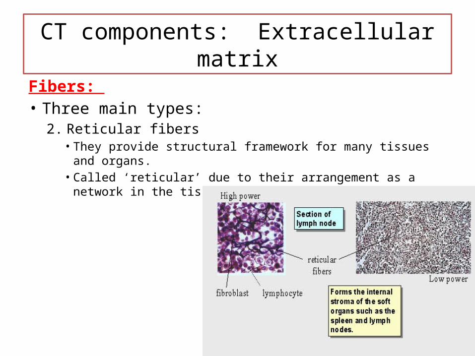
Fibers: • Three main types:
2. Reticular fibers• They provide structural framework for many tissues and organs.• Called ‘reticular’ due to their arrangement as a network in the
tissue they support.
CT components: Extracellular matrix

Fibers: • Three main types:
3. Elastic fibers:• Found where the tissue
needs to stretch and distend (vertebral ligament, larynx, elastic cartilage, arteries …)
• Fibers (elastin) are arranged in a network of coiled branching manner.
• Can be affected by excessive sun exposure.
CT components: Extracellular matrix

CT classification: CT properThese two tissues are distinguished according to the
relative amounts of fibers they contain. Dense connective tissues are completely dominated by
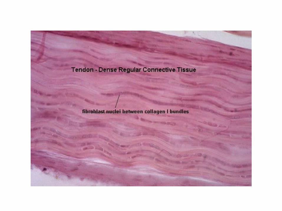
fibers: dense irregular connective tissue, example is the dermis of the skin. regular dense connective tissue, examples of regular dense
connective tissue are tendons, ligaments and apponeurosisLoose connective tissue is relatively cell rich, soft and
compliant. It is also rich in vessels and nerves. Examples: mucous connective tissue, reticular connective tissue and adipose tissue.

Dense irregular CT (dermis of skin)

Loose areolar CT

Adipose tissue (AT)
• Adipose tissue is specialized to store fat in its adipocytes.
• According to the color of the stored fat, AT is divided into:– White AT: found mainly in ADULTS– Brown AT: in present during fetal life and first year of life
• Functions of AT:– White AT: energy starage, insulation, cushioning of vital
organs and hormone secretion.– Brown AT: heat generation
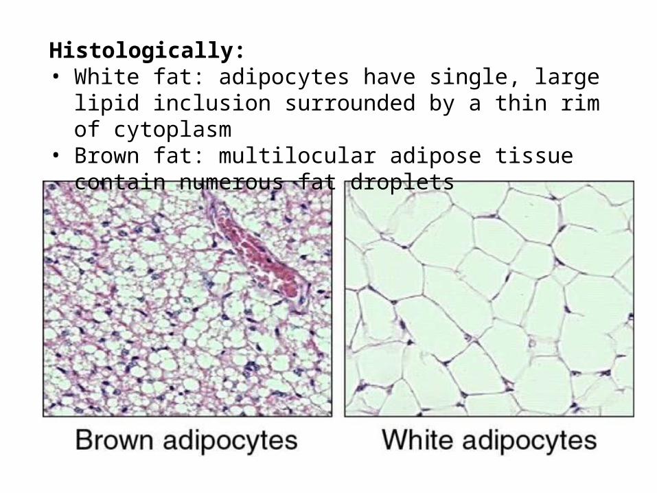
Histologically: • White fat: adipocytes have single, large lipid inclusion
surrounded by a thin rim of cytoplasm• Brown fat: multilocular adipose tissue contain numerous fat
droplets
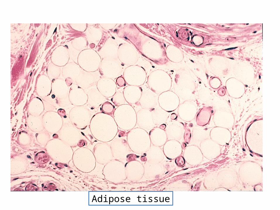
Adipose tissue

Thanks