dr agbonifo m consultant e.n.t surgeon isth, irrua. edo state. dizziness, vertigo & imbalance
TRANSCRIPT

DR AGBONIFO M
CONSULTANT E .N.T SURGEONISTH, IRRUA.EDO STATE.
DIZZINESS, VERTIGO & IMBALANCE

PRETEST ABOUT VERTIGOIT IS ALSO REFFERED TO AS DIZZINESSIT IS AN INNER EAR DISEASEIT IS DEFINED AS HALLUCINATION OF
MOVEMENTA VERY IMPORTANT COMPONENT OF IT IS
NYSTAGMUS

BPPV:MEANS BENIGH PAROXYSMAL
POSITIONAL VERTIGOIT IS THE COMMONEST CAUSE OF
PERIPHERAL VERTIGOIT IS ASSOCIATED WITH HEARING LOSSTREATMENT IS SURGICAL

DIX HALLPIKE TEST IS AN OFFICE PROCEDURE TO DIAGNOSE MENIERE’S DX
MEDICAL TREATMENT FOR VERTIGO INCLUDE THE USE OF BETAHISTINE
CAUSE OF PERIPHERAL VERTIGO INCLUDE MENIERE’S DX
CENTRAL VERTIGO COULD BE CAUSED BY C.V.A
SURGICAL TREATMENT IS THE FIRST LINE OF TREATMENT FOR BPPV
CAUSES OF VERTIGO IS VERTIGO

OUTLINE
INTRODUCTIONANATOMY &PATHOPHYSIOLOGY
(VESTIBULAR APPARATUS)AETIOLOGYCLINICAL FEATURESINVESTIGATIONTREATMENTREHABILITATIONCONCLUSION

INTRODUCTION
For the otolaryngologist the dizzy Pt often presents a diagnostic challenge, therefore the clinical evaluation has the following basic objectives:
It aims to present the basic knowledge and concept of symptom of dizziness
It presents the clinical approach to evaluating the dizzy patient. To differentiate different causes of dizziness, vertigo and imbalance
It explains the skills needed in primary care and secondary care settings of managing a dizzy patient
It aims to bring about positive attitudinal changes in the management of dizzy patients
Introduction cont…
It is a common symptom especially with increasing age
We have to distinguish between VERTIGO andNONVERTIGO
A critical distinction is differentiating vertigo from nonvertigo
Central and peripheral vertigo

Definitions
Vertigo is the true rotational movement of self or the surroundings.
Nonvertigo includes light-headedness, unsteadiness, motion intolerance, imbalance, floating, or a tilting sensation.
This dichotomy is helpful because true vertigo is often due to inner-ear disease, whereas symptoms of nonvertigo may be due to CNS, cardiovascular, or systemic diseases.
Nystagmus is involuntary rapid movt of the eyes.

Definitions cont
It can be horizontal (back & forth ; left & right), vertical (up & down) or rotatory. It has 2 phases ; slow & fast . The slow phase originates from the vestibular system and fast corrective phase from the brain.
10
ANATOMY/ PATHOPHYSIOLOGY
MAITENANCE OF BALANCE RELIES UPON
INPUTS FROM
VISUAL(70%)
VESTIBULAR(15%)
PROPRIOCEPTIVE(15%)
PATHOLOGY IN A WIDE VARIETY OF SYSTEMS
MAY GIVE RISE TO DYSEQUILIBRIUM
.

ANATOMY/PATHOPHYSIOLOGY CONT
Vestibular apparatus is made up of
Membranous and bony labyrinth embedded in petrous part of the temporal bone
5 distinct end organs3 semicircular canals: superior, lateral, posterior
2 otolith organs: utricle and saccule

Anatomy

Copyright ©2003 Canadian Medical Association or its licensors
Parnes, L. S. et al. CMAJ 2003;169:681-693
Osseous (grey/white) and membranous (lavender) labyrinth of the left inner ear

ANATOMY/PATHOPHYSIOLOGY CONT
There are five openings into area of utricle
Saccule in spherical recess
Utricle in elliptical recess

15
ANATOMY/PATHOPHYSIOLOGY CONT
Vestibular labyrinth - detects linear and angular head movements
Semicircular canals - angularHair cells organized under cupula
Otolithic organs (utricle, sacule) - linearHair cells attached to a layer of
otoconiaVestibular nerve - superior, inferior
branchAfferent nerve fibers are bipolar -
cell bodies lie within Scarpa’s ganglion

ANATOMY/PATHOPHYSIOLOGY CONT
Afferent fibers terminate in the vestibular nuclei in floor of fourth ventricleSuperior vestibular nucleusLateral vestibular nucleusMedial vestibular nucleusDescending vestibular nucleus

ANATOMY/PATHOPHYSIOLOGY CONT
Vestibular nuclei project toCerebellumExtraocular nucleiSpinal cordContralateral vestibular nuclei

ANATOMY/PATHOPHYSIOLOGY CONT
Superior vestibular nerve: superior canal, lateral canal, utricle
Inferior vestibular nerve: posterior canal and saccule
19
Pathophysiology
Balance requires – Normal functioning vestibular system Input from visual system (vestibulo-ocular) Input from proprioceptive system (vestibulo-
spinal)Central causes compromise central
circuits that mediate vestibular influences on posture, gaze control, autonomic fx
Disruption of balance between inputs results in vertigo
Goal of treatment: restore balance between different inputs

20
Pathophysiology
Vestibular system influences autonomic system
Intimate linkage in brainstem pathways between vestibular and visceral inputs
Alteration of vestibular inputs results in:nausea, vomitingPallorRespiratory/circulatory changes

VERTIGO
CAUSES:VascularEndocrine/Epilepsy℞eceived (℞)TraumaInfection/InflammatoryGrowth (Tumour)Others (Ophthalmologic, Miscellaneous)

CAUSES OF VERTIGOVASCULAR
Hypertension Orthostatic hypotensionSubclavian steal syndromeCarotid-artery diseaseVertebral-basilar artery insufficiencyArrhythmiaAortic stenosisBradycardia

CAUSES OF VERTIGOENDOCRINE
Diabetis mellitus Hypoglcaemia Thyromegaly Hyperthyroidism/Hypothyroidism Salt losing syndrome
EPILEPSY Temporal lobe epilepsy
HAEMATOLOGIC Anemia Polycythemia Leukemia
CAUSES OF VERTIGORECEIVED DRUGS
Streptomycin Kanamycine Diazepam Sedatives Opiates Alcohol Neuroleptics Aspirin Nicotine Caffeine Prochlorperazine

CAUSES OF VERTIGOTRAUMA
Head injuryNeck injuryFracture semicircular canal
CAUSES OF VERTIGO
INFECTIONS/INFLAMMATORY Influenza Herpes zoster oticus Measles Mumps Syphilis , neurosyphilis Encephalitis Meningitis
( see more under otologic causes)
CAUSES OF VERTIGOGROWTHS (TUMOURS)
Posterior fossa tumoursMetastatic tumours to brainAcoustic neuroma (intracranial)Primary intracranial tumors
GLIAL Multiple sclerosis

CAUSES OF VERTIGOOTHERS
Heat strokeTemporomandibular joint syndromeOsteoarthritisStrokeTransient ischemic attacks
OTOLOGIC

CAUSES OF VERTIGOOTOLOGIC
Bening paroxysmal positional vertigo Vestibular neuronitis Acute serous otitis media Acute labyrinthitis Choleasteatoma Purulent otitis media Petrositis Poststapedectomy syndrome Perilymph fistula Meniere’s syndrome Acoustic neuroma (intracranial) Ototoxic drugs Auto-immune ear diseases

TYPES OF VERTIGO
Peripheral Vertigo: BPPV Vestibular neuronitis Ménière disease Auto-immune inner ear disease Labyrinthitis Acoustic neuroma syphilis
Central Vertigo: Migraine Cerebropontine angle tumours Multiple sclerosis Falls Cerebrovascular disease

CLINICAL FEATURES
HISTORY:A patient who presents with dizziness should
be questioned to distinguish true vertigo and nonvertigo. Ask patient to use other words other than dizziness in describing symptom
The rationale for using other words is that patients may use word dizzy nonspecifically to describe vertigo, unsteadiness, generalized weakness, syncope, presyncope, or falling.

Associated symptoms
Sudden onset and vivid memory of vertiginous episodes are often due to inner-ear disease, especially if hearing loss, ear pressure, or tinnitus is also present.
Gradual and ill-defined symptoms are most common in CNS, cardiac, and systemic diseases.

The time course of vertigo is also important.
Episodic true vertigo that lasts for seconds and is associated with head or body position changes is probably due to benign paroxysmal positional vertigo (BPPV).
Vertigo that lasts for hours or days is probably caused by Ménière disease or vestibular neuronitis.
Vertigo of sudden onset that lasts for minutes can be due to brain or vascular disease, especially if cerebrovascular risk factors are present.
The history should include
review of systems (especially head trauma and/or ear diseases) and screening for anxiety and/or depression.
History of prescription medicines, over-the-counter medications, herbal medicines, and recreational drugs (including smoking and alcohol) can help to identify pharmacologically induced syndromes.

Physical examination
General examination should emphasize
vital signs, supine and standing blood-pressure measurement,
evaluation of the cardiovascular and
neurologic systems.
Ear and Neck examinations
Examine the ears for visible external- and/or middle-ear infection and/or inflammation.
Test hearing by using a tuning fork or by whispering.
( Audiometric tests)Examine the neck for range of motion.

Other Tests
Specific examination of the vestibular system, beyond the ears, nose, throat and neurologic examination, is fundamental to the evaluation of the patient with dizziness.
Clinical assessment of the Vestibular system
The Romberg and single leg standing tests
Assessment of gait with eyes open and closed
A search for past pointingEvidence of spontaneous nystagmusEvidence of positional nystagmusThe fistula tests

Office Examination of the Dizzy Patient
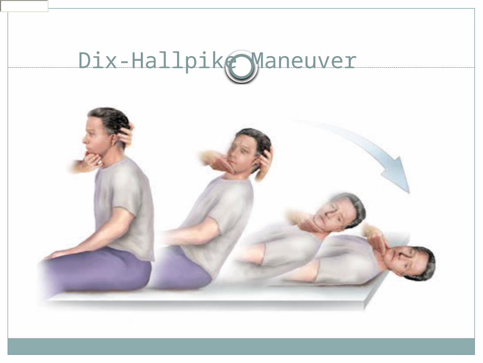
Dix-Hallpike ManeuverUsed to provoke nystagmus and
vertigo commonly associated with BPPV
Head turned 45 degrees to maximally stimulate posterior semicircular canal
Head supported and rapidly placed into head hanging position
Frenzel glasses eliminate visual fixation suppression of response
Dix-Hallpike Maneuver

Dix-Hallpike Maneuver
Positive testUp-beating nystagmusNystagmus to the stimulated sideRotary component to the affected earLasts 15-45 secondsLatency of 2-15 secondsFatigues easily
Pneumatic Otoscopy
Positive and negative pressure applied to middle ear
Hennebert’s sign/symptom – nystagmus and vertigo with pressure, alternates with positive and negative pressure
Can be present in patients with perilymphatic fistula, syphilis, Meninere’s disease, SCC dehiscence syndrome

Dynamic Visual Acuity
Used for bilateral vestibular weakness
Visual acuity checked on Snellen chart
Rechecked while rotating head back and forth at 1-2 Hz.
Loss of 2-3 lines considered abnormal

Romberg Test
Patient asked to stand with feet together and eyes closed
Fall or step is positive testEqual sway with eyes open and closed suggests proprioceptive or cerebellar site
More sway with eyes closed suggests vestibular weakness

Romberg Test

Dysdiadochokinesia Testing
Most commonly tested with the hand slapping test
Abnormalities seen in patients with cerebellar dysfunction
Poor sensitivity and specificity

Tandem Gait Test
Patients are asked to walk heal to toe in a straight line or in a circle
Complex function evaluates many aspects of balance
Poor performance seen in cerebellar lesions, but can be seen in many disorders
Poor sensitivity and specificity

Quantitative Vestibular Testing
Diagnosis unclearProlonged symptoms unresponsive to conservative treatment
Screen for central disordersEvaluate prior to surgical ablation procedures
Documentation of vestibular deficits

Electronystagmography (ENG)
Divided into oculomotor tests, positional and positioning tests, and caloric tests
Only vestibular test with the ability to test individual labyrinths separately
Relies on the vestibulo-ocular reflex (VOR) to test the peripheral vestibular function
Mostly a test of HSCC function

Electronystagmography (ENG)
Oculomotor testsAll test eye movements that originate in the cerebellum
Saccadic trackingSmooth pursuit trackingOptokinetic testing

Oculomotor Tests
Saccadic trackingPatients concentrates on a randomly
moving targetLatency – difference in time between
movement of object and eye (150-250 ms)
Velocity – speed of saccade 200-400 degrees/second low end of normal
Accuracy – amount of undershoot/overshoot of target (75-120%)
Smooth Pursuit Test
Tests ability to accurately and smoothly pursue a target
Gain of eyes compared to movement of target
Saccade movements eliminated from calculations
Asymmetrical pursuit highly suggestive of central disorders

Optokinetic Tests
Vestibular system and optokinetic nystagmus allow steady focus on objects
Target is rapidly passed in front of subject in one direction, then the other
Eye movements are recorded and compared in each direction
Asymmetry suggestive of CNS lesionHigh rate of false positive results

Caloric Testing
Established and widely accepted method of vestibular testing
Most sensitive test of unilateral vestibular weakness
Patient positioned 30 degrees from prone (HSCC vertical allowing max stim)
Cold and warm water/air flushed into EAC

Caloric Testing
COWS (cold opposite, warm same) – direction of the nystagmus
Stimulation in 0.002-0.004 Hz range (Head movements in 1-6 Hz range)
Visual fixation should reduce strength of caloric responses 50-70%
% caloric paresis = 100 * [(LC + LW) – (RC + RW)/(LC + LW + RC + RW)]
Posturography
Used to tests integration of balance systemsUseful in quantification of fall riskMost useful in following conditions:
Chronic disequilibrium and normal examsSuspected malingeringSuspected multifactorial disequilibriumPoorly compensated vestibular injuries
INVESTIGATIONS
Audiogram is routinely needed
Electronystagmography
Caloric testing Radiological Investigation
Blood examination
Gadolinium-enhance MRI – pinpoints site of lesion.
Dynamic posturography - quantifies balancing response to induced sways.

58
TREATMENT
TREAT THE CAUSE IF KNOWN
MEDICAL
SURGICAL
PHYSICAL THERAPY

59
Medical Treatment
SymptomaticSpecific therapyVestibular rehabilitation

Medical TreatmentMedications are most useful for treating
acute vertigo that lasts a few hours to several days
They have limited benefit in patients with benign paroxysmal positional vertigo, because the vertiginous episodes usually last less than one minute.
Vertigo lasting more than a few days is suggestive of permanent vestibular injury (e.g., stroke), and medications should be stopped to allow the brain to adapt to new vestibular input.

Categories of Drugs commonly used in treatment of dizziness
Antihistamines: eg meclizine, stugerone,• Betahistidine (serc)
Anticholinergics: e.g.Scopolamine (Isopto),
glycopyrrolate (Robinul)Phenothiazines : eg Promethazine,
prochlorperazineBenzodiazepines: eg diazepam (Valium) Monoaminergics: eg ephedrine

Vestibular rehabilitation exercises
Semont manoeuvreParticle repositioning manoeuvre (Epley)
Dix-Hallpike manoevreCawthorne exercisesBrandt-Daroff exercises (dispersing
otolithic debris in the semicircular canals)
Lempert, and Hamid maneuvers, among others.

Copyright ©2003 Canadian Medical Association or its licensors
Parnes, L. S. et al. CMAJ 2003;169:681-693
Fig 9: Liberatory manoeuvre of Semont (right ear)

Fig. 9: Liberatory manoeuvre of Semont (right ear).
The top panel shows the effect of the manoeuvre on the labyrinth as viewed from the front and the induced movement of the canaliths (from blue to black). This manoeuvre relies on inertia, so that the transition from position 2 to 3 must be made very quickly.
Photo: Christine Kenney

Epley maneuver The patient sits on the examination table, with eyes
open and head turned 45 degrees to the right (A). The physician supports the patient's head as the patient
lies back quickly from a sitting to supine position, ending with the head hanging 20 degrees off the end of the examination table (B).
The physician turns the patient's head 90 degrees to the left side. The patient remains in this position for 30 seconds (C).
The physician turns the patient's head an additional 90 degrees to the left while the patient rotates his or her body 90 degrees in the same direction. The patient remains in this position for 30 seconds (D).
The patient sits up on the left side of the examination table. (E)
The procedure may be repeated on either side until the patient experiences relief of symptoms.

Copyright ©2003 Canadian Medical Association or its licensors
Parnes, L. S. et al. CMAJ 2003;169:681-693
Particle repositioning manoeuvre (right ear) (Epley)

Particle repositioning manoeuvre (right ear).
Schema of patient and concurrent movement of posterior/ superior semicircular canals and utricle. The patient is seated on a table as viewed from the right side (A). The remaining parts show the sequential head and body positions of a patient lying down as viewed from the top. Before moving the patient into position B, turn the head 45° to the side being treated (in this case it would be the right side). Patient in normal Dix–Hallpike head-hanging position (B). Particles gravitate in an ampullofugal direction and induce utriculofugal cupular displacement and subsequent counter-clockwise rotatory nystagmus. This position is maintained for 1–2 minutes. The patient's head is then rotated toward the opposite side with the neck in full extension through position C and into position D in a steady motion by rolling the patient onto the opposite lateral side. The change from position B to D should take no longer than 3–5 seconds. Particles continue gravitating in an ampullofugal direction through the common crus into the utricle. The patient's eyes are immediately observed for nystagmus. Position D is maintained for another 1–2 minutes, and then the patient sits back up to position A. D = DIRECTION OF VIEW OF LABYRINTH, DARK CIRCLE = POSITION OF PARTICLE CONGLOMERATE, OPEN CIRCLE = PREVIOUS POSITION. ADAPTED FROM PARNES AND ROBICHAUD (Otolaryngol Head Neck Surg 1997;116: 238-43).45 Photo: Christine Kenney

Surgical Care:Surgery is usually reserved for those in whom CRP fails. It is not a first-line treatment because it is invasive and holds the possibility of complications such as hearing loss and facial nerve damage.

Surgical Care:
Options includelabyrinthectomy, posterior canal occlusion, singular neurectomy, Vestibular nerve section, and transtympanic aminoglycoside application.
All have a high chance of vertigo control.
70
Conclusions
Vestibular complaints common to ENTThorough evaluation and
understandingDx and treat acute symptomsWean vestibular suppressantsSpecific pharmacotherapy institutedChronic, uncompensated disease
benefits from early VRT