dr. animesh mondal dept. of botany replication.pdf · replication, recombination, and repair. short...
TRANSCRIPT


Dr. Animesh Mondal
Dept. of Botany
B B College, Asansol-03

Discovery of Replication
Watson and Crick’s seminal paper describing the
DNA double helix ended with the statement:
“It has not escaped our notice that the specific
pairing we have postulated immediately suggests
a possible copying mechanism for the genetic
material.”

The Nobel Prize in Physiology or Medicine 1962
James Dewey Watson
Francis Harry Compton Crick
Maurice Hugh Frederick Wilkins
(From extreme Left to Right)
Prize share: 1/3 (each)
The Nobel Prize in Physiology or Medicine 1962 was awarded jointly to Francis Harry Compton Crick, James Dewey Watson and Maurice Hugh Frederick Wilkins "for their discoveries concerning the molecular structure of nucleic acids and its significance for information transfer in living material".


CsCl equilibrium density gradient

Sucrose velocity density gradient


(1) DNA polymerase.
(2) enzymes known as helicases that separate the DNA strands at the replication fork.
(3) proteins that prevent them from re-annealing before they are replicated.
(4) Enzymes that synthesize RNA primers.
(5) DNA topo-isomerases.
(6) An enzyme to remove the RNA primers, and
(7) an enzyme to covalently link successive Okazaki fragments.


DNA Polymerases

DNA Polymerase-I In 1957, Arthur Kornberg reported that he had
discovered an enzyme that catalyzes the synthesis of DNA in extracts of E. coli through its ability to incorporate the radioactive label from [14C] thymidine triphosphate into DNA. This enzyme, which has since become known as DNA polymerase I or Pol I, consists of a monomeric 928-residue polypeptide.

Properties of DNA Pol-I A. Pol I Recognizes the Incoming dNTP According to the Shape of
the Base Pair It Forms with the Template DNA.
B. Pol I Can Edit Its Mistakes In addition to its polymerase activity, Pol I has two independent hydrolytic activities:
1. It can act as a 3’ → 5’ exonuclease.
2. It can act as a 5’ → 3’ exonuclease.
C. Pol I’s Polymerase and Two Exonuclease Functions Each Occupy Separate Active Sites
D. The X-Ray Structure of Klenow Fragment Indicates How It Binds DNA.
E. DNA Polymerase Distinguishes Watson–Crick Base Pairs via Sequence-Independent Interactions That Induce Domain Movements

Contd.
F. The DNA Polymerase Catalytic Mechanism Involves Two Metal Ions.
Their active sites all contain two metal ions, usually Mg2+, that are liganded by two invariant Asp side chains in the palm domain. Metal ion B is liganded by all three phosphate groups of the bound dNTP, whereas metal ion A bridges the –phosphate group of this dNTP and the primer’s 3’-OH group.
G. Editing Complexes Contain the Primer Strand in the 3’ → 5 ‘ Exonuclease Site.
H. Pol- I Functions Physiologically to Repair DNA.
I. Pol- I Catalyzes Nick Translation.
J. Pol I’s 5’ → 3’ Exonuclease Functions Physiologically to Excise RNA Primers


Sub units of DNA Pol-III

Structure of DNA Pol-III


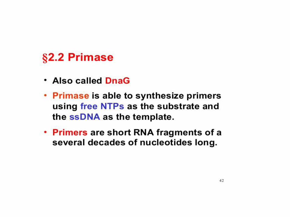
RNA Primers
• DNA polymerases’ all but universal requirement for a free 3’-OH group to extend a DNA chain poses a question that was emphasized by the establishment of the semidiscontinuous model of DNA replication: How is DNA synthesis initiated? Careful analysis of Okazaki fragments revealed that their 5’ ends consist of RNA segments of 1 to 60 nt (a length that is species dependent) that are complementary to the template DNA chain (Fig. 30-7). E. coli has two enzymes that can catalyze the formation of these RNA primers: RNA polymerase, the 459-kD multisubunit enzyme that mediates transcription (Section 31-2), and the much smaller primase (60 kD), the monomeric product of the dnaG gene.

Function


Single-strand DNA-binding protein (SSB) is a protein, 178 amino acids long, found in Escherichia coli (E. coli) bacteria, that binds to single-stranded regions of deoxyribonucleic acid (DNA). Single-stranded DNA is produced during all aspects of DNA metabolism: replication, recombination, and repair.


Short List of proteins & their Mr. involved in DNA Replication

Bacteriophage M13

M13 carries a 6408-nt single-stranded circular
DNA known as its viral or (+) strand. On infecting an
E. coli cell, this strand directs the synthesis of its
complementary or (-) strand to form the circular duplex
replicative form (RF), which may be either nicked (RF
II) or supercoiled (RF I). This replication process (Fig.
30-23) may be taken as a paradigm for leading strand
synthesis in duplex DNA.
As the M13 (+) strand enters the E. coli cell, it becomes
coated with SSB except at a palindromic 57-nt segment
that forms a hairpin. RNA polymerase commences primer
synthesis 6 nt before the start of the hairpin and extends
the RNA 20 to 30 residues to form a segment of
RNA–DNA hybrid duplex. The DNA that is displaced
from the hairpin becomes coated with SSB so that when
RNA polymerase reaches it, primer synthesis stops. Pol III
holoenzyme then extends the RNA primer around the circle
to form the (–) strand. The primer is removed by
Pol-I catalyzed
nick translation, thereby forming RF II, which is
converted to RF I by the sequential actions of DNA ligase
and DNA gyrase.

Phy X 174 (-) Strand Replication Is a Paradigm for Lagging Strand Synthesis

Phi X174 (+) Strand Replication Serves as a Model
for Leading Strand Synthesis


E. coli replication of origin


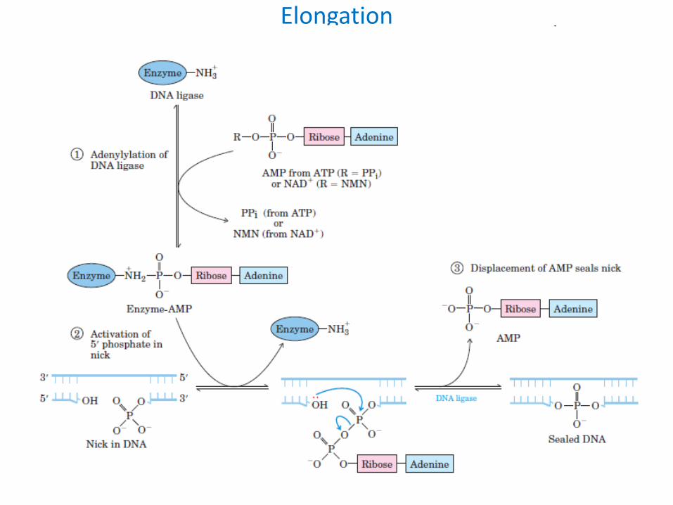
Elongation

Since a single polypeptide as small as the Pol I Klenow fragment can replicate DNA by itself, why does E. coli maintain a battery of 20 intricately coordinated proteins to replicate its chromosome?

THANKS
THANKS