dr. gaurav kaushik
TRANSCRIPT

Dr. Gaurav Kaushik
Resident, Department of Radiodiagnosis
Padmashree Dr. D.Y. Patil Medical college and Hospital.
ULTRASONOGRAPHY AND COLOR
DOPPLER EVALUATION OF THYROID
DISEASES

INTRODUCTION
The role of high resolution Ultrasonography in
the evaluation of the neck region is becoming
increasingly important due to the availability of
high frequency probes.
Detection of thyroid lesions by sonography
are common and the incidence is much higher
than the incidence of palpable thyroid
nodules.
Thyroid sonography is frequently asked to be
performed in the work up of a palpable thyroid
lesion.
Thyroid lesions are often found unrelated to
palpable neck masses.
Along with being much safer and non-ionising,
ultrasound is also much cheaper alternative to
C.T. and M.R.I. which are used in the evaluation
of thyroid masses, but are not as sensitive as
ultrasound in the detection of intrathyroid
lesions but are used for mediastinal extension
of thyroid masses.
Thyroid ultrasound differentiates solid from
cystic lesions, solitary from multinodular and
diffuse enlargement, and extrathyroid lesions.
Nearly 50% of patients with clinically solitary
thyroid nodule have avoided surgery by thyroid
scanning.
The newly developed high resolution
Ultrasonography and color Doppler flow
mapping can reveal fine details of the thyroid
gland and the hemodynamic features of thyroid
neoplasms.
Thus the combination of conventional
sonography and color Doppler provides benefits
in increasing the screening sensitivity and
improves accuracy in distinguishing different
thyroid abnormalities.
An attempt was made in present study to
evaluate and establish certain sonographic
criteria for distinguishing benign and malignant
lesions by evaluating a large spectrum of thyroid
diseases.
Aim and Objectives
1. To study spectrum and role of Ultrasound &
Color Doppler in differentiating different
thyroid diseases.
2. To co-relate Ultrasound & Color Doppler with
fine needle aspiration cytology (FNAC)

Materials and Methods
Type of Study: A Prospective Observational
study
Study Area and Duration: The Study was
conducted in a tertiary care institute and
Medical College of Mumbai for a period of two
years.
Sampling Technique: Consecutive type of
Non-probability Sampling.
Sample Size: A total of 140 patients with
thyroid & parathyroid diseases were referred
& USG of thyroid gland was performed.
Data Analysis: Data was analysed using SPSS
software Ver. 19.
The characteristics studied were the presence
of the nodule, internal characteristic of
lesions, the internal echogenecity of the
nodule, presence of cystic changes, number
of focal lesions,involvement of
gland, calcifications and type of vascularity on
color & power doppler.
The presence of any significant neck nodes
was also recorded.
Patients who had suspicious lesions or
change in image characteristic over time or
with equivocal findings were recommended
USG guided FNAC for further definitive
diagnosis.
Observations
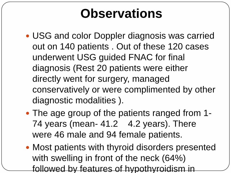
USG and color Doppler diagnosis was carried
out on 140 patients . Out of these 120 cases
underwent USG guided FNAC for final
diagnosis (Rest 20 patients were either
directly went for surgery, managed
conservatively or were complimented by other
diagnostic modalities ).
The age group of the patients ranged from 1-
74 years (mean- 41.2 4.2 years). There
were 46 male and 94 female patients.
Most patients with thyroid disorders presented
with swelling in front of the neck (64%)
followed by features of hypothyroidism in
another 28% of cases.

Main Symptoms Frequency Percent
Asymtomatic 8 5.71%
Constitutional
symptoms 10 7.14%
Difficulty in breathing 4 2.86%
Difficulty in swallowing 8 5.71%
Features of
hyperthyroidism 11 7.86%
Features of
hypothyroidism 28 20.00%
Hoarseness of voice 1 0.71%
Hypercalcemia, raised
PTH 6 4.29%
Swelling in front of neck 64 45.71%
Total 140 100.00%

0.00%
5.00%
10.00%
15.00%
20.00%
25.00%
30.00%
35.00%
10.71%
32.14%
25.00%
8.57%
3.57%
9.29%
3.57% 3.57% 3.57%
Diagnosis (USG/Doppler)

0.00%
5.00%
10.00%
15.00%
20.00%
25.00%
30.00%
10.00%
27.86%
22.14%
7.86%8.57%7.86%
3.57%3.57%1.43%0.71%
2.86%3.57%
Final Diagnosis
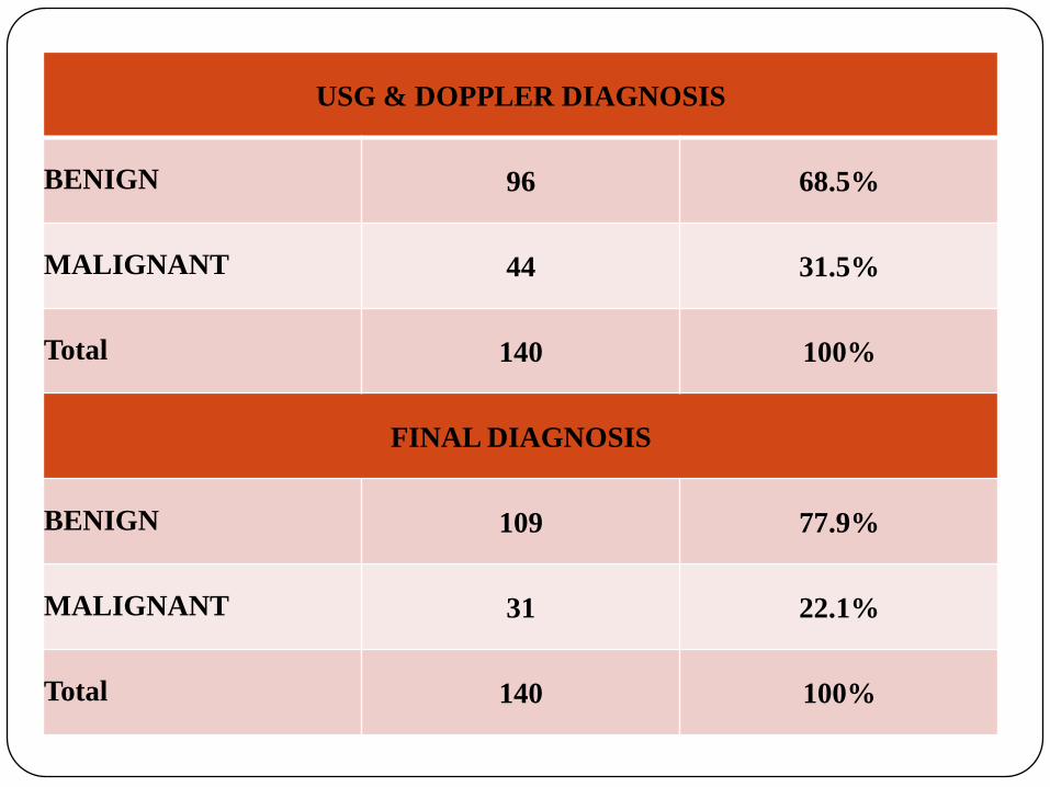
USG & DOPPLER DIAGNOSIS
BENIGN 96 68.5%
MALIGNANT 44 31.5%
Total 140 100%
FINAL DIAGNOSIS
BENIGN 109 77.9%
MALIGNANT 31 22.1%
Total 140 100%
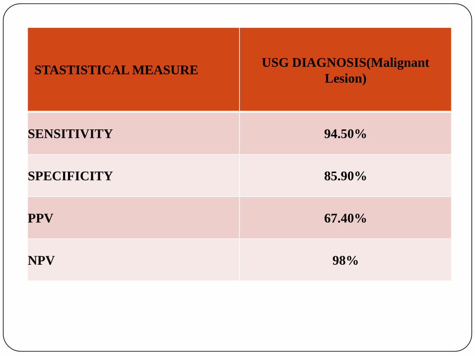
STASTISTICAL MEASUREUSG DIAGNOSIS(Malignant
Lesion)
SENSITIVITY 94.50%
SPECIFICITY 85.90%
PPV 67.40%
NPV 98%
Conclusion
The Present study aimed at determining the accuracy of the combination of USG and ColorDoppler in delineating different thyroid diseases.
Amongst them the most important distinction was between relatively benign lesions vssuspicious lesions of malignancy.
The present study shows that combined use of Ultrasound and Color Doppler as a primary investigation in symptomatic patients of thyroid disease allows safe management decisions to be made and can further direct patient for USG guided FNAC for reaching a definitive diagnosis.
We therefore recommend USG and ColorDoppler to be the first Investigation of choice in all suspected thyroid disorders.
Bibliography
1. Usha Menon, Sundaran JR: High prevalance of undetecdthyroid disorders in iodine sufficient adult south indianpopulation. Journal of Indian Medical Assoc, 2009 Feb; 107(2):72-7
2. Walker J, findlay D et al : A prospective study of thyroid ultrasound scan in the clinical solitary thyroid nodule. British Journal Of Radiology 1985, 58(691): 617-619.
3. Lundgren CI, Zedenius J, Skoog L. Fine-needle aspiration biopsy of benign thyroid nodules: an evidence-based eview. World J Surg. 2008;32:1247-52.
4. Crocker EF, Jellins J. Grey scale ultrasonic examination of the thyroid gland Med J Aust 1978;9:244.
5. Scheible W, Leopold GR, Woo VL, Gosink BB (1979) High resolution real-time ultrasonography of thyroid nodules. Radiology 133:413–417
6. Fujimoto F, Oka A, Omoto R, Hirsoe M (1967)Ultrasound scan- ning of the thyroid gland as a new diagnostic approach. Ultrasonics 5:177–180
7. Thijs LG (1971) Diagnostic ultrasound in clinical thyroid investigation. J Clin Endocrinol Metab 32(6):709–716.