dr i. bojak section neurophysiology and neuroinformatics computational brain models of eeg / meg...
TRANSCRIPT
Dr I. BojakSection Neurophysiology and Neuroinformatics
http://www.neuropi.org
Computational brain models of EEG / MEG and fMRI signals in health and disease
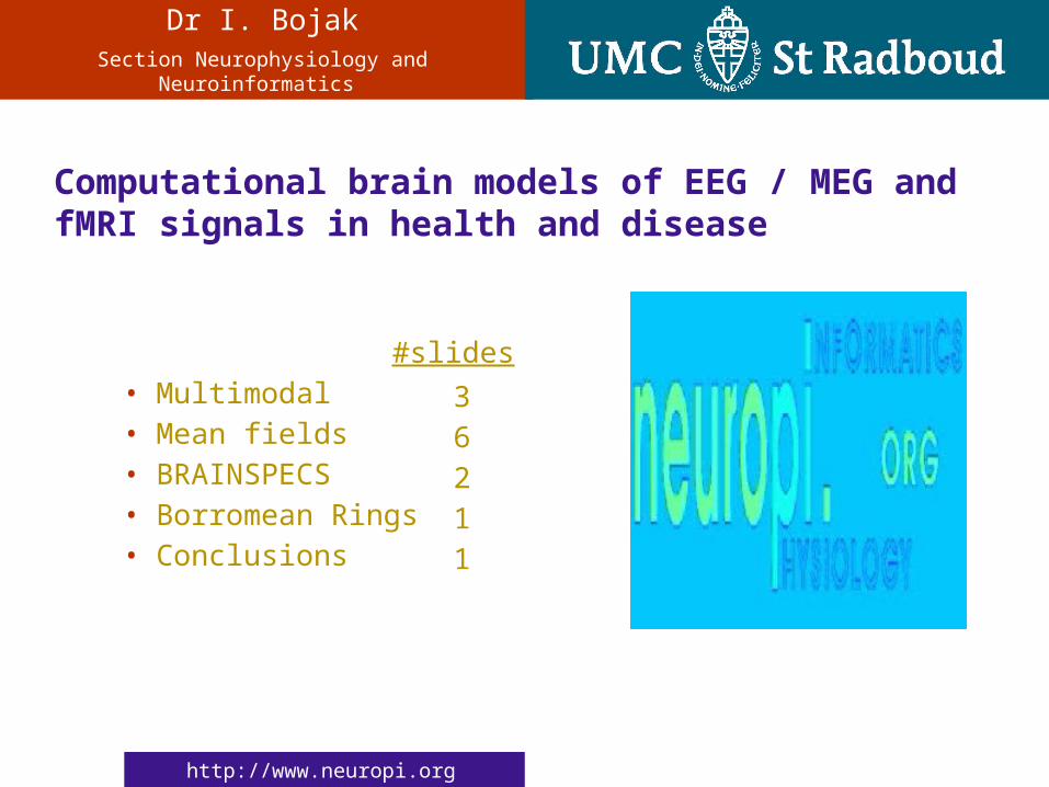
#slides• Multimodal• Mean fields• BRAINSPECS• Borromean Rings• Conclusions
• 3• 6• 2• 1• 1

2
Dr I. Bojak
Section Neurophysiology and Neuroinformatics
Computational brain models of EEG/MEG and fMRI signals in health and disease
Multimodal – the six blind men and the elephant
Pics - EEG: Brain Sci. Institute Swinburne; MEG: Dept. of Psychology NYU; fMRI: Dept. Cog. Neurology, MPI Leipzig; SPECT: C. Studholme UCSF; PET: N.D. Volkov et al.; Anatomy: NTVH MRI Lab; Poem: Wordinfo.
SPECT
EEG
MEG
fMRI
PET
anatomy
3
Dr I. Bojak
Section Neurophysiology and Neuroinformatics
Computational brain models of EEG/MEG and fMRI signals in health and disease
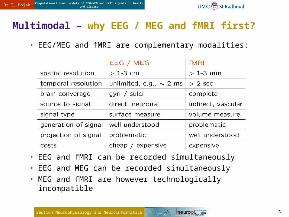
Multimodal – why EEG / MEG and fMRI first?
• EEG/MEG and fMRI are complementary modalities:
• EEG and fMRI can be recorded simultaneously• EEG and MEG can be recorded simultaneously• MEG and fMRI are however technologically incompatible
4
Dr I. Bojak
Section Neurophysiology and Neuroinformatics
Computational brain models of EEG/MEG and fMRI signals in health and disease
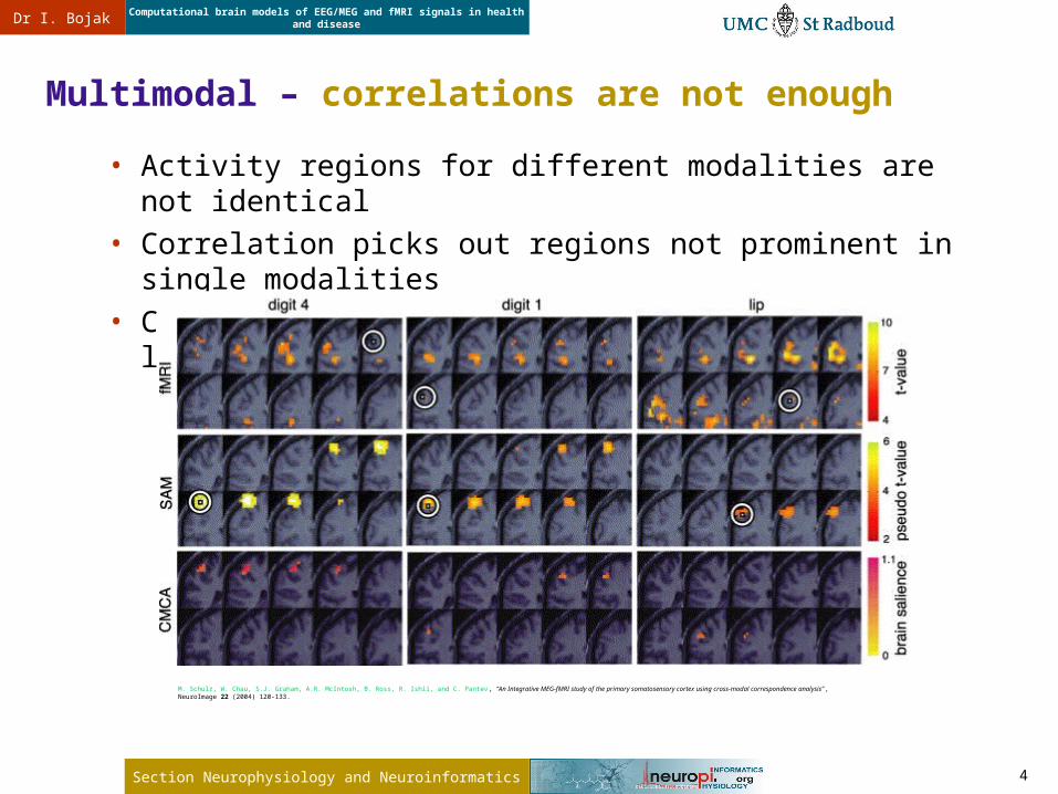
• Activity regions for different modalities are not identical• Correlation picks out regions not prominent in single modalities• Correlated activity regions are much more localized
Multimodal – correlations are not enough
M. Schulz, W. Chau, S.J. Graham, A.R. McIntosh, B. Ross, R. Ishii, and C. Pantev, “An Integrative MEG-fMRI study of the primary somatosensory cortex using cross-modal correspondence analysis” , NeuroImage 22 (2004) 120-133.

5
Dr I. Bojak
Section Neurophysiology and Neuroinformatics
Computational brain models of EEG/MEG and fMRI signals in health and disease
Mean fields – sources for non-invasive imaging
• “in phase” neurons contribute , “out of phase” • 105 neurons, 1% “in phase”: 32x stronger signal – seen only.• Imaging behaviour neuronal mass action
averaging mean field theories

6
Dr I. Bojak
Section Neurophysiology and Neuroinformatics
Computational brain models of EEG/MEG and fMRI signals in health and disease
Mean fields – our model
flattened
simplified
averaged spatially

7
Dr I. Bojak
Section Neurophysiology and Neuroinformatics
Computational brain models of EEG/MEG and fMRI signals in health and disease
• 145 Dell Power Edge 1950 blades: 2 x quad-core 2.33 GHz Clovertown, 16 GB RAM
• 145 TB blade hard drives, 100 TB Raid 5 disks, 77 TB robot tape
• Cisco 6509 gigabit ethernet about to be upgraded to 20Gb/s infiniband
• 4 -128 nodes run in parallel using MPI Fortran
Mean fields – whole cortex computing
0framesdisk
10noise
1 2
4 5 6
3
7 8 9Linux: CentOS 5,queue: PBS,manager: Torque,scheduler: Moab,compilers: Intel 9.1.
Green Machine

8
Dr I. Bojak
Section Neurophysiology and Neuroinformatics
Computational brain models of EEG/MEG and fMRI signals in health and disease
• PSP response under Isoflurane :
Mean fields – works well for EEG, e.g., anesthesia
Banks & Pearce, MacIver et al.
f
req
ue
ncy
[H
z]
0
6
.0
12
.0
1
8.0
24
.0
0 0.13 0.27 0.40
wavelength-1 [cm-1]
MAC
EEG ~ mean excitatory soma membrane potential
9
Dr I. Bojak
Section Neurophysiology and Neuroinformatics
Computational brain models of EEG/MEG and fMRI signals in health and disease
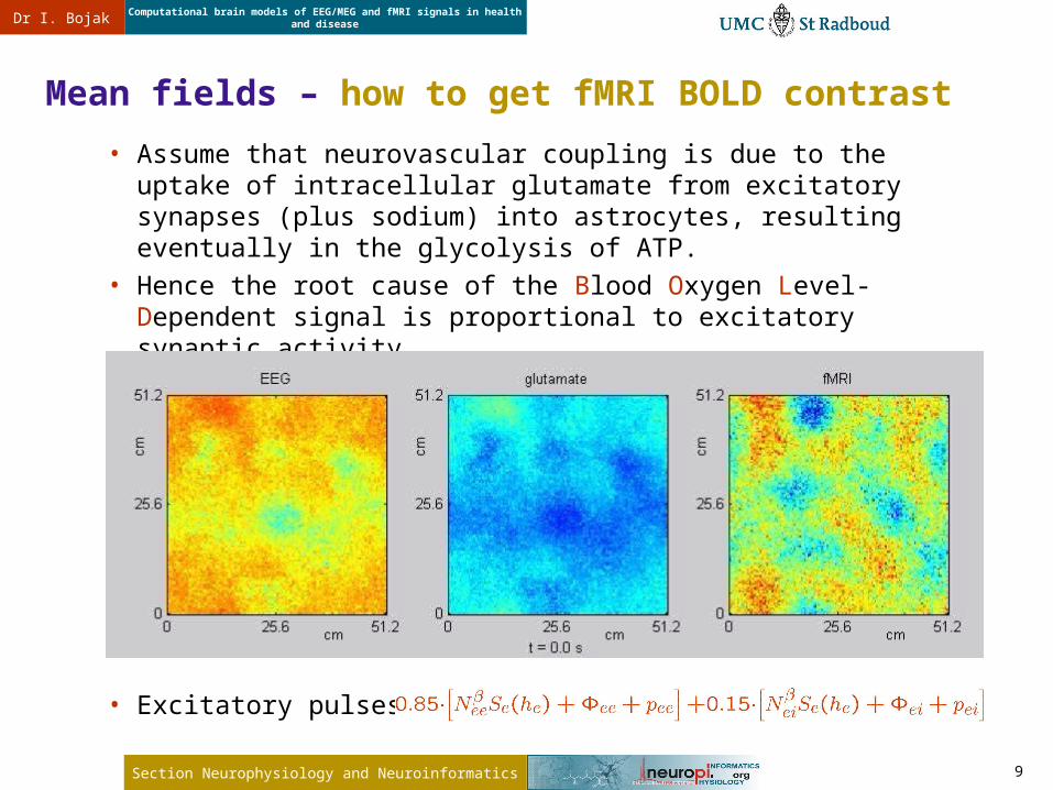
Mean fields – how to get fMRI BOLD contrast
• Assume that neurovascular coupling is due to the uptake of intracellular glutamate from excitatory synapses (plus sodium) into astrocytes, resulting eventually in the glycolysis of ATP.
• Hence the root cause of the Blood Oxygen Level-Dependent signal is proportional to excitatory synaptic activity.
• Excitatory pulses:

10
Dr I. Bojak
Section Neurophysiology and Neuroinformatics
Computational brain models of EEG/MEG and fMRI signals in health and disease
• Isotropic, homogeneous, exp. connectivity:
• But there’s also specific one:
Mean fields – how to implement connectivity?
# synapses
Felleman & Van Essen

11
Dr I. Bojak
Section Neurophysiology and Neuroinformatics
Computational brain models of EEG/MEG and fMRI signals in health and disease
BRAINSPECS – the proposal
• BRain Activity Imaging and Network Simulations for the Prediction and Evaluation of Clinical Syndromes - a personalizable brain model of EEG/MEG and fMRI signals in health and disease (Integrating Project for FP7-ICT-2007-2)
• 40 principal researchers, budget € 9.5 million, 5 years runtime
Nijmegen Amsterdam London Cambridge Barcelona Warsaw Sankt Augustin Ås Lausanne
iel Stockholm
1
SME Localite 1

12
Dr I. Bojak
Section Neurophysiology and Neuroinformatics
Computational brain models of EEG/MEG and fMRI signals in health and disease
BRAINSPECS – working packages
projection software
main fieldprograms
networkprograms
data integration
connectivity database
WP3Model fitting
WP8Advanced modeling tools
WP4Local and detailed models
WP2Connectivity
WP10Data acquisition and visualization
WP9Data management and ontology
WP7Epilepsy
WP6Drug effects
WP5Lesions and dementia
data interface
connectivity constraints
visualisation interface
data access
clinical data
func
tion
al c
onne
ctiv
ity
WP1Forward and inverse modeling
com
puta
tion
al
mea
n fi
eld
para
met
ers
experimental main field parameters
10 scientific Working Packages
WP12Project management
WP11Dissemination
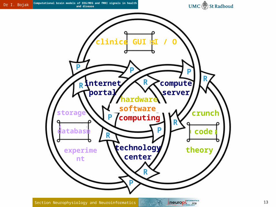
clinics
experiment
theory
computing
public relations
13
Dr I. Bojak
Section Neurophysiology and Neuroinformatics
Computational brain models of EEG/MEG and fMRI signals in health and disease
experi-ment
clinics
theory
computing
clinics
computing
experiment theory
storage
I / O
crunch
hardware storage
hardware
crunch
I / O
database
GUI
code
software
technologycenter
internetportal
computeserver
P
P
P
PP
P
R
R
R
RRR
14
Dr I. Bojak
Section Neurophysiology and Neuroinformatics
Computational brain models of EEG/MEG and fMRI signals in health and disease
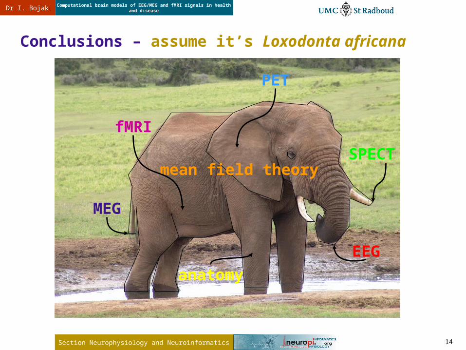
Conclusions – assume it’s Loxodonta africana
mean field theory
EEG
MEG
fMRI
PET
SPECT
anatomy