drosophila as a genetic model for hematopoiesis · as in mammalian systems, multiple sites of...
TRANSCRIPT

| FLYBOOK
DEVELOPMENT AND GROWTH
Drosophila as a Genetic Model for HematopoiesisUtpal Banerjee,*,†,‡,§,1,2 Juliet R. Girard,*,1,2 Lauren M. Goins,*,1,2 and Carrie M. Spratford*,1,2
*Department of Molecular, Cell, and Developmental Biology, †Molecular Biology Institute, ‡Department of Biological Chemistry, and §Eli and EdytheBroad Center of Regenerative Medicine and Stem Cell Research, University of California, Los Angeles, California 90095
ABSTRACT In this FlyBook chapter, we present a survey of the current literature on the development of the hematopoietic system inDrosophila. The Drosophila blood system consists entirely of cells that function in innate immunity, tissue integrity, wound healing, andvarious forms of stress response, and are therefore functionally similar to myeloid cells in mammals. The primary cell types arespecialized for phagocytic, melanization, and encapsulation functions. As in mammalian systems, multiple sites of hematopoiesisare evident in Drosophila and the mechanisms involved in this process employ many of the same molecular strategies that exemplifyblood development in humans. Drosophila blood progenitors respond to internal and external stress by coopting developmentalpathways that involve both local and systemic signals. An important goal of these Drosophila studies is to develop the tools andmechanisms critical to further our understanding of human hematopoiesis during homeostasis and dysfunction.
KEYWORDS crystal cell; Drosophila; FlyBook; hematopoiesis; hemocyte; innate immunity; lamellocyte; lymph gland; plasmatocyte; stress response
TABLE OF CONTENTS
Abstract 367
Introduction 369
Evolution of Blood 3
Drosophila Blood Cell Types 5Plasmatocytes 6
Summary 6Specifics 7
Crystal cells 7Summary 7Specifics 8
Lamellocytes 9Summary 9Specifics 9
Sites of Hematopoietic Development 10Continued
Copyright © 2019 Banerjee et al.doi: https://doi.org/doi: https://doi.org/10.1534/genetics.118.300223Manuscript received August 31, 2017; accepted for publication December 5, 2018.Available freely online through the author-supported open access option.This is an open-access article distributed under the terms of the Creative Commons Attribution 4.0 International License (http://creativecommons.org/licenses/by/4.0/), which permitsunrestricted use, distribution, and reproduction in any medium, provided the original work is properly cited.1These authors contributed equally to this work.2Corresponding authors: 610 Charles E Young Drive East, University of California, Los Angeles, CA 90095. E-mail: [email protected]; [email protected]; [email protected];and [email protected]
Genetics, Vol. 211, 367–417 February 2019 367

CONTENTS, continued
Embryonic procephalic mesoderm 10Summary 10Specifics 11
Sessile pools and circulating larval hemocytes 12Summary 12Specifics 12
Dorsal mesoderm 14Summary 14Specifics 14
Larval Lymph Gland: Zones, Cells, and Signals 16Summary 16
Specifics 17
Preprogenitors 18Summary 18Specifics 18
Progenitors and differentiation: the MZ and CZ 19ECM 20Hh signaling 20Wg/Wnt signaling 20Calcium signaling 20JAK/STAT pathway 21ROS 21FGF pathway 22Collier 22Equilibrium signal 22
The PSC 23Summary 23Specifics 23
The Posterior lobes 24
Pupal and Adult Blood Cells 24Summary 24
Specifics 25
Nutritional and Sensory Control of Hematopoiesis 25Nutritional control 25
Summary 25Specifics 26
Sensory control 27Olfactory control 27Control by environmental gases 28
Stress Response and the Hematopoietic System 28Cellular response to intrinsic stress 29
Summary 29Specifics 29
Cellular response to sterile injury 30Summary 30Specifics 31
Cellular response to wasp parasitization 32Summary 32Specifics 32
Continued
368 U. Banerjee et al.

CONTENTS, continued
Cellular response to pathogens and septic injury 34Summary 34Specifics 35
Development and the Stress Response: Two Sides of the Same Coin 37
Final Words 38
Evolution of Blood
THE study of invertebrate hemocytes, and their likelyevolutionary link to mammalian blood, has a long and
chequered past. It seems likely that the earliest metazoanhemocytes are analogous to mammalian blood cells, i.e., theyshare similar functions but might have arisen through a poly-phyletic system of independent evolutionary events. Homol-ogy between the two evolutionarily distant systems wouldimply a monophyletic pathway, and it seems very likely thatsome cell types evolved only once while others might haveevolved independently [reviewed in Millar and Ratcliffe(1989)]. Historically, this debate is further complicated dueto the variety of names that have been assigned to blood cellswithin the invertebrate phyla, even as they represent identi-cal cell types [reviewed in Ratcliffe and Rowley (1981)]. Thisis in contrast to the well-established nomenclature forcells of the hematopoietic system in mammals [reviewed inWeissman et al. (2001)]. Molecular and genetic approachesare now accessible for use broadly across metazoans and suchinvestigations will shed further light onto this important evo-lutionary question [reviewed in Hartenstein (2006)].
Debates over analogy and homology are not specific tothe blood. For example, homology in eye developmentremained elusive despite clear functional and molecularsimilarities between them [reviewed in Gehring (1996)]. Visualtransduction by invertebrate rhabdomeric-Rhodopsin (r-R)(Arendt et al. 2004) and vertebrate ciliary-Rhodopsin (c-R)were thought to have evolved independently, until the un-expected finding that both r-R and c-R are found in the in-vertebrate ragworm (Arendt et al. 2004). This nonmodelsystem study was critical to the findings that rhodopsins arespecialized through evolution for photoreceptors, retinalganglion cells, and cells that control circadian rhythms, asneeded [reviewed in Ernst et al. (2014)].
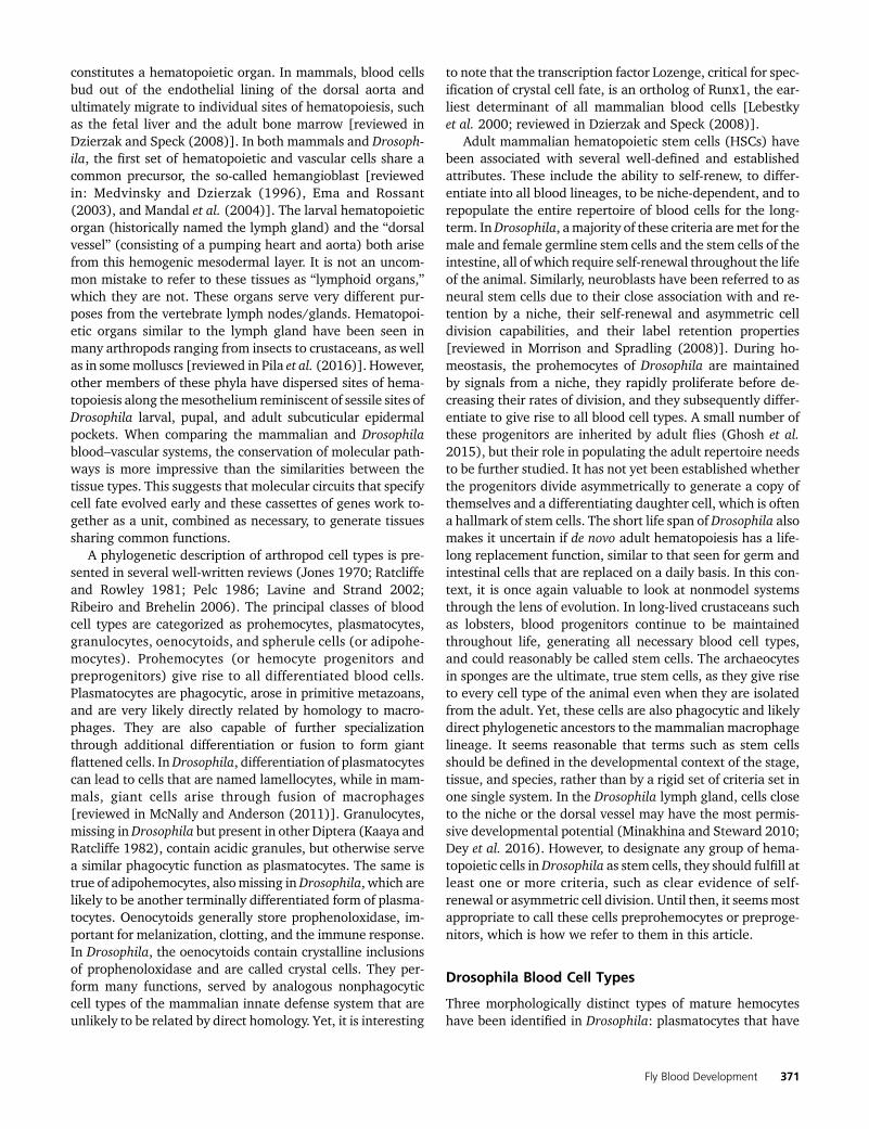
We can anticipate a similar scenario for the evolution ofmetazoan hematopoiesis (Figure 1). Blood cells likely arosein the choanoflagellate ancestors of metazoans since they arereadily apparent in several species of diploblastic sponges,which lack a mesoderm. These species contain a group ofcells, termed archaeocytes, that can efficiently generate allof the 10 cell types that give rise to the entire animal (DeSutter and Buscema 1977; De Sutter and Van de Vyver 1977;Simpson 1984). The rest of the cell types lack this regenerative
potential and, thus, the archaeocytes are bona fide stem cellsthat are maintained through the life of the animal. Interest-ingly, these circulating archaeocytes are phagocytic, not un-like those seen in more evolved animals, such as themammalian macrophages and microglia. The primary func-tion of these phagocytic cells is to gather nutrition throughengulfment and deliver this to the rest of the cells of theanimal. Phagocytes are considered to be the only blood celltype that has been maintained throughout evolution in amonophyletic manner, radiating out for specialized functionsthat reflect the adaptive needs of each separate clade. Phago-cytes in higher animals are neither totipotent, nor gatherersof nutrition, but they have retained the specialized functionthat allows them to recognize and engulf pathogens, or ves-tiges of apoptotic and nonself tissue. In general, the conceptof a multifunctional cell type that has then compartmental-ized a subset of its functions to formmore specialized cells is acommon theme seen in metazoan evolution [reviewed inMillar and Ratcliffe (1989)].
Like sponges, cnidariansarealsodiploblastic,witha largelyacellular layer of mesoglea in between the ectoderm and theendoderm.Manyspecieswithin thisphylumdonothavebloodcells sincediffusionofwaterandnutrients is fairlyunrestrictedin the mesoglea, often aided by symbiotic interactions withalgae (for example, in corals). However, in a cnidarian such asthe hydra, phagocytic blood cells populate andmove throughthe mesoglea distributing nutrition (Cooper 1976). Recentstudies provide evidence of Toll/NFΚB signaling in sea anem-ones, which raises the possibility that innate immunity pre-ceded the traditional cnidarian–bilaterian split and mighthave evolved at about the same time as the most ancientblood cells (Brennan et al. 2017).
The first signs of additional differentiated blood cell typesare seen with the evolution of the “pseudocoelom” in flat-worms and nematodes, but the most rapid diversificationand evolution of the blood tissue is observed with the adventof the true coelom in triploblastic animals that have evolved awell-defined mesodermal germ layer. Annelids have a closedloop circulatory system. Erythrocytes or “red blood cells” thatcarry oxygen to other body parts first appeared in marine(polychaete) annelids (Cooper 1976). Additionally, annelidblood contains cells that have been referred to as leukocytes,which are functionally akin to granulocytes, lymphocytes,and monocytes, as components of an immune system that
Fly Blood Development 369

can distinguish self from nonself [reviewed in Vetvicka andSíma (2009)]. It is hypothesized that ancestors of annelidsand other bilateriansmight also have been coelomic, and thatprimitive blood cells arose from its linings; but in the absenceof fossil data, it cannot be ruled out that the common ancestorhad a “solid” mesenchyme within which the blood cells firstarose. In either case, the bilaterians all built upon a basicancestral framework, and generated diversity through thegain and loss of cell types depending on their respective adap-tive strategies. This basic framework of the hematopoietic/vascular/innate immune system laid down in segmentedworms is identifiable in molluscs [reviewed in Pila et al.(2016)], arthropods, echinoderms (starfish and sea urchins),and tunicates (sea squirts and ciona).
The phagocytic cell type first evident in sponges is consid-ered homologous to macrophages. Additionally, the proto-stome invertebrates possess several blood cell types that areanalogous to those seen in vertebrates. For example, crystalcells and lamellocytes in Drosophila have functions in com-monwith platelets and giant cells in humans, but it is unlikelythat these fly and mammalian cells arose through a mono-phyletic path. Finally, the lymphoid system is generally be-lieved to have made its first appearance in bony fishes(osteichthyes), but scattered elements of this lineage are seenin the invertebrate deuterostomes (echinoderms, hemichor-dates, and chordates). There is good evidence that the
vertebrate hematopoietic system, from fishes on to the mam-mals, follows a monophyletic evolutionary scheme that arosefrom the deuterostome invertebrates (echinoderms and tuni-cates) (Cooper 1976). For the purposes of this review onDrosophila hematopoiesis, based on currently available infor-mation, it is safe to assume that arthropod blood cells arerestricted in their similarities to the myeloid, but not any ofthe lymphoid, cell types in mammals.
There is a close relationship between the development ofthe hematopoietic and vascular systems.With the appearanceof the third body layer (the mesoderm) in triploblastic ani-mals, a body cavity (the coelom) formed separating theendodermal and ectodermal layers. In most triploblasts, theblood–vascular system develops and functions in addition tothe coelomic system. With the advent of vasculature, thecirculating blood cells could reach longer distances of bodylength carrying nutrients and gases. Hartenstein and cowork-ers have reviewed the blood–vascular system from a phylo-genetic point of view (Hartenstein 2006; Hartenstein andMandal 2006; Grigorian and Hartenstein 2013). They pointto the similarities of the endothelial layer lining the verte-brate vascular system with the mesothelium that lines(sometimes discontinuously) the coelom. Depending on theorganism, the endothelial and/or mesothelial layers eitherdirectly give rise to blood precursors or to groups of cells thatform structures such as the lymph gland in Drosophila, which
Figure 1 Phylogenetic tree depicting key eventsduring the evolution of metazoan blood cells. HSCs,hematopoietic stem cells.
370 U. Banerjee et al.
constitutes a hematopoietic organ. In mammals, blood cellsbud out of the endothelial lining of the dorsal aorta andultimately migrate to individual sites of hematopoiesis, suchas the fetal liver and the adult bone marrow [reviewed inDzierzak and Speck (2008)]. In both mammals and Drosoph-ila, the first set of hematopoietic and vascular cells share acommon precursor, the so-called hemangioblast [reviewedin: Medvinsky and Dzierzak (1996), Ema and Rossant(2003), and Mandal et al. (2004)]. The larval hematopoieticorgan (historically named the lymph gland) and the “dorsalvessel” (consisting of a pumping heart and aorta) both arisefrom this hemogenic mesodermal layer. It is not an uncom-mon mistake to refer to these tissues as “lymphoid organs,”which they are not. These organs serve very different pur-poses from the vertebrate lymph nodes/glands. Hematopoi-etic organs similar to the lymph gland have been seen inmany arthropods ranging from insects to crustaceans, as wellas in somemolluscs [reviewed in Pila et al. (2016)]. However,other members of these phyla have dispersed sites of hema-topoiesis along themesothelium reminiscent of sessile sites ofDrosophila larval, pupal, and adult subcuticular epidermalpockets. When comparing the mammalian and Drosophilablood–vascular systems, the conservation of molecular path-ways is more impressive than the similarities between thetissue types. This suggests that molecular circuits that specifycell fate evolved early and these cassettes of genes work to-gether as a unit, combined as necessary, to generate tissuessharing common functions.
A phylogenetic description of arthropod cell types is pre-sented in several well-written reviews (Jones 1970; Ratcliffeand Rowley 1981; Pelc 1986; Lavine and Strand 2002;Ribeiro and Brehelin 2006). The principal classes of bloodcell types are categorized as prohemocytes, plasmatocytes,granulocytes, oenocytoids, and spherule cells (or adipohe-mocytes). Prohemocytes (or hemocyte progenitors andpreprogenitors) give rise to all differentiated blood cells.Plasmatocytes are phagocytic, arose in primitive metazoans,and are very likely directly related by homology to macro-phages. They are also capable of further specializationthrough additional differentiation or fusion to form giantflattened cells. InDrosophila, differentiation of plasmatocytescan lead to cells that are named lamellocytes, while in mam-mals, giant cells arise through fusion of macrophages[reviewed in McNally and Anderson (2011)]. Granulocytes,missing inDrosophila but present in other Diptera (Kaaya andRatcliffe 1982), contain acidic granules, but otherwise servea similar phagocytic function as plasmatocytes. The same istrue of adipohemocytes, alsomissing inDrosophila, which arelikely to be another terminally differentiated form of plasma-tocytes. Oenocytoids generally store prophenoloxidase, im-portant for melanization, clotting, and the immune response.In Drosophila, the oenocytoids contain crystalline inclusionsof prophenoloxidase and are called crystal cells. They per-form many functions, served by analogous nonphagocyticcell types of the mammalian innate defense system that areunlikely to be related by direct homology. Yet, it is interesting
to note that the transcription factor Lozenge, critical for spec-ification of crystal cell fate, is an ortholog of Runx1, the ear-liest determinant of all mammalian blood cells [Lebestkyet al. 2000; reviewed in Dzierzak and Speck (2008)].
Adult mammalian hematopoietic stem cells (HSCs) havebeen associated with several well-defined and establishedattributes. These include the ability to self-renew, to differ-entiate into all blood lineages, to be niche-dependent, and torepopulate the entire repertoire of blood cells for the long-term. InDrosophila, a majority of these criteria aremet for themale and female germline stem cells and the stem cells of theintestine, all of which require self-renewal throughout the lifeof the animal. Similarly, neuroblasts have been referred to asneural stem cells due to their close association with and re-tention by a niche, their self-renewal and asymmetric celldivision capabilities, and their label retention properties[reviewed in Morrison and Spradling (2008)]. During ho-meostasis, the prohemocytes of Drosophila are maintainedby signals from a niche, they rapidly proliferate before de-creasing their rates of division, and they subsequently differ-entiate to give rise to all blood cell types. A small number ofthese progenitors are inherited by adult flies (Ghosh et al.2015), but their role in populating the adult repertoire needsto be further studied. It has not yet been established whetherthe progenitors divide asymmetrically to generate a copy ofthemselves and a differentiating daughter cell, which is oftena hallmark of stem cells. The short life span of Drosophila alsomakes it uncertain if de novo adult hematopoiesis has a life-long replacement function, similar to that seen for germ andintestinal cells that are replaced on a daily basis. In this con-text, it is once again valuable to look at nonmodel systemsthrough the lens of evolution. In long-lived crustaceans suchas lobsters, blood progenitors continue to be maintainedthroughout life, generating all necessary blood cell types,and could reasonably be called stem cells. The archaeocytesin sponges are the ultimate, true stem cells, as they give riseto every cell type of the animal even when they are isolatedfrom the adult. Yet, these cells are also phagocytic and likelydirect phylogenetic ancestors to the mammalian macrophagelineage. It seems reasonable that terms such as stem cellsshould be defined in the developmental context of the stage,tissue, and species, rather than by a rigid set of criteria set inone single system. In the Drosophila lymph gland, cells closeto the niche or the dorsal vessel may have the most permis-sive developmental potential (Minakhina and Steward 2010;Dey et al. 2016). However, to designate any group of hema-topoietic cells inDrosophila as stem cells, they should fulfill atleast one or more criteria, such as clear evidence of self-renewal or asymmetric cell division. Until then, it seemsmostappropriate to call these cells preprohemocytes or preproge-nitors, which is how we refer to them in this article.
Drosophila Blood Cell Types
Three morphologically distinct types of mature hemocyteshave been identified in Drosophila: plasmatocytes that have
Fly Blood Development 371

phagocytic and antimicrobial functions; crystal cells that fa-cilitate wound healing, innate immunity, and the hypoxic re-sponse; and lamellocytes, which are specialized cells thatprimarily respond to wasp parasitization [Rizki and Rizki1979; reviewed in Evans et al. (2003) and Letourneau et al.(2016)]. Plasmatocytes comprise �90–95% of hemocytesand crystal cells account for �2–5% during embryogenesis,in larvae, and in adults (Tepass et al. 1994; Lebestky et al.2000; Ghosh et al. 2015; Leitão and Sucena 2015). Lamello-cytes are rarely seen in healthy larvae, but are induced upondeposition of eggs by parasitic wasps (Rizki and Rizki 1992).The three cell types were first distinguished based on theirultrastructure and cytochemistry, and later refined throughstudies of biological function, as well as expression patternsof cell surface antigens, enhancer traps, and transcriptionfactors that specify hemocyte fate (Shrestha and Gateff 1982;Rodriguez et al. 1996; Braun et al. 1997; Lo et al. 2002;Tirouvanziam et al. 2004; Kurucz et al. 2007b).
Many of the genes used as markers are also functionallysignificant for the individual blood cell types. Similar tomammalian macrophages, plasmatocytes eliminate both ap-optotic cells and invading particles (Franc et al. 1996). Duringembryogenesis, plasmatocytes primarily function to endocy-tose apoptotic cells and secrete extracellular matrix (ECM)proteins as they aid in tissue remodeling (Olofsson and Page2005; Bunt et al. 2010). Plasmatocytes express the free rad-ical scavenging enzyme Peroxidasin as well as several cell-surface molecules involved in phagocytosis, such as thereceptors Nimrod C1 (P1 antigen) and Eater (Nelson et al.1994; Kocks et al. 2005; Kurucz et al. 2007a,b; Bretscher et al.2015). Crystal cells contain crystalline inclusions of theprophenoloxidase (ProPO) enzymes, which mediate melani-zation, and these cells primarily function to initiate the mel-anization cascade during injury and the innate immuneresponse (Binggeli et al. 2014; Dudzic et al. 2015). Melani-zation leads to a darkening and hardening of damaged tissuethat assists in wound healing, while the free radicals pro-duced during the melanization process neutralize pathogens.Lamellocytes are large, flat cells that encapsulate wasp eggsthat are injected by the parasite’s ovipositor through the Dro-sophila larval cuticle (Rizki and Rizki 1992). bPS-integrin(encoded by myospheroid; mys) is highly expressed in lamel-locytes, and while not required for differentiation, it isrequired for the encapsulation response by lamellocytes(Irving et al. 2005).
Plasmatocytes
Summary: Over the years, ultrastructural analyses haverevealed intriguing differences between morphologically dis-tinct subsets of plasmatocytes [Shrestha and Gateff 1982;Rizki and Rizki 1984; Lanot et al. 2001; Grigorian et al.2011b; reviewed in Grigorian and Hartenstein (2013) andGrigorian et al. (2013)]. These likely represent differentstages of plasmatocyte maturation as it has proven more dif-ficult to distinguish between them using molecular markers.Such markers include Hemolectin (Hml), Peroxidasin (Pxn),
NimC1 (P1 antigen), Croquemort (Crq), Collagen, and Eater(Tepass et al. 1994; Franc et al. 1996; Asha et al. 2003;Sinenko and Mathey-Prevot 2004; Jung et al. 2005;Olofsson and Page 2005; Kurucz et al. 2007a,b; Sorrentinoet al. 2007; Stofanko et al. 2008; Tokusumi et al. 2009a;Makhijani et al. 2011; Evans et al. 2014). While each of theseis a reasonably representative identifier of the plasmatocytefate, none is 100% effective in marking every plasmatocyteand, at least in some instances, this might represent differ-ences in the differentiation/maturation process. This vari-ability usually does not pose a serious problem for mostanalyses, although future studies combining ultrastructurewith marker analysis may help elucidate the basis for thisvariability inmarker expression patterns. An antibody againstthe P1 antigen is widely used to identify mature plasmato-cytes in larvae and adult flies, but does not recognize embry-onic plasmatocytes (Kurucz et al. 2007a). On a practical note,it is worth keeping in mind that a large number of commonlyused Drosophila stocks and chromosomes are “P1-negative”(Honti et al. 2013). This variation is inherited as a recessivetrait, and can therefore confound clonal and other forms ofimmunohistochemical analyses that utilize staining of theheterozygous tissue as a control. Coupled with the fact thatthe total blood cell number in an animal can show a consider-able degree of variation, one cannot overstate the importanceof proper statistical analysis in studies of blood phenotypes.
In addition to phagocytosis of apoptotic cells, embryonicplasmatocytes function in the secretion of ECM proteins.During the second-half of embryogenesis, all cell surfaces thatare in contact with the hemolymph become covered withbasement membranes due to the widespread secretion ofECM molecules from the blood cells and the fat body[reviewed in Fessler and Fessler (1989), Tepass et al.(1994), and Martinek et al. (2008)]. An exception is the cellsurface of the circulating hemocytes themselves that con-stitute a major source of these ECM molecules, includingPapilin, Laminin, Collagen IV, Glutactin, and Tiggrin (Tig)[reviewed in Fessler and Fessler (1989), Kusche-Gullberget al. (1992), and Fogerty et al. (1994)]. Regulated macro-phage migration is essential for uniform delivery of matrixproteins such as Collagen IV, Perlecan, and Laminin A(Matsubayashi et al. 2017; Sánchez-Sánchez et al. 2017).Deposition of these basement membrane components byhemocytes is crucial for embryonic renal tubule morphogen-esis (Bunt et al. 2010). These key functions may explain whyembryos depleted of plasmatocytes have very low viability(Defaye et al. 2009; Shia et al. 2009). Basement membranedeposition by plasmatocytes is also important for later stagesof development. For example, plasmatocytes are known toassociate with the adult ovarian stem cell niche where theydeposit ECM components to form the basement membrane(Van De Bor et al. 2015). Niche signaling, morphology,and stem cell number are all adversely affected if ECMcomponents are knocked down in hemocytes, or if theplasmatocytes are depleted (Van De Bor et al. 2015). Thus,although larvae depleted of a majority of plasmatocytes can
372 U. Banerjee et al.
survive to adulthood, these animals might have defects inorganogenesis.
Specifics: A detailed mechanistic description of how embry-onic plasmatocytes detect, engulf, and degrade apoptoticcorpses is beyond the scope of this article, but this is anintensely studied field, recently reviewed by experts (Ulvilaet al. 2011; Wood and Martin 2017). In brief, plasmatocytesexpress several proteins in combination that identify entitieson the surface of cells they engulf, including lipids such asphosphatidylserine (Tung et al. 2013). The cell-surfaceproteins involved in this recognition process include, butare not limited to, Crq, an ortholog of the vertebrate CD36scavenger receptor, the CED-1 homolog Draper, isoforms ofthe immunoglobulin-superfamily receptor Dscam (Downsyndrome cell adhesion molecule), and the a-PS3 (encodedby scab)/Integrin bn heterodimer (Franc et al. 1996; Manakaet al. 2004; Watson et al. 2005; Nonaka et al. 2013). Six-microns-under (encoded by NimC4), a CED-1/Draper familyphagocytosis receptor, is expressed in both phagocytic glialcells and plasmatocytes (Kurant et al. 2008). A prominentsignaling cascade downstream of apoptotic engagementinvolves Pallbearer, an F-box E3 ubiquitin ligase that is asubunit of a Skp/Cullin/F-box complex, which promotesproteasomal degradation (Xiao et al. 2015). Interaction ofan apoptotic cell with plasmatocytes bearing receptors suchas Draper induces the release of intracellular calcium and thismechanism is essential for efficient phagocytosis of the apo-ptotic cell (Cuttell et al. 2008). This calcium flash also causesa further increase in the expression of Draper, which primesthe macrophages during development for further engulfmentactivity during injury and infection (Weavers et al. 2016a).When phagocytosis is blocked either by depleting plasmato-cytes or eliminating Crq, the embryonic ventral nerve cordfails to condense, illustrating the importance of phagocytosisof apoptotic cells for proper embryogenesis (Sears et al. 2003;Defaye et al. 2009; Guillou et al. 2016). The most prominentdevelopmental function in the absence of any infection isevident during pupal development when plasmatocytesremove large amounts of cellular debris that result from theremodeling activities associated with metamorphosis (Lanotet al. 2001; Regan et al. 2013).
The transmembrane receptor Eater binds to bacterial sur-faces, and is expressed in both mature and immature plasma-tocytes (Kocks et al. 2005; Kroeger et al. 2012). This proteinassists in efficient phagocytosis of both Eschericia coli andStaphylococcuc aureus bacteria in vitro and in vivo. eater nullflies are more susceptible than wild-type to infection by bac-terial pathogens (Kocks et al. 2005; Charroux and Royet2009; Defaye et al. 2009). Mature plasmatocytes produceantimicrobial peptides (AMPs) in response to bacterial chal-lenge of larvae (Irving et al. 2005; Kurucz et al. 2007b).However, the major roles of hemocytes in the host defensesystem are the phagocytosis of microorganisms, surveillanceof damaged tissue, and the first stages of encapsulation oflarge parasitic invaders (Russo et al. 1996; Babcock et al.
2008; Pastor-Pareja et al. 2008; Charroux and Royet 2009;Defaye et al. 2009; Kelsey et al. 2012; Anderl et al. 2016).
The Drosophila GATA protein Serpent (Srp) is a masterregulator of early hemocyte specification. Srp directly con-trols eater expression at the transcriptional level. The eater-minimal enhancer transgene often serves as a marker formature plasmatocytes; however, the endogenous expressionof the gene seems less restricted. Widespread distribution ofeater mRNA is seen throughout the primary and secondarylobes of the lymph gland, consistent with their ubiquitous Srpexpression (Kocks et al. 2005; Tokusumi et al. 2009a; Kroegeret al. 2012). A different GATA transcription factor, Pannier(Pnr), along with JAK/STAT signaling plays a role in plasma-tocyte development in the lymph gland (Minakhina et al.2011). The gene pair glide/glial cells missing (gcm) and glialcells missing 2 (gcm2), a critical determinant of the embryonicplasmatocyte fate, is also controlled by Srp (Bernardoni et al.1997). A genome-wide screen determined that transcrip-tional targets of Gcm include components of the Notch,Hedgehog (Hh),Wingless (Wg)/Wnt, Fibroblast Growth Fac-tor (FGF) Receptor (FGFR), and JAK/STAT signaling path-ways, all known to be important for hemocyte differentiation(Cattenoz et al. 2016). Gcm and Gcm2 play a role in theterminal differentiation of functional plasmatocytes, sincehemocytes in double-mutant animals express the earlymarker Pxn but fail to express the late marker Crq. Singlemutants of gcm or gcm2 reduce the number of hemocytes thatexpress Crq, while a large deficiency removing both geneseliminates all Crq+ cells (Alfonso and Jones 2002). Similarly,gcm/gcm2 double mutants lack expression of the phagocyto-sis receptor Draper, another indication that the hemocytesare not terminally differentiated (Freeman et al. 2003). gcmis expressed prior to Pxn in embryonic hemocytes and over-expression of gcm results in an increase in Pxn+ cells(Bernardoni et al. 1997). During stages 12–13 of wild-typeembryogenesis, functional macrophages begin to terminallydifferentiate and spread throughout the embryo, and at thisstage hemocytes cease expression of gcm/gcm2. However,plasmatocytes fail to migrate in a gcm/ gcm2 double-mutantbackground, perhaps reflecting a secondary effect due to im-proper maturation of the plasmatocytes (Alfonso and Jones2002). While Gcm is critical for embryonic hematopoiesis, itremains unclear whether it plays a role in the lymph glanddue to the fact that gcm-GAL4, one of the few reagents avail-able to study Gcm function, is not expressed in the lymphgland or circulating cells in larvae (Avet-Rochex et al. 2010).
Crystal cells
Summary: Crystal cells can bemorphologically distinguishedfrom plasmatocytes due to their lack of cytoplasmic processesand by their less electron dense cytoplasm due to fewerribosomes (Shrestha and Gateff 1982). A quick and easy,though coarse, way to visualize larval crystal cells throughthe cuticle is by heating/boiling the larvae, which allowsactivation of the melanization cascade within the crystal cells(Rizki 1957; Lanot et al. 2001). Crystal cells can be more
Fly Blood Development 373
reliably identified in the Black cells (Babcock et al. 2008)mutant (a dominant mutation in PPO1), as well as by usingreporter constructs and antibodies against early markerssuch as Lozenge (Lz) or Pebbled/Hindsight (Hnt), and thelate marker ProPO (Lebestky et al. 2000; Jung et al. 2005;Gajewski et al. 2007; Tokusumi et al. 2009a; Benmimounet al. 2012; Terriente-Felix et al. 2013; Evans et al. 2014).ProPO is inactive within crystal cells but, upon release, isactivated by a proteolytic cascade that involves Hayan andSp7 (also known as MP2 or PAE1) (Tang et al. 2006; Namet al. 2012). Serine protease inhibitors (Serpins) prevent ab-errant activation of this cascade. When released from crystalcells, activated phenoloxidase (PO) converts tyrosine-de-rived phenols to quinones, which in turn polymerize to formmelanin [reviewed in Cerenius et al. (2008)]. Three separategenes—PPO1, PPO2, and PPO3—encode ProPO enzymes inDrosophila (Binggeli et al. 2014; Dudzic et al. 2015). PPO3 isexpressed in lamellocytes while crystal cells express bothPPO1 and PPO2, which are the primary enzymes involvedin melanization after injury (Irving et al. 2005; Binggeliet al. 2014; Dudzic et al. 2015). Although both PPO1 andPPO2 are expressed in crystal cells, only PPO2 has beenshown to colocalize with the crystalline inclusions, sug-gesting that PPO1 might be secreted into the hemolymph(Binggeli et al. 2014).
Crystal cells are first seen in the embryo, but their functionat this stage is unclear. However, during injury or infection inthe larval stages, crystal cells help in wound healing and theimmune response to pathogens through their PO activity.Larvae deficient for PPO1 and PPO2 are rendered moresusceptible to microbial infection (Binggeli et al. 2014;Dudzic et al. 2015), possibly due to an absence of cytotoxicreactive oxygen species (ROS) generated as by-products ofthe melanization cascade [reviewed in Nappi et al. (1995)].ROS also play a role in signaling during wound healing (Namet al. 2012). Larvae and adult flies deficient in melanization(e.g., in Bc or lzmutants) exhibit less-efficient wound healingand a high mortality rate after wounding (Rizki et al. 1980;Rämet et al. 2002; Galko and Krasnow 2004; Neyen et al.2015).
The Runx domain transcription factor Lz, the closest in-vertebrate ortholog of Runx1 (also called acute myeloidleukemia-1 or AML1 in humans), is the critical transcriptionfactor necessary for crystal cell specification, as well as dif-ferentiation during both embryonic and larval hematopoiesis(Lebestky et al. 2000). Both the transcript and the protein aredetected by stage 10 of embryogenesis, and mature crystalcells become evident shortly thereafter in stage 11/12(Fossett et al. 2003; Bataille et al. 2005). Temperature shiftexperiments involving lzts1;Bc embryos demonstrate that Lzfunction is continuously required during stages 10–14 forcrystal cell development. Lz+ crystal cell precursors arisefrom a subset of Serpent-expressing cells, both in the embry-onic head mesoderm and in the larval lymph gland, wherecells expressing lz are first seen during the second instar, andcontinuously increase in number through the third instar.
These Lz+ crystal cell precursors are scattered among thedifferentiating cells of the lymph gland (Lebestky et al.2000). Crystal cells are completely lost in both embryosand larvae when Lz activity or expression is blocked, whileplasmatocyte development remains unaffected. Wg signalingalso seems to play a role in crystal cell formation since ex-pression of a dominant negative form of the Wg receptorFrizzled-2 (Fz2) [but not Frizzled (Fz)] in Lz+ crystal cellsreduces their number.Wg is expressed in some but not all Lz+
cells, and its overexpression causes an increased number ofcrystal cells (Sinenko et al. 2009).
Throughout the early Drosophila life cycle, crystal cellscomprise only 2–5% of total hemocyte numbers under nor-mal conditions (Lanot et al. 2001; Kurucz et al. 2007b). Acomplex regulatory circuit involving Srp, Lz, and the Friendof GATA homolog U-shaped (Ush) functions to both specifycrystal cells, and also limit their number. Srp and Lz physi-cally interact, and synergize to activate an autoregulatoryloop that controls lz transcription and specifies crystal cellfate. Thus, forced expression of Lz can only induce crystalcells in Srp+ cells and loss of Srp reduces Lz expression.Following the initial specification of the crystal cell lineage,Srp and Lz function together to upregulate Ush expression,which in turn functions with Srp to limit Lz expression, andthus control the number of crystal cells. Srp and Lz togetherplay a later role in the control of ProPO expression in maturecrystal cells (Fossett et al. 2001, 2003; Waltzer et al. 2002,2003; Muratoglu et al. 2006; Ferjoux et al. 2007; Gajewskiet al. 2007).
The major signaling pathway required for crystal celldifferentiation is Notch. Crystal cells are reduced in the headmesoderm and lymph glands in Notch and Suppressor of Hair-less [Su(H)] mutants (Duvic et al. 2002; Lebestky et al. 2003;Bataille et al. 2005). Lz is a direct transcriptional target of thispathway and its expression is decreased in these mutants.Notch transcriptional activity remains unchanged in an lzmutant background showing that Lz functions downstreamof Notch (Lebestky et al. 2003). Notch and Lz function to-gether to activate target genes klumpfuss and pebbled/hind-sight, which promote crystal cell differentiation, and preventthem from assuming alternate fates (Terriente-Felix et al.2013). In addition to specifying crystal cell fate, Notch sig-naling also plays an earlier role in cell proliferation aslymph gland clones mutant for Notch or Su(H) are smallerthan their wild-type counterparts (Lebestky et al. 2003;Dey et al. 2016).
Specifics: Serrate (Ser), rather than Delta is the Notch ligandinvolved in promoting Lz expression in the lymph gland(Duvic et al. 2002; Lebestky et al. 2003). Ser protein isexpressed in scattered cells found in close proximity to crystalcells throughout the lymph gland, which arise from Dome+
progenitors but do not contribute to the crystal cell lineage(Lebestky et al. 2003; Crozatier et al. 2004; Ferguson andMartinez-Agosto 2014). Instead, the Ser+ cells also expressYorkie (Yki) and Scalloped (Sd), and the Yki pathway is
374 U. Banerjee et al.

essential for proper Ser expression and crystal cell formation(Milton et al. 2014). Depletion of Yki or Sd in Ser+ cellsresults in a significant decrease in ProPO+ cells (Fergusonand Martinez-Agosto 2014).
Following the initial specification and differentiation of aLz+ cell by the canonical Ser/Notch signal, the maturationand maintenance of the crystal cell fate requires a second,noncanonical function of Notch. Lz induces nitric oxide (NO)synthase, which uses arginine as a substrate to produce NO inthe developing crystal cell. In a manner similar to ROS, NOstabilizes Sima, the Drosophila ortholog of the mammalianhypoxia-inducible factor-a (Hif-a), protein by inactivating itsbinding partner involved in Sima degradation. The stabilizedSima protein is able to form a complex with the intracellulardomain of Notch, and together this complex binds Su(H) andactivates a unique set of genes that is not involved in thehypoxia response. Nevertheless, upon exposure to hypoxicconditions, additional Sima protein is stabilized, giving riseto more robust crystal cell formation and maintenance. Lossof Sima has no effect on the initial specification of the crystalcell, but such loss leads to a bursting phenotype due to thecrystal cell’s inability to maintain its integrity. Overexpressionof Sima in either Lz+ or Hml+ cells causes a dramatic expan-sion of crystal cells. Removal of Ser early (50–60 hr after egglay) decreases crystal cell numbers, but late removal (60–76 hr after egg lay) does not affect this population. Additionalexperiments manipulating downstream effectors of Ser/Notch interaction support the idea that Sima functions in aligand-independent Notch signaling pathway to stabilize full-length Notch and maintain crystal cell numbers (Mukherjeeet al. 2011).
Lamellocytes
Summary:While not typically seen in healthy animals, lamel-locytes are induced during larval stages under stress condi-tions such as wasp parasitization, injury, or in the presence ofabnormal/damaged tissues. These cells are large, flat, disc-shaped, and show irregularmarginswith cytoplasmic process-es and a relatively small nucleus (Shrestha and Gateff 1982).Lamellocytes typically contain more lysosomes and phago-cytic vacuoles than plasmatocytes although they do notexhibit phagocytic ability (Lanot et al. 2001; Kurucz et al.2007b). Lamellocytes are detected in white prepupae, butnot in adult animals (Rizki 1957; Shrestha and Gateff1982; Lemaitre et al. 1995). Good markers for lamellocytesinclude Atilla, b-PS integrin (encoded by myospheroid, mys),a-PS4 integrin, Misshapen (Msn), Puckered, PPO3, and L6 orL2 antigens [Irving et al. 2005; Kurucz et al. 2007b; Nam et al.2008; Honti et al. 2009; Tokusumi et al. 2009b; reviewed inEvans et al. (2014), Honti et al. (2014), Dudzic et al. (2015),and Anderl et al. (2016)]. Mys is also expressed in hemocyteprogenitors and in plasmatocytes, and while Mys is not re-quired for lamellocyte differentiation, the encoded integrinis essential for the encapsulation of parasitoid wasp eggsand the formation of melanotic tumors (Irving et al. 2005;Stofanko et al. 2008). The Rho-GTPase Rac1 allows proper
localization of Mys to the cellular periphery of lamellocytesand participates in the activation of Focal adhesion kinase inlamellocytes during the encapsulation process (Williamset al. 2006; Xavier and Williams 2011). The L1-type cell ad-hesion protein Neuroglian is also localized to the cell periph-ery and is required for proper encapsulation of wasp eggs(Williams 2009). As detailed below, the Notch, JAK/STAT,JNK, Toll, EGFR, and ecdysone pathways all contribute tothe production of this single cell type, the lamellocyte(Sorrentino et al. 2002, 2004; Kurucz et al. 2003; Zettervallet al. 2004; Williams et al. 2006; Small et al. 2014; Louradouret al. 2017).
Specifics: Larval circulating plasmatocytes are activated uponwasp parasitization. These plasmatocytes adhere to the waspegg and are reported to transdifferentiate into “type II” lamel-locytes. This is indicated by their expression of Msn. How-ever, these cells retain Eater and P1 expression, normallyseen only in plasmatocytes. In contrast, circulating “type I”lamellocytes do not show significant levels of Eater ex-pression and arise from “lamelloblast” or “prelamellocyte”precursor populations. Unlike mature lamellocytes, the pro-genitor populations incorporate EdU and proliferate. Thepreviously identified L-antigens show different temporalpatterns in the different hemocyte populations after para-sitization and can be used in some cases to identify thesubpopulations of cells (Anderl et al. 2016).
The JNK (Basket, Bsk) pathway plays a pivotal role inspecifying lamellocyte fate. Components of this pathway—Bsk, Msn, Puckered (Puc), Hemipterous (Hep), Kayak (Kay;Fos), and FOXO—are all involved in this process (Braun et al.1997; Zettervall et al. 2004; Williams et al. 2006; Stofankoet al. 2008; Tokusumi et al. 2009b, 2017). Overexpression ofKay, Rac1, FOXO, or a constitutively active Hep in hemocytescauses lamellocytes to differentiate in the absence of waspparasitization (Zettervall et al. 2004; Williams et al. 2006;Stofanko et al. 2008; Tokusumi et al. 2017). Kay is requiredfor lamellocyte-specific msn enhancer activity. Loss of eitherKay or FOXO impairs lamellocyte production in parasitizedlarvae (Tokusumi et al. 2009b, 2017).
Toll signaling also plays a role in lamellocyte formation asconstitutiveactivationofToll (usingToll10BorToll9Qmutations),overexpression of Dorsal, or loss of Cactus causes increasednumbers of circulating lamellocytes by a process that involvesboth the fat body and blood cells (Lemaitre et al. 1995;Qiu et al.1998; Schmid et al. 2014). Overexpression of Toll10B or loss ofJumeau (Jumu), a member of the forkhead transcription factorfamily, throughout the lymph gland, induces lamellocyte differ-entiation as well as nuclear translocation of Dif (Dorsal-relatedimmunity factor), indicating a cell autonomous role of Toll ac-tivation in lamellocyte differentiation in the lymph gland (Haoand Jin 2017). Loss of ird1 (immune response-deficient 1) en-hances lamellocyte formation in Toll10B mutants and in factinduces lamellocyte formation on its own (Schmid et al.2016). Toll also plays a role in the cellular response to waspparasitization (Louradour et al. 2017).
Fly Blood Development 375

TheGATAproteinSrpand theFriendofGATAhomologUshare also involved in lamellocyte differentiation. Loss of even asingle copy of ush results in a significant increase in the num-ber of lamellocytes in circulation, a phenotype suppressed bya concurrent single-copy loss of srp (Gajewski et al. 2007).Overactivation of the JAK/STAT signal in hemocytes in-creases lamellocyte differentiation in the absence of waspinfestation (Harrison et al. 1995; Luo et al. 1995, 1997;Zettervall et al. 2004). This induction of supernumerarylamellocytes is in part due to the positive regulation of Ushby JAK/STAT signaling (Gao et al. 2009). Overexpression ofUsh and loss of the Drosophila JAK (encoded by hopscotch,hop) both reduce circulating lamellocyte numbers in re-sponse to wasp parasitization (Sorrentino et al. 2004,2007). However, the role of JAK/STAT signaling in lamello-cyte differentiation is more complex and might involve non-autonomous and systemic signaling. Single-cell clones thatoverexpress hop cause both autonomous and nonautono-mous induction of lamellocytes within the lymph gland(Minakhina et al. 2011). As an example of systemic effects,knockdown of JAK/STAT signaling in body wall muscle cellsimpedes postparasitization differentiation of lamellocytes.One explanation for these results is that JAK/STAT activationin hemocytes causes them to secrete Upd2/Upd3, which inturn activates JAK/STAT signaling in body wall muscles(Yang et al. 2015). In contrast, JAK/STAT signaling isswitched off in lymph gland progenitors following wasp par-asitization to allow lamellocyte differentiation. Unlike JAK/STAT, the role of Notch signaling in lamellocyte differentia-tion is less well studied, although it is reported that lamello-cyte formation in the lymph gland is inhibited by Notch(Small et al. 2014).
Sites of Hematopoietic Development
The process of hematopoiesis, defined as the segregation ofblood cells from a broader group of mesodermal precursors,occurs in twowaves in theDrosophila embryo (Figure 2). Thefirst wave originates in the procephalic or head mesoderm,and gives rise to both circulating and sessile pools of hemoc-ytes that populate all four life stages of this holometabolousinsect (Tepass et al. 1994; Holz et al. 2003; Honti et al. 2010;Ghosh et al. 2015). The second wave begins in the dorsalmesoderm giving rise to the dorsal vessel and the lymphgland that are, respectively, the heart–aorta that controlsthe open circulation of hemolymph and the major hemato-poietic organ during the larval stages (Rugendorff et al.1994). Cells from the lymph gland, together with cells de-rived from the head mesoderm, eventually contribute to theadult hemocyte population (Holz et al. 2003; Ghosh et al.2015).
While the exact total number of blood cells in Drosophilavaries with stage and environmental effects, it is generallyrecognized that several hundred blood cells are made in theembryo (Tepass et al. 1994). This number decreases some-what at hatching and then expands through the larval stages
to . 5000 hemocytes during the pupal stage (Lanot et al.2001; Makhijani et al. 2011). At this point, there is a highdemand for blood cells to accommodate the extensive his-tolysis and tissue reengineering during metamorphosis(Lanot et al. 2001; Regan et al. 2013). The total numberof hemocytes in adults likely ranges between 1000 and2000 cells per animal (Lanot et al. 2001).
Embryonic procephalic mesoderm
Summary:During thefirstwaveofhematopoiesis, hemocytesoriginate from the procephalic or head mesoderm of theembryo (Tepass et al. 1994; Holz et al. 2003) (Figure 2A).In contrast to other later-developing mesodermal cell types,transplantation studies show that embryonic hemocytes arealready specified at the cellular blastoderm stage (stage 5)(Holz et al. 2003). The head mesoderm undergoes four divi-sions during embryonic stages 8–11. After the final division,the majority of head mesoderm cells (�300 on either side of
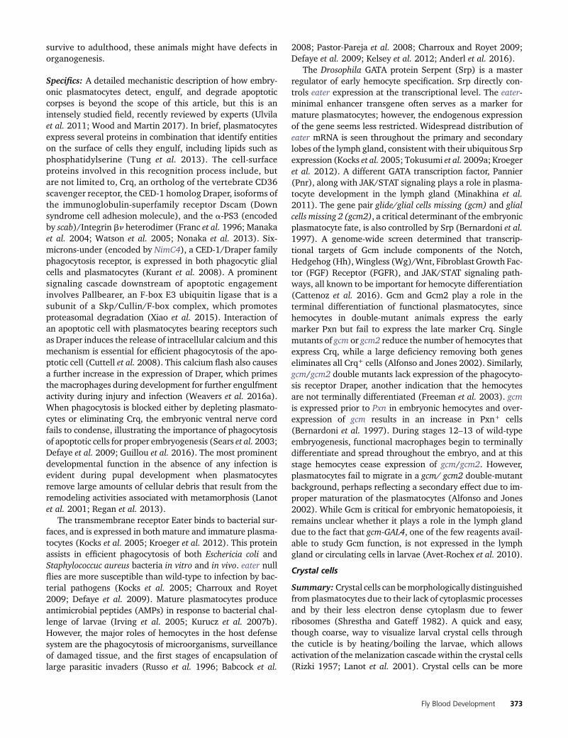
Figure 2 Embryonic hematopoiesis. (A) Stage 5 embryo. Precursors forembryonic hemocytes (yellow) are specified from the head mesoderm,while lymph gland precursors (blue) arise from the thoracic region of thedorsal mesoderm. BR, gray. (B) Stage 11 embryo. Embryonic prohemo-cytes migrate and differentiate into plasmatocytes (green) and crystal cells(red). The lymph gland anlage proliferate and are seen in the trunk re-gion. (C) Stage 17 embryo. Plasmatocytes migrate throughout the em-bryo, while crystal cells accumulate near the proventriculus. During dorsalclosure, the lymph gland precursors on either side of the embryo movedorsally and are positioned flanking the DV. Later, these cells will consti-tute the lymph gland with pairs of distinguishable primary and posteriorlobes. Schematics in (A–C) adapted from Volker Hartenstein, see Lebestkyet al. (2000). BR, brain; DV, dorsal vessel.
376 U. Banerjee et al.

the embryo) are recognizable as hemocytes and this numberremains constant throughout the rest of embryogenesis(Tepass et al. 1994). During stages 10–12, the head meso-derm also contains a cluster of 20–30 crystal cell precursors(Lebestky et al. 2000).
Several factors first identified as important regulators ofmammalian hematopoiesis are also essential for embryonichematopoiesis in Drosophila. In particular, the GATA, Friendof GATA (FOG), and RUNX protein families are conservedhematopoietic regulators that act combinatorially to regulatelineage commitment in Drosophila hematopoiesis. The GATAfactor Serpent (Srp) is required for the development anddifferentiation of both plasmatocytes and crystal cells. Thisregulator controls key proteins such as Gcm, Lz, and Ush,which are necessary for hemocyte development [Bernardoniet al. 1997; Lebestky et al. 2000; Fossett et al. 2001; Alfonsoand Jones 2002; Waltzer et al. 2002; reviewed in Evans et al.(2003)].
Specifics: In many ways, the process of procephalic hemato-poiesis in Drosophila is, at least superficially, similar to initialwaves of mammalian hematopoiesis that give rise to earlyand intermediate progenitors. Analogous to primitive hema-topoiesis, the first wave that occurs in mammals shares manycommon molecular strategies observed during this phase ofDrosophila blood development [reviewed in Evans et al.(2003)]. Of the six known mammalian GATA factors,GATA-1, -2, and -3 are involved in hematopoiesis [reviewedin Orkin and Zon (2008)]. Mammalian GATA-1/2 are re-quired early during the specification of blood progenitors un-dergoing primitive erythropoiesis (Fujiwara et al. 2004).Depending on the context, members of the FOG family ofproteins enhance or inhibit GATA transcription factor activity[reviewed in Cantor and Orkin (2005)]. Another earlymarker for mammalian hematopoiesis is the RUNX familytranscription factor RUNX1 (also known as AML1), which isessential for the very first steps of blood formation within thedorsal aorta and later inmany other hematopoietic processes.Chromosomal translocations into this locus are responsiblefor a variety of human leukemias (Okuda et al. 1996;Yokomizo et al. 2001; Chen et al. 2009). Each of these tran-scription factor classes also plays critical roles in Drosophilahematopoiesis, including the GATA factors Serpent (Srp) andPannier (Pnr), the FOG homolog Ush, and the RUNX domainprotein Lozenge (Lz).
Srp is a critical regulator of hematopoiesis that is firstexpressed in the head mesoderm of stage-5 embryos (Abelet al. 1993; Sam et al. 1996; Brückner et al. 2004). Later-stagesrp mutant embryos are devoid of all mature hemocytes(Rehorn et al. 1996). Srp expression in hemocyte precursorsis controlled through the combinatorial activity of Snail,Buttonhead, Empty spiracles, and Even-skipped, which to-gether confine Srp expression to the head mesoderm (Yinet al. 1997; Spahn et al. 2014). Indeed, ectopic expression ofSrp in the trunk mesoderm, even in the absence of headmesoderm as in bicoid (bcd) mutant embryos, induces the
formation of hemocytes and the fat body at the expense ofother mesodermal tissues. Snail and Buttonhead, with aminor contribution from Empty spiracles, drive early Srpexpression while Even-skipped, expressed posterior (P) tothe Srp region, negatively regulates Srp expression limitingit to the head mesoderm (Spahn et al. 2014).
At the end of blastoderm stage 5, Srp+ hemocyte precur-sors begin to form distinct subsets from which the plasma-tocyte and crystal cell populations are derived. The majorityof Srp+ cells begin to express Gcm and Gcm2, which areboth required for terminal plasmatocyte differentiation(Bernardoni et al. 1997; Alfonso and Jones 2002). A smallsubset of Srp+ cells in the embryonic head mesoderm do notexpress Gcm but express Lz, the key crystal cell determinant(Lebestky et al. 2000). It is essential for Gcm expression to bedownregulated to form amature crystal cell. A combined lossof both Gcm and Gcm2 leads to an increase in the Lz+ pop-ulation. Gcm/Gcm2 are initially expressed in all blood pre-cursors, but are then downregulated specifically in the mostanterior (A) row of cells, which begin to express Lz. There arethen two potential fates for Lz+ cells: in the continued ab-sence of Gcm/Gcm2 in crystal cell progenitors near the pro-ventriculus they becomemature crystal cells, but when Gcm/Gcm2 is expressed in Lz+ cells distant from the proventricu-lus they become plasmatocytes (Bataille et al. 2005). Over-expression of Gcm with lz-GAL4 represses crystal cell fate byinhibiting Lz expression, while Lz is unable to override theplasmatocyte fate even when ubiquitously expressed in thehead mesoderm (Lebestky et al. 2000; Waltzer et al. 2003).
As early as stage 8 of embryogenesis, Srp also controls theexpression of Ush, which together with Srp and Lz playsimportant roles in crystal cell production. The contributionof these proteins has been carefully refined to reveal that inSrp+Lz+ crystal cell precursors, Srp and Lz promote crystalcell lineage commitment. In Srp+Gcm+ plasmatocyte precur-sors, Srp functions with Ush to suppress crystal cell fate andinduce plasmatocyte differentiation genes. A complex feed-back circuit with Srp, Lz, and Ush functions to both specifycrystal cells and limit their production (Fossett et al. 2001,2003; Waltzer et al. 2002, 2003; Muratoglu et al. 2006).
Prior to embryonic stage 12, the morphology of the bloodcells is similar to the ultrastructure of prohemocytes, whichare typically small, round mesodermal cells (Tepass et al.1994). A total of �700 hemocytes begin spreading through-out the embryo at the beginning of germ band retraction(early stage 12). These prohemocytes migrate and begin todifferentiate, morphologically resembling plasmatocytes andbecome highly polarized with dynamic, large, actin-rich filo-podia and lamellipodia, which continually extend and retract(Tepass et al. 1994; Wood et al. 2006). These cells now ex-hibit phagocytic activity as they engulf apoptotic cells withinthe developing tissues (Tepass et al. 1994) (Figure 2, B andC). Plasmatocytes continue to spread through stages 13–14,migratingmedially from either end of the embryo along threespecific developmentally regulated routes directed by thePDGF/VEGF receptor (Pvr) ligands Pvf2 and Pvf3 [Tepass
Fly Blood Development 377

et al. 1994; Heino et al. 2001; Alfonso and Jones 2002; Choet al. 2002; Wood et al. 2006; Parsons and Foley 2013;reviewed in Ratheesh et al. (2015)]. However, this conclusionmay have to be interpreted in the context of an additionalproposed antiapoptotic function of Pvr in embryonic bloodcells. Apoptosis seen in Pvr1 mutant embryos can be reversedthrough expression of the viral caspase inhibitor p35 and thisinhibition also restores hemocyte migration (Brückner et al.2004; Parsons and Foley 2013). By late stage 14, plasmato-cytes are evenly distributed throughout the embryo, with theexception of dense clusters around the head, foregut, andhindgut (Tepass et al. 1994). In contrast, stage 17 embryoshave crystal cells clustered around the proventriculus (Lebestkyet al. 2000).
Sessile pools and circulating larval hemocytes
Summary: During the larval instars, hemocytes derived fromtheheadmesodermduring embryogenesis spread throughoutthe animal and are found in two locations: a subset of themcirculates in the hemolymph and the rest are attached to thebody wall in sessile pools (Shrestha and Gateff 1982; Lanotet al. 2001) (Figure 3A). Unlike circulating hemocytes, whichare readily released upon bleeding, release of sessile hemoc-ytes requires physical disruption by pressure applied to thelarval cuticle. The sessile hemocytes are secluded from theopen hemocoel, and are positioned in between the epidermaland muscle layers of the larva in clusters of cells termedepidermal–muscular pockets (Makhijani et al. 2011) (Figure3C). These cells initially form a pattern of lateral patches thatlater extend into dorsal stripes. Formation of this pattern isdependent upon hemocytes homing to the pockets, followedby adhesion and lateral migration. Peripheral neurons thatinnervate these pockets are required for hemocyte homing tothe sites and provide Activin-b/TGF-b (Transforming GrowthFactor) signals to promote adhesion and proliferation(Makhijani et al. 2011, 2017). Both Rac1 GTPase and theJun N-terminal kinase (JNK; Basket, Bsk) are required forproper adhesion and targeting of hemocytes to these sessilepools (Williams et al. 2006). The Nimrod family transmem-brane receptor Eater is also required for hemocyte attach-ment to the sessile compartment (Bretscher et al. 2015).The final step of lateral migration is mediated by Rho1 andthe actin cytoskeleton (Makhijani et al. 2011). Following ex-ternal mechanical disruption, the sessile pools disperse butspontaneously reform after 30–60 min, presumably throughthe same mechanisms that regulate initial pattern formation.Peripheral neurons that innervate these pockets have beenproposed to act as a niche to help control hematopoiesis inthis tissue through the inhibition of apoptosis and mainte-nance of sessile hemocytes (Makhijani et al. 2011). Ecdysonesignaling induces dispersal and activation of sessile hemoc-ytes upon pupariation, and this facilitates tissue remodelingduring metamorphosis (Regan et al. 2013).
Mature plasmatocytes transdifferentiate into crystal cellswithin these sessile pools, a process mediated by a Notch-dependent signaling mechanism (Leitão and Sucena 2015).
The number of crystal cells in the sessile pools increases bymeans of transdifferentiation of mature Hml+ Lz2 plasmato-cytes, initially into Hml+ Lz+ cells that continue to be P1+
and retain some phagocytic activity. However, as they matureand express higher levels of Lz, they lose this phagocyticpotential and also lose all plasmatocyte markers to becomemature crystal cells. During this transdifferentiation process,Hml+ Lz2 plasmatocytes signal through Notch to physicallyadjacent cells that are to become Hml+ Lz+, likely mediatedby the ligand Serrate. This process requires cell-to-cell con-tact, which is achievable in sessile pools, but not when thesepools are repeatedly disrupted by mechanical means. Theresult of such a manipulation is a decrease in the overallnumber of circulating crystal cells (Leitão and Sucena2015). Mature plasmatocytes within sessile pools can alsoform lamellocytes upon wasp infestation (Honti et al.2009). Therefore, although de novo conversion of a mesoder-mal precursor to blood tissue does not seem to occur withinthe sessile pools, the sessile hemocyte pool is a hematopoieticcompartment that contributes to differentiation of hemocytesduring larval development as well as in response to immunechallenge.
Plasmatocytes can also transdifferentiate into lamello-cytes, although the mechanism is not yet fully clear (Avet-Rochex et al. 2010; Honti et al. 2010; Stofanko et al. 2010;Anderl et al. 2016). Knockdown of ush causes plasmatocytesto transdifferentiate into lamellocytes even in the absenceof wasp infestation (Avet-Rochex et al. 2010). In addition,overexpression of Srp, Vinculin-RNAi (RNA interference), ora dominant negative form of ecdysone receptor induces theformation of lamellocytes in nonparasitized larvae that simul-taneously express the lamellocyte marker L1 and the plasma-tocyte marker Eater, suggesting a plasmatocyte–lamellocyteconversion (Kroeger et al. 2012). However, transdifferentia-tion of plasmatocytes is unlikely to be the exclusive mecha-nism for lamellocyte formation, particularly in the lymphgland where lamellocytes can arise directly from a prohemo-cyte pool in response to wasp infestation (Makki et al. 2010;Oyallon et al. 2016).
Specifics: In second-instar larvae, the sessile hemocytes areprimarily located at the posterior end. In the third instar, theycontinue to remainas two large clumpsof 100–200hemocyteson segments A8 and A9, forming an organ-like structuretermed the posterior hematopoietic tissue (Kurucz et al.2007b;Markus et al. 2009). This concentration of hemocytes,which appears early in development, is in addition to thesegmentally distributed sessile cells in the epidermal–muscularpockets that are most apparent in the third instar (Makhijaniet al. 2011). Within the sessile pools, the hemocytes are denselypacked and form stable cell-to-cell contacts, and these cellsconstitute at least a one-third of all larval hemocytes (Lanotet al. 2001).
Themajority of circulating and sessile larval hemocytes arederived directly from Pxn+ cells formed during embryonichematopoiesis and not from a pool of undifferentiated lymph
378 U. Banerjee et al.

gland prohemocytes (Makhijani et al. 2011). The circulatinghemocytes do proliferate, but the sessile hemocytes incorpo-rate EdU at a much higher rate (Makhijani et al. 2011; Anderlet al. 2016). Overall, the sessile pool is dynamic in its hema-topoietic activity and has therefore been thought to functionas a compartment that exchanges with the circulating cells,but is independently regulated. Large hepatocyte-like cellscalled oenocytes, as well as peripheral neurons, reside in ornear these hematopoietic pockets, but it is only the peripheralneurons that are required for proper sessile hemocyte clusterformation (Lanot et al. 2001; Makhijani et al. 2011) (Figure3C). When the peripheral nervous system (PNS) is perturbedusing atonal (ato1) mutants or if the neurons are ablated withdiphtheria toxin, the number and pattern of sessile hemocyteclusters is severely altered. Furthermore, hemocytes can berecruited to ectopic sites by misexpression of the proneuralgene scute (sc), which creates supernumerary peripheral neu-rons (Makhijani et al. 2011). The multidendritic sensory neu-rons and chordotonal organs of the PNS express Activin-b, aligand for the TGF-b family, which promotes the adhesionand proliferation of hemocytes within the hematopoieticpockets (Makhijani et al. 2017). A functional connectionbetween the PNS and the hematopoietic system mighthave been conserved in vertebrates, since signals from
the sympathetic nervous system help regulate HSC prolifer-ation and egress from the bonemarrow (Hanoun et al. 2015).
Several classes of mutants that either increase the numberof circulating hemocytes, affect the dorsal sessile compart-ments, or induce the spreading of sessile hemocytes through-out the cuticle were identified in an overexpression screenfor candidate genes. For example, overexpression of theaPS3 integrin Scab in Pxn+ cells disrupts the dorsal sessilecompartments, and decreases circulating and lymph glandhemocyte numbers, but also results in hemocyte accumula-tion on the dorsal vessel. Similarly, overexpression of Kruppelor the CBP homolog Nejire disrupts sessile hemocyte com-partments, but surprisingly also induces lamellocyte for-mation (Stofanko et al. 2008). A correlation betweenlamellocyte formation and release from sessile pools is alsoseen when Wnt/Wg signaling is disrupted by overexpressionof Shaggy (Sgg) or a dominant negative form of Pangolin(Pan)/T-cell factor (TCF), and also upon wasp parasitization(Zettervall et al. 2004). In response to wasp infestation,hemocytes in these sessile and circulating pools differentiateinto lamellocytes. During this process, the circulating hemoc-yte population also increases as the sessile pool is releasedinto the hemolymph (Honti et al. 2009). Constitutive activa-tion of Toll signaling in Toll10B mutants also disrupts the
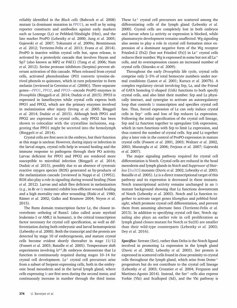
Figure 3 Larval hematopoiesis. (A) In the third-instar larva, the LG lobes are positioned spanning theDV (gray) posterior to the BR. Plasmatocytes (green)and crystal cells (red) circulate in the hemolymphthroughout the larva and are also seen in segmen-tally distributed sessile pools. (B) The lobes of the LGflank the DV. The primary lobes are the largest andmost anterior within the LG, and consist of severaldistinct cell types and zones (see Figure 4). The pos-terior lobes (blue) are smaller and remain largelyundifferentiated. Pericardial cells (gray spheres) sep-arate the individual lobes of the LG. (C) Detailedview of sessile hematopoietic pockets. The majorityof sessile hemocytes, including plasmatocytes andcrystal cells, reside along the dorsal side of the larvain stereotypically arranged lateral patches termedhematopoietic pockets. Clusters of oenocytes (gray)also reside within these regions, but are dispensablefor the formation of the sessile pools. Activity ofexternal sensory and multidendritic neurons (purple)is necessary for adherence, proliferation, andmaintenance of hemocytes within hematopoieticpockets. Schematic in (A) adapted from VolkerHartenstein, in (C) adapted from Gold and Brueckner(2015). BR, brain; DV, dorsal vessel; EA, eye-antennaldisc; LG, lymph gland; MH, mouth hooks; PV, pro-ventriculus; SG, salivary gland.
Fly Blood Development 379
sessile hemocyte pools, a phenotype that is suppressed in anird1 mutant (Schmid et al. 2016).
In eater null mutant larvae, both plasmatocytes and crystalcells are virtually absent in the sessile pockets, which resultsin an apparent increase in the number of circulating hemoc-ytes. Specific knockdown of eater in the plasmatocyte lineageusing Hml-GAL4 disrupts plasmatocyte adhesion, which non-autonomously impedes crystal cell attachment to the sessilecompartment. This effect is not observed when eater is de-pleted in crystal cells using lz-GAL4 (Bretscher et al. 2015).These experiments illustrate that the sessile plasmatocytesprovide an instructive cue for crystal cells to adhere to thesessile compartment, in addition to the Serrate-dependentcue required for transdifferentiation of crystal cells (Bretscheret al. 2015; Leitão and Sucena 2015).
Release of hemocytes from the sessile compartments iscontrolled by several pathways. For example, overexpressionofwild-typeRac1disrupts the sessile populationand increasesthe number of circulating cells (Zettervall et al. 2004). Rac1GTPase requires both JNK activation and actin polymeriza-tion to release sessile hemocytes (Williams et al. 2006). Ec-dysone signaling is another example of a pathway involved insessile hemocyte release. An ecdysone pulse that occurs atthe onset of pupariation is received by hemocytes, resulting inchanges in hemocyte morphology, migration, and dispersal,all of which are disrupted upon expression of a dominantnegative form of an ecdysone receptor (EcRB1DN) in hemoc-ytes. EcR can transcriptionally activate several genes in-volved in Rac GTPase-mediated actin remodeling, whichlikely contributes to its effect on hemocyte dispersal duringpupariation (Regan et al. 2013).
Dorsal mesoderm
Summary: During the second wave of embryonic hemato-poiesis, a regionof thedorsalmesodermcalled thecardiogenicmesoderm gives rise to both the lymph gland and the dorsalvessel (Rugendorff et al. 1994) (Figure 2C and Figure 3B).Lymph gland progenitors and cardioblasts are closely related,and clonal analysis provides evidence for the presence of ahemangioblast population consisting of cells, which in a sin-gle division gives rise to one cell that differentiates into thedorsal vessel and another that differentiates into blood(Mandal et al. 2004). This is reminiscent of the hemangio-blast population in vertebrates that constitutes progenitorcells in the aorta–gonad–mesonephros (AGM) mesenchyme,and produces both blood and vascular cells (Medvinsky andDzierzak 1996). Several additional molecular and develop-mental similarities have been noted between these two sys-tems [reviewed in Evans et al. (2003)].
Precursors of the lymph gland appear as a local bulgewithin the cardiogenic mesoderm during stage 13 of embry-onic development (Rugendorff et al. 1994; Holz et al. 2003).These precursors then migrate dorsally to form a tight clusterassociated with the dorsal vessel and eventually form apaired chain comprising multiple lobes flanking the dorsalvessel. Cell clusters positive for the zinc finger protein
Odd-skipped (Odd) in the three thoracic segments, T1–T3,coalesce to form the lymph gland, while Odd+ clusters in theabdominal segments form pericardial cells (Mandal et al.2004). By stages 11–12, mesodermal expression of the ho-meotic gene Antennapedia (Antp) is restricted to the T3 seg-ment. This Antp expression is further restricted to 5–6 cells atthe posterior boundary of the lymph gland as these cell clus-ters coalesce during stages 13–16. Antp+ cells are the first toproliferate within the larval lymph gland, giving rise to apopulation of �30 cells that have been named the posteriorsignaling center (PSC) (Mandal et al. 2007). These cells pro-vide signals that control the development of the rest of thelymph gland and also participate in the larval response towasp parasitization. Antp is maintained in the PSC through-out larval development, similar to the expression pattern ofthe Drosophila early B-cell factor (EBF) ortholog, Collier/Knot (Crozatier et al. 2004). The rest of the lymph gland cellsthat form the primary lobes develop from the Odd+ clustersthat arise from segments T1–T2. The homeodomain cofactorHomothorax (Hth) is initially expressed throughout the em-bryonic lymph gland and is later downregulated within thePSC. Antp and Hth function in a mutually antagonistic man-ner, with Antp specifying the PSC and Hth specifying theblood primordium (Mandal et al. 2007).
The cardiogenic mesoderm is a subcompartment of thedorsal mesoderm and therefore multiple factors that controldorsal mesoderm formation are also critical for lymph glanddevelopment. Examples include BMP/Dpp (bone morphoge-netic protein/Decapentaplegic) and FGFR/Heartless (Htl),which control expression of the homeodomain transcriptionfactor Tinman (Tin) and the GATA factor Pannier (Pnr). Inaddition, Wingless (Wg/Wnt1) positively regulates cardio-genic mesoderm specification and Notch negatively regulatesit. Mutations in any of these entities—dpp, htl, tin, pnr, orwg—cause loss of lymph gland and other associated struc-tures derived from the cardiogenic mesoderm. In contrast,loss of Notch has the opposite effect with substantially morecells arising within the cardiogenic mesoderm (Mandal et al.2004).
Specifics: Following gastrulation and during stages 6–9, themesodermal cells are not committed to any particular lineageand express mixed markers such as Tin, required for heartdevelopment, and Mef2, which regulates muscle formation.These genes are controlled by the mesoderm determinantsTwist (Twi) and Snail (Sna) (Bodmer et al. 1990; Leptin1991; Lilly et al. 1994; Taylor et al. 1995; Yin et al. 1997;Cripps et al. 1998; Nguyen and Xu 1998). Later in develop-ment, the lineage-specific genes will become restricted intheir expression, controlled by signals from the segmentedectoderm.
At stage 11, the mesoderm splits into the various lineagesthat will give rise to the organs derived from them [Eastham1930; Bate and Rushton 1993; Borkowski et al. 1995;Riechmann et al. 1997; reviewed in Hartenstein and Chip-man (2015)]. Like the ectoderm, the mesoderm is also
380 U. Banerjee et al.

segmented into myomeres, further split into A and P do-mains, and abuts the overlying ectoderm. Each of these seg-mental units moves the A domain toward the ectoderm andthe P domains are pushed inside toward the endoderm. Dur-ing stage 12 (germ band retraction), the A domains fuse toform a continuously linear primordium that will give rise tosomatic muscles, dorsal vessel, the lymph gland, and otherassociated tissues. The P domains, pushed inside, also fuseand will give rise to the visceral mesoderm as well as the fatbody. Maintenance of the A domain requires Wg signaling,while the P domain is maintained by Hh (Azpiazu et al. 1996;Park et al. 1996). These signals are interpreted in the contextof pair-rule transcription factors that form the 14 segmentalstripes. Each of these fused metameric structures is then dif-ferentially specified along the dorsal/ventral axis to positionthe formation of various organs. This description holds forthe mesoderm in the segmented parts of the embryo. Thehead mesoderm, from which the circulating and sessilehemocytes are derived, have a very different developmentallogic. In fact, hemocytes, but not fat andmuscles, are themajorderivatives of the head mesoderm [reviewed in Hartensteinand Chipman (2015)].
Dpp expressed in the ectoderm specifies the fate of thedorsal mesoderm that will give rise to dorsal vessel/lymphgland (anteriorly) and visceral muscles (posteriorly) in eachsegment. The ventral part will give rise to somatic mesoderm(A) and fat body (P). Loss-of-function mutations in dpp lackthe dorsal vessel while overexpression causes heart cells toform from ventral cells (Frasch 1995). The transcription fac-tor Tin is a direct target of the Dpp signal, and its expressionalso requires the function of FGFR (Shishido et al. 1997;Zaffran et al. 2002). At this stage of development, expressionof Tin defines the region that is designated dorsal mesoderm(Bodmer 1993). Within this dorsal mesoderm, the A quad-rant, which is high in both Wg and Dpp activity, defines thecardiogenic mesoderm from which the heart, blood, and thepericardial nephrocytes will arise. This bears many similari-ties to the AGM region in vertebrate definitive hematopoiesisthat arises from the lateral plate mesoderm, also in responseto BMP and FGF (Marshall et al. 2000; Nishikawa et al.2001). Furthermore, cells sharing an immediate commonancestor within the cardiogenic mesoderm can be fated tobecome either a dorsal vessel or a lymph gland precursor,leading to the designation of such cells as hemangioblastsin comparison with similar cells in mammals, which can be-come components of either the blood vessel or the hemato-poietic system [reviewed in Marshall and Thrasher (2001)and Mandal et al. (2004)].
The homeodomain protein Tin, initially expressed broadlyin the mesoderm, is later restricted to the cardiogenic meso-derm. Interestingly, Tin is a homolog of the vertebrateNkx2.5,which is consideredaheart-specificmarker inbothvertebratesand in Drosophila (Lien et al. 1999). In reality, Drosophila Tinis expressed in the common progenitor for both heart andblood cells, and then becomes heart-specific only when theselineages diverge (Mandal et al. 2004). The dorsal mesoderm
requires Tin and Pnr, both of which are controlled by FGFRand Dpp signaling (Frasch 1995; Beiman et al. 1996;Klinedinst and Bodmer 2003). Prior to the specification oflymph gland precursors from cardioblasts at the time of germband retraction (stages 12–13), the entire cardiogenic meso-derm expresses Tin, but by stage 13, Tin and Pnr becomeconfined to cardioblasts. This refinement is essential forlymph gland specification because ectopic expression of Tinor Pnr throughout the entire mesoderm, or in the cardiogenicmesoderm, reduces the numbers of both lymph gland andpericardial cells. Odd continues to be expressed throughoutthe cardiogenic mesoderm while Srp is upregulated in lymphgland precursors (Mandal et al. 2004).
Notch is active during stages 11–13 and plays a dual role inlymph gland specification. At stage 11/12, Notch is requiredfor specification of the cardiogenic mesoderm, while duringstages 12/13 Notch inhibits expression of Tin and upregu-lates Odd and Srp in a Delta-dependent manner (Mandalet al. 2004; Grigorian et al. 2011a). Consequently, duringstages 12/13, reduction of Notch causes an increased numberof cardioblasts at the expense of lymph gland precursors,while expression of activated Notch (Nact) gives rise to alarger lymph gland (Mandal et al. 2004). Null mutations inDelta convert all cells of the cardiogenic mesoderm into car-dioblasts (Grigorian et al. 2011a). Delta ligand expression iswidespread until stage 12, but then becomes spatially re-stricted to cardioblasts and persists through stage 14. EGFRand FGFR are also required for specification and mainte-nance of the cardiogenic mesoderm, since expression of aconstitutively active form of Ras in these cells increases theirnumbers (Grigorian et al. 2011a).
Following the split of the cardioblast and lymph glandlineages, the pericardial cells are distinguished from lymphgland precursors through regulation by Srp. In srp null em-bryos, Odd+ cells still form a cluster resembling the earlylymph gland; however, these Odd+ cells now express thepericardial marker Pericardin. On the other hand, Srp expres-sion throughout the cardiogenic mesoderm induces pericar-dial cells to adopt the lymph gland fate (Mandal et al. 2004).
Tin, Pnr, and Srp control the conserved basic helix-loop-helix transcription factor Hand,which is critical for both heartand lymph gland development. Hand expression is initiatedin the cardiogenic region in late stage 12 and, while it isexpressed in cardioblasts, pericardial cells, and lymph glandprecursors, it is regulated differently in these cell types. Incardioblasts and pericardial cells, Tin and Pnr control Handexpression, while in lymph gland precursors, Srp controlsHand expression (Han and Olson 2005). Hand null mutantembryos and larvae exhibit complete loss of lymph gland,pericardial cell, and cardiac precursors through apoptosis.Thus, it is likely that a primary function of Hand is to promotecell survival in the cardiogenic mesoderm (Han et al. 2006).
Cells of the lymph gland primary lobe that will eventuallygive rise to hemocytes arise from embryonic segments T1–T2,while the cells of the PSC arise from 5–6 Antp+ cells thatoriginate from the T3 segment. The expression of Antp is
Fly Blood Development 381

maintained in the PSC throughout larval development(Mandal et al. 2007). Collier is expressed in two clusters ofcells in T2 and T3 that later coalesce. Following germ bandretraction, Collier expression remains high in 3–5 cells at theP tip of the lymph gland and at low levels throughout the restof the lymph gland cells (Crozatier et al. 2004). The PSC cellsinitially form in colliermutants but are lost by the third instar,indicating that Collier is specifically required for PSC main-tenance and not for its specification (Crozatier et al. 2004).Collier expression is seen prior to, and is independent of, Srpas it continues to be expressed in srp6G mutant embryos.However, Collier expression in the PSC depends on Antpand is not maintained in Antp mutant embryos. In contrast,loss of collier does not affect initial Antp expression (Mandalet al. 2007).
In addition to T1–T3, Odd is also expressed in the abdom-inal segments A1–A6 in stage 11 embryos (Ward and Skeath2000). These latter clusters form pericardial cells (Rugendorffet al. 1994; Holz et al. 2003; Mandal et al. 2007). Positionalcues provided by the homeobox protein, Ultrabithorax(Ubx), in segments A1–A5, restricts primary lobe lymphgland formation to the thoracic regions of the cardiogenicmesoderm. Loss of Ubx results in abnormal expansion oflymph gland cells into the abdominal segments [Rodriguezet al. 1996; Mandal et al. 2004; reviewed in Hartensteinand Chipman (2015)].
Later in development, following dorsal closure (stage 17),the rows of cardioblasts on either side of the embryo cometogether and fuse. The dorsal vessel forms as a double row ofcardioblasts lining a central lumen through which the hemo-lymph circulates (Rugendorff et al. 1994). In the mature Dro-sophila embryo, the lymph gland appears as a paired clusterof �20 cells flanking the anterior region of the dorsal vessel(Mandal et al. 2004). The cells of the lymph gland continue toexpress both Srp and Odd throughout larval development(Jung et al. 2005).
Based on the nomenclature used for the larval heart, thewide, posterior part of the dorsal vessel is referred to as theheart, and the anterior part as the aorta (Miller 1950; Rizki1978). The hemolymph is pumped through the lumen ofthe dorsal vessel in the P/A direction, exiting the aortaalong with the circulating hemocytes, which are releasedat the anterior end into open circulation within the bodycavity. In addition to the incurrent opening at the posteriorend of the heart through which the majority of the bloodenters, the rest enters through small, segmentally distrib-uted lateral openings or ostia along the length of the heart.The aorta part of the dorsal vessel does not have these ostiaand maintains unidirectional flow. Together, the aorta andheart represent a contractile tube lined by a layer of myoe-pithelial vascular cardioblasts. Alary muscles connect thedorsal vessel to the epidermis. Encircling the anterior tip ofthe dorsal vessel is the ring gland, an organ involved inmaintaining a complex endocrine system, including ste-roid hormones that maintain growth cycles of the develop-ing larva and pupa. Along the edge of the dorsal vessel and
posterior to the ring gland are the primary lobes of thelymph gland (Figure 3B). This is followed by a series ofsmaller lobes that flank the dorsal vessel (Rugendorff et al.1994). In between the major lobes, and also in a loose rowposterior to the lymph gland on either side of the dorsalvessel, are large, nonpolarized pericardial nephrocytes.Although collectively termed pericardial cells, they arenot all derived from identical precursors (Zmojdzianet al. 2018). Similar to vertebrate nephrocytes, maturepericardial cells have extensive arrays of folding mem-branes to provide greater surface area, and are in-volved in the ultrafiltration and excretion of hemolymph(Weavers et al. 2009). While all structures described aboveoriginate from the cardiogenic mesoderm, it is only thelymph gland lobes where hematopoietic progenitorsreside.
Larval Lymph Gland: Zones, Cells, and Signals
Summary
Structurally, the larval lymph gland is divided into multiplelobes. The anterior-most lobe, also called the primary lobe, isthe best characterized (Figure 3B). Located posterior to theprimary lobe are the secondary, tertiary, and quaternarylobes, which are much smaller in size and in the absence ofimmune challenge exhibit significantly lower levels of hema-topoietic activity compared to the primary lobes. Based onextensive morphological and molecular marker analysis, theprimary lobe of the lymph gland can be divided into zones,each containing cells that are functionally distinct. Thesezones include the PSC, which acts as a niche to regulate pro-genitor maintenance; the Medullary Zone (MZ) consisting ofmedially located hemocyte progenitors; and the distal CorticalZone (CZ), which is populated by maturing hemocytes(Jung et al. 2005) (Figure 4). This subdivision has revealedan underlying spatial and temporal regulation of blood celldevelopment within the primary lobe throughout the larvalstages. Technically, the first zone to arise as a separate cellpopulation is the PSC since it can be distinguished as a sep-arate cluster of cells in the embryo, as described above. Inlarval development, the PSC is located at the posterior cusp ofthe primary lobe, adjacent to a single pericardial cell thatseparates it from the secondary lobe. Akin to the purely sig-naling function ascribed to the stroma in mammalian devel-opment, clonal analysis demonstrates that the cells of thePSC do not give rise to any progenitor or differentiated bloodcell types, but do provide signals to regulate progenitor main-tenance or differentiation (Crozatier et al. 2004; Krzemieńet al. 2007; Mandal et al. 2007; Mondal et al. 2011;Benmimoun et al. 2012; Pennetier et al. 2012; Tokusumiet al. 2012; Khadilkar et al. 2014, 2017; Dey et al. 2016).Prior to the midsecond instar, the cells within the lymphgland primary lobe are progenitors that express a reporterfor domeless (dome). These Dome+ progenitors will populatethe MZ and differentiate at their distal edge to initiate
382 U. Banerjee et al.

formation of the CZ (Jung et al. 2005; Evans et al. 2009;Krzemien et al. 2010; Minakhina and Steward 2010; Deyet al. 2016) (Figure 4). As yet, there is no direct evidencethat these Dome+ cells self-renew while simultaneously pro-ducing a differentiated offspring, as is evident during germ-line differentiation (Fuller and Spradling 2007).
Specifics
The first- and early second-instar lymph glands are largelypopulated by progenitor cells that express Dome, the receptorupstream of JAK/STAT (Jung et al. 2005; Dey et al. 2016).During these early stages, the primary lymph gland lobe has asmooth appearance and consists of a rapidly dividing popu-lation of Dome+ progenitors that are tightly packed withclose cell-to-cell contacts (Jung et al. 2005) (Figure 4A). Inthe first larval instar, a small population of cells on the medialside of the lymph gland, along the dorsal vessel and perhapsextending adjacent to the PSC, do not express Dome as themore distal cells do. These cells are preprogenitors and were
named as such based on marker expression (Jung et al.2005). In more current literature, genetic and molecularanalyses have sought to determine the nature of this cellpopulation, and have found that the preprogenitors give riseto the Dome+ cells of the MZ (Minakhina and Steward 2010;Dey et al. 2016).
Dome+ progenitors attenuate their rate of proliferationduring themidsecond instar as they change in overall density.Cells at the periphery begin to increase their spacing andgranularity coincident with loss of E-cad (encoded by shot-gun) as they initiate differentiation. It is from this point on indevelopment that the separate zones are readily identifiablewithin the primary lobe. In addition to morphological crite-ria, the zones become distinct based on their marker expres-sion patterns. The PSC cells are distinguished by theirexpression of Antp, Hh, and Collier (Lebestky et al. 2003;Crozatier et al. 2004; Mandal et al. 2007). The MZ expresseshigh levels of dome and upd3 reporters, ROS, Wg, E-cad, andvery low levels of Collier (Hombría et al. 2005; Jung et al.
Figure 4 LG zones and cell types in the primarylobe. (A) Schematic diagrams of LG primary lobesfrom first (left), late second (middle), and third(right) instar larvae. By convention, we designatethe region close to the dorsal vessel as medial andthe opposite edge as distal. Anterior is up in all LGimages. The PSC (green) acts as a niche for LGprogenitors and is present throughout develop-ment in all three instars. The majority of cells inthe first-instar primary lobe are undifferentiatedDomeless+ progenitors that belong to the MZ (darkblue). A small number of domeless2 and Notch+
preprohemocytes (light blue) lie on the medial edgeof the lobe adjacent to the dorsal vessel (gray), andare capable of contributing to the MZ populationduring the first 5–20 hr of first instar development.The second-instar LG develops mature hemocytesthat make up the CZ (red). This zone mostly in-cludes plasmatocytes and a small number of crystalcells. Additionally, an IZ (yellow) at the interface ofthe MZ and the CZ contains cells that express bothprogenitor (domeless) and differentiating hemocyte(Peroxidasin and Hemolectin) markers, but they lackmature hemocyte markers such as P1 or Lozenge/Hindsight. A population of domeless2 cells remainsalong the medial edge of the lobe, although theyno longer retain active Notch signaling. Duringthe third instar, the overall LG size increasesand a larger number of differentiated hemocytesare seen. (B) Image of a third-instar primary lobeobtained using fluorescence microscopy. Hh-GFP(PSC; green), domeMESO-BFP (MZ; blue), Hml-DsRed (CZ; red), and intermediate progenitors(pseudocolored as yellow based on overlap ofdomeMESO-BFP and Hml-DsRed). (C) Computerrendering of the confocal data shown in (B) usingImaris software. This software generates accuratethree-dimensional models from which quantitativedata can be readily derived. CZ, cortical zone; Hh,Hedhehog; Hml, Hemolectin; IZ, intermediate zone;LG, lymph gland; MZ, medullary zone; PSC, poste-rior signaling center.
Fly Blood Development 383

2005; Krzemień et al. 2007; Owusu-Ansah and Banerjee2009; Benmimoun et al. 2015; Oyallon et al. 2016). ECMproteins, including Viking (Vkg), a component of CollagenIV, and the Drosophila homolog of mammalian Perlecan(Trol), are densely interwoven between individual cells ofthe MZ and are sparser in surrounding groups of differenti-ated cells of the CZ (Krzemien et al. 2010; Grigorian et al.2013). Plasmatocytes within the CZ are identified by Hml,Eater, the NimC antigen P1, and Peroxidasin (Pxn) (Junget al. 2005; Kurucz et al. 2007a,b; Stofanko et al. 2008;Sinenko et al. 2009; Makhijani et al. 2011). Crystal cellsare identified by the expression of Lozenge (Lz), Hindsight(Hnt), Sima (Drosophila Hif-a), prophenoloxidases (PPO1and PPO2), and by the Bc mutation/reporter (Rizki et al.1980; Lebestky et al. 2000; Jung et al. 2005; Tokusumiet al. 2009a; Mukherjee et al. 2011; Binggeli et al. 2014).In the rare instance that lamellocytes are detected withinlymph glands of nonparasitized larvae, they are easily iden-tified as giant cells within the CZ that express L1/Atilla,Misshapen, a-PS4 integrin, and its partner Myospheroid(Braun et al. 1997; Crozatier et al. 2004; Irving et al.2005; Kurucz et al. 2007b; Honti et al. 2009; Tokusumiet al. 2009b). The individual zones and the patterns of geneexpression of cells occupying them remain distinguishablethrough the late third instar, becoming less so as the lymphgland begins to dissociate during early pupal stages (Grigorianet al. 2011b).
Positioned between the progenitors of theDome+MZandthe differentiating cells of the Pxn+ CZ, there is a smallpopulation of cells that simultaneously express markersfor both zones. These were initially described as being in a“transition state” as these cells are both Dome+ and Pxn+
(Sinenko et al. 2009). Simultaneous expression of dominantnegative forms of Fz and Fz2 increases the number of thesetransitioning cells (Sinenko et al. 2009). More recent liter-ature has referred to these cells as belonging to a separatezone termed the Intermediate Zone (IZ) (Krzemien et al.2010) (Figure 4). This nomenclature is now widely used(including in this review) and the cells therein are referredto as intermediate progenitors. While these cells are positivefor the earliest differentiation markers Hml and Pxn, theylack later, mature markers for plasmatocytes (P1) and crys-tal cells (Lz) (Krzemien et al. 2010). This intermediate pro-genitor population is not yet fully characterized, but giventhat cells within the IZ transit from a relatively quiescentmultipotent state to one in which their fate is specified, thiszone is likely quite important, and deserving of a more com-prehensive and mechanistic characterization of pathwaysgoverning their fate (Kalamarz et al. 2012; Kroeger et al.2012).
Preprogenitors
Summary: A small group of cells located at the most medialportion of the lymph gland near the dorsal vessel just abovethe PSC are Dome2 and were designated preprogenitors(Jung et al. 2005) (Figure 4A). Clones generated in the lymph
gland during embryogenesis and the first larval instar wereanalyzed to determine if large single clones, such as thosearising from stem cells, can form during development(Minakhina and Steward 2010). The authors report thatclones generated near the PSC can occupy up to one-thirdof the lymph gland and are maintained in position near thePSC. The authors suggest that the size of these clones is de-pendent on the Zfrp8 protein, and that cells derived fromthese clones are competent to become either plasmatocytesor crystal cells. Clones distant from the PSC are small andscattered (Minakhina and Steward 2010). Large clones arealso induced from the region of the Dome2 preprogenitors,adjacent to the dorsal vessel. These Dome2 preprogenitorsare seen 5–22 hr after hatching and have been termed HSCsbased on this clonal analysis (Dey et al. 2016). As statedearlier, the widely recognized vertebrate term HSC is associ-ated with additional characteristics not yet established inDrosophila, and while these results are consistent with astem-like identity for the Dome2 cells, given the very tran-sient nature of this population, it is preferable to retain theirdesignation as preprogenitors until future studies settle theissue of self-renewal linked to asymmetric cell division withinthis population.
Specifics: About 4–5 Dome2 cells are seen 8 hr after egghatching in first-instar larvae. They are larger in size thantheir Dome+ neighbors and express low levels of Dorothy(Dot) (Dey et al. 2016). Importantly, these cells also expressseveral indicators of active Notch pathway signaling includ-ing Notch-GAL4, Su(H)-lacZ, and a target gene for the path-way, Enhancer of split mb (E(spl)mb). In addition to theNotch pathway reporters, Trio, a guanyl-nucleotide exchangefactor previously implicated in stem cell maintenance inmammals, is also expressed in these cells. A majority of theseNotch+ cells are in S phase and, as they progress through G2and mitosis, they transition to become Dome+ progenitors by22 hr after egg hatching during the late first larval instar.Clonal analysis shows that these Notch+ preprogenitors aremultipotent and contribute to the Dome+ population (Deyet al. 2016).
This preprogenitor fate is also dependent upon Dpp sig-naling from the niche (Figure 5A). Phosphorylated Mothersagainst dpp (pMad) is elevated within these preprogenitorsand is critical for their Notch-induced E(spl)mb expression.Knockdown of dpp in the PSC or depletion of Mad in theNotch+ preprogenitors causes a threefold decrease in lymphgland size at third instar, consistent with a role of Notch inpreprogenitor proliferation. This population of Dome2 andNotch-pathway active cells is not seen after the first instar,and thus far represents the earliest postembryonic hemato-poietic preprogenitor population within the lymph gland (Deyet al. 2016).
Later in the second and third instars, Dome2 lymph glandcells neighboring the dorsal vessel express a GFP reporterfor the TEAD family transcription factor Scalloped (Sd)(Ferguson andMartinez-Agosto 2017). Pvf2, one of the three
384 U. Banerjee et al.

ligands known to bind and activate the PDGF/VEGF receptorPvr, is dependent on Sd and expressed in cells positive for thistranscription factor. An RNAi knockdown of Pvf2 specificallywithin these cells, but not in the Dome+ cells, causes an earlyproliferation defect resulting in a small lymph gland.Overexpression of Pvf2 in these cells rescues the lymphgland growth defect of sd hypomorphic mutant animals.These data suggest that Pvf2 expression is essential fornormal lymph gland proliferation during early larvalstages (Ferguson and Martinez-Agosto 2017). Althoughthese Dome2 Sd+ cells are located in a similar region tothe Dome2 Notch+ cells seen in the first instar (Dey et al.2016), and loss of either population affects the total num-ber of cells in the mature lymph gland, further experimen-tation is needed to establish the relationship betweenthese two populations to fully understand the progenitorhierarchy during blood cell development.
Progenitors and differentiation: the MZ and CZ
The MZ cells are the common progenitors for all matureblood cell types (Jung et al. 2005; Defaye et al. 2009;Krzemien et al. 2010; Minakhina and Steward 2010; Deyet al. 2016). These progenitors are critically dependent on amultitude of signals from three different origins to retaintheir quiescence and multipotency. These are the autono-mous/autocrine signals that originate within the zone andare received by the same population, signalling via secretedfactors that originate from a different zone within the lymphgland that affect the MZ and, finally, systemic signals thatresult from active sensing of the internal and externalenvironment.
A truly unique feature of the MZ is the strict control ofprogenitor proliferation during development. During thefirst and early second larval instars, all non-PSC cells withinthe lymph gland, except the small number of preprogenitorsdescribed above, are Dome+. Bromodeoxyuridine (BrdU)incorporation studies show that during these stages, theDome+ cells proliferate extensively in an asynchronousmanner. Then, coincident with the first appearance of dif-ferentiated cells during the mid-to-late second instar, pro-liferation slows dramatically and very few of the cellsincorporate BrdU. As development proceeds, and the sepa-ration of the MZ and CZ becomes more prominent, theDome+ cells of the MZ continue to maintain a low prolifer-ative state, while the CZ maintains higher proliferative ratesthroughout the third instar to expand its population (Junget al. 2005; Krzemien et al. 2010). This control of prolifer-ation is critical for the MZ cells to remain multipotent andundifferentiated. Progenitor maintenance requires a niche-derived signal, as well as one that emanates from the differ-entiating cells, termed an “equilibrium signal.” Loss of ei-ther signal causes the MZ cells to lose multipotency, as theyproliferate and subsequently differentiate (Mandal et al.2007; Mondal et al. 2011). An important question to resolveis the mechanism by which the proliferative and fate-determinative events are coupled during hematopoiesis. Areasonable hypothesis to test in the future is that differenti-ation signals are received by all cells, but are not interpretedas such by cells that are maintained in a nonproliferativestate. A relief in the proliferation block at the boundary be-tween the zones would then allow these cells to differentiate.The molecular mechanisms underlying the maintenance and
Figure 5 Interzonal signaling. (A) During the firstinstar, a Dpp signal originates from the PSC andactivates the Notch pathway by an unknown mech-anism in 5–8 preprogenitor cells along the medialedge of the primary lobe. The preprogenitors aredomeless2, proliferate, and give rise to the Dome-less+ progenitors of the MZ. (B) Late second instaronward, the PSC secretes both Hh and Pvf1, whichare needed to maintain progenitor quiescence. Theniche-derived Hh signal is important for generatingan active version of Ci (CiACT) in the MZ. The Pvf1-derived signal is sensed by the cells of the CZ, whichtrigger a molecular cascade known as the equilib-rium signal that also further enhances the stabilityof CiACT in the MZ. The combination of the niche-derived Hh signal and the equilibrium signal arisingfrom newly differentiated cells together maintain aprogenitor cell population that is adaptable to bothhomeostatic and stress-induced conditions. All ar-rows represent genetic regulation and not necessar-ily direct molecular steps within a transductioncascade. Adgf-A, Adenosine deaminase growth fac-tor-A; AdoR, adenosine receptor; Ci, Cubitus inter-ruptus; CZ, cortical zone; Dpp, Decapentaplegic;Hh, Hedhehog; MZ, medullary zone; PSC, posteriorsignaling center; Pvr, PDGF/VEGF receptor.
Fly Blood Development 385

release of progenitors from quiescence is a current topic ofinvestigation within the field.
In the following sections, we elaborate upon some of thekey molecular events that control the balance betweenprogenitors and mature hemocytes within the larval lymphgland.
ECM: Differences in the expression of ECM proteins contrib-ute to morphological distinctions between the zones of thelymph gland (Lanot et al. 2001; Jung et al. 2005; Krzemienet al. 2010; Grigorian et al. 2013). The progenitor cells ofthe MZ are densely arranged and express the cell adhesionmolecule E-cad (Jung et al. 2005). In other systems, theintercellular mechanical tension caused by E-Cad bindingacross junctions has been shown to lead to reorganizationof integrin molecules and the deposition of ECM compo-nents (Weber et al. 2011). During normal lymph gland de-velopment, progenitors maintain their close contact untilmaturation and dispersal. Loss of E-cad in the progenitorsinduces differentiation while overexpression promotes en-hanced progenitor maintenance (Jung et al. 2005; Gao et al.2009, 2013). The CZ expresses high levels of the collagen-binding protein SPARC (secreted protein acidic cysteine-rich) (Irving et al. 2005). This is also true of mature embry-onic hemocytes, in which SPARC expression is essential forthe proper deposition of collagen during the formation ofthe basal lamina (Martinek et al. 2008). A thin layer ofcollagen encapsulates the outer boundary of the lymphgland lobes, separating the cells from the hemolymph(Lanot et al. 2001; Krzemien et al. 2010). In addition, theCollagen IV subunits Viking (Vkg) and a1 (Cg25C) are de-tected in an interwoven pattern throughout the lymphgland, although transcriptional reporters for a1 are de-tected only in the CZ (Jung et al. 2005; Sorrentino et al.2007; Benmimoun et al. 2012; Grigorian et al. 2013). Thismay indicate that Collagen IV is secreted by the maturehemocytes then distributed throughout the entire lymphgland. The Drosophila homolog of the heparin sulfate pro-teoglycan Perlecan (encoded by terribly reduced optic lobes;Trol) also forms a thin layer that coats the periphery ofthe lymph gland. Within the lymph gland proper, Trol ishighly concentrated between MZ progenitors but also sur-rounds either individual or groups of cells within the CZ(Grigorian et al. 2013). Loss of trol function throughoutthe entire animal leads to a loss of circulating blood cellsand a smaller lymph gland (Lindner et al. 2007; Grigorianet al. 2013). Specific knockdown of trol within progenitors,but not CZ cells, leads to premature differentiation and lossof progenitor maintenance (Grigorian et al. 2013). Whetheradditional sources of ECM proteins, such as from the fatbody or circulating hemocytes, contribute to the lymphgland is not yet clear. Together, the appropriate distributionof ECM proteins among the zones provides the proper localenvironment and cell-to-cell interactions for hemocytes tobecome functionally active while they are still confinedwithin the hematopoietic organ.
Hh signaling: Hh binds to its receptor Patched (Ptc) andconsequently activates the downstream transcription factorCubitus interruptus (CiACT). In the lymph gland, Hh isexpressed in the cells of the PSC in the second and thirdinstar (Figure 5B). In hhTS mutants or when hh-RNAi isdriven in the PSC, the specification and maintenance ofthe PSC are unaffected, as indicated by normal Antp expres-sion. However, both genotypes cause a significant increasein the differentiation of mature hemocytes (Mandal et al.2007; Baldeosingh et al. 2018). Expression of Ptc and CiACT
is elevated within progenitors. Loss of Ci function in theprogenitors phenocopies Hh loss-of-function in an other-wise normal PSC and the resulting lymph gland lacks aproper MZ (Mandal et al. 2007; Baldeosingh et al. 2018).Projections visualized using a membrane-bound form ofGFP are seen emanating from the PSC cells and help toextend Hh distribution within the MZ (Krzemień et al.2007; Mandal et al. 2007).
Wg/Wnt signaling: Wg/Wnt pathway activity plays a com-plex role in lymph gland development (Sinenko et al. 2009;Zhang et al. 2014). An antibody raised against Wg (al-though it might cross-react with other Wnts; Doumpaset al. 2013) detects Wg throughout the lymph gland at earlylarval stages. This staining, as well as that of one of itsreceptors, Fz2, is less prominent in the cells of the CZ(Sinenko et al. 2009). Differentiating cells that express re-porters for Hml strongly attenuate Wg staining, and whendominant negative versions of both Fz and Fz2 are simulta-neously driven in the MZ, the boundary between the MZand CZ becomes disorganized. The cell adhesion moleculeE-Cad, which glues the MZ cells together, is downregulatedwhen Wg signaling is decreased and this may possibly con-tribute to the disruption of zones. No obvious change indifferentiation markers is evident except for an increase inthe number of cells of the IZ displaying both MZ and CZmarkers (Sinenko et al. 2009). While this could suggestthat, in the absence of Wg signaling, cells are unable toescape a transition state, this conclusion is likely an over-simplification since unpublished work from our laboratorysuggests that multiple Wnts, from different sources withinthe lymph gland and following multiple modes of down-stream function, influence progenitor maintenance. Onthe other hand, hyperactivation of the pathway by eitheroverexpression of Wg or a constitutively active form ofb-Catenin [Armadillo (Arm)] has a dramatic phenotype inwhich the MZ expands to fill the entire lymph gland at thecost of differentiated cells, resulting in a virtual loss of theCZ (Sinenko et al. 2009). The ECM protein Tig is repressedin the progenitors by Wg signaling in a TCF/Pan- and Arm-dependent manner. Tig can be readily detected within CZ cellsand this expression is repressed upon overactivation of theWgpathway. Lymph glands that lack Tig are overall smaller thanwild-type and without Tig, plasmatocyte differentiation oc-curs earlywithin the primary and posterior lobes (Zhang et al.2014). Overexpression of Tig in Hml+ cells does not alter
386 U. Banerjee et al.

crystal cell and lamellocyte formation, but it does inhibit P1and Eater expression, and thus plasmatocyte maturation(Zhang and Cadigan 2017).
Calcium signaling: GABABR, a metabotropic receptor forg-aminobutyric acid (GABA), is expressed in both the PSCand MZ. In the PSC, it plays a role in maintaining lymphgland size without affecting hemocyte differentiation. Thisregulation occurs early in lymph gland development, beforemidsecond instar, and is independent of the nonautonomousfunction of the PSC in progenitor maintenance. Disruption ofthe calcium signal within the cells of the PSC phenocopies thesmall lymph gland defect, indicating that the calcium/calmodulin pathway promotes early lymph gland developmentin a role that is distinct from its function in progenitor main-tenance. Within the MZ, calcium signaling is required for thenormal maintenance of progenitor cells. Decreased calciumsignaling induced by blockage of ER-dependent release ofCa2+, reduction of stored ER calcium, or reduction of down-stream calcium signaling components all lead to an increasein differentiated cells at the expense of the progenitor pop-ulation. The opposite effect is seen when the calcium signal isincreased relative to wild-type. In these genetic backgrounds,the MZ is larger and the lymph gland contains many fewermature hemocytes than seen in wild-type (Shim et al. 2013).
JAK/STAT pathway: The JAK/STAT pathway, also activewithin the MZ, helps maintain progenitor identity and pre-vents differentiation (Jung et al. 2005; Krzemień et al. 2007;Ferguson and Martinez-Agosto 2017). The receptor Dome,which is highly expressed in progenitors, is able to bind thecytokines Unpaired 1–3 (Upd1–3) to activate downstreamgene transcription (Krzemień et al. 2007; Makki et al.2010). STAT92e activity is high in the CZ but low in theMZ, as determined by the expression of its downstream tar-get Chinmo in scattered cells within the CZ (Flaherty et al.2010). Loss of STAT activity using a temperature-sensitivemutant indicates that STAT function is required to maintainthe MZ population (Krzemień et al. 2007). However, loss ofDome, Hop (the Drosophila JAK), or STAT specifically withinthe MZ shows no phenotypic effect on lymph gland differen-tiation (Minakhina et al. 2011; Mondal et al. 2011). Whilebasal expression of JAK/STAT pathway components withinthe progenitors may not be essential for normal develop-ment, the pathway plays an important role during the differ-entiation of progenitors into lamellocytes in response toimmune stress. Latran (encoded by eye transformer), alsoexpressed within the progenitors, can heterodimerize withDome and inhibit its function (Makki et al. 2010). Althougha loss of Latran does not lead to a differentiation phenotype, itdoes have an effect on lamellocyte formation upon wasp par-asitization (Makki et al. 2010).
Downstream of JAK/STAT, Ush promotes E-cad and Ptcexpression within the MZ to impede differentiation (Gaoet al. 2009, 2013). Prior to hemocyte differentiation, Ushis expressed in all immature cells and is subsequently
downregulated in the maturing cells of the CZ. A single-copyloss of ush increases numbers of plasmatocytes and crystalcells, and lowers levels of E-cad and Ptc within the MZ (Gaoet al. 2009). Biochemical analysis suggests that Ush physicallybinds Srp to overcome Srp-dependent inhibition of E-cad ex-pression (Gao et al. 2013).
Another downstream factor required for inhibition of dif-ferentiation in the MZ is the endosomal trafficking proteinAsrij (Arj) (Kulkarni et al. 2011; Sinha et al. 2013). Arj, theDrosophila homolog of Ociad1 (Ovarian carcinoma immuno-reactive antigen domain-containing 1), a hematopoietic stemcell marker in humans, is present within all hemocytes duringembryonic, larval, and adult stages (Phillips et al. 2000;Inamdar 2002; Mukhopadhyay et al. 2003; Kulkarni et al.2011). Arj is expressed throughout the lymph gland andbinds STAT to promote its phosphorylation and activity(Kulkarni et al. 2011; Sinha et al. 2013). A loss of this proteincauses a reduction in the number of MZ and PSC cells, ele-vated numbers of plasmatocytes and crystal cells in the CZ,and a corresponding decrease in E-cad within the MZ. Loss ofArj also raises the number of larval circulating cells. Interest-ingly, arj mutants also show abnormally high proliferationand crystal cell differentiation in the posterior lobes of thelymph gland, giving rise to a large increase in their size(Kulkarni et al. 2011; Sinha et al. 2013).
The mammalian homolog of Arj is involved in endosomaltrafficking and recycling (Mukhopadhyay et al. 2003). Con-sistent with the possibility that this might be an evolutionarilyconserved function, Drosophila Arj interacts in significantways with the Ras family GTPase Arf79F. Loss of Arf79Fphenocopies the effects of loss of Arj function, including asmaller PSC size, increased differentiation, loss of progeni-tors, and large posterior lobes. Both Arf79F and arj mutantanimals display mislocalized NotchICD protein and a decreasein the ability of hemocytes to uptake fluorescent probes in-dicative of decreased endocytic function. This aberrant Notchtrafficking leads to increased crystal cell differentiation in arjmutants (Kulkarni et al. 2011; Sinha et al. 2013; Khadilkaret al. 2014).
ROS: Physiologically generated ROS are seen in a gradientpattern in the lymph gland, with high levels in the MZgradually fading into lower levels in the differentiated cellsof the CZ (Owusu-Ansah and Banerjee 2009). At first glance,it appears counterintuitive that a progenitor population inhomeostasis should have higher ROS levels than its differen-tiated descendants. However, this relatively high ROS isphysiologically generated in wild-type flies and is clearly im-portant for hematopoietic development (Owusu-Ansah andBanerjee 2009; Gao et al. 2014). This high progenitor ROSproduction is conserved in mammals where the common my-eloid precursor shows three orders of magnitude higher ROSlevels than the HSC precursor (Tothova et al. 2007). In Dro-sophila hematopoiesis, ROS function as signaling molecules,a phenomenon that has multiple well-established counter-parts in other systems [reviewed in Rovira and Finkel
Fly Blood Development 387
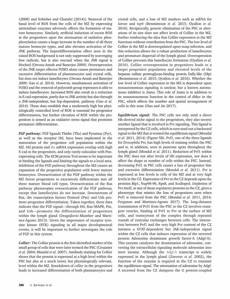
(2008) and Schieber and Chandel (2014)]. Removal of thebasal level of ROS from the cells of the MZ by expressingantioxidant enzymes adversely affects the formation of ma-ture hemocytes. Similarly, artificial induction of excess ROSin the progenitors upon the attenuation of oxidative phos-phorylation causes a huge increase in the number of all threemature hemocyte types, and also elevates activation of theJNK pathway. The hyperdifferentiation effect seen in theraised ROS background is not only suppressed by scavengingfree radicals, but is also rescued when the JNK signal isblocked (Owusu-Ansah and Banerjee 2009). Overexpressionof the JNK target effector FOXO alone causes premature andexcessive differentiation of plasmatocytes and crystal cells,but does not induce lamellocytes (Owusu-Ansah and Banerjee2009; Gao et al. 2014). However, simultaneous activation ofFOXO and the removal of polycomb group repressors is able toinduce lamellocytes. Increased ROS also result in a reductionin E-cad expression, partly due to JNK activity but also througha JNK-independent, but Srp-dependent, pathway (Gao et al.2014). These data establish that a moderately high but phys-iologically controlled level of ROS is essential for progenitordifferentiation, but further elevation of ROS within the pro-genitors is sensed as an oxidative stress signal that promotespremature differentiation.
FGF pathway: FGF ligands Thisbe (Ths) and Pyramus (Pyr),as well as the receptor Htl, have been implicated in thematuration of the progenitor cell population within theMZ. Htl protein and ths mRNA expression overlap with highE-cad in the progenitors, and only rarely colocalize with Pxn-expressing cells. The ECM protein Trol seems to be importantin binding the ligands and limiting the signals to a local area.Inhibition of the FGF pathway throughout the MZ causes anexpansion of the progenitor population with fewer maturehemocytes. Overactivation of the FGF pathway within theMZ forces progenitors to excessively differentiate into allthree mature blood cell types. Overactivation of the Raspathway phenocopies overactivation of the FGF pathway,except that lamellocytes do not increase. Downstream ofRas, the transcription factors Pointed (Pnt) and Ush pro-mote progenitor differentiation. Taken together, these dataindicate that the FGF signal—through Htl, Ras/MAPK, Pnt,and Ush—promotes the differentiation of progenitorswithin the lymph gland (Dragojlovic-Munther and Marti-nez-Agosto 2013). Given the importance of receptor tyro-sine kinase (RTK) signaling in all major developmentalevents, it will be important to further investigate the roleof FGF in this system.
Collier: The Collier protein is the first identifiedmarker of thesmall group of cells that were later termed the PSC (Crozatieret al. 2004; Mandal et al. 2007). Antibody staining for Colliershows that the protein is expressed at a high level within thePSC but also at a much lower, but physiologically relevant,level within the MZ. Knockdown of collier in the progenitorsleads to increased differentiation of both plasmatocytes and
crystal cells, and a loss of MZ markers such as mRNA forlatran and tep4 (Benmimoun et al. 2015; Oyallon et al.2016). Reciprocally, genetic elimination of the PSC or alter-ations of its size does not affect levels of Collier in the MZ,further reinforcing the idea that Collier expression in the MZfunctions without contribution from the PSC. The low level ofCollier in the MZ is downregulated upon wasp infection, andthis reduction allows for a robust production of lamellocytesand premature dispersal of the lymph gland. Overexpressionof Collier prevents this lamellocyte formation (Oyallon et al.2016). Collier overexpression in progenitors leads to alarger progenitor population and elevated levels of theheparan sulfate proteoglycan-binding protein Dally-like (Dlp)(Benmimoun et al. 2015; Oyallon et al. 2016). Whether thelow level of Collier expression in the MZ is dependent uponnonautonomous signaling is unclear, but a known autono-mous inhibitor is Jumu. This role of Jumu is in addition toits nonautonomous function in the control of dMyc in thePSC, which affects the number and spatial arrangement ofcells in this zone (Hao and Jin 2017).
Equilibrium signal: The PSC cells not only send a directHh-derived niche signal to the progenitors, they also secreteanother ligand that is involved in Pvr signaling. This ligand isinterpreted by the CZ cells, which in turn send out a backwardsignal to theMZ that is termed the equilibrium signal (Mondalet al. 2011, 2014) (Figure 5B). Pvf1, one of the three ligandsfor Drosophila Pvr, has high levels of staining within the PSCand is, in addition, seen in punctate spots throughout thelymph gland (Mondal et al. 2011). Reduction of Pvf1 withinthe PSC does not alter levels of Hh expression, nor does itaffect the shape or number of cells within the PSC. Instead,decreasing Pvf1 in PSC cells causes a loss of progenitor fateand excessive differentiation (Mondal et al. 2011). Pvr isexpressed at low levels in cells of the MZ and at very highlevels in the CZ. Expression of Pvr in the CZ depends upon theproteins Bip1, Nup98-96, Rps8, and Scalloped. Depletion ofPvr itself, or any of these regulatory proteins in the CZ, gives aphenotype that mimics the loss of progenitors seen whenPvf1 is removed from the PSC (Mondal et al. 2011, 2014;Ferguson and Martinez-Agosto 2017). The long-distancetransmission of Pvf1 from the PSC to the CZ involves trans-port vesicles, binding of Pvf1 to Pvr at the surface of MZcells, and transcytosis of the complex through repeatedrounds of vesicular exchanges between cells. The interac-tion between Pvf1 and the very high Pvr content of the CZinitiates a STAT-dependent but JAK-independent signalwithin the CZ cells that induces expression of the secretedprotein Adenosine deaminase growth factor-A (Adgf-A).This enzyme catalyzes the deamination of adenosine, con-verting the extracellular signaling molecule adenosine intoinert inosine. Although the Adgf-A transcript is widelyexpressed in the lymph gland (Zurovec et al. 2002), thefunction of the enzyme is required in the CZ to transmitthe equilibrium signal. The attenuation of adenosine by Adgf-A secreted from the CZ dampens the G protein-coupled
388 U. Banerjee et al.

adenosine receptor (AdoR) signal in the MZ, resulting inreduced PKA (cAMP-dependent protein kinase A) activityand the consequent maintenance of active Ci. Interestingly,PKA function is also inhibited when Hh binds its receptor,Ptc, activating Ci. Thus, the Hh-dependent signal from thePSC and the adenosine signal controlled by factors withinthe CZ synergize to inhibit PKA activity and stabilize theactivated form of Ci, leading to the maintenance of progen-itors within the MZ (Mandal et al. 2007; Mondal et al.2011). This two-way communication between the zonesreinforces a balance between the progenitor and the maturehemocyte populations.
The PSC
Summary: The PSC sends out a plethora of signaling li-gands and is relatively sparse in its expression of thecorresponding receptors, which are largely confined tothe MZ. This fact is at the core of the designation of thePSC as a hematopoietic niche. Loss of these ligands leads todisruption of progenitor maintenance in the MZ. In addi-tion,mechanisms of autonomous regulation of PSC cell-fatespecification, proliferation, maintenance, and growth havealso been explored.
In a pioneering set of experiments, the PSC cells weregenetically ablated by artificially inducing the proapoptoticprotein Reaper in these cells from the earlyfirst instar of larvaldevelopment. Surprisingly, this manipulation does not alterCollier expression in theMZanddoesnot appreciably increasehemocyte differentiation, as would be expected upon lossof a Hh-driven signaling center under normal conditions(Benmimoun et al. 2015). Others have repeated this manip-ulation and, at least qualitatively, reported the same results(Oyallon et al. 2016; Baldeosingh et al. 2018). This findingseems to be at odds with the concept of the PSC cells consti-tuting a hematopoietic niche (Benmimoun et al. 2015), andyet multiple laboratories utilizing a variety of genetic back-grounds have demonstrated that when present, the PSC in-fluences the fate of the MZ cells in significant ways (Crozatieret al. 2004; Krzemień et al. 2007; Mandal et al. 2007; Mondalet al. 2011; Benmimoun et al. 2012; Pennetier et al. 2012;Tokusumi et al. 2012; Khadilkar et al. 2014, 2017; Dey et al.2016; Baldeosingh et al. 2018). Several alternative explana-tions could be put forth to explain these differences. Onereasonable starting point in resolving the inconsistency isprovided by the Fossett laboratory, which showed that theMZ cells are not all identical as they are composed of a mix-ture of Hh-sensitive (Odd+ Col2) and Hh-insensitive (Odd+
Col+) progenitors (Baldeosingh et al. 2018). This is consis-tent with earlier data that Col+ cells constitute a subset of theDome+ progenitors (Oyallon et al. 2016), but the criticalobservation in the current context is the demonstrationof their heterogeneity in responding to the niche signal(Baldeosingh et al. 2018). For instance, when an Antp driveris used to express hh-RNAi in the PSC, the Odd+ Col2 cells arelost while the Odd+ Col+ population remains (Baldeosinghet al. 2018). Based on these studies, ablation of the PSC
would still retain some progenitors as a back-up system, per-haps to be used in the event of stress or infection. Given theirlocation and developmental history, it is tempting to specu-late that these Col+ cells are direct derivatives of the prepro-genitor population that is induced and maintained by signalsrelated to the dorsal vessel (Dey et al. 2016; Ferguson andMartinez-Agosto 2017), but further experiments will test thevalidity of this conjecture.
Specifics: Antp and Collier are used as primary markers forthe PSC and, although Serrate-lacZ can also be used as alater marker for a subset of these cells, Serrate protein hasno known function in the PSC (Lebestky et al. 2003;Crozatier et al. 2004; Mandal et al. 2007). Both Antp andCollier are initially expressed in 3–5 cells of the embryoniclymph gland at stage 16. The PSC cells are the first to pro-liferate during early larval development and generate thecluster of 30–40 cells seen in the third instar. Antp andCollier expression is maintained in PSC cells throughoutall larval instars. Antp is an autonomous determinant ofPSC cells, while Collier functions downstream but is re-quired for the maintenance of Antp expression in these cells.Overexpression of Antp causes an expansion in the numberof PSC cells, suggesting that Antp specifies both the fate andsize of the PSC. This manipulation indirectly increases pro-genitor maintenance and further reduces progenitor prolifera-tion (Lebestky et al. 2000; Crozatier et al. 2004; Mandal et al.2007). Dorothy (Dot), a UDP glycosyltransferase, is expressedin all cells of the early lymph gland but retracts to the PSCduring later stages (Zhou et al. 2001; Kimbrell et al. 2002;Jung et al. 2005). However, the role of Dot in the PSC is notentirely clear.
TheDppandWgsignalingpathways regulate the sizeof thePSC in an opposing fashion (Sinenko et al. 2009; Pennetieret al. 2012). A GFP reporter for a downstream target of Dppsignaling, Daughters against dpp (Ddad), and pMad (a down-stream transcription factor) are expressed in the PSC cells.Genetic manipulations that inhibit Dpp pathway activity in-crease the number of these cells and this is attributed to anincreased frequency of proliferation in the absence of thesignal (Pennetier et al. 2012). Dpp pathway activity is mod-ulated by the heparan sulfate proteoglycan-binding proteinDally-like (Dlp or Dly), which is highly expressed within thePSC (Belenkaya et al. 2004; Sinenko et al. 2011; Pennetieret al. 2012). A dlp mutant fails to activate pMad in the PSCand leads to an increase in PSC size (Sinenko et al. 2011;Pennetier et al. 2012). In summary, these data provide evi-dence that the Dpp pathway plays an inhibitory role in PSCproliferation.
The glycoprotein Slit (Labrosse et al. 2005a) is expressedin, and secreted by, the dorsal vessel, while its receptors,Roundabout 1 and 2 (Robo 1–2), are highly expressed inthe PSC. Binding of secreted Slit to Robo promotes PSC pro-liferation and clustering (Morin-Poulard et al. 2016). Roboloss increases PSC cell number and disrupts their tight clus-tering, causing them to scatter to more distal and anterior
Fly Blood Development 389
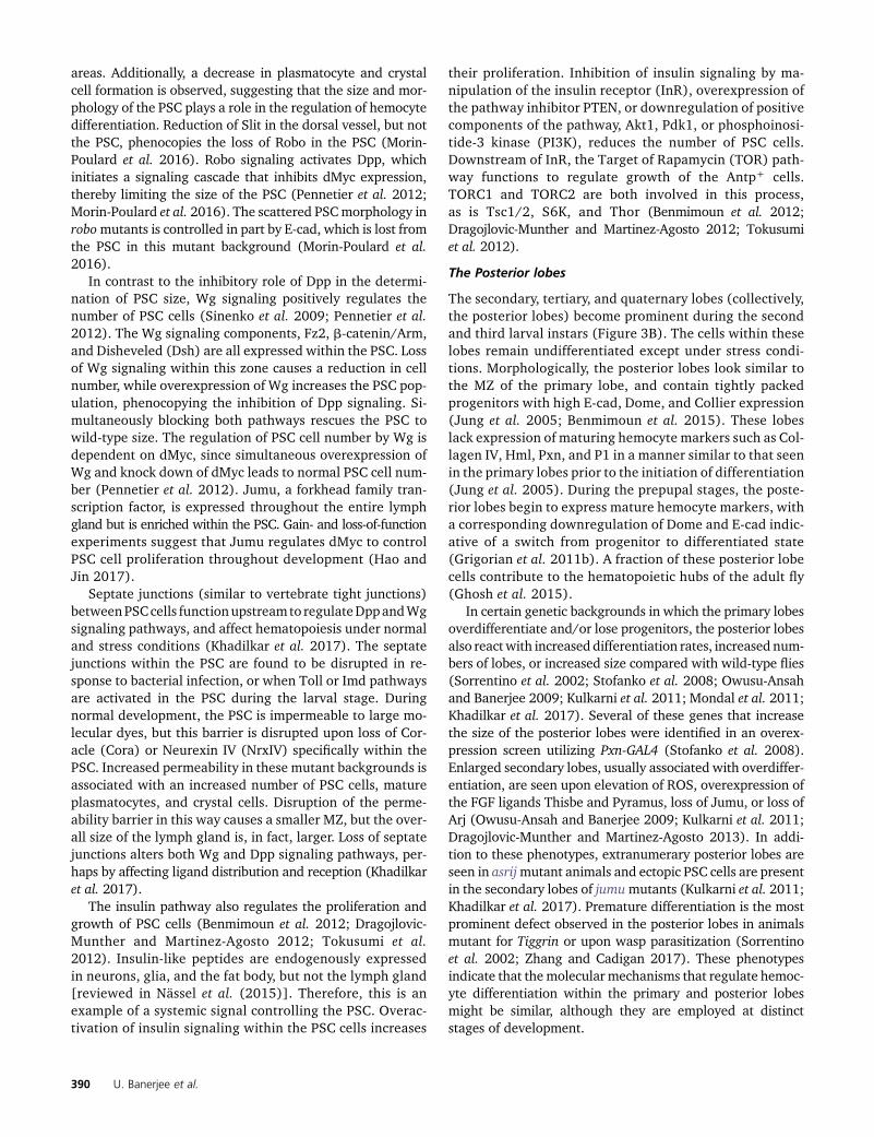
areas. Additionally, a decrease in plasmatocyte and crystalcell formation is observed, suggesting that the size and mor-phology of the PSC plays a role in the regulation of hemocytedifferentiation. Reduction of Slit in the dorsal vessel, but notthe PSC, phenocopies the loss of Robo in the PSC (Morin-Poulard et al. 2016). Robo signaling activates Dpp, whichinitiates a signaling cascade that inhibits dMyc expression,thereby limiting the size of the PSC (Pennetier et al. 2012;Morin-Poulard et al. 2016). The scattered PSCmorphology inrobomutants is controlled in part by E-cad, which is lost fromthe PSC in this mutant background (Morin-Poulard et al.2016).
In contrast to the inhibitory role of Dpp in the determi-nation of PSC size, Wg signaling positively regulates thenumber of PSC cells (Sinenko et al. 2009; Pennetier et al.2012). The Wg signaling components, Fz2, b-catenin/Arm,and Disheveled (Dsh) are all expressed within the PSC. Lossof Wg signaling within this zone causes a reduction in cellnumber, while overexpression of Wg increases the PSC pop-ulation, phenocopying the inhibition of Dpp signaling. Si-multaneously blocking both pathways rescues the PSC towild-type size. The regulation of PSC cell number by Wg isdependent on dMyc, since simultaneous overexpression ofWg and knock down of dMyc leads to normal PSC cell num-ber (Pennetier et al. 2012). Jumu, a forkhead family tran-scription factor, is expressed throughout the entire lymphgland but is enriched within the PSC. Gain- and loss-of-functionexperiments suggest that Jumu regulates dMyc to controlPSC cell proliferation throughout development (Hao andJin 2017).
Septate junctions (similar to vertebrate tight junctions)betweenPSCcells functionupstream to regulateDppandWgsignaling pathways, and affect hematopoiesis under normaland stress conditions (Khadilkar et al. 2017). The septatejunctions within the PSC are found to be disrupted in re-sponse to bacterial infection, or when Toll or Imd pathwaysare activated in the PSC during the larval stage. Duringnormal development, the PSC is impermeable to large mo-lecular dyes, but this barrier is disrupted upon loss of Cor-acle (Cora) or Neurexin IV (NrxIV) specifically within thePSC. Increased permeability in these mutant backgrounds isassociated with an increased number of PSC cells, matureplasmatocytes, and crystal cells. Disruption of the perme-ability barrier in this way causes a smaller MZ, but the over-all size of the lymph gland is, in fact, larger. Loss of septatejunctions alters both Wg and Dpp signaling pathways, per-haps by affecting ligand distribution and reception (Khadilkaret al. 2017).
The insulin pathway also regulates the proliferation andgrowth of PSC cells (Benmimoun et al. 2012; Dragojlovic-Munther and Martinez-Agosto 2012; Tokusumi et al.2012). Insulin-like peptides are endogenously expressedin neurons, glia, and the fat body, but not the lymph gland[reviewed in Nässel et al. (2015)]. Therefore, this is anexample of a systemic signal controlling the PSC. Overac-tivation of insulin signaling within the PSC cells increases
their proliferation. Inhibition of insulin signaling by ma-nipulation of the insulin receptor (InR), overexpression ofthe pathway inhibitor PTEN, or downregulation of positivecomponents of the pathway, Akt1, Pdk1, or phosphoinosi-tide-3 kinase (PI3K), reduces the number of PSC cells.Downstream of InR, the Target of Rapamycin (TOR) path-way functions to regulate growth of the Antp+ cells.TORC1 and TORC2 are both involved in this process,as is Tsc1/2, S6K, and Thor (Benmimoun et al. 2012;Dragojlovic-Munther and Martinez-Agosto 2012; Tokusumiet al. 2012).
The Posterior lobes
The secondary, tertiary, and quaternary lobes (collectively,the posterior lobes) become prominent during the secondand third larval instars (Figure 3B). The cells within theselobes remain undifferentiated except under stress condi-tions. Morphologically, the posterior lobes look similar tothe MZ of the primary lobe, and contain tightly packedprogenitors with high E-cad, Dome, and Collier expression(Jung et al. 2005; Benmimoun et al. 2015). These lobeslack expression of maturing hemocyte markers such as Col-lagen IV, Hml, Pxn, and P1 in a manner similar to that seenin the primary lobes prior to the initiation of differentiation(Jung et al. 2005). During the prepupal stages, the poste-rior lobes begin to express mature hemocyte markers, witha corresponding downregulation of Dome and E-cad indic-ative of a switch from progenitor to differentiated state(Grigorian et al. 2011b). A fraction of these posterior lobecells contribute to the hematopoietic hubs of the adult fly(Ghosh et al. 2015).
In certain genetic backgrounds in which the primary lobesoverdifferentiate and/or lose progenitors, the posterior lobesalso reactwith increased differentiation rates, increased num-bers of lobes, or increased size compared with wild-type flies(Sorrentino et al. 2002; Stofanko et al. 2008; Owusu-Ansahand Banerjee 2009; Kulkarni et al. 2011; Mondal et al. 2011;Khadilkar et al. 2017). Several of these genes that increasethe size of the posterior lobes were identified in an overex-pression screen utilizing Pxn-GAL4 (Stofanko et al. 2008).Enlarged secondary lobes, usually associated with overdiffer-entiation, are seen upon elevation of ROS, overexpression ofthe FGF ligands Thisbe and Pyramus, loss of Jumu, or loss ofArj (Owusu-Ansah and Banerjee 2009; Kulkarni et al. 2011;Dragojlovic-Munther and Martinez-Agosto 2013). In addi-tion to these phenotypes, extranumerary posterior lobes areseen in asrijmutant animals and ectopic PSC cells are presentin the secondary lobes of jumumutants (Kulkarni et al. 2011;Khadilkar et al. 2017). Premature differentiation is the mostprominent defect observed in the posterior lobes in animalsmutant for Tiggrin or upon wasp parasitization (Sorrentinoet al. 2002; Zhang and Cadigan 2017). These phenotypesindicate that the molecular mechanisms that regulate hemoc-yte differentiation within the primary and posterior lobesmight be similar, although they are employed at distinctstages of development.
390 U. Banerjee et al.

Pupal and Adult Blood Cells
Summary
At the onset of pupariation, the lymph gland undergoes adramatic change. The progenitor population differentiatesleaving very few, if any, cells that are Dome+ (Grigorianet al. 2011b). The lymph gland disintegrates, dispersing theblood cells into the hemolymph (Lanot et al. 2001; Grigorianet al. 2011b). The blood cells released in circulation engulfthe large number of apoptotic cells generated during tissueremodeling associated with pupal development and removebacteria released during the remodeling of the gut. This pro-cess of dispersion also allows a mixing of the blood cells fromthe two major hematopoietic waves that together populatethe circulating and sessile pools of blood cells in the adult fly(Lanot et al. 2001; Grigorian et al. 2011b). While most of thedispersed lymph gland cells, together with those in larvalcirculation, go on to become adult circulating blood cells(Holz et al. 2003), some of them populate hubs along thebody wall of the adult abdomen (Ghosh et al. 2015). Thesehubs are proposed to serve as active sites of adult hemato-poiesis, an important finding that is worthy of furtherinvestigation.
Pulses of the fly steroid hormone 20-hydroxyecdysterone(commonly referred to as ecdysone for simplicity) that initiatepupariation [reviewed in Thummel (2001)] also trigger thefinal burst of differentiation and disintegration of the lymphgland. In fact, lymph glands are severely hypertrophic andfail to disperse in temperature-sensitive ecdysonelessmutants(Sorrentino et al. 2002). Hemocytes in circulation also dis-play ecdysone-related alterations to their morphology andfunction. When ecdysone is injected into midthird instar lar-vae, plasmatocytes become more flattened, adherent, andmore phagocytic, similar to those seen in the white prepupalstage (Lanot et al. 2001; Regan et al. 2013). Expression ofa dominant negative version of the ecdysone receptor(EcRB1DN) in blood cells also prevents their dispersal fromsessile clusters on the body wall (Regan et al. 2013). Thus,ecdysone signaling plays diverse roles in directing hemato-poiesis during pupal development that together cause thedissociation, dispersal, and activation of blood cells, fulfillingthe demands of metamorphosis and eclosion.
Specifics
Under normal culture conditions, the lymph gland does notcontribute to the blood cells in circulation and remains intactthroughout all larval stages, with cells encased in ECM com-ponents (Lanot et al. 2001; Grigorian et al. 2011b). Duringthe pupal stage, lymph gland cells break away from thesematrix components as they exit the lymph gland and entercirculation (Grigorian et al. 2011b). Upon their release, theplasmatocytes phagocytose larval muscle cells, the ECM sur-rounding lymph gland cells, and even other plasmatocytes(Lanot et al. 2001). Markers such as Pxn and P1 are detectedthroughout the lymph gland at 4 and 8 hr after puparium for-mation, respectively, indicating that progenitors differentiate
prior to dispersal. Trol protein is usually more concentratedaround progenitors of the MZ than the differentiated cells inthe CZ. However, during pupation, this distinction in density islost as Trol becomes uniformly distributed throughout thelymph gland. The Antp+ PSC cells are also affected, as theyno longer remain confined in a small cluster in the posteriorregion of the lymph gland, and begin to spread throughoutthe primary lobes by 8–12 hr after puparium formation(Grigorian et al. 2011b). The significance of this observation isas yet unclear.
As the primary lobes differentiate and disperse duringpupation, the secondary and tertiary lobes proliferate, en-large, initiate differentiation, and express Pxn, but not the latemarker P1 (Grigorian et al. 2011b). The cells within theselobes then disperse and enter into circulation beginning at10 hr after puparium formation (Grigorian et al. 2011b).Lineage trace analysis suggests that progenitors from the ter-tiary and quaternary lobes of the lymph gland do not fullydifferentiate, and instead migrate to hubs on the body wall inthe adult, which may serve as an adult hematopoietic niche(Ghosh et al. 2015).
Early studies established that adult hemocytes do notproliferate during homeostasis (Lanot et al. 2001). However,upon immune challenge, several clusters of hemocytes lo-cated in hubs along the dorsal abdomen incorporate BrdU(Ghosh et al. 2015). Despite lacking proliferative capacityduring normal development, these hubs increase in size forup to 5 days posteclosion, presumably due to localized hom-ing of circulating cells to these sites. All adult hemocytes inthe hubs express Srp and are dependent on ECM componentsin the neighboring microenvironment for their localization.Lineage tracing shows that the plasmatocytes and crystalcells in these hubs are derived from hemocytes of embryonicand larval origin. Interestingly, this study also detects hemoc-yte progenitors among the hub cells that are Srp+ but lackHml or Hnt, suggesting that the hubs could serve as a site ofhematopoiesis (Ghosh et al. 2015). This is a foundationalstudy on adult hematopoiesis in Drosophila and, given theinterest in, and the importance of, adult stem and progenitorpopulations across species, these intriguing results warrantfurther investigation.
Nutritional and Sensory Control of Hematopoiesis
Drosophila blood cells respond to a lack of nutritional con-tent (Benmimoun et al. 2012; Dragojlovic-Munther andMartinez-Agosto 2012; Shim et al. 2012), lack of favorableodors (Shim et al. 2013), or the depletion of environmentalCO2 or O2 (Cho et al. 2018) (Figure 6). The response to alack of nutrition involves insulin production from the brainand its reception by lymph gland progenitors. The other twocases involve the detection of small odorants or gaseousligands by external sensory neurons in the terminal organ.These signals are both transduced through a multiorganhormone-dependent signaling cascade to the cells of thelymph gland. However, when these two pathways are
Fly Blood Development 391
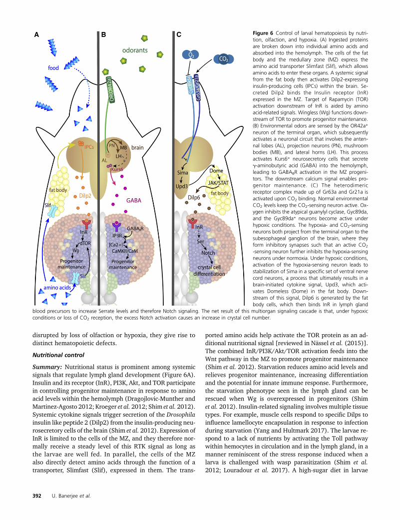
disrupted by loss of olfaction or hypoxia, they give rise todistinct hematopoietic defects.
Nutritional control
Summary: Nutritional status is prominent among systemicsignals that regulate lymph gland development (Figure 6A).Insulin and its receptor (InR), PI3K, Akt, and TOR participatein controlling progenitor maintenance in response to aminoacid levels within the hemolymph (Dragojlovic-Munther andMartinez-Agosto 2012; Kroeger et al. 2012; Shim et al. 2012).Systemic cytokine signals trigger secretion of the Drosophilainsulin like peptide 2 (Dilp2) from the insulin-producing neu-rosecretory cells of the brain (Shim et al. 2012). Expression ofInR is limited to the cells of the MZ, and they therefore nor-mally receive a steady level of this RTK signal as long asthe larvae are well fed. In parallel, the cells of the MZalso directly detect amino acids through the function of atransporter, Slimfast (Slif), expressed in them. The trans-
ported amino acids help activate the TOR protein as an ad-ditional nutritional signal [reviewed in Nässel et al. (2015)].The combined InR/PI3K/Akt/TOR activation feeds into theWnt pathway in the MZ to promote progenitor maintenance(Shim et al. 2012). Starvation reduces amino acid levels andrelieves progenitor maintenance, increasing differentiationand the potential for innate immune response. Furthermore,the starvation phenotype seen in the lymph gland can berescued when Wg is overexpressed in progenitors (Shimet al. 2012). Insulin-related signaling involves multiple tissuetypes. For example, muscle cells respond to specific Dilps toinfluence lamellocyte encapsulation in response to infectionduring starvation (Yang and Hultmark 2017). The larvae re-spond to a lack of nutrients by activating the Toll pathwaywithin hemocytes in circulation and in the lymph gland, in amanner reminiscent of the stress response induced when alarva is challenged with wasp parasitization (Shim et al.2012; Louradour et al. 2017). A high-sugar diet in larvae
Figure 6 Control of larval hematopoiesis by nutri-tion, olfaction, and hypoxia. (A) Ingested proteinsare broken down into individual amino acids andabsorbed into the hemolymph. The cells of the fatbody and the medullary zone (MZ) express theamino acid transporter Slimfast (Slif), which allowsamino acids to enter these organs. A systemic signalfrom the fat body then activates Dilp2-expressinginsulin-producing cells (IPCs) within the brain. Se-creted Dilp2 binds the Insulin receptor (InR)expressed in the MZ. Target of Rapamycin (TOR)activation downstream of InR is aided by aminoacid-related signals. Wingless (Wg) functions down-stream of TOR to promote progenitor maintenance.(B) Environmental odors are sensed by the OR42a+
neuron of the terminal organ, which subsequentlyactivates a neuronal circuit that involves the anten-nal lobes (AL), projection neurons (PN), mushroombodies (MB), and lateral horns (LH). This processactivates Kurs6+ neurosecretory cells that secreteg-aminobutyric acid (GABA) into the hemolymph,leading to GABABR activation in the MZ progeni-tors. The downstream calcium signal enables pro-genitor maintenance. (C) The heterodimericreceptor complex made up of Gr63a and Gr21a isactivated upon CO2 binding. Normal environmentalCO2 levels keep the CO2-sensing neuron active. Ox-ygen inhibits the atypical guanylyl cyclase, Gyc89da,and the Gyc89da+ neurons become active underhypoxic conditions. The hypoxia- and CO2-sensingneurons both project from the terminal organ to thesubesophageal ganglion of the brain, where theyform inhibitory synapses such that an active CO2
-sensing neuron further inhibits the hypoxia-sensingneurons under normoxia. Under hypoxic conditions,activation of the hypoxia-sensing neuron leads tostabilization of Sima in a specific set of ventral nervecord neurons, a process that ultimately results in abrain-initiated cytokine signal, Upd3, which acti-vates Domeless (Dome) in the fat body. Down-stream of this signal, Dilp6 is generated by the fatbody cells, which then binds InR in lymph gland
blood precursors to increase Serrate levels and therefore Notch signaling. The net result of this multiorgan signaling cascade is that, under hypoxicconditions or loss of CO2 reception, the excess Notch activation causes an increase in crystal cell number.
392 U. Banerjee et al.

induces lamellocytes via JNK, and also decreases phagocyto-sis of large particles such as fungal spores and latex beads (Yuet al. 2018). In adult flies, a high-fat diet causes global acti-vation of the JAK/STAT pathway and the adult circulatingcells secrete Upd3, which triggers a systemic immune re-sponse (Woodcock et al. 2015).
Specifics: Larvae raised under starvation conditions haveexcessive differentiation at the expense of lymph gland pro-genitors. There is an obvious increase in the number ofcirculating plasmatocytes and lamellocytes. Plasmatocytesare also often seen interspersed between fat body cells ina starved larva (Shim et al. 2012). In response to starva-tion, crystal cells prematurely differentiate in the second in-star and subsequently rupture during the third instar(Benmimoun et al. 2012; Shim et al. 2012). Furthermore,larvae respond to starvation by activating the Toll pathway,which leads to accumulation of the transcription factor Dorsalin the nucleus in circulating hemocytes and in lymph glandcells (Shim et al. 2012).
There are eight insulin-like peptides in Drosophila (Dilp1–8) that act as ligands for the InR [reviewed in Nässel et al.(2015)]. Dilp2, in particular, is involved in regulating hema-topoiesis during normal development and upon starvation,and a decrease in Dilp2 is the major cause of blood cell over-differentiation. Consistent with this explanation, eliminationof insulin-producing cells (IPCs) in the brain causes a loss ofprogenitors similar to that seen upon starvation, and consti-tutive activation of the IPC neurons induces increased pro-genitor maintenance. Similarly, loss of InR or its downstreameffector Chico, or Akt from the hemocyte progenitors, in-creases differentiation of the progenitor population withinthe lymph gland (Shim et al. 2012). TOR kinase functionsfurther downstream in the InR pathway, but it also respondsto amino acid availability (Oldham et al. 2000a,b; Zhang et al.2000; Kim et al. 2008; Teleman et al. 2008; Oldham 2011).TOR integrates these two inputs through a pathway that in-cludes the translation initiation factor 4E-binding protein(4EBP, also known as Thor) and ribosomal protein S6 kinase(S6K) (Rodriguez et al. 1996; Montagne et al. 1999). Up-stream effectors of TOR, Rheb, and Tsc2, as well as the down-stream component p-4EBP, are expressed in high amounts inthe progenitors and this is supportive of active TOR signaling.Gain- and loss-of-function experiments involving TSC1/2,Rheb, and PTEN suggest a relationship between the entireTOR pathway and ROS, and this regulation is likely linked tothe starvation-induced lymph gland phenotype (Dragojlovic-Munther and Martinez-Agosto 2012; Shim et al. 2012).
The fat body also detects amino acid levels within thehemolymph via an amino acid transporter called Slif, and thisresults in a paracrine signal to the brain that activates Dilp2secretion. In addition to the Dilp2/InR response to nutrition,blood progenitors also respond directly to essential aminoacids imported into the cell by Slif (Shim et al. 2012). Thus,amino acid levels are detected both directly by the lymphgland progenitors and also indirectly via the fat body to
maintain the pool of hemocyte progenitors (Benmimounet al. 2012; Shim et al. 2012). Similar to the lack of nutrition,a lack of amino acid detection leads to a loss of the progenitorpopulation and an increase in the number of mature bloodcells.
Increasing theamountof fat in thediet of adultflies leads tochanges in hemocyte gene expression (Woodcock et al.2015).Under these conditions, adult hemocytes in circulation havehigher expression of Upd3. This result suggests that plasma-tocytes are able to sense lipid levels in the hemolymph. Ge-netic dissection shows that this nutrient sensing phenomenonrequires the Crq receptor, which presumably allows for theuptake of lipids from the hemolymph into the plasmatocytes.Reduction of crq within hemocytes causes decreases inboth Upd3 secretion and Socs36e transcription withinhemocytes. Excess lipid consumption does not activate theToll or Imd pathways, and also does not alter the number ofplasmatocytes in circulation. Rather, the affected plasmato-cytes increase their lipid stores anddisplay an alteredmorphol-ogy. The resulting increase in Upd3 expression in hemocytes iseither directly or indirectly involved in decreasing the adultlife span when flies are fed a high-fat diet (Woodcock et al.2015).
Sensory control
Environmental odors and gases sensed by “gustatory” recep-tors also trigger a cascade of signaling events involving ex-ternal sensory organs, the brain, and the developing lymphgland to regulate the blood progenitor population. GABABR,a metabotropic receptor for GABA, is expressed in both thePSC and MZ. Within the PSC, GABABR plays an early role,before midsecond instar, in promoting lymph gland growth.Importantly, the MZ-expressed GABABR is responsible forprogenitor maintenance in response to sensory cues. Environ-mental odiferous ligands are sensed through the larval ter-minal organ and detected by a neuron expressing thereceptor OR42a that senses fruity odors. This neuron projectsinto the antennal lobe of the brain and interacts via a networkof interneurons that ultimately activate a small number ofneurosecretory cells to secrete GABA into the hemolymph.The binding of GABA to GABABR expressed in the MZ isessential for progenitor maintenance that is dependent ondownstream Ca2+ signaling. Loss of olfaction is sensed as astress and the progenitors respond by losing their quiescenceand multipotency (Shim et al. 2013) (Figure 6B). Environ-mental gases are also detected and interpreted in a similarmanner utilizing external receptors in the terminal organ.This leads to secretion of a cytokine across the blood–brainbarrier and, with the fat body as an intermediary organ, pro-duction of an insulin-like peptide that ultimately influencescrystal cell development within the lymph gland through theregulation of Notch signaling (Figure 6C).
Olfactory control: Olfaction is relayed by multiple organsusing signaling peptides to exert an effect on hematopoiesis.Lymph glands of larvae raised on food lacking odorants have a
Fly Blood Development 393

decreased pool of blood progenitors and increased number ofdifferentiated cells, despite the PSC being unaffected. Amaz-ingly, themutant lymph gland phenotype causedwhen larvaeare grown on food lacking odorants can be rescued when asmall dialysis bag containing odorant food is placedwithin theodor-free growth medium. In this experiment, larvae are ableto detect the food smell, but are unable to ingest the odorantfood due to the barrier presented by the dialysis membrane(Shim et al. 2013).
Odorants are detected by external sensory neurons, whichthen transmit aneuronal signal to the subesophageal ganglionof thebrain.This information is further relayed todownstreamprojection neurons that interact with Kurs6-GAL4+ neurose-cretory cells to activate GABA secretion into the hemolymph.A high level of GABA is detected within Kurs6+ neuronsand also at the surface of lymph gland progenitors. A loss ofGABA synthesis or inhibition of its proper vesicular packagingwithin the Kurs6+ neurons causes a reduction of GABA incirculation, decreased detection of GABA on the MZ cells,and increased hemocyte differentiation with a loss of progen-itors. Genetic ablation, inhibition of neuronal activity, or de-pletion of olfactory coreceptor Orco within all olfactoryneurons decreases GABA staining in the Kurs6+ neurons, aswell as in circulation and on the MZ cells. This ultimatelyresults in a loss of lymph gland progenitors, mimicking theloss-of-odorant phenotype. These data suggest that Kurs6+
GABA-producing cells function downstream of olfactory per-ception to translate the sensory signal to one that controlsblood cell development (Shim et al. 2013).
Reception of GABA secreted from the Kurs6+ neurons byprogenitors in the MZ activates cytosolic calcium signaling,which promotes progenitor maintenance. Calcium signalingactivity, indicated by a GCaMP sensor, decreases in progeni-tors expressing RNAi against either subunit of the GABAreceptor (GABABR1 or GABABR2). This suggests that theultimate step in olfactory regulation downstream of Or42ain larval hematopoiesis is the modulation of calcium withinthe progenitors to control levels of hemocyte differentiation.Olfactory perception is independent of insulin signaling anddoes not affect food intake. Unlike starvation, olfactory dep-rivation does not affect levels of Dilp2 secretion. Further-more, Dilp2 expression within the IPCs is unaltered whenGABA production is decreased in the Kurs6+ neurons (Shimet al. 2013).
Control by environmental gases: Although classicallynamed as gustatory receptors, the heterodimeric partnersGr63a and Gr21a have been shown to be activated by lowlevels of CO2 in adults and in larvae (Jones et al. 2007; Kwonet al. 2007). Hypoxia and low levels of ROS molecules havebeen known to activate an atypical guanylyl cyclase, Gyc89da(Morton 2004; Evgenov et al. 2006; Morton et al. 2008;Vermehren-Schmaedick et al. 2010). Disruption of any ofthese three molecules (Gr63a, Gr21a, or Gyc89da) in theirrespective neurons leads to an increase in lymph gland pro-duction of crystal cells (Cho et al. 2018).
The CO2- and hypoxia-sensing neurons interact in thebrain to convert the external sensation of gases into a signalthat can cross the blood–brain barrier to have a systemiceffect on the animal. Dendritic labeling reveals that theCO2-sensing neuron and the hypoxia-sensing neurons arelocated near each other in the terminal organ of the larvalhead, and then overlap within the subesophageal ganglion ofthe brain. It is at this juxtaposition that these two neuronsutilize GABA signaling to form synaptic connections witheach other, which can be visualized using themolecular prox-imity detector GRASP (Feinberg et al. 2008; Macphersonet al. 2015). Larvae grown in a normoxic environment haveregular Sima accumulation in particular neurons of the ven-tral nerve cord (VNC). However, when CO2 or oxygen levelsare decreased during development, the Sima protein is stabi-lized in Gyc89da+ cells in the VNC. This elevated Sima leads toincreased production of the Upd3 cytokine in hypoxia-sensingneurons. The inhibition of neuronal expression of either Simaor Upd3 can suppress the increased crystal cell phenotype seenunder low CO2 conditions (Cho et al. 2018).
After secretion into the hemolymph, the Upd3 released bythe brain is received by theDome receptor expressed in the fatbody.Thisbinding triggers theJAK/STATsignalingpathway inthis organ, which can be detected by higher levels of the STATtranscriptional target Socs36E. Also dependent upon in-creased JAK/STAT activity in the fat body is the augmentedlevel of Dilp6. Upon inhibition of CO2 reception, this Dilp6 isrequired for the downstream lymph gland production of crys-tal cells. Finally, the high amounts of Dilp6 are received by theInR expressed in the lymph gland to increase the productionof crystal cells. Decreasing the amount of CO2 detection leadsto high levels of pAKT and p4EBP/Thor throughout thelymph gland. Upregulation of Serrate in the second-instarmutant lymph gland promotes increased crystal cell differen-tiation (Cho et al. 2018). It is through this multisignal andmultiorgan relay system that both external gaseous and odi-ferous ligands are able to affect hematopoietic developmentto respond to the environment.
Stress Response and the Hematopoietic System
It can be argued that all cells are programmed to respond tostresses such as changes in redox levels, unfolded proteins,DNA damage, infection, injury, or metabolic dysfunction. In-ternal circuitry relieves such stresses by activating mecha-nisms ranging from antioxidant pathways, the unfoldedprotein response, autophagy, cell cycle arrest, repair, and aplethora of cell death pathways. However,myeloid precursorsand the cells derived from them are specialists at mitigatingmany formsof stresses thatdisrupt homeostasis. This becomesreadily apparent in Drosophila (and invertebrates in general)as their entire blood cell repertoire is similar to the myeloidcells in vertebrates [reviewed in Evans et al. (2003)]. Evenwithin the context of innate immunity, the strongest re-sponses are systemic, arising outside the hematopoietic tissue[reviewed in Lemaitre and Hoffmann (2007)]. Also, oxygen
394 U. Banerjee et al.

distribution is not an essential cellular function in Drosophila,although gaseous sensing is certainly important (Mondalet al. 2011; Cho et al. 2018). The myeloid-like blood cellsare sentinels of stress sensation and mitigation, oftenresponding faster than system-wide immunity afforded bythe fat body (Kocks et al. 2005; Bidla et al. 2007; Babcocket al. 2008; Cuttell et al. 2008). In this context, we broadlydefine stress as any event or condition that threatens to dis-rupt homeostasis. These stresses can be intrinsic, arising fromabnormal internal processes such as genetic mutations, aber-rant tissue characteristics, and growth, or they might initiatefrom abnormal external processes such as infection, parasit-ization, a lack of essential nutrients, or injury caused by me-chanical, chemical, or heat damage. Interestingly, althoughthese events have very different inducers and follow distincttransduction processes, the blood cell response is often quitesimilar. Thus, multiple independent stress signals might feedinto a few key blood cell response pathways. This is particu-larly true for the hematopoietic system since stress responsesfrequently coopt the same pathways that direct homeostaticdevelopment, except in an exaggerated form suitable for thespecific kind of stress.
Cellular response to intrinsic stress
Summary: Internal stresses often result from genetic muta-tions that induce tissue abnormalities. For example, ECMcomponents keep healthy tissues isolated from the hemo-lymph and their loss can cause blood cell responses that havebeen likened to an “auto-immune response.” These responsesinclude: the attachment of plasmatocytes onto the affectedsurface and their invasion into the tissue, activation of plas-matocyte signaling, and encapsulation of the tissue by lamel-locytes to form melanotic masses (Pastor-Pareja et al. 2008;Cordero et al. 2010; Kim and Choe 2014; Fogarty et al. 2016).Depletion of basement membrane components, particularlyin combination with loss of appropriate cell polarity markersand decreased cell-to-cell contacts, leads to the formation ofmelanotic masses around self-tissue (Kim and Choe 2014).Similarly, in a metastatic tumor model in the eye/antennaldisc, loss of basement membrane components causes the en-hanced recruitment and proliferation of plasmatocytes thathelp limit tumor growth (Pastor-Pareja et al. 2008).
Similar phenotypes are seenwhenmutationsdirectly affectblood cell differentiation, proliferation, or signaling. In thiscase, hyperactive blood cells respond inappropriately to nor-mal tissues. For example, this occurs inflymodels of leukemia,in which oncogenes are introduced into the blood tissue andthis results in an increase in circulating blood cells, lamello-cyte differentiation, and the formation of melanotic massesaround normal tissue (Osman et al. 2009; Sinenko et al. 2010;Reitman et al. 2015). Signs of immune activation includinglamellocyte differentiation, lymph gland hypertrophy, in-creased proliferation, and encapsulation of healthy tissuesare also seen when blood differentiation pathways such asToll, JAK/STAT, and adenosine signaling are overactivatedthrough mutations in pathway components (Gerttula et al.
1988; Hanratty and Dearolf 1993; Harrison et al. 1995;Lemaitre et al. 1995; Luo et al. 1995; Qiu et al. 1998;Dolezal et al. 2005). Interestingly, the fat body and hemoc-ytes respond differently to knockdown of Toll pathway com-ponents (Schmid et al. 2014). Ultimately, screening forfactors that cause these types of overactive blood cell pheno-types has provided insights into pathways involved in theirdevelopment and stress responses (Watson et al. 1991;Zettervall et al. 2004; Minakhina and Steward 2006; Avet-Rochex et al. 2010).
Specifics: Blood cells respond to abnormal or damaged tissuesin several ways. For example, . 100 genetic mutations havebeen identified that cause encapsulation of self-tissue bylamellocytes and subsequent melanization, forming mela-notic masses in the bodies of the fly sometimes called mela-notic capsules, melanotic nodules, melanotic tumors, ormelanotic pseudotumors (Watson et al. 1991; Minakhinaand Steward 2006; Avet-Rochex et al. 2010; Gramates et al.2017). Melanotic masses often resemble one another, butthey have been categorized into two broad classes based ondifferences in the underlying mechanisms of their formation.Class I melanotic mass mutants have apparently normal cel-lular immune systems that respond to an abnormal self-tissue. For example, mutations in kurtz, encoding an arrestinprotein that is involved in the internalization of G protein-coupled receptors, causes fat body cells to dissociate andmelanotic masses to form around these dissociating fat bodycells (Roman et al. 2000). Blood cells identify normal tissuethrough the detection of an intact basement membrane(Rizki and Rizki 1974). Knockdown of Collagen IV subunitsVkg and a1, or Laminin subunits in fat body cells, disruptstheir basement membrane and leads to encapsulation bylamellocytes and the formation of melanotic masses (Kimand Choe 2014). Similarly, knockdown of a receptor for thebasement membrane, the integrin-b subunit Myospheroid,also causes a similar phenotype. Reliable encapsulation oc-curs when basement membrane disruption is combined witha loss of cell integrity, cell-to-cell contacts, and improper lo-calization of polarity proteins such as Cora and Discs large(Kim and Choe 2014).
Class II melanotic masses have been defined as thosearising in the context of an overactive immune system thatcauses ectopic activation of hemocytes and an inappropriateresponse tonormal tissue.Often, class IImutants also exhibit alarge increase in the number of blood cells and aberrantlamellocyte differentiation. Pathways such as Toll, JAK/STAT,and adenosine signaling, known to be involved in blood cellhomeostasis, give rise to class II phenotypes if they are madeoveractive (Gerttula et al. 1988; Hanratty and Dearolf 1993;Harrison et al. 1995; Lemaitre et al. 1995; Luo et al. 1995; Qiuet al. 1998; Dolezal et al. 2005; Matova and Anderson 2006;Krzemień et al. 2007; Minakhina et al. 2011; Zhang et al.2016). A melanotic tumor phenotype similar to class IIis induced when human oncogenic aberrations, such astranslocations giving rise to the fusion protein AML1-ETO
Fly Blood Development 395

or gain-of-function mutations in isocitrate dehydrogenase(IDH1 and 2), are expressed in Drosophila hemocytes to sim-ulate models for blood-related disorders (Osman et al. 2009;Sinenko et al. 2010; Reitman et al. 2015). Melanotic tumorsare terminal phenotypes that are easy to identify in a geneticscreen. Their origins are often linked to defects in proliferation,differentiation, or autoimmunity. Genetic screens are valuablefor identifying new factors that contribute to normal and de-fective hematopoiesis, and also to the cellular stress response.
Drosophila has emerged as a powerful system in which tostudy the complex role that macrophages play in disease pro-gression, such as in a metastatic cancer model induced by theexpression of overactive Ras along with a loss-of-functionmutation in the scribbled (scrib) gene that is importantfor the maintenance of epithelial polarity. These geneticbackgrounds cause massive tumors that can become meta-static (Pagliarini and Xu 2003; Pastor-Pareja et al. 2008).Importantly for this discussion, mature plasmatocytes mi-grate to the tumor sites and adhere to them, preferentiallybinding to areas where the basement membrane is disrupted.Surprisingly, melanotic masses are not observed aroundRasV12; scrib2/2 tumors or in a RasV12-driven salivary glandtumor model. In the latter case, plasmatocytes, crystal cells,and lamellocytes migrate to the tumorous tissue, yet no mel-anotic masses are seen (Hauling et al. 2014). Tissue damageinduced by a tumor, an aseptic wound, or UV irradiationcauses an increase in proliferation and the number of bloodcells in circulation that is dependent on JAK/STAT signalingin the blood cells (Pastor-Pareja et al. 2008; Karpac et al.2011; Kelsey et al. 2012). While modulation of JAK/STATin hemocytes represses this excessive proliferation, it does nothave any effect on the attachment of the existing hemocytes tothe tumor surface (Pastor-Pareja et al. 2008).
Overall, it appears that stress-induced plasmatocyte mi-gration, proliferation, and signaling aids the restriction oftumor growth and tissue maintenance (Pastor-Pareja et al.2008; Kelsey et al. 2012). This is evidenced by the fact thatthe size of scrib2/2 tumors in the imaginal epithelium in-creases if blood cells are unable to receive a JAK/STAT signal(Pastor-Pareja et al. 2008). Similarly, blood cells also restricttumor growth induced in the wing disc in a discs largemutantbackground, and in these mutant animals, plasmatocytes ex-press Eiger (Egr), the Drosophila homolog of tumor necrosisfactor (TNF). Egr expression activates JNK signaling in tumorcells, which induces apoptosis, thus decreasing tumor size(Parisi et al. 2014).
Although plasmatocytes normally function to mitigate in-fection, injury, and apoptotic decay, while fighting artificiallyinduced disease, their attempt tominimize the damage could,in fact, lead to enhanced mortality or dysfunction at thecellular and systems level. For example, in the aforemen-tioned tumor model, if, in addition to the loss of polaritygenes, the tumor also expresses RasV12, this second mutationleads to a block in apoptosis (Bergmann et al. 1998; Pérezet al. 2017). In this case, the expression of Egr by plasmato-cytes causes very high levels of JNK signaling in the tumor
without causing apoptosis, which results in increased meta-static growth (Cordero et al. 2010; Pérez et al. 2017). Inaddition, high levels of ROS in the RasV12; scrib2/2 tumorsrecruit plasmatocytes, which consequently amplify JNK sig-naling and metastatic growth (Pérez et al. 2017). Reductionof Egr or the inhibition of hemocyte recruitment throughexpression of ROS-scavenging enzymes attenuates such met-astatic events (Cordero et al. 2010; Pérez et al. 2017). In adifferent but analogous scenario, “undead” cells created bysimultaneously activating the proapoptotic protein Hidand the antiapoptotic caspase inhibitor p35 cause a charac-teristic overgrowth known as apoptosis-induced proliferation.Hemocytes expressing Egr, detected in the vicinity of the un-dead tissue, in fact aid in apoptosis-induced proliferation byactivating JNK signaling, and this can be reduced in the epi-dermal tissues if hemocyte differentiation is prevented usinga serpent mutant background (Fogarty et al. 2016). In a flymodel of Alzheimer’s, neurodegeneration is observed whenhuman Ab42 is overexpressed in the brain. This defect isenhanced following bacterial infection due to the activationof JNK signaling in the brain, in part by Egr-expressing plas-matocytes that bind to the brain (Wu et al. 2017). The aboveresults demonstrate the constantly evolving conflict betweena growing tumor and the cellular immune response. To de-fend against the growing tumor, macrophages proliferate andinduce apoptosis, but if an additional mutation arises withinthe tumor to block apoptosis, the same mechanism that theplasmatocyte successfully employed before ends up causingmetastasis of the tumor.
Cellular response to sterile injury
Summary: A protective response against a breach of theepidermis by sterile injury is a necessity for all multicellularorganisms, and is likely to have evolved even earlier than thedefense mechanisms against infections by foreign particles.However, for an insect larva in the wild, a breach of epidermisis likely to be accompanied by an infection, unlike the sterileneedle injuries carriedout in laboratory settings. The responseto an aseptic injury is therefore a combination of woundhealing and immune challenge anticipation. The reaction toinjury bears remarkable resemblance to the mammalian “in-flammatory response,” although tissue types such as the vas-cular system that are critical in the mammalian context aremissing in arthropods.
As the mechanistic details of injury response in Drosophilaare deciphered, it has become increasingly clear that hemoc-ytes play an important role in this process (Rizki and Rizki1992; Rämet et al. 2002; Goto et al. 2003;Márkus et al. 2005;Bidla et al. 2007; Ayres and Schneider 2008; Babcock et al.2008; Lindgren et al. 2008; Pastor-Pareja et al. 2008; Defayeet al. 2009; Nam et al. 2012; Chakrabarti et al. 2016). Some ofthe seminal work on this topic comes from injury studies inthe embryo (Wood et al. 2002, 2006; Mace et al. 2005;Stramer et al. 2005). The first molecular response to wound-ing is the propagation of a pulse of calcium at the wound siteimmediately following injury (Razzell et al. 2013). This
396 U. Banerjee et al.

activates the NADPH oxidase enzyme Dual oxidase (DUOX)to produce hydrogen peroxide (H2O2) (Moreira et al. 2010;Razzell et al. 2013). Circulating hemocytes respond to thisH2O2 signal by migrating to the wound site (Wood et al.2006; Moreira et al. 2010; Razzell et al. 2013). Once there,plasmatocytes phagocytose damaged cells (Stramer et al.2005).
A similar scenario is observed for larval injuries, where theemphasis thus far is largely on the homing of hemocytes andtheir role in resolving the wound (Goto et al. 2003; Babcocket al. 2008; Shibata et al. 2017). Here, plasmatocytes areattracted to the wound site, where they phagocytose dam-aged cells and secrete factors that help form a clot to preventleakage of the hemolymph (Goto et al. 2003; Babcock et al.2008; Shibata et al. 2017). Crystal cells rupture and releaseprophenoloxidase enzymes and proteases such as Mp2/Sp7/PAE1, which also contribute to the formation of melanin(Castillejo-López and Häcker 2005; Bidla et al. 2007;Binggeli et al. 2014; Dudzic et al. 2015). This aids in proteincross-linking and in the formation of a hardened clot that aidsthe reepithelialization of the wound (Galko and Krasnow2004).
Specifics: JNK signaling is required for crystal cell ruptureafter injury (Bidla et al. 2007). The melanization reactiongenerates ROS as a by-product (Nappi et al. 1995) that func-tions as a systemic wound signal to activate protective path-ways such as JNK signaling in the brain (Nam et al. 2012). Asa late response to wounding, blood cells in circulation differ-entiate into lamellocytes (Márkus et al. 2005). While no func-tion has been attributed to these lamellocytes in woundhealing, puncturing the cuticle with a sterile needle has ob-vious resemblance to a breach of the epidermis by the waspovipositor, and could have evolved in anticipation of eventsthat follow in the wild. Sterile injury also activates JAK/STATsignaling in the circulating blood cells, fat body, and woundsite, and this activation depends on JNK signaling (Pastor-Pareja et al. 2008). The Toll pathway is activated in the fatbody after injury, but this activation does not require bloodcells (Kenmoku et al. 2017).
The role of blood cells in the inflammatory response hasbeen studied by causing injuries using a variety of techniques,including puncture of the epidermis with a sterile needle,pinching, cutting, laser-wounding, the injection of foreignsubstances, and by feeding damage-inducing chemicals(Rizki and Rizki 1992; Rämet et al. 2002; Wood et al. 2002,2006; Goto et al. 2003; Galko and Krasnow 2004; Mace et al.2005; Márkus et al. 2005; Stramer et al. 2005; Bidla et al.2007; Ayres and Schneider 2008; Babcock et al. 2008;Lindgren et al. 2008; Pastor-Pareja et al. 2008; Amcheslavskyet al. 2009; Chatterjee and Ip 2009; Defaye et al. 2009; Jiangand Edgar 2009; Nam et al. 2012; Chakrabarti et al. 2016;Kenmoku et al. 2017). Hemocytes migrate to a wound sitewithin minutes and continue to accumulate up to 3 hr post-injury (Stramer et al. 2005; Wood et al. 2006; Babcock et al.2008). Following migration, the hemocytes experience a
refractory period lasting a few hours, during which time theycannot migrate to the site of a subsequent wound (Weaverset al. 2016b). In the embryo, directed migration toward thewound site is essential, but larval hemocytes are freely circu-lating and can arrive at the wound site by direct capture fromthe hemolymph (Babcock et al. 2008).
It was first noted in zebrafish that macrophages areattracted to a wound site by a H2O2 signal and that thischemotaxis requires the Src family kinase Lyn (Niethammeret al. 2009; Yoo et al. 2011). This process is remarkably con-served as chemotaxis of hemocytes in Drosophila also in-volves a H2O2 signal and Src42a (Stramer et al. 2005;Moreira et al. 2010; Razzell et al. 2013; Evans et al. 2015).In embryos, hemocyte migration to the wound requires Rac-and Rho-dependent formation of lamellipodial protrusions,but the Rho-family protein Cdc42 is dispensable (Strameret al. 2005). Embryonic hemocytes also require PI3K to mi-grate toward wounds (Wood et al. 2006).
In larvae and adults, the hemolymph near the wound sitecoagulates into aplug or clot shortly after initial bleeding froman injury, which prevents further bleeding. This clot is com-posed of proteins secreted by both plasmatocytes and the fatbody (Goto et al. 2003; Scherfer et al. 2004; Lindgren et al.2008; Wang et al. 2010; Shibata et al. 2017). For example,Hml is an abundant clot protein in Drosophila that containsdomains similar to those found in the human clotting proteinvonWillebrand factor (Goto et al. 2003; Scherfer et al. 2004).Blood cells also secrete transglutaminase, the fly homolog ofthe human coagulation factor VIII, which cross-links clot pro-teins and can entrap microbes introduced during injury(Lindgren et al. 2008; Wang et al. 2010; Shibata et al. 2017).
Once formed, the clotproteins are thenmelanized to formadark, stabilizedclot that isnecessary forproperwoundhealing(Rämet et al. 2002; Galko and Krasnow 2004; Bidla et al.2005, 2007; Nam et al. 2012). This process depends on therupture of crystal cells and dissolution of their crystallineinclusions, which releases ProPOs that are activated by pro-teolytic cleavage, ultimately leading to deposition of melaninat the wound site (Bidla et al. 2005, 2007). Crystal cellsrupture in response to the apoptosis of hemocytes and themelanization cascade requires the activation of the JNK path-way through Egr (Bidla et al. 2007). Mutants that disrupt themelanization cascade fail to survive after severe wounding asthe wounded epidermis fails to close properly, and this dem-onstrates the beneficial function of prophenoloxidase activityin wound healing (Braun et al. 1998; Rämet et al. 2002; Galkoand Krasnow 2004; Bidla et al. 2005; Ayres and Schneider2008; Nam et al. 2012).
Asmentioned above, the NADPH enzymeDUOX generatesROS at the wound site (Razzell et al. 2013). The PO-inducedmelanization reaction that occurs after injury produces addi-tional ROS as a byproduct (Nam et al. 2012). Overexpressionof ROS scavengers (Jafrac1 or Irc) or feeding of an antioxi-dant chemical (N-acetylcysteine) decreases survival afterneedle puncture, suggesting a positive role for ROS in injuryresponse. Consistent with this notion, the injury-induced
Fly Blood Development 397

lethality in melanization mutants (Bc or Hayan1) is rescuedby knockdown of Jafrac1, exposure to the prooxidant chem-ical paraquat, or the overexpression of PO (Nam et al. 2012).
It is well known that JNK activation at the injury site is keyfor wound healing (Rämet et al. 2002; Galko and Krasnow2004). In addition, epidermal injury causes a systemic effectthat induces JNK in the brain. The neural activation of JNKis dependent on ROS and the function of the melanization-promoting Hayan protein (Nam et al. 2012). Taken together,these data suggest a model in which ROS, generated as abyproduct of the melanization process mediated by Hayan,lead to JNK activation in the brain, which ultimately im-proves survival following epidermal injury.
The JAK/STATpathway is also involved inepidermal injuryresponse. Sterile injury causes plasmatocytes to secrete Updligands that activate the Dome receptor and the JAK/STATpathway in the fat body. As a consequence, this organ ex-presses the downstream target protein Turandot A, whichis a humoral factor that plays a role in stress tolerance(Chakrabarti et al. 2016). When Upd cytokine production isknocked down in plasmatocytes or when plasmatocytes aredepleted by expression of a proapoptotic gene, flies becomemore susceptible to death from injury (Defaye et al. 2009;Chakrabarti et al. 2016). Thus, it is apparent that Drosophilablood cells respond to injury in multiple ways and that ahemocyte response is critical for the degree of postinjurysurvival.
Cellular response to wasp parasitization
Summary: Historically, the role of hemocytes in the immuneresponse was studied most extensively in the context ofinfestationbywaspeggsdepositedbeneath the larval cuticle.Some 50 or so hymenopteran species are reported to para-sitize Drosophila (Carton et al. 1986). The best character-ized is Leptopilina boulardi, which is a specialist parasite forDrosophila melanogaster (Carton and Nappi 1991). Duringwasp infestation, female wasps use an ovipositor to insert anegg into a first- or second-instar Drosophila larva (Rizki andRizki 1991; Lanot et al. 2001). In the absence of a properlymounted host defense, these eggs proceed through theirdevelopmental stages, using the fly as their nutritionalsource. The ovipositor deposits chemical agents to restrictthe immune response in some cases, but the process of eggdeposition itself initiates a cascade of events by which thelarva attempts to neutralize the wasp egg through encapsu-lation before the fly reaches the pupal stage (Labrosse et al.2005a) (Figure 7). Fly hemocytes are crucial for the result-ing immune response [reviewed in Letourneau et al.(2016)]. In fact, the fly involves its entire hematopoieticrepertoire in its attempt to counter wasp parasitization(Russo et al. 1996). The breach of the epidermis by theovipositor induces local changes that are systemicallysensed by the PSC of the lymph gland as oxidative stress.Circulating lamellocytes that aid in the encapsulation of theparasitic wasp egg are induced by the PSC cells through amechanism that involves ROS and the Drosophila Epidermal
Growth Factor-like protein Spitz (Sinenko et al. 2011;Louradour et al. 2017).
Progenitors of the lymph gland differentiate, the circulat-ing hemocyte numbers increase, andmuch of the lymphglanddisintegrates earlier than it would without parasitization[reviewed in Letourneau et al. (2016)]. Plasmatocytes arethe initial responders to foreign invasion, as they are the firstto recognize and bind the wasp egg. Then, lamellocytes incirculation encapsulate the plasmatocyte-coated egg andform septate junctions between themselves (Russo et al.1996; Anderl et al. 2016). If they are successful, the final stepof the parasite-neutralization process involves the melaniza-tion reaction triggered by prophenoloxidase enzymes fromcrystal cells and lamellocytes (Irving et al. 2005; Dudzicet al. 2015).
Specifics: One of themost severe naturally occurring immunechallenges a fly larva faces is infestation by parasitoid wasps,which lay eggs in larvae and cause death in the absence of aneffective cellular immune response [reviewed in Letourneauet al. (2016)] (Figure 7). As the first line of immune super-vision, plasmatocytes respond to the wasp egg by elevatingintracellular calcium levels. This process activates the plas-matocytes and promotes their attachment to the chorion ofthe egg 2–10 hr postwasp attack (Russo et al. 1996;Mortimeret al. 2013; Bajgar et al. 2015; Anderl et al. 2016). Later, astrong cellular reaction is seen with enhanced hemocyteproliferation �6 hr after egg deposition. Differentiation oflamellocytes within the circulating hemolymph and the larvallymph gland begins �10 hr after attack (Rizki and Rizki1979, 1983, 1991; Lanot et al. 2001; Sorrentino et al.2002; Crozatier et al. 2004; Makki et al. 2010; Sinenkoet al. 2011). The lamellocytes, in particular, have the abilityto encapsulate the foreign egg and prevent it from hatching.The lymph glands of parasitized animals prematurely dis-perse during the third instar (24–48 hr postwasp attack) torelease lamellocytes that aid in this mode of defense (Lanotet al. 2001; Sorrentino et al. 2002; Louradour et al. 2017).Hemocytes in sessile clusters are also released into circula-tion beginning 27–29 hr after infestation (Zettervall et al.2004; Markus et al. 2009; Vanha-Aho et al. 2015).
During encapsulation, the plasmatocyte-coated egg is sur-rounded by successive layers of lamellocytes that rapidlydevelop septate junctions between themselves, sequesteringthe wasp egg from the surrounding hemocoel (Russo et al.1996; Lanot et al. 2001). The accumulation of lamellocytesaround the egg is accompanied by flattening of the differentblood cell layers, which are then melanized to further isolate,immobilize, and kill the parasite (Russo et al. 1996). Crystalcells and lamellocytes play a role in this process of melaniza-tion, both contributing PO activity encoded by the genesPPO2 and PPO3, respectively (Irving et al. 2005; Binggeliet al. 2014; Dudzic et al. 2015).
Parasitized larvae begin expressing genes associated withmolting and pupariation later than nonparasitized controls,and, accordingly, pupal development is delayed by �7 hr
398 U. Banerjee et al.

(Wertheim et al. 2005; Bajgar et al. 2015). Metabolic la-beling experiments with 14C-glucose reveal that only one-tenth of the 14C is distributed into blood cells under normalgrowth conditions, but this increases dramatically to al-most one-third of the glucose utilized following wasp par-asitization. This is suggestive of a significant bioenergeticrequirement within the hemocytes for the cellular immuneresponse. After wasp infestation, blood cells in circulationand in the lymph gland upregulate the expression of glu-cose and trehalose transporters, and glycolytic genes. Incontrast, the expression of these genes is partially re-pressed in the fat body. This metabolic shift of the bloodcells at the expense of the other larval organs requires re-ception of an adenosine signal secreted by blood cells. Aloss-of-function mutation in AdoR reduces this shift, de-creases lamellocyte formation, and rescues the delay inpupariation (Bajgar et al. 2015). This metabolic shift inhemocytes is also observed after bacterial infection with
Streptococcus pneumoniae and Listeria monocytogenes (Bajgarand Dolezal 2018).
Interestingly, the increase in lamellocyte differentiation incirculation is orchestrated by the PSC of the lymph gland. Lossof PSC cells in a collier mutant background or by expressionof the proapoptotic protein Reaper prevents differentiationof lamellocytes following wasp infestation (Crozatier et al.2004; Benmimoun et al. 2015). Genetic dissection of thepathway that facilitates the response to wasp infestation sug-gests that elevation of ROS within the PSC is a key triggeringevent (Sinenko et al. 2011). During normal development,PSC cells have undetectable levels of ROS. However, uponwasp parasitization, ROS levels rise dramatically, and scav-enging ROS in the PSC using Superoxide dismutase 2 sup-presses lamellocyte formation within the lymph gland and incirculation. Artificially increasing ROS in the PSC cells byaltering metabolic activity increases the number of lamello-cytes in circulation and the lymph gland in the absence of
Figure 7 Larval hemocyte response to wasp infestation. Egg deposition beneath the larval cuticle by a parasitic wasp (top right) initiates a systemicsignal that increases ROS levels in the cells of the PSC that causes Rhomboid and Spitz (Spi) activation. Secreted Spitz activates EGFR and promoteslamellocyte formation. The lymph gland disperses prematurely, and sessile hemocytes are dislodged from the cuticle and increase the number ofmature hemocytes in circulation. Plasmatocytes adhere and spread along the outer shell of the wasp egg. Lamellocytes encapsulate and sequesterthe plasmatocyte-coated egg. Finally, crystal cell rupture aids in the melanization of the lamellocyte-encapsulated egg. This succession of eventsallows for the successful neutralization of the parasitic eggs in a fraction of infestation attempts. PSC, posterior signaling center; ROS, reactiveoxygen species.
Fly Blood Development 399
wasp infestation (Sinenko et al. 2011). High ROS activatesthe Toll pathway in PSC cells, which contributes to lamello-cyte induction in response to wasp parasitization (Louradouret al. 2017). Toll activity reporters demonstrate that thispathway is active in both the PSC and the rest of the primarylobe after wasp infestation (Gueguen et al. 2013; Louradouret al. 2017). Loss of Toll signaling in Dif and pelle mutantsimpairs the formation of lamellocytes and the encapsulationof parasitic wasp eggs (Louradour et al. 2017). In parallel,Spitz/EGFR signaling is also an essential component down-stream of ROS within the PSC that mediates lamellocyte dif-ferentiation. The proteins Star and Rhomboid, necessary fortrafficking and cleavage of the EGFR ligand Spitz, are essen-tial for lamellocyte induction. EGFR activity is elevatedwithin circulating cells, including lamellocytes, followingparasitization, and a dominant negative form of EGFRexpressed in the hemocytes suppresses lamellocyte formation(Sinenko et al. 2011).
Genes that affect the maintenance and differentiation ofprohemocytes also affect lamellocyte differentiation duringwasp infestation. For example, Collier expression is requiredwithin lymph gland progenitors for their maintenance(Benmimoun et al. 2015), and downregulation of Collierin progenitors is essential for lamellocyte differentiationand lymph gland dispersal following wasp parasitization(Oyallon et al. 2016). Similarly, JAK/STAT signaling inprohemocytes affects progenitor maintenance and the re-sponse to wasp infestation (Krzemień et al. 2007). Latranbinds the Domeless receptor to form a heterodimeric com-plex and prevents signaling through the JAK/STAT path-way (Kallio et al. 2010). When larvae are parasitized bywasps, the expression of Latran increases, which inhibitsJAK/STAT signaling and promotes the ability of progeni-tors to form lamellocytes. In latran mutant larvae, JAK/STAT signaling remains active in progenitors and lamello-cytes are less frequently induced upon parasitization(Makki et al. 2010). In an interesting twist, insulin andJAK/STAT signaling are also both required in muscles forthe formation of lamellocytes and the encapsulation re-sponse upon wasp parasitization. This immune responseis nutritionally controlled by carbohydrate availabilityand involves the muscle as a target organ, but not the fatbody or hemocytes (Yang and Hultmark 2017)
In addition to hematopoietic factors that affect lamello-cyte differentiation, a number of external signals affect thecellular immune response to wasp parasitization. For ex-ample, ecdysone plays a role in the lymph gland response towasp parasitization, and induces premature lymph glanddispersal and lamellocyte differentiation (Sorrentino et al.2002). Both lymph gland dispersal and lamellocyte forma-tion are inhibited in the ecdysoneless (ecd) mutant. Further-more, both the ecd mutant and a mutant of the BroadComplex, a downstream regulator of ecdysone signaling,have decreased encapsulation ability (Sorrentino et al.2002). Secretion of Edin (Elevated during infection) fromthe fat body promotes wasp egg encapsulation, not by
altering the number of circulating lamellocytes, but by af-fecting the encapsulation process itself (Vanha-Aho et al.2015). Knockout of the complement-like thioester-containing proteins (Tep1–4) suppresses the effectivenessof the immune response to wasp parasitization (Dostálováet al. 2017). Additionally, several factors are injected bythe wasp that suppress the cellular immune response. Forexample, a factor released during L. heterotoma infestationcauses lysis of lamellocytes and thus prevents encapsula-tion (Rizki and Rizki 1984). Similarly, L. boulardi andother species utilize RhoGAP proteins to interfere withthe cytoskeleton of lamellocytes through inhibition ofthe Rho GTPases Rac1 and Rac2. This changes lamellocytemorphology and adhesive properties, which also inhibitseffective encapsulation (Reineke et al. 2002; Labrosseet al. 2005a,b; Colinet et al. 2007, 2013). L. boulardivenom also contains two inhibitors of PO activity, a serpinand an extracellular superoxide dismutase enzyme thatscavenges ROS (Colinet et al. 2009, 2011, 2013). Effectivewasp egg encapsulation also requires an elevated level ofintracellular calcium in plasmatocytes. Ganaspis sp.1blocks this process by utilizing a protein component in itsvenom that is homologous to SERCA (sarco/ER calciumATPase) (Mortimer et al. 2013). Several wasp species in-cluding Culicoides sonorensis and Microplitis demolitor in-ject viral ankyrin proteins similar to Drosophila IkB(Cactus), which can bind to NF-kB proteins and suppressToll activation, thus preventing lamellocyte differentia-tion and encapsulation of wasp eggs (Bitra et al. 2012;Gueguen et al. 2013).
Cellular response to pathogens and septic injury
Summary: In addition to wasp parasitization, Drosophilais infected by many pathogens including bacteria andfungi. Drosophila innate immunity is best characterizedas a humoral response in adult flies that is initiated by thefat body, the arthropod equivalent of the mammalian liverand adipose tissue. The extensive body of literature de-voted to these studies is addressed elsewhere [reviewedin Hoffmann and Reichhart (2002), Brennan and Ander-son (2004), Wasserman (2004), Senger et al. (2006),Lemaitre and Hoffmann (2007), and Ganesan et al.(2011)]. These studies in Drosophila were critical to ourunderstanding of the innate immune response in any org-anism, including humans. In brief, infection by gram-positive bacteria or fungi activates Toll signaling in thefat body, while gram-negative infections activate the Im-mune deficiency (Imd) pathway in this organ. These sig-nals act through different transcription factors of theNF-kB family to induce the expression of downstreamimmune response genes, especially several AMPs thatare secreted into the hemolymph and allow flies to resistinfections. Therefore, Toll and Imd pathway mutants arevery susceptible to infection with many different mi-crobes. Blood cells also secrete AMPs, but the humoralresponse is clearly dominant in the context of infection.
400 U. Banerjee et al.
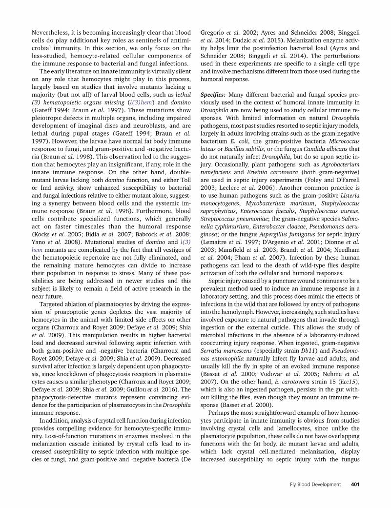
Nevertheless, it is becoming increasingly clear that bloodcells do play additional key roles as sentinels of antimi-crobial immunity. In this section, we only focus on theless-studied, hemocyte-related cellular components ofthe immune response to bacterial and fungal infections.
The early literature on innate immunity is virtually silenton any role that hemocytes might play in this process,largely based on studies that involve mutants lacking amajority (but not all) of larval blood cells, such as lethal(3) hematopoietic organs missing (l(3)hem) and domino(Gateff 1994; Braun et al. 1997). These mutations showpleiotropic defects in multiple organs, including impaireddevelopment of imaginal discs and neuroblasts, and arelethal during pupal stages (Gateff 1994; Braun et al.1997). However, the larvae have normal fat body immuneresponse to fungi, and gram-positive and -negative bacte-ria (Braun et al. 1998). This observation led to the sugges-tion that hemocytes play an insignificant, if any, role in theinnate immune response. On the other hand, double-mutant larvae lacking both domino function, and either Tollor Imd activity, show enhanced susceptibility to bacterialand fungal infections relative to either mutant alone, suggest-ing a synergy between blood cells and the systemic im-mune response (Braun et al. 1998). Furthermore, bloodcells contribute specialized functions, which generallyact on faster timescales than the humoral response(Kocks et al. 2005; Bidla et al. 2007; Babcock et al. 2008;Yano et al. 2008). Mutational studies of domino and l(3)hem mutants are complicated by the fact that all vestiges ofthe hematopoietic repertoire are not fully eliminated, andthe remaining mature hemocytes can divide to increasetheir population in response to stress. Many of these pos-sibilities are being addressed in newer studies and thissubject is likely to remain a field of active research in thenear future.
Targeted ablation of plasmatocytes by driving the expres-sion of proapoptotic genes depletes the vast majority ofhemocytes in the animal with limited side effects on otherorgans (Charroux and Royet 2009; Defaye et al. 2009; Shiaet al. 2009). This manipulation results in higher bacterialload and decreased survival following septic infection withboth gram-positive and -negative bacteria (Charroux andRoyet 2009; Defaye et al. 2009; Shia et al. 2009). Decreasedsurvival after infection is largely dependent upon phagocyto-sis, since knockdown of phagocytosis receptors in plasmato-cytes causes a similar phenotype (Charroux and Royet 2009;Defaye et al. 2009; Shia et al. 2009; Guillou et al. 2016). Thephagocytosis-defective mutants represent convincing evi-dence for the participation of plasmatocytes in the Drosophilaimmune response.
In addition, analysis of crystal cell functionduring infectionprovides compelling evidence for hemocyte-specific immu-nity. Loss-of-function mutations in enzymes involved in themelanization cascade initiated by crystal cells lead to in-creased susceptibility to septic infection with multiple spe-cies of fungi, and gram-positive and -negative bacteria (De
Gregorio et al. 2002; Ayres and Schneider 2008; Binggeliet al. 2014; Dudzic et al. 2015). Melanization enzyme activ-ity helps limit the postinfection bacterial load (Ayres andSchneider 2008; Binggeli et al. 2014). The perturbationsused in these experiments are specific to a single cell typeand involve mechanisms different from those used during thehumoral response.
Specifics: Many different bacterial and fungal species pre-viously used in the context of humoral innate immunity inDrosophila are now being used to study cellular immune re-sponses. With limited information on natural Drosophilapathogens, most past studies resorted to septic injury models,largely in adults involving strains such as the gram-negativebacterium E. coli, the gram-positive bacteria Micrococcusluteus or Bacillus subtilis, or the fungus Candida albicans thatdo not naturally infect Drosophila, but do so upon septic in-jury. Occasionally, plant pathogens such as Agrobacteriumtumefaciens and Erwinia carotovora (both gram-negative)are used in septic injury experiments (Foley and O’Farrell2003; Leclerc et al. 2006). Another common practice isto use human pathogens such as the gram-positive Listeriamonocytogenes, Mycobacterium marinum, Staphylococcussaprophyticus, Enterococcus faecalis, Staphylococcus aureus,Streptococcus pneumoniae; the gram-negative species Salmo-nella typhimurium, Enterobacter cloacae, Pseudomonas aeru-ginosa; or the fungus Aspergillus fumigatus for septic injury(Lemaitre et al. 1997; D’Argenio et al. 2001; Dionne et al.2003; Mansfield et al. 2003; Brandt et al. 2004; Needhamet al. 2004; Pham et al. 2007). Infection by these humanpathogens can lead to the death of wild-type flies despiteactivation of both the cellular and humoral responses.
Septic injury caused by a puncturewound continues to be aprevalent method used to induce an immune response in alaboratory setting, and this process does mimic the effects ofinfections in the wild that are followed by entry of pathogensinto thehemolymph.However, increasingly, such studies haveinvolved exposure to natural pathogens that invade throughingestion or the external cuticle. This allows the study ofmicrobial infections in the absence of a laboratory-inducedcooccurring injury response. When ingested, gram-negativeSerratia marcescens (especially strain Db11) and Pseudomo-nas entomophila naturally infect fly larvae and adults, andusually kill the fly in spite of an evoked immune response(Basset et al. 2000; Vodovar et al. 2005; Nehme et al.2007). On the other hand, E. carotovora strain 15 (Ecc15),which is also an ingested pathogen, persists in the gut with-out killing the flies, even though they mount an immune re-sponse (Basset et al. 2000).
Perhaps the most straightforward example of how hemoc-ytes participate in innate immunity is obvious from studiesinvolving crystal cells and lamellocytes, since unlike theplasmatocyte population, these cells do not have overlappingfunctions with the fat body. Bc mutant larvae and adults,which lack crystal cell-mediated melanization, displayincreased susceptibility to septic injury with the fungus
Fly Blood Development 401

Beauveria bassiana (Braun et al. 1998; Leclerc et al. 2006). Bcmutant animals also have increased susceptibility to thegram-negative bacteria A. tumefaciens and E. coli, and thegram-positive bacteria E. faecalis and M. luteus (Braun et al.1998; Leclerc et al. 2006). Furthermore, a double knockoutof PPO1 and PPO2 is more susceptible to septic injury withseveral species of fungi (C. albicans and A. fumigatus), gram-positive bacteria (S. aureus, Li. monocytogenes, S. saprophyticus,E. faecalis, and B. subtilis), and the gram-negative bacte-rium E. carotovora, but has only minor effects on survivalafter septic injury with several other gram-negative bacterialspecies (S. typhimurium and E. cloacae). These PPO1/PPO2double mutants are also more susceptible to natural infectionwith fungal species such as B. bassiana andMetarhizium ani-sopliae, which invade fly larvae through the cuticle. Thesemutants also carry a higher bacterial load after S. aureusseptic injury, implying that PO activity helps keep bacterialinfection in check (Binggeli et al. 2014). This crystal cell-mediated immune response to fungal and bacterial infectionis not related to AMP production, which remains unchangedin mutants incapable of melanization. Instead, PO-mediatedROS formation is the likely mechanism for this antimicrobialdefense system (Tang et al. 2006; Nam et al. 2012; Binggeliet al. 2014).
Mutations in proteases that activate PO activity have alsobeen used to study the role of melanization in defense againstmicrobes. For example, Hayan is the protease that activatesPPO1 and PPO2 through cleavage of these zymogens (Namet al. 2012). Microbes introduced during septic injury en-hance Hayan transcription in a Toll- and Imd-dependentmanner (Nam et al. 2012). Two other proteases that play arole in PPO1 and PPO2 activation are Melanization protease1 (MP1) and Melanization protease 2 (MP2, also known asPAE1 and Sp7). MP2 is specifically expressed in crystal cellsand is upregulated in response to infection (Castillejo-Lópezand Häcker 2005). Knockdown of MP2 enhances lethalityfollowing fungal infection (Tang et al. 2006). Likewise,MP2-deficient animals are very susceptible to S. typhimurium(gram-negative) and L. monocytogenes (gram-positive) bac-teria (Ayres and Schneider 2008).MP1- orMP2-deficient fliesbecome even more susceptible to infections when combinedwith mutations in the Toll or Imd pathways, suggesting thatthe systemic response from the fat body synergizes with thehemocyte-derived cellular response to provide full immunityagainst microbes.
Excessive melanization in flies deficient in Serpin27A(Spn27A), a negative regulator of PO function, results inpoorer survival outcomes than for wild-type flies when theseflies encounter septic infection by B. bassiana, E. coli, M.luteus, or A. fumigatus (De Gregorio et al. 2002). PO activityis strictly regulated in homeostasis by Spn27A to preventspontaneous melanization (De Gregorio et al. 2002;Ligoxygakis et al. 2002). When the need for active defenseagainst pathogen infection arises, the activated Toll pathwaydownregulates Spn27A, and this facilitates the PO-mediatedmelanization response (Ligoxygakis et al. 2002). These
results further demonstrate the interplay between the hu-moral and cellular response systems as an essential elementof innate immunity.
Anotheruniquepropertyof the cellular immuneresponse isexemplified by plasmatocyte function in combating opportu-nistic infection by bacteria or fungi present under normalculture conditions that donot negatively affect survival unlessthe immune system is compromised. Depletion of blood cellsreduces the animal’s ability to survive such invasions andnumerous bacteria are seen in the hemolymph (Braun et al.1998; Matova and Anderson 2006). Similarly, specific deple-tion of plasmatocytes through expression of proapoptoticgenes or mutation of the phagocytic receptor croquemort(crq) causes a decrease in survival to adulthood under normalculture conditions (Charroux and Royet 2009; Defaye et al.2009; Shia et al. 2009; Guillou et al. 2016). This phenotype isreversed either by growing flies in axenic (germ-free) condi-tions or by including antibiotics in the culture medium. Thisemphasizes the importance of the phagocytic response tobacteria that are frequently ingested in the typical cultureenvironment. This mechanism is especially important for in-fections by bacteria such as the gram-negative S. marcescens,which does not elicit a strong fat body immune response, andtherefore phagocytosis plays a dominant role in combatingthis pathogen (Nehme et al. 2007).
Depletion of plasmatocytes from either larvae or adultsincreases susceptibility to septic infection with pathogenssuch as S. aureus and E. faecalis (gram-positive), S. typhi-murium and P. entomophila (gram-negative), and C. albi-cans (fungus) (Jin et al. 2008; Charroux and Royet 2009;Defaye et al. 2009). When phagocytosis is blocked, eitherby injection of latex beads into the hemocoel or by muta-tions in eater, nimrod, crq, or Rac2, a similar decrease insurvival is seen after infection with S. aureus, M. luteus, E.faecalis, P. aeruginosa, and S. marascens (Kocks et al. 2005;Avet-Rochex et al. 2007; Nehme et al. 2007; Schneideret al. 2007; Jin et al. 2008; Charroux and Royet 2009;Defaye et al. 2009; Nehme et al. 2011; Guillou et al.2016; Bajgar and Dolezal 2018). Since blocking phagocy-tosis does not affect AMP induction in the fat body, thisemphasizes the importance of cellular immunity in resist-ing infection (Elrod-Erickson et al. 2000; Kocks et al. 2005;Charroux and Royet 2009). Indeed, P. aeruginosa directlyinhibits phagocytosis through injection of the virulencefactor ExoS toxin, a GAP domain-containing enzyme,which binds and inhibits Rac2 GTPase resulting in de-creased survival (Avet-Rochex et al. 2005, 2007). Whenconditions that lead to a loss of phagocytosis are combinedwith mutations in the Toll or Imd pathways, the resultinglarvae and adults display even greater susceptibility toseptic injury relative to either defect alone (Braun et al.1998; Elrod-Erickson et al. 2000; Nehme et al. 2007,2011; Charroux and Royet 2009; Defaye et al. 2009). Ad-ditionally, chemically synthesized Cecropin A, an AMP nor-mally secreted by the fat body after immune challenge,also enhances Eater binding to live bacteria (Chung and
402 U. Banerjee et al.

Kocks 2011). These observations point to a synergy be-tween the humoral and cellular immune responses thatprovides optimal resistance to infection.
During oral infection of larvae with Ecc15, hemocytesare necessary to induce the AMP Diptericin (Dpt) in the fatbody downstream of the Imd pathway (Basset et al. 2000;Foley and O’Farrell 2003; Dijkers and O’Farrell 2007;Charroux and Royet 2009; Shia et al. 2009). In this case,Calcineurin A1 is required in hemocytes to respond to anNO signal from the gut, which ultimately causes activationof the Imd pathway in the fat body and the transcription ofdownstream AMPs (Foley and O’Farrell 2003; Dijkers andO’Farrell 2007).
Both hemocytes and the fat body express Spatzle (Spz),a Toll ligand that is proteolytically cleaved to allow forToll pathway activation; however, upon bacterial infection,hemocytes, but not the fat body, upregulate Spz (Irving et al.2005). Hemocytes also upregulate PGRP-SA, a circulatingpattern recognition receptor upstream of the Toll pathwayin response to septic infection (Irving et al. 2005). Theseresults suggest that hemocytes play a role in promoting Tollpathway activation. However, several studies have been con-ducted that have combined septic injury with depletion ofhemocytes. The results using bacterial and fungal specieshave obtained different results. Most suggest no effect ofhemocytes on AMP production (Braun et al. 1998; Bassetet al. 2000; Matova and Anderson 2006; Charroux and Royet2009; Defaye et al. 2009). However, one study using severaldifferent strategies for hemocyte depletion as well as hemoc-yte-specific depletion of Spz concluded that blood cells areindeed required for the induction of Dpt and Drosomycin(Shia et al. 2009). Furthermore, blood cells have also beenshown to express Psidin (Phagocyte signaling impaired),which is specifically required for the induction of Defensinin the fat body following septic injury with E. coli (Brennanet al. 2007).
Unexpectedly, the lymph gland proper also plays a role inimmunity against microbial infection. Bacterial challenge(such as by E. coli or B. subtilis) causes loss of proper septatejunctions between the cells of the PSC (Khadilkar et al.2017). This causes increased differentiation of plasmato-cytes and crystal cells, and leads to premature dispersal ofthe lymph gland cells. Thus, disruption of PSC septate junc-tions during bacterial infection ultimately increases thenumber of circulating hemocytes in the larva that then con-tinue into the adult. This improves the survival of adult fliessubjected to E. coli and B. subtilis infection. While infectionleads to increased plasmatocytes, overexpression of Cora orNrxIV in the PSC reforms the septate junctions, and therebyinhibits the increase in differentiation. Interestingly, Toll orImd activation in the PSC also causes the perturbation ofseptate junctions between these cells in the absence of in-fection, and artificial activation of either the Toll or Imdpathways in the PSC leads to the breakdown of septatejunctions within the PSC and increased plasmatocyte differ-entiation. Disrupting septate junctions in larvae increases
the number of circulating hemocytes in adults. This im-proves the survival of adult flies after E. coli infection(Khadilkar et al. 2017).
As described for aseptic injury and wasp infestation,Upd3 is also activated in hemocytes in response to septicinjury (for example with the gram-negative bacteriumEcc15) and consequently leads to the activation of theDome/JAK/STAT pathway in multiple tissues, includingthe fat body, muscle, and gut (Agaisse et al. 2003;Chakrabarti et al. 2016; Yang and Hultmark 2017). Asexpected, knockdown of upd3 in plasmatocytes causes in-creased susceptibility to septic injury (Chakrabarti et al.2016). In the gut, activation of this pathway upon infectioncauses increased proliferation of intestinal stem cells,while in the fat body, active STAT cooperates with theImd pathway leading to expression of the stress-responseprotein Turandot A (TotA) and the C3 complement-like pro-tein Tep1 (Lagueux et al. 2000; Agaisse et al. 2003;Chakrabarti et al. 2016). Tep1–4 represent a set of thio-ester-containing proteins that act as opsonins and promotephagocytosis. Their loss renders flies more susceptible to in-fection (Stroschein-Stevenson et al. 2006; Dostálová et al.2017). This process is akin to the human acute-phase re-sponse to infection in which blood cell cytokine signalingcauses the liver to secrete stress-response proteins, such asthe opsonin C-reactive protein that promotes the phagocyto-sis of microbes [reviewed in Cray et al. (2009)]. During oralinfection with either Ecc15 or P. entomophila (both gram-negative), plasmatocytes migrate to the gut and secreteDpp, which is required for intestinal stem cell proliferationin response to infection (Ayyaz et al. 2015). These resultsindicate that hemocytes communicate with multiple tissuesto restore homeostasis and mitigate stress caused byinfection.
Development and the Stress Response: Two Sides ofthe Same Coin
As a core principle of homeostasis, any tissue that experi-ences stress is expected to react in a local or systemic fashionto bring its function back to normalcy. The brain, for ex-ample, will secrete hormones to mitigate the effects ofoxidative stress. However, signals responsible for the de-velopment of a specific tissue are typically distinct from theones that are sent out to indicate distress. In contrast,independent studies over the years suggest that myeloidprogenitors, at least in Drosophila, often make dual use ofthe same pathways during stress and homeostasis. It is notsurprising that differentiated myeloid cells are particularlysensitive to environmental and systemic cues, given theirinnate immune function as proverbial watchmen that sur-veil the body for signs of trouble and respond to situationsthat threaten homeostasis such as injury, tissue damage,infection, and disease. What is interesting and novel is therealization that it is the quiescent multipotent progenitors,largely confined within the hematopoietic organ, that
Fly Blood Development 403

directly sense and respond to stress. This response involvesa loss of quiescence as progenitors proliferate and differ-entiate, giving rise to hemocytes capable of stress mitiga-tion. The circulating cells also respond to stress, but it isless clear whether this is due to the rapid amplification ofexisting blood cells or a de novo hematopoietic response.
Dual use of the same pathway for development and thestress response is commonplace within the myeloid system.The Toll and JAK/STAT pathways, critically responsible forinnate immunity against bacteria, wasp eggs, aseptic injury,and epithelial tumors, are also important for blood celldifferentiation and survival (Harrison et al. 1995; Qiuet al. 1998; Sorrentino et al. 2002, 2004; Zettervall et al.2004; Matova and Anderson 2006; Senger et al. 2006;Krzemień et al. 2007; Makki et al. 2010; Minakhina et al.2011; Yang et al. 2015; Chakrabarti et al. 2016; Louradouret al. 2017). In the lymph gland, a balance between pro-genitors and differentiated cells is maintained during lar-val homeostasis. The pathways employed for maintenanceof the progenitor population are often adapted to alter thisbalance during stress. In addition to the use of well-char-acterized developmental pathways, more esoteric and evo-lutionarily ancient signaling molecules such as ROS, NO,adenosine, odorants, atmospheric gases, steroids, GABA,and amino acids are used as developmental signals, andreutilized as stress sensors.
We highlight some of the above strategies in this section.As mentioned earlier, the progenitor population physiolog-ically generates higher levels of ROS than are seen in mostother tissues during development, and fails to differentiate ifthis ROS is entirely scavenged. Thus, ROS are used as atrigger for differentiation during normal Drosophila hema-topoiesis. This is convenient since during oxidative stress,ROS levels rise and the progenitor population can rapidlydifferentiate to expand the mature hemocyte pool to miti-gate the effects of the oxidative stress (Owusu-Ansah andBanerjee 2009; Gao et al. 2013). As another example, ecdy-sone signaling initiates many of the transitions in the con-trolled growth of the larva, including that of the lymphgland, and is also essential for the dispersal and activationof lymph gland cells during pupariation (Sorrentino et al.2002; Regan et al. 2013). In addition, this same pathway isthen used to minimize the effects of wasp parasitization(Sorrentino et al. 2002).
An interesting aspect of dual use is seen for the function ofthe InR pathway, which is critical for progenitor maintenanceduring development. Loss of insulin signaling causes loss oftheprogenitor population through the actionof thePI3K/Akt/TOR pathways (Benmimoun et al. 2012; Dragojlovic-Munther and Martinez-Agosto 2012; Shim et al. 2012;Tokusumi et al. 2012). The same pathway combinationsenses a loss of nutrients, largely amino acids, as a stresssignal during starvation, and reacts in multiple ways tomount an immune response and also to utilize fat cells forsurvival (Dragojlovic-Munther and Martinez-Agosto 2012;Shim et al. 2012).
An unusual form of Notch signaling in the context ofhypoxia involves the formation of a Notch/Hif-a (Sima inDrosophila) complex that activates specialized target re-sponse elements (Mukherjee et al. 2011). This mechanismis utilized to maintain the integrity of crystal cells undernormoxic conditions. This is achieved by stabilizing Hif-ausing the normally high NO levels in crystal cells whenthe O2 levels are normal (Mukherjee et al. 2011). If, inaddition, the animal encounters hypoxia, or an imbalancebetween CO2 and O2, additional amounts of Hif-a are sta-bilized and this causes an increase in the crystal cell popu-lation (Mukherjee et al. 2011; Cho et al. 2018). Largevariations in O2 and CO2 concentrations are encounteredas the larva digs deep into food, particularly when attractedby yeast. By mechanisms not yet clear, the increased num-bers of crystal cells provide protection during hypoxia orother gaseous imbalances.
The Drosophila hematopoietic system receives sensory in-formation from the outside world and also reacts to it usingan elegant strategy of dual control (Dragojlovic-Munther andMartinez-Agosto 2012; Shim et al. 2012, 2013; Cho et al.2018). The progenitor population of the MZ is maintainedin its relatively quiescent state by a G protein-coupledGABA-B receptor (GABABR) system that creates a proper bal-ance between the levels of cytosolic and ER-stored Ca2+ ions.During homeostasis, proper calcium levels are maintainedby binding of the neurotransmitter GABA to its receptorexpressed in the cells of the MZ. Interestingly, the hemo-lymph of the fly contains significant levels of secreted GABA,similar to the blood serum of humans. The secretion of GABAinto the hemolymph is under the control of an olfactory re-ceptor that detects fruity odors. Thus, during homeostasis,appropriate levels of odorant ligands in the immediate vicin-ity of the larva maintain appropriate GABA levels in its circu-lating blood to signal favorable conditions for progenitormaintenance in the MZ during development. Strikingly, agenetically altered larva unable to smell, or a normal larvasuffering odor deprivation, has a very strong immune re-sponse due to a decrease in cytosolic calcium in the MZ ofthe lymph gland, which causes a loss of progenitor quies-cence and increased differentiation (Shim et al. 2013). Thus,the same Ca2+-dependent mechanism used during develop-ment to maintain progenitors is reused to overcome sensorystress by rapidly expanding the hematopoietic repertoire.
It is known that intrinsic, oxidative, infectious, systemic,and environmental stresses elicit strong reactions by thehuman myeloid system [reviewed in Muralidharan andMandrekar (2013)]. However, due to the complexity of thesystem, the lack of appropriate genetic tools, and the in-volvement of multiple organs in the process, it is extremelydifficult to establish the mechanistic steps that underlie thecausal link between a stress and the resulting myeloid re-sponse. The Drosophila studies suggest that conserved de-velopmental and stress-related pathways may be moreinterrelated than previously appreciated. We hope thatsuch studies will help model the molecular mechanisms
404 U. Banerjee et al.

underlying the human myeloid system during normal de-velopment and stress.
Final Words
The classical biological literature on invertebrate blood cellshas created a solid foundation on which current studies onmolecular genetic mechanisms rest. The Dipteran lymphgland was first identified and named as such by Weismannin the early 1860s [described in Weismann (1864)], but itsfunction remained controversial over a century later [dis-cussed in Shrestha and Gateff (1982)]. The blood cell typesin Drosophila, and aspects of their development and function,including immune defense against wasp parasitization, wereall very carefully cataloged by the venerable team work ofTahir M. Rizki and Rose M. Rizki. T. Rizki received his earlyeducation in Afghan-India and, during his career spanningwell over three decades, mostly at the University of Michigan,he published 129 seminal papers, more than one-half of whichare coauthored with Rose Rizki. It is fairly clear that Rizki andRizki are the founders of this field, as is Elisabeth Gateff, along-time former Director of the Max Planck Institute inMainz, Germany, who analyzed the ultrastructure of bloodcells and isolated a large collection of mutants that affect thehematopoietic system, ushering the field into the modern era.In this context, it is noteworthy to mention Istvan Ando at theHungarian Academy of Sciences in Szeged and Dan Hultmarkat Umea, Sweden,who during the early days of research in thisfield, collaboratively developed antibody markers and identi-fied the corresponding genetic loci respectively, and facilitatedall future mechanistic genetic analysis within the field. By nomeans is this meant to be a comprehensive list, even from ahistorical perspective. The combined effort of many laborato-ries has now led to a reasonably detailed picture of the hema-topoietic process at different developmental stages. Theauthors of this chapter have attempted to include as muchinformation as possible within the constraints of a publication.Weapologize in advance to all thosewhose important researchwas not included in this chapter.
The virtues of using Drosophila as a model system to pro-vide hints about human diseases have been eloquently statedelsewhere. This review has a narrower goal: to present acompilation of results that explore the full potential of theDrosophilamodel system in providing a bona fide tool kit thatwill help understand human blood cell development andfunction at a molecular level. Many new insights, concepts,and pathways have already emerged from molecular geneticanalyses of Drosophila hematopoiesis. As with most otherstudies that have successfully exploited genetic dissectionas a strategy to understand development, the analogy andsimilarity of Drosophila blood to the mammalian hematopoi-etic system is expected to be at the level of fundamentalprinciples. The logic and algorithm that controls the system,as well as the conserved molecular networks that support thedevelopmental strategies, are likely to be more importantthan the exact matches or lack thereof in cell types that have
been selected during evolution due to their specific adaptivefunctions. For example, it is no doubt convenient and usefulto point to the similarities of plasmatocytes to macrophages;of crystal cells to platelets and granulocytes; or of lamello-cytes to giant cells. However, themore useful analogy is in theconservation of specific transcription factors as well as signal-ing pathways involved in the construction of a hematopoieticsystem. In this review alone, we have explored dozens of suchconserved signaling components and specific transcriptionfactors between flies and humans. Also important is the con-servation of function, such as phagocytosis, innate immunity,wound healing, gas sensing, and engulfment of large parti-cles, that is shared with the mammalian blood system. A fullunderstanding of the Drosophila hematopoietic system is stillin its infancy, but given the rapid progress in recent years,we expect this system to facilitate breakthroughs in ourunderstanding of myeloid cell development and function inhumans.
Acknowledgments
We thank Volker Hartenstein for his advice and suggestions,as many of the illustrations in this manuscript are adapta-tions from his work; Carl Thummel and Trudi Schupbach fortheir patience and support during the delays in ourpreparation of this chapter; the past and present membersof the Banerjee laboratory; FlyBase; the Bloomington Dro-sophila Stock Center; the Vienna Drosophila Resource Cen-ter; the Kyoto Stock Center, and the fly community. Weacknowledge the help of the Broad Stem Cell Research Cen-ter (BSCRC) and the Department of Molecular, Cell, andDevelopmental Biology BSCRC Core Facilities in Microscopy.We apologize to any researcher whose work we failed tocite. U.B. is supported by National Institutes of Health grantsR01 HL-067395 and R01 CA-217608; J.R.G. by Ruth L.Kirschstein National Research Service Award numberT32HL69766 and UPLIFT (UCLA Postdocs’ Longitudinal In-vestment in Faculty) Award number K12GM106996; L.M.G.by Ruth L. Kirschstein Institutional National ResearchService Award number T32CA009056; and C.M.S. byRuth L. Kirschstein National Research Service Award numberT32HL863458.
Literature Cited
Abel, T., A. M. Michelson, and T. Maniatis, 1993 A DrosophilaGATA family member that binds to Adh regulatory sequencesis expressed in the developing fat body. Development 119:623–633.
Agaisse, H., U. M. Petersen, M. Boutros, B. Mathey-Prevot, and N.Perrimon, 2003 Signaling role of hemocytes in DrosophilaJAK/STAT-dependent response to septic injury. Dev. Cell 5:441–450. https://doi.org/10.1016/S1534-5807(03)00244-2
Alfonso, T. B., and B. W. Jones, 2002 gcm2 promotes glial celldifferentiation and is required with glial cells missing for mac-rophage development in Drosophila. Dev. Biol. 248: 369–383.https://doi.org/10.1006/dbio.2002.0740
Fly Blood Development 405

Amcheslavsky, A., J. Jiang, and Y. T. Ip, 2009 Tissue damage-in-duced intestinal stem cell division in Drosophila. Cell Stem Cell4: 49–61. https://doi.org/10.1016/j.stem.2008.10.016
Anderl, I., L. Vesala, T. O. Ihalainen, L. M. Vanha-Aho, I. Andoet al., 2016 Transdifferentiation and proliferation in two dis-tinct hemocyte lineages in Drosophila melanogaster larvae afterwasp infection. PLoS Pathog. 12: e1005746. https://doi.org/10.1371/journal.ppat.1005746
Arendt, D., K. Tessmar-Raible, H. Snyman, A. W. Dorresteijn, and J.Wittbrodt, 2004 Ciliary photoreceptors with a vertebrate-typeopsin in an invertebrate brain. Science 306: 869–871. https://doi.org/10.1126/science.1099955
Asha, H., I. Nagy, G. Kovacs, D. Stetson, I. Ando et al.,2003 Analysis of Ras-induced overproliferation in Drosophilahemocytes. Genetics 163: 203–215.
Avet-Rochex, A., E. Bergeret, I. Attree, M. Meister, and M. O. Fau-varque, 2005 Suppression of Drosophila cellular immunity bydirected expression of the ExoS toxin GAP domain of Pseudo-monas aeruginosa. Cell. Microbiol. 7: 799–810. https://doi.org/10.1111/j.1462-5822.2005.00512.x
Avet-Rochex, A., J. Perrin, E. Bergeret, and M. O. Fauvarque,2007 Rac2 is a major actor of Drosophila resistance to Pseudo-monas aeruginosa acting in phagocytic cells. Genes Cells 12:1193–1204. https://doi.org/10.1111/j.1365-2443.2007.01121.x
Avet-Rochex, A., K. Boyer, C. Polesello, V. Gobert, D. Osman et al.,2010 An in vivo RNA interference screen identifies gene net-works controlling Drosophila melanogaster blood cell homeo-stasis. BMC Dev. Biol. 10: 65. https://doi.org/10.1186/1471-213X-10-65
Ayres, J. S., and D. S. Schneider, 2008 A signaling protease re-quired for melanization in Drosophila affects resistance and tol-erance of infections. PLoS Biol. 6: 2764–2773. https://doi.org/10.1371/journal.pbio.0060305
Ayyaz, A., H. Li, and H. Jasper, 2015 Haemocytes control stem cellactivity in the Drosophila intestine. Nat. Cell Biol. 17: 736–748.https://doi.org/10.1038/ncb3174
Azpiazu, N., P. A. Lawrence, J. P. Vincent, and M. Frasch,1996 Segmentation and specification of the Drosophila meso-derm. Genes Dev. 10: 3183–3194. https://doi.org/10.1101/gad.10.24.3183
Babcock, D. T., A. R. Brock, G. S. Fish, Y. Wang, L. Perrin et al.,2008 Circulating blood cells function as a surveillance systemfor damaged tissue in Drosophila larvae. Proc. Natl. Acad. Sci. USA105: 10017–10022. https://doi.org/10.1073/pnas.0709951105
Bajgar, A., and T. Dolezal, 2018 Extracellular adenosine modulateshost-pathogen interactions through regulation of systemic metab-olism during immune response in Drosophila. PLoS Pathog. 14:e1007022. https://doi.org/10.1371/journal.ppat.1007022
Bajgar, A., K. Kucerova, L. Jonatova, A. Tomcala, I. Schneedorfer-ova et al., 2015 Extracellular adenosine mediates a systemicmetabolic switch during immune response. PLoS Biol. 13:e1002135. https://doi.org/10.1371/journal.pbio.1002135
Baldeosingh, R., H. Gao, X. Wu, and N. Fossett, 2018 Hedgehogsignaling from the posterior signaling center maintainsU-shaped expression and a prohemocyte population in Drosoph-ila. Dev. Biol. 441: 132–145. https://doi.org/10.1016/j.ydbio.2018.06.020
Basset, A., R. S. Khush, A. Braun, L. Gardan, F. Boccard et al.,2000 The phytopathogenic bacteria Erwinia carotovora infectsDrosophila and activates an immune response. Proc. Natl. Acad. Sci.USA 97: 3376–3381. https://doi.org/10.1073/pnas.97.7.3376
Bataille, L., B. Auge, G. Ferjoux, M. Haenlin, and L. Waltzer,2005 Resolving embryonic blood cell fate choice in Drosoph-ila: interplay of GCM and RUNX factors. Development 132:4635–4644. https://doi.org/10.1242/dev.02034
Bate, M., and E. Rushton, 1993 Myogenesis and muscle pattern-ing in Drosophila. C. R. Acad. Sci. III 316: 1047–1061.
Beiman, M., B. Z. Shilo, and T. Volk, 1996 Heartless, a DrosophilaFGF receptor homolog, is essential for cell migration and estab-lishment of several mesodermal lineages. Genes Dev. 10: 2993–3002. https://doi.org/10.1101/gad.10.23.2993
Belenkaya, T. Y., C. Han, D. Yan, R. J. Opoka, M. Khodoun et al.,2004 Drosophila Dpp morphogen movement is independent ofdynamin-mediated endocytosis but regulated by the glypicanmembers of heparan sulfate proteoglycans. Cell 119: 231–244.https://doi.org/10.1016/j.cell.2004.09.031
Benmimoun, B., C. Polesello, L. Waltzer, and M. Haenlin,2012 Dual role for Insulin/TOR signaling in the control ofhematopoietic progenitor maintenance in Drosophila. Develop-ment 139: 1713–1717. https://doi.org/10.1242/dev.080259
Benmimoun, B., C. Polesello, M. Haenlin, and L. Waltzer,2015 The EBF transcription factor Collier directly promotesDrosophila blood cell progenitor maintenance independentlyof the niche. Proc. Natl. Acad. Sci. USA 112: 9052–9057.https://doi.org/10.1073/pnas.1423967112
Bergmann, A., J. Agapite, K. McCall, and H. Steller, 1998 TheDrosophila gene hid is a direct molecular target of Ras-depen-dent survival signaling. Cell 95: 331–341. https://doi.org/10.1016/S0092-8674(00)81765-1
Bernardoni, R., V. Vivancos, and A. Giangrande, 1997 Glide/gcmis expressed and required in the scavenger cell lineage. Dev.Biol. 191: 118–130. https://doi.org/10.1006/dbio.1997.8702
Bidla, G., M. Lindgren, U. Theopold, and M. S. Dushay,2005 Hemolymph coagulation and phenoloxidase in Drosoph-ila larvae. Dev. Comp. Immunol. 29: 669–679. https://doi.org/10.1016/j.dci.2004.11.007
Bidla, G., M. S. Dushay, and U. Theopold, 2007 Crystal cell rup-ture after injury in Drosophila requires the JNK pathway, smallGTPases and the TNF homolog Eiger. J. Cell Sci. 120: 1209–1215. https://doi.org/10.1242/jcs.03420
Binggeli, O., C. Neyen, M. Poidevin, and B. Lemaitre,2014 Prophenoloxidase activation is required for survival tomicrobial infections in Drosophila. PLoS Pathog. 10: e1004067.https://doi.org/10.1371/journal.ppat.1004067
Bitra, K., R. J. Suderman, and M. R. Strand, 2012 PolydnavirusAnk proteins bind NF-kappaB homodimers and inhibit process-ing of Relish. PLoS Pathog. 8: e1002722. https://doi.org/10.1371/journal.ppat.1002722
Bodmer, R., 1993 The gene tinman is required for specification ofthe heart and visceral muscles in Drosophila. Development 118:719–729.
Bodmer, R., L. Y. Jan, and Y. N. Jan, 1990 A new homeobox-containing gene, msh-2, is transiently expressed early during me-soderm formation of Drosophila. Development 110: 661–669.
Borkowski, O. M., N. H. Brown, andM. Bate, 1995 Anterior-posteriorsubdivision and the diversification of the mesoderm in Drosophila.Development 121: 4183–4193.
Brandt, S. M., M. S. Dionne, R. S. Khush, L. N. Pham, T. J. Vigdalet al., 2004 Secreted bacterial effectors and host-producedEiger/TNF drive death in a Salmonella-infected fruit fly. PLoSBiol. 2: e418. https://doi.org/10.1371/journal.pbio.0020418
Braun, A., B. Lemaitre, R. Lanot, D. Zachary, and M. Meister,1997 Drosophila immunity: analysis of larval hemocytes byP-element-mediated enhancer trap. Genetics 147: 623–634.
Braun, A., J. A. Hoffmann, and M. Meister, 1998 Analysis of theDrosophila host defense in domino mutant larvae, which aredevoid of hemocytes. Proc. Natl. Acad. Sci. USA 95: 14337–14342. https://doi.org/10.1073/pnas.95.24.14337
Brennan, C. A., and K. V. Anderson, 2004 Drosophila: the geneticsof innate immune recognition and response. Annu. Rev. Immu-nol. 22: 457–483. https://doi.org/10.1146/annurev.immunol.22.012703.104626
Brennan, C. A., J. R. Delaney, D. S. Schneider, and K. V. Anderson,2007 Psidin is required in Drosophila blood cells for both
406 U. Banerjee et al.
phagocytic degradation and immune activation of the fatbody. Curr. Biol. 17: 67–72. https://doi.org/10.1016/j.cub.2006.11.026
Brennan, J. J., J. L. Messerschmidt, L. M. Williams, B. J. Matthews,M. Reynoso et al., 2017 Sea anemone model has a single Toll-like receptor that can function in pathogen detection, NF-kBsignal transduction, and development. Proc. Natl. Acad. Sci.USA 114: E10122–E10131. https://doi.org/10.1073/pnas.1711530114
Bretscher, A. J., V. Honti, O. Binggeli, O. Burri, M. Poidevin et al.,2015 The Nimrod transmembrane receptor Eater is requiredfor hemocyte attachment to the sessile compartment in Dro-sophila melanogaster. Biol. Open 4: 355–363. https://doi.org/10.1242/bio.201410595
Brückner, K., L. Kockel, P. Duchek, C. M. Luque, P. Rorth et al.,2004 The PDGF/VEGF receptor controls blood cell survivalin Drosophila. Dev. Cell 7: 73–84. https://doi.org/10.1016/j.devcel.2004.06.007
Bunt, S., C. Hooley, N. Hu, C. Scahill, H. Weavers et al.,2010 Hemocyte-secreted type IV collagen enhances BMP signal-ing to guide renal tubule morphogenesis in Drosophila. Dev. Cell19: 296–306. https://doi.org/10.1016/j.devcel.2010.07.019
Cantor, A. B., and S. H. Orkin, 2005 Coregulation of GATA factorsby the Friend of GATA (FOG) family of multitype zinc fingerproteins. Semin. Cell Dev. Biol. 16: 117–128. https://doi.org/10.1016/j.semcdb.2004.10.006
Carton, Y., and A. Nappi, 1991 The Drosophila immune-reactionand the parasitoid capacity to evade it - genetic and coevolu-tionary aspects. Acta Oecologica-Int. J. Ecol. 12: 89–104.
Carton, Y., M. Bouletreau, J. Alphen, and J. van Lenteren,1986 The Drosophila parasitic wasps, pp. 348–394 in The Ge-netics and Biology of Drosophila, Vol. III, edited by M. Ashburner,H. L. Carson, and J. N. Thompson. Academic Press, London.
Castillejo-López, C., and U. Häcker, 2005 The serine protease Sp7is expressed in blood cells and regulates the melanization re-action in Drosophila. Biochem. Biophys. Res. Commun. 338:1075–1082. https://doi.org/10.1016/j.bbrc.2005.10.042
Cattenoz, P. B., A. Popkova, T. D. Southall, G. Aiello, A. H. Brandet al., 2016 Functional conservation of the Glide/Gcm regula-tory network controlling glia, hemocyte, and tendon cell differ-entiation in Drosophila. Genetics 202: 191–219. https://doi.org/10.1534/genetics.115.182154
Cerenius, L., B. L. Lee, and K. Soderhall, 2008 The proPO-sys-tem: pros and cons for its role in invertebrate immunity.Trends Immunol. 29: 263–271. https://doi.org/10.1016/j.it.2008.02.009
Chakrabarti, S., J. P. Dudzic, X. Li, E. J. Collas, J. P. Boquete et al.,2016 Remote control of intestinal stem cell activity by haemocytesin Drosophila. PLoS Genet. 12: e1006089. https://doi.org/10.1371/journal.pgen.1006089
Charroux, B., and J. Royet, 2009 Elimination of plasmatocytes bytargeted apoptosis reveals their role in multiple aspects of theDrosophila immune response. Proc. Natl. Acad. Sci. USA 106:9797–9802. https://doi.org/10.1073/pnas.0903971106
Chatterjee, M., and Y. T. Ip, 2009 Pathogenic stimulation of in-testinal stem cell response in Drosophila. J. Cell. Physiol. 220:664–671. https://doi.org/10.1002/jcp.21808
Chen, M. J., T. Yokomizo, B. M. Zeigler, E. Dzierzak, and N. A.Speck, 2009 Runx1 is required for the endothelial to haema-topoietic cell transition but not thereafter. Nature 457: 887–891.https://doi.org/10.1038/nature07619
Cho, B., C. M. Spratford, S. Yoon, N. Cha, U. Banerjee et al.,2018 Systemic control of immune cell development by integratedcarbon dioxide and hypoxia chemosensation in Drosophila. Nat.Commun. 9: 2679. https://doi.org/10.1038/s41467-018-04990-3
Cho, N. K., L. Keyes, E. Johnson, J. Heller, L. Ryner et al.,2002 Developmental control of blood cell migration by the
Drosophila VEGF pathway. Cell 108: 865–876. https://doi.org/10.1016/S0092-8674(02)00676-1
Chung, Y. S., and C. Kocks, 2011 Recognition of pathogenic mi-crobes by the Drosophila phagocytic pattern recognition recep-tor Eater. J. Biol. Chem. 286: 26524–26532. https://doi.org/10.1074/jbc.M110.214007
Colinet, D., A. Schmitz, D. Depoix, D. Crochard, and M. Poirie,2007 Convergent use of RhoGAP toxins by eukaryotic para-sites and bacterial pathogens. PLoS Pathog. 3: e203. https://doi.org/10.1371/journal.ppat.0030203
Colinet, D., A. Dubuffet, D. Cazes, S. Moreau, J. M. Drezen et al.,2009 A serpin from the parasitoid wasp Leptopilina boularditargets the Drosophila phenoloxidase cascade. Dev. Comp. Immu-nol. 33: 681–689. https://doi.org/10.1016/j.dci.2008.11.013
Colinet, D., D. Cazes, M. Belghazi, J. L. Gatti, and M. Poirie,2011 Extracellular superoxide dismutase in insects: character-ization, function, and interspecific variation in parasitoid waspvenom. J. Biol. Chem. 286: 40110–40121. https://doi.org/10.1074/jbc.M111.288845
Colinet, D., E. Deleury, C. Anselme, D. Cazes, J. Poulain et al.,2013 Extensive inter- and intraspecific venom variation inclosely related parasites targeting the same host: the case ofLeptopilina parasitoids of Drosophila. Insect Biochem. Mol. Biol.43: 601–611. https://doi.org/10.1016/j.ibmb.2013.03.010
Cooper, E. L., 1976 Evolution of blood cells. Ann. Immunol.(Paris) 127: 817–825.
Cordero, J. B., J. P. Macagno, R. K. Stefanatos, K. E. Strathdee, R. L.Cagan et al., 2010 Oncogenic Ras diverts a host TNF tumorsuppressor activity into tumor promoter. Dev. Cell 18: 999–1011. https://doi.org/10.1016/j.devcel.2010.05.014
Cray, C., J. Zaias, and N. H. Altman, 2009 Acute phase response inanimals: a review. Comp. Med. 59: 517–526.
Cripps, R. M., B. L. Black, B. Zhao, C. L. Lien, R. A. Schulz et al.,1998 The myogenic regulatory gene Mef2 is a direct target fortranscriptional activation by Twist during Drosophila myogenesis.Genes Dev. 12: 422–434. https://doi.org/10.1101/gad.12.3.422
Crozatier, M., J. M. Ubeda, A. Vincent, and M. Meister,2004 Cellular immune response to parasitization in Drosoph-ila requires the EBF orthologue collier. PLoS Biol. 2: E196.https://doi.org/10.1371/journal.pbio.0020196
Cuttell, L., A. Vaughan, E. Silva, C. J. Escaron, M. Lavine et al.,2008 Undertaker, a Drosophila Junctophilin, links Draper-me-diated phagocytosis and calcium homeostasis. Cell 135: 524–534. https://doi.org/10.1016/j.cell.2008.08.033
D’Argenio, D. A., L. A. Gallagher, C. A. Berg, and C. Manoil,2001 Drosophila as a model host for Pseudomonas aeruginosainfection. J. Bacteriol. 183: 1466–1471. https://doi.org/10.1128/JB.183.4.1466-1471.2001
Defaye, A., I. Evans, M. Crozatier, W. Wood, B. Lemaitre et al.,2009 Genetic ablation of Drosophila phagocytes reveals theircontribution to both development and resistance to bacterialinfection. J. Innate Immun. 1: 322–334. https://doi.org/10.1159/000210264
De Gregorio, E., S. J. Han, W. J. Lee, M. J. Baek, T. Osaki et al.,2002 An immune-responsive Serpin regulates the melaniza-tion cascade in Drosophila. Dev. Cell 3: 581–592. https://doi.org/10.1016/S1534-5807(02)00267-8
De Sutter, D., and M. Buscema, 1977 Isolation of a highly purearcheocyte fraction from the fresh-water sponge Ephydatia flu-viatilis. Wilehm Roux Arch Dev Biol 183: 149–153. https://doi.org/10.1007/BF00848784
De Sutter, D., and G. Van de Vyver, 1977 Aggregative propertiesof different cell types of the fresh-water sponge Ephydatia flu-viatilis isolated on ficoll gradients. Wilehm Roux Arch Dev Biol181: 151–161. https://doi.org/10.1007/BF00848439
Dey, N. S., P. Ramesh, M. Chugh, S. Mandal, and L. Mandal,2016 Dpp dependent Hematopoietic stem cells give rise to
Fly Blood Development 407
Hh dependent blood progenitors in larval lymph gland of Dro-sophila. Elife 5: e18295. https://doi.org/10.7554/eLife.18295
Dijkers, P. F., and P. H. O’Farrell, 2007 Drosophila calcineurinpromotes induction of innate immune responses. Curr. Biol.17: 2087–2093. https://doi.org/10.1016/j.cub.2007.11.001
Dionne, M. S., N. Ghori, and D. S. Schneider, 2003 Drosophilamelanogaster is a genetically tractable model host for Mycobac-terium marinum. Infect. Immun. 71: 3540–3550. https://doi.org/10.1128/IAI.71.6.3540-3550.2003
Dolezal, T., E. Dolezelova, M. Zurovec, and P. J. Bryant, 2005 Arole for adenosine deaminase in Drosophila larval development.PLoS Biol. 3: e201. https://doi.org/10.1371/journal.pbio.0030201
Dostálová, A., S. Rommelaere, M. Poidevin, and B. Lemaitre,2017 Thioester-containing proteins regulate the Toll pathwayand play a role in Drosophila defence against microbial patho-gens and parasitoid wasps. BMC Biol. 15: 79. https://doi.org/10.1186/s12915-017-0408-0
Doumpas, N., G. Jekely, and A. A. Teleman, 2013 Wnt6 is re-quired for maxillary palp formation in Drosophila. BMC Biol.11: 104 [corrigends: BMC Biol. 13: 100 (2015)]. https://doi.org/10.1186/1741-7007-11-104
Dragojlovic-Munther, M., and J. A. Martinez-Agosto, 2012 Multifacetedroles of PTEN and TSC orchestrate growth and differentiation ofDrosophila blood progenitors. Development 139: 3752–3763.https://doi.org/10.1242/dev.074203
Dragojlovic-Munther, M., and J. A. Martinez-Agosto, 2013 Extracellularmatrix-modulated heartless signaling in Drosophila blood progen-itors regulates their differentiation via a Ras/ETS/FOG pathwayand target of rapamycin function. Dev. Biol. 384: 313–330. https://doi.org/10.1016/j.ydbio.2013.04.004
Dudzic, J. P., S. Kondo, R. Ueda, C. M. Bergman, and B. Lemaitre,2015 Drosophila innate immunity: regional and functionalspecialization of prophenoloxidases. BMC Biol. 13: 81. https://doi.org/10.1186/s12915-015-0193-6
Duvic, B., J. A. Hoffmann, M. Meister, and J. Royet, 2002 Notchsignaling controls lineage specification during Drosophila larvalhematopoiesis. Curr. Biol. 12: 1923–1927. https://doi.org/10.1016/S0960-9822(02)01297-6
Dzierzak, E., and N. A. Speck, 2008 Of lineage and legacy: thedevelopment of mammalian hematopoietic stem cells. Nat. Im-munol. 9: 129–136. https://doi.org/10.1038/ni1560
Eastham, L. E. S., 1930 The formation of germ layers in insects.Biol. Rev. Camb. Philos. Soc. 5: 1–29. https://doi.org/10.1111/j.1469-185X.1930.tb00891.x
Elrod-Erickson, M., S. Mishra, and D. Schneider, 2000 Interactionsbetween the cellular and humoral immune responses in Dro-sophila. Curr. Biol. 10: 781–784. https://doi.org/10.1016/S0960-9822(00)00569-8
Ema, M., and J. Rossant, 2003 Cell fate decisions in early bloodvessel formation. Trends Cardiovasc. Med. 13: 254–259. https://doi.org/10.1016/S1050-1738(03)00105-1
Ernst, O. P., D. T. Lodowski, M. Elstner, P. Hegemann, L. S. Brownet al., 2014 Microbial and animal rhodopsins: structures, func-tions, and molecular mechanisms. Chem. Rev. 114: 126–163.https://doi.org/10.1021/cr4003769
Evans, C. J., V. Hartenstein, and U. Banerjee, 2003 Thicker thanblood: conserved mechanisms in Drosophila and vertebrate he-matopoiesis. Dev. Cell 5: 673–690. https://doi.org/10.1016/S1534-5807(03)00335-6
Evans, C. J., J. M. Olson, K. T. Ngo, E. Kim, N. E. Lee et al.,2009 G-TRACE: rapid Gal4-based cell lineage analysis in Dro-sophila. Nat. Methods 6: 603–605. https://doi.org/10.1038/nmeth.1356
Evans, C. J., T. Liu, and U. Banerjee, 2014 Drosophila hematopoi-esis: markers and methods for molecular genetic analysis.Methods 68: 242–251. https://doi.org/10.1016/j.ymeth.2014.02.038
Evans, I. R., F. S. Rodrigues, E. L. Armitage, and W. Wood,2015 Draper/CED-1 mediates an ancient damage responseto control inflammatory blood cell migration in vivo. Curr. Biol.25: 1606–1612. https://doi.org/10.1016/j.cub.2015.04.037
Evgenov, O. V., P. Pacher, P. M. Schmidt, G. Hasko, H. H. Schmidtet al., 2006 NO-independent stimulators and activators of solu-ble guanylate cyclase: discovery and therapeutic potential. Nat.Rev. Drug Discov. 5: 755–768. https://doi.org/10.1038/nrd2038
Feinberg, E. H., M. K. Vanhoven, A. Bendesky, G. Wang, R. D. Fetteret al., 2008 GFP reconstitution across synaptic partners(GRASP) defines cell contacts and synapses in living nervoussystems. Neuron 57: 353–363. https://doi.org/10.1016/j.neuron.2007.11.030
Ferguson, G. B., and J. A. Martinez-Agosto, 2014 Yorkie and Scal-loped signaling regulates Notch-dependent lineage specificationduring Drosophila hematopoiesis. Curr. Biol. 24: 2665–2672.https://doi.org/10.1016/j.cub.2014.09.081
Ferguson, G. B., and J. A. Martinez-Agosto, 2017 The TEAD fam-ily transcription factor Scalloped regulates blood progenitormaintenance and proliferation in Drosophila through PDGF/VEGFR receptor (Pvr) signaling. Dev. Biol. 425: 21–32.https://doi.org/10.1016/j.ydbio.2017.03.016
Ferjoux, G., B. Auge, K. Boyer, M. Haenlin, and L. Waltzer, 2007 AGATA/RUNX cis-regulatory module couples Drosophila bloodcell commitment and differentiation into crystal cells. Dev. Biol.305: 726–734. https://doi.org/10.1016/j.ydbio.2007.03.010
Fessler, J. H., and L. I. Fessler, 1989 Drosophila extracellular ma-trix. Annu. Rev. Cell Biol. 5: 309–339. https://doi.org/10.1146/annurev.cb.05.110189.001521
Flaherty, M. S., P. Salis, C. J. Evans, L. A. Ekas, A. Marouf et al.,2010 chinmo is a functional effector of the JAK/STAT pathwaythat regulates eye development, tumor formation, and stem cellself-renewal in Drosophila. Dev. Cell 18: 556–568. https://doi.org/10.1016/j.devcel.2010.02.006
Fogarty, C. E., N. Diwanji, J. L. Lindblad, M. Tare, A. Amcheslavskyet al., 2016 Extracellular reactive oxygen species drive apopto-sis-induced proliferation via Drosophila macrophages. Curr.Biol. 26: 575–584. https://doi.org/10.1016/j.cub.2015.12.064
Fogerty, F. J., L. I. Fessler, T. A. Bunch, Y. Yaron, C. G. Parker et al.,1994 Tiggrin, a novel Drosophila extracellular matrix proteinthat functions as a ligand for Drosophila alpha PS2 beta PSintegrins. Development 120: 1747–1758.
Foley, E., and P. H. O’Farrell, 2003 Nitric oxide contributes toinduction of innate immune responses to gram-negative bacteriain Drosophila. Genes Dev. 17: 115–125. https://doi.org/10.1101/gad.1018503
Fossett, N., S. G. Tevosian, K. Gajewski, Q. Zhang, S. H. Orkin et al.,2001 The Friend of GATA proteins U-shaped, FOG-1, andFOG-2 function as negative regulators of blood, heart, and eyedevelopment in Drosophila. Proc. Natl. Acad. Sci. USA 98:7342–7347. https://doi.org/10.1073/pnas.131215798
Fossett, N., K. Hyman, K. Gajewski, S. H. Orkin, and R. A. Schulz,2003 Combinatorial interactions of serpent, lozenge, andU-shaped regulate crystal cell lineage commitment during Dro-sophila hematopoiesis. Proc. Natl. Acad. Sci. USA 100: 11451–11456. https://doi.org/10.1073/pnas.1635050100
Franc, N. C., J. L. Dimarcq, M. Lagueux, J. Hoffmann, and R. A.Ezekowitz, 1996 Croquemort, a novel Drosophila hemocyte/macrophage receptor that recognizes apoptotic cells. Immunity4: 431–443. https://doi.org/10.1016/S1074-7613(00)80410-0
Frasch, M., 1995 Induction of visceral and cardiac mesoderm byectodermal Dpp in the early Drosophila embryo. Nature 374:464–467. https://doi.org/10.1038/374464a0
Freeman, M. R., J. Delrow, J. Kim, E. Johnson, and C. Q. Doe,2003 Unwrapping glial biology: Gcm target genes regulatingglial development, diversification, and function. Neuron 38:567–580. https://doi.org/10.1016/S0896-6273(03)00289-7
408 U. Banerjee et al.

Fujiwara, Y., A. N. Chang, A. M. Williams, and S. H. Orkin,2004 Functional overlap of GATA-1 and GATA-2 in primitivehematopoietic development. Blood 103: 583–585. https://doi.org/10.1182/blood-2003-08-2870
Fuller, M. T., and A. C. Spradling, 2007 Male and female Dro-sophila germline stem cells: two versions of immortality. Science316: 402–404. https://doi.org/10.1126/science.1140861
Gajewski, K. M., R. P. Sorrentino, J. H. Lee, Q. Zhang, M. Russellet al., 2007 Identification of a crystal cell-specific enhancer ofthe black cells prophenoloxidase gene in Drosophila. Genesis45: 200–207. https://doi.org/10.1002/dvg.20285
Galko, M. J., and M. A. Krasnow, 2004 Cellular and genetic anal-ysis of wound healing in Drosophila larvae. PLoS Biol. 2: E239.https://doi.org/10.1371/journal.pbio.0020239
Ganesan, S., K. Aggarwal, N. Paquette, and N. Silverman,2011 NF-kappaB/Rel proteins and the humoral immune re-sponses of Drosophila melanogaster. Curr. Top. Microbiol. Im-munol. 349: 25–60. https://doi.org/10.1007/82_2010_107
Gao, H., X. Wu, and N. Fossett, 2009 Upregulation of the Dro-sophila friend of GATA gene U-shaped by JAK/STAT signalingmaintains lymph gland prohemocyte potency. Mol. Cell. Biol.29: 6086–6096. https://doi.org/10.1128/MCB.00244-09
Gao, H., X. Wu, and N. Fossett, 2013 Drosophila E-cadherin func-tions in hematopoietic progenitors to maintain multipotencyand block differentiation. PLoS One 8: e74684. https://doi.org/10.1371/journal.pone.0074684
Gao, H., X. Wu, L. Simon, and N. Fossett, 2014 Antioxidantsmaintain E-cadherin levels to limit Drosophila prohemocyte dif-ferentiation. PLoS One 9: e107768. https://doi.org/10.1371/journal.pone.0107768
Gateff, E., 1994 Tumor-suppressor and overgrowth suppressorgenes of Drosophila-Melanogaster - developmental aspects.Int. J. Dev. Biol. 38: 565–590.
Gehring, W. J., 1996 The master control gene for morphogenesisand evolution of the eye. Genes Cells 1: 11–15. https://doi.org/10.1046/j.1365-2443.1996.11011.x
Gerttula, S., Y. S. Jin, and K. V. Anderson, 1988 Zygotic expres-sion and activity of the Drosophila Toll gene, a gene requiredmaternally for embryonic dorsal-ventral pattern formation. Ge-netics 119: 123–133.
Ghosh, S., A. Singh, S. Mandal, and L. Mandal, 2015 Active he-matopoietic hubs in Drosophila adults generate hemocytes andcontribute to immune response. Dev. Cell 33: 478–488. https://doi.org/10.1016/j.devcel.2015.03.014
Gold, K. S., and K. Brückner, 2015 Macrophages and cellular im-munity in Drosophila melanogaster. Semin. Immunol 6: 357–368. https://doi.org/doi.org/10.1016/j.smim.2016.03.010
Goto, A., T. Kadowaki, and Y. Kitagawa, 2003 Drosophila hemo-lectin gene is expressed in embryonic and larval hemocytes andits knock down causes bleeding defects. Dev. Biol. 264: 582–591. https://doi.org/10.1016/j.ydbio.2003.06.001
Gramates, L. S., S. J. Marygold, G. D. Santos, J. M. Urbano, G.Antonazzo et al., 2017 FlyBase at 25: looking to the future.Nucleic Acids Res. 45: D663–D671. https://doi.org/10.1093/nar/gkw1016
Grigorian, M., and V. Hartenstein, 2013 Hematopoiesis and he-matopoietic organs in arthropods. Dev. Genes Evol. 223: 103–115. https://doi.org/10.1007/s00427-012-0428-2
Grigorian, M., L. Mandal, M. Hakimi, I. Ortiz, and V. Hartenstein,2011a The convergence of Notch and MAPK signaling specifiesthe blood progenitor fate in the Drosophila mesoderm. Dev. Biol.353: 105–118. https://doi.org/10.1016/j.ydbio.2011.02.024
Grigorian, M., L. Mandal, and V. Hartenstein, 2011b Hematopoie-sis at the onset of metamorphosis: terminal differentiationand dissociation of the Drosophila lymph gland. Dev. GenesEvol. 221: 121–131. https://doi.org/10.1007/s00427-011-0364-6
Grigorian, M., T. Liu, U. Banerjee, and V. Hartenstein, 2013 Theproteoglycan Trol controls the architecture of the extracellularmatrix and balances proliferation and differentiation of bloodprogenitors in the Drosophila lymph gland. Dev. Biol. 384: 301–312. https://doi.org/10.1016/j.ydbio.2013.03.007
Gueguen, G., M. E. Kalamarz, J. Ramroop, J. Uribe, and S. Govind,2013 Polydnaviral ankyrin proteins aid parasitic wasp survivalby coordinate and selective inhibition of hematopoietic and im-mune NF-kappa B signaling in insect hosts. PLoS Pathog. 9:e1003580. https://doi.org/10.1371/journal.ppat.1003580
Guillou, A., K. Troha, H. Wang, N. C. Franc, and N. Buchon,2016 The Drosophila CD36 homologue croquemort is requiredto maintain immune and Gut homeostasis during developmentand aging. PLoS Pathog. 12: e1005961. https://doi.org/10.1371/journal.ppat.1005961
Han, Z., and E. N. Olson, 2005 Hand is a direct target of Tinmanand GATA factors during Drosophila cardiogenesis and hemato-poiesis. Development 132: 3525–3536. https://doi.org/10.1242/dev.01899
Han, Z., P. Yi, X. Li, and E. N. Olson, 2006 Hand, an evolutionarilyconserved bHLH transcription factor required for Drosophilacardiogenesis and hematopoiesis. Development 133: 1175–1182.https://doi.org/10.1242/dev.02285
Hanoun, M., M. Maryanovich, A. Arnal-Estape, and P. S. Frenette,2015 Neural regulation of hematopoiesis, inflammation, andcancer. Neuron 86: 360–373. https://doi.org/10.1016/j.neu-ron.2015.01.026
Hanratty, W. P., and C. R. Dearolf, 1993 The Drosophila tumor-ous-lethal hematopoietic oncogene is a dominant mutation inthe hopscotch locus. Mol. Gen. Genet. 238: 33–37.
Hao, Y., and L. H. Jin, 2017 Dual role for Jumu in the control ofhematopoietic progenitors in the Drosophila lymph gland. Elife6: e25094. https://doi.org/10.7554/eLife.25094
Harrison, D. A., R. Binari, T. S. Nahreini, M. Gilman, and N. Perri-mon, 1995 Activation of a Drosophila Janus kinase (JAK)causes hematopoietic neoplasia and developmental defects.EMBO J. 14: 2857–2865. https://doi.org/10.1002/j.1460-2075.1995.tb07285.x
Hartenstein, V., 2006 Blood cells and blood cell development inthe animal kingdom. Annu. Rev. Cell Dev. Biol. 22: 677–712.https://doi.org/10.1146/annurev.cellbio.22.010605.093317
Hartenstein, V., and A. D. Chipman, 2015 Hexapoda: a Drosoph-ila’s view of development, pp. 1–91 in Evolutionary Developmen-tal Biology of Invertebrates 5: Ecdysozoa III: Hexapoda, editedby A. Wanninger. Springer Verlag, Vienna. https://doi.org/10.1007/978-3-7091-1868-9_1
Hartenstein, V., and L. Mandal, 2006 The blood/vascular systemin a phylogenetic perspective. BioEssays 28: 1203–1210. https://doi.org/10.1002/bies.20497
Hauling, T., R. Krautz, R. Markus, A. Volkenhoff, L. Kucerova et al.,2014 A Drosophila immune response against Ras-inducedovergrowth. Biol. Open 3: 250–260. https://doi.org/10.1242/bio.20146494
Heino, T. I., T. Karpanen, G. Wahlstrom, M. Pulkkinen, U. Erikssonet al., 2001 The Drosophila VEGF receptor homolog is ex-pressed in hemocytes. Mech. Dev. 109: 69–77. https://doi.org/10.1016/S0925-4773(01)00510-X
Hoffmann, J. A., and J. M. Reichhart, 2002 Drosophila innateimmunity: an evolutionary perspective. Nat. Immunol. 3: 121–126. https://doi.org/10.1038/ni0202-121
Holz, A., B. Bossinger, T. Strasser, W. Janning, and R. Klapper,2003 The two origins of hemocytes in Drosophila. Develop-ment 130: 4955–4962. https://doi.org/10.1242/dev.00702
Hombría, J. C., S. Brown, S. Hader, and M. P. Zeidler,2005 Characterisation of Upd2, a Drosophila JAK/STAT path-way ligand. Dev. Biol. 288: 420–433. https://doi.org/10.1016/j.ydbio.2005.09.040
Fly Blood Development 409

Honti, V., E. Kurucz, G. Csordas, B. Laurinyecz, R. Markus et al.,2009 In vivo detection of lamellocytes in Drosophila mela-nogaster. Immunol. Lett. 126: 83–84. https://doi.org/10.1016/j.imlet.2009.08.004
Honti, V., G. Csordas, R. Markus, E. Kurucz, F. Jankovics et al.,2010 Cell lineage tracing reveals the plasticity of the hemocytelineages and of the hematopoietic compartments in Drosophilamelanogaster. Mol. Immunol. 47: 1997–2004. https://doi.org/10.1016/j.molimm.2010.04.017
Honti, V., G. Cinege, G. Csordas, E. Kurucz, J. Zsamboki et al.,2013 Variation of NimC1 expression in Drosophila stocksand transgenic strains. Fly (Austin) 7: 263–266. https://doi.org/10.4161/fly.25654
Honti, V., G. Csordas, E. Kurucz, R. Markus, and I. Ando, 2014 Thecell-mediated immunity of Drosophila melanogaster: hemocytelineages, immune compartments, microanatomy and regulation.Dev. Comp. Immunol. 42: 47–56. https://doi.org/10.1016/j.dci.2013.06.005
Inamdar, M. S., 2002 Stem cell identity: life is plastic, it’s fantas-tic! J. Biosci. 27: 93–95. https://doi.org/10.1007/BF02703764
Irving, P., J. M. Ubeda, D. Doucet, L. Troxler, M. Lagueux et al.,2005 New insights into Drosophila larval haemocyte functionsthrough genome-wide analysis. Cell. Microbiol. 7: 335–350.https://doi.org/10.1111/j.1462-5822.2004.00462.x
Jiang, H., and B. A. Edgar, 2009 EGFR signaling regulates theproliferation of Drosophila adult midgut progenitors. Develop-ment 136: 483–493. https://doi.org/10.1242/dev.026955
Jin, L. H., J. Shim, J. S. Yoon, B. Kim, J. Kim et al.,2008 Identification and functional analysis of antifungal im-mune response genes in Drosophila. PLoS Pathog. 4: e1000168.https://doi.org/10.1371/journal.ppat.1000168
Jones, J. C., 1970 Hemocytopoiesis in insects, pp. 7–65 in Regu-lation of Hematopoiesis, Vol. 1, edited by A. S. Gordon. Apple-ton-Century-Crofts, New York.
Jones, W. D., P. Cayirlioglu, I. G. Kadow, and L. B. Vosshall,2007 Two chemosensory receptors together mediate carbondioxide detection in Drosophila. Nature 445: 86–90. https://doi.org/10.1038/nature05466
Jung, S. H., C. J. Evans, C. Uemura, and U. Banerjee, 2005 TheDrosophila lymph gland as a developmental model of hemato-poiesis. Development 132: 2521–2533. https://doi.org/10.1242/dev.01837
Kaaya, G. P., and N. A. Ratcliffe, 1982 Comparative study ofhemocytes and associated cells of some medically importantdipterans. J. Morphol. 173: 351–365. https://doi.org/10.1002/jmor.1051730310
Kalamarz, M. E., I. Paddibhatla, C. Nadar, and S. Govind,2012 Sumoylation is tumor-suppressive and confers prolifera-tive quiescence to hematopoietic progenitors in Drosophilamelanogaster larvae. Biol. Open 1: 161–172. https://doi.org/10.1242/bio.2011043
Kallio, J., H. Myllymaki, J. Gronholm, M. Armstrong, L. M. Vanha-aho et al., 2010 Eye transformer is a negative regulator ofDrosophila JAK/STAT signaling. FASEB J. 24: 4467–4479.https://doi.org/10.1096/fj.10-162784
Karpac, J., A. Younger, and H. Jasper, 2011 Dynamic coordinationof innate immune signaling and insulin signaling regulates sys-temic responses to localized DNA damage. Dev. Cell 20: 841–854. https://doi.org/10.1016/j.devcel.2011.05.011
Kelsey, E. M., X. Luo, K. Bruckner, and H. Jasper, 2012 Schnurriregulates hemocyte function to promote tissue recovery afterDNA damage. J. Cell Sci. 125: 1393–1400. https://doi.org/10.1242/jcs.095323
Kenmoku, H., A. Hori, T. Kuraishi, and S. Kurata, 2017 A novelmode of induction of the humoral innate immune response inDrosophila larvae. Dis. Model. Mech. 10: 271–281. https://doi.org/10.1242/dmm.027102
Khadilkar, R. J., D. Rodrigues, R. D. Mote, A. R. Sinha, V. Kulkarniet al., 2014 ARF1-GTP regulates Asrij to provide endocyticcontrol of Drosophila blood cell homeostasis. Proc. Natl. Acad. Sci.USA 111: 4898–4903. https://doi.org/10.1073/pnas.1303559111
Khadilkar, R. J., W. Vogl, K. Goodwin, and G. Tanentzapf,2017 Modulation of occluding junctions alters the hematopoi-etic niche to trigger immune activation. Elife 6: e28081. https://doi.org/10.7554/eLife.28081
Kim, E., P. Goraksha-Hicks, L. Li, T. P. Neufeld, and K. L. Guan,2008 Regulation of TORC1 by Rag GTPases in nutrient re-sponse. Nat. Cell Biol. 10: 935–945. https://doi.org/10.1038/ncb1753
Kim, M. J., and K. M. Choe, 2014 Basement membrane and cellintegrity of self-tissues in maintaining Drosophila immunologi-cal tolerance. PLoS Genet. 10: e1004683. https://doi.org/10.1371/journal.pgen.1004683
Kimbrell, D. A., C. Hice, C. Bolduc, K. Kleinhesselink, and K. Beck-ingham, 2002 The Dorothy enhancer has Tinman binding sitesand drives hopscotch-induced tumor formation. Genesis 34: 23–28. https://doi.org/10.1002/gene.10134
Klinedinst, S. L., and R. Bodmer, 2003 Gata factor Pannier is requiredto establish competence for heart progenitor formation. Development130: 3027–3038. https://doi.org/10.1242/dev.00517
Kocks, C., J. H. Cho, N. Nehme, J. Ulvila, A. M. Pearson et al.,2005 Eater, a transmembrane protein mediating phagocytosisof bacterial pathogens in Drosophila. Cell 123: 335–346.https://doi.org/10.1016/j.cell.2005.08.034
Kroeger, P. T., Jr., T. Tokusumi, and R. A. Schulz, 2012 Transcrip-tional regulation of eater gene expression in Drosophilablood cells. Genesis 50: 41–49. https://doi.org/10.1002/dvg.20787
Krzemień, J., L. Dubois, R. Makki, M. Meister, A. Vincent et al.,2007 Control of blood cell homeostasis in Drosophila larvaeby the posterior signalling centre. Nature 446: 325–328.https://doi.org/10.1038/nature05650
Krzemien, J., J. Oyallon, M. Crozatier, and A. Vincent, 2010 Hema-topoietic progenitors and hemocyte lineages in the Drosophilalymph gland. Dev. Biol. 346: 310–319. https://doi.org/10.1016/j.ydbio.2010.08.003
Kulkarni, V., R. J. Khadilkar, S. S. Magadi, and M. S. Inamdar,2011 Asrij maintains the stem cell niche and controls differ-entiation during Drosophila lymph gland hematopoiesis. PLoSOne 6: e27667 [corrigenda: PLoS One 7 (2012)]. https://doi.org/10.1371/journal.pone.0027667
Kurant, E., S. Axelrod, D. Leaman, and U. Gaul, 2008 Six-microns-under acts upstream of Draper in the glial phagocytosis of apo-ptotic neurons. Cell 133: 498–509. https://doi.org/10.1016/j.cell.2008.02.052
Kurucz, E., C. J. Zettervall, R. Sinka, P. Vilmos, A. Pivarcsi et al.,2003 Hemese, a hemocyte-specific transmembrane protein, af-fects the cellular immune response in Drosophila. Proc. Natl.Acad. Sci. USA 100: 2622–2627. https://doi.org/10.1073/pnas.0436940100
Kurucz, E., R. Markus, J. Zsamboki, K. Folkl-Medzihradszky, Z. Dar-ula et al., 2007a Nimrod, a putative phagocytosis receptorwith EGF repeats in Drosophila plasmatocytes. Curr. Biol. 17:649–654. https://doi.org/10.1016/j.cub.2007.02.041
Kurucz, E., B. Vaczi, R. Markus, B. Laurinyecz, P. Vilmos et al.,2007b Definition of Drosophila hemocyte subsets by cell-typespecific antigens. Acta Biol. Hung. 58: 95–111. https://doi.org/10.1556/ABiol.58.2007.Suppl.8
Kusche-Gullberg, M., K. Garrison, A. J. MacKrell, L. I. Fessler, andJ. H. Fessler, 1992 Laminin A chain: expression during Drosoph-ila development and genomic sequence. EMBO J. 11: 4519–4527.https://doi.org/10.1002/j.1460-2075.1992.tb05553.x
Kwon, J. Y., A. Dahanukar, L. A. Weiss, and J. R. Carlson,2007 The molecular basis of CO2 reception in Drosophila.
410 U. Banerjee et al.

Proc. Natl. Acad. Sci. USA 104: 3574–3578. https://doi.org/10.1073/pnas.0700079104
Labrosse, C., P. Eslin, G. Doury, J. M. Drezen, and M. Poirie,2005a Haemocyte changes in D-Melanogaster in response tolong gland components of the parasitoid wasp Leptopilina bou-lardi: a Rho-GAP protein as an important factor. J. Insect Physiol.51: 161–170. https://doi.org/10.1016/j.jinsphys.2004.10.004
Labrosse, C., K. Staslak, J. Lesobre, A. Grangeia, E. Huguet et al.,2005b A RhoGAP protein as a main immune suppressive factorin the Leptopilina boulardi (Hymenoptera, Figitidae) - Drosoph-ila melanogaster interaction. Insect Biochem. Mol. Biol. 35: 93–103. https://doi.org/10.1016/j.ibmb.2004.10.004
Lagueux, M., E. Perrodou, E. A. Levashina, M. Capovilla, and J. A.Hoffmann, 2000 Constitutive expression of a complement-likeprotein in toll and JAK gain-of-function mutants of Drosophila.Proc. Natl. Acad. Sci. USA 97: 11427–11432. https://doi.org/10.1073/pnas.97.21.11427
Lanot, R., D. Zachary, F. Holder, and M. Meister, 2001 Postembryonichematopoiesis in Drosophila. Dev. Biol. 230: 243–257. https://doi.org/10.1006/dbio.2000.0123
Lavine, M. D., and M. R. Strand, 2002 Insect hemocytes and theirrole in immunity. Insect Biochem. Mol. Biol. 32: 1295–1309.https://doi.org/10.1016/S0965-1748(02)00092-9
Lebestky, T., T. Chang, V. Hartenstein, and U. Banerjee, 2000 Spec-ification of Drosophila hematopoietic lineage by conserved tran-scription factors. Science 288: 146–149. https://doi.org/10.1126/science.288.5463.146
Lebestky, T., S. H. Jung, and U. Banerjee, 2003 A Serrate-express-ing signaling center controls Drosophila hematopoiesis. GenesDev. 17: 348–353. https://doi.org/10.1101/gad.1052803
Leclerc, V., N. Pelte, L. El Chamy, C. Martinelli, P. Ligoxygakis et al.,2006 Prophenoloxidase activation is not required for survivalto microbial infections in Drosophila. EMBO Rep. 7: 231–235.https://doi.org/10.1038/sj.embor.7400592
Leitão, A. B., and É. Sucena, 2015 Drosophila sessile hemocyteclusters are true hematopoietic tissues that regulate larval bloodcell differentiation. Elife 4: e06166. https://doi.org/10.7554/eLife.06166
Lemaitre, B., and J. Hoffmann, 2007 The host defense of Dro-sophila melanogaster. Annu. Rev. Immunol. 25: 697–743.https://doi.org/10.1146/annurev.immunol.25.022106.141615
Lemaitre, B., M. Meister, S. Govind, P. Georgel, R. Steward et al.,1995 Functional analysis and regulation of nuclear import ofdorsal during the immune response in Drosophila. EMBO J. 14:536–545. https://doi.org/10.1002/j.1460-2075.1995.tb07029.x
Lemaitre, B., J. M. Reichhart, and J. A. Hoffmann, 1997 Drosophilahost defense: differential induction of antimicrobial peptide genesafter infection by various classes of microorganisms. Proc. Natl.Acad. Sci. USA 94: 14614–14619. https://doi.org/10.1073/pnas.94.26.14614
Leptin, M., 1991 Twist and snail as positive and negative regula-tors during Drosophila mesoderm development. Genes Dev. 5:1568–1576. https://doi.org/10.1101/gad.5.9.1568
Letourneau, M., F. Lapraz, A. Sharma, N. Vanzo, L. Waltzer et al.,2016 Drosophila hematopoiesis under normal conditions andin response to immune stress. FEBS Lett. 590: 4034–4051.https://doi.org/10.1002/1873-3468.12327
Lien, C. L., C. Wu, B. Mercer, R. Webb, J. A. Richardson et al.,1999 Control of early cardiac-specific transcription of Nkx2–5 by a GATA-dependent enhancer. Development 126: 75–84.
Ligoxygakis, P., N. Pelte, C. Ji, V. Leclerc, B. Duvic et al., 2002 Aserpin mutant links Toll activation to melanization in the hostdefence of Drosophila. EMBO J. 21: 6330–6337. https://doi.org/10.1093/emboj/cdf661
Lilly, B., S. Galewsky, A. B. Firulli, R. A. Schulz, and E. N. Olson,1994 D-MEF2: a MADS box transcription factor expressedin differentiating mesoderm and muscle cell lineages during
Drosophila embryogenesis. Proc. Natl. Acad. Sci. USA 91:5662–5666. https://doi.org/10.1073/pnas.91.12.5662
Lindgren, M., R. Riazi, C. Lesch, C. Wilhelmsson, U. Theopold et al.,2008 Fondue and transglutaminase in the Drosophila larvalclot. J. Insect Physiol. 54: 586–592. https://doi.org/10.1016/j.jinsphys.2007.12.008
Lindner, J. R., P. R. Hillman, A. L. Barrett, M. C. Jackson, T. L. Perryet al., 2007 The Drosophila Perlecan gene trol regulates mul-tiple signaling pathways in different developmental contexts.BMC Dev. Biol. 7: 121. https://doi.org/10.1186/1471-213X-7-121
Lo, P. C., J. B. Skeath, K. Gajewski, R. A. Schulz, and M. Frasch,2002 Homeotic genes autonomously specify the anteroposte-rior subdivision of the Drosophila dorsal vessel into aorta andheart. Dev. Biol. 251: 307–319. https://doi.org/10.1006/dbio.2002.0839
Louradour, I., A. Sharma, I. Morin-Poulard, M. Letourneau, A. Vin-cent et al., 2017 Reactive oxygen species-dependent Toll/NF-kB activation in the Drosophila hematopoietic niche confersresistance to wasp parasitism. Elife 6: e25496. https://doi.org/10.7554/eLife.25496
Luo, H., W. P. Hanratty, and C. R. Dearolf, 1995 An amino acidsubstitution in the Drosophila hopTum-l Jak kinase causes leu-kemia-like hematopoietic defects. EMBO J. 14: 1412–1420.https://doi.org/10.1002/j.1460-2075.1995.tb07127.x
Luo, H., P. Rose, D. Barber, W. P. Hanratty, S. Lee et al.,1997 Mutation in the Jak kinase JH2 domain hyperactivatesDrosophila and mammalian Jak-Stat pathways. Mol. Cell. Biol.17: 1562–1571. https://doi.org/10.1128/MCB.17.3.1562
Mace, K. A., J. C. Pearson, and W. McGinnis, 2005 An epidermalbarrier wound repair pathway in Drosophila is mediated bygrainy head. Science 308: 381–385. https://doi.org/10.1126/science.1107573
Macpherson, L. J., E. E. Zaharieva, P. J. Kearney, M. H. Alpert, T.Y. Lin et al., 2015 Dynamic labelling of neural connectionsin multiple colours by trans-synaptic fluorescence comple-mentation. Nat. Commun. 6: 10024. https://doi.org/10.1038/ncomms10024
Makhijani, K., B. Alexander, T. Tanaka, E. Rulifson, and K. Bruckner,2011 The peripheral nervous system supports blood cell homingand survival in the Drosophila larva. Development 138: 5379–5391. https://doi.org/10.1242/dev.067322
Makhijani, K., B. Alexander, D. Rao, S. Petraki, L. Herboso et al.,2017 Regulation of Drosophila hematopoietic sites by activin-beta from active sensory neurons. Nat. Commun. 8: 15990.https://doi.org/10.1038/ncomms15990
Makki, R., M. Meister, D. Pennetier, J. M. Ubeda, A. Braun et al.,2010 A short receptor downregulates JAK/STAT signallingto control the Drosophila cellular immune response. PLoSBiol. 8: e1000441. https://doi.org/10.1371/journal.pbio.1000441
Manaka, J., T. Kuraishi, A. Shiratsuchi, Y. Nakai, H. Higashida et al.,2004 Draper-mediated and phosphatidylserine-independentphagocytosis of apoptotic cells by Drosophila hemocytes/macrophages. J. Biol. Chem. 279: 48466–48476. https://doi.org/10.1074/jbc.M408597200
Mandal, L., U. Banerjee, and V. Hartenstein, 2004 Evidence for afruit fly hemangioblast and similarities between lymph-glandhematopoiesis in fruit fly and mammal aorta-gonadal-meso-nephros mesoderm. Nat. Genet. 36: 1019–1023 (erratum:Nat. Genet. 36: 1126) https://doi.org/10.1038/ng1404
Mandal, L., J. A. Martinez-Agosto, C. J. Evans, V. Hartenstein, andU. Banerjee, 2007 A Hedgehog- and Antennapedia-dependentniche maintains Drosophila haematopoietic precursors. Nature446: 320–324. https://doi.org/10.1038/nature05585
Mansfield, B. E., M. S. Dionne, D. S. Schneider, and N. E. Freitag,2003 Exploration of host-pathogen interactions using Listeria
Fly Blood Development 411

monocytogenes and Drosophila melanogaster. Cell. Microbiol.5: 901–911. https://doi.org/10.1046/j.1462-5822.2003.00329.x
Márkus, R., E. Kurucz, F. Rus, and I. Andó, 2005 Sterile woundingis a minimal and sufficient trigger for a cellular immune re-sponse in Drosophila melanogaster. Immunol. Lett. 101: 108–111. https://doi.org/10.1016/j.imlet.2005.03.021
Márkus, R., B. Laurinyecz, E. Kurucz, V. Honti, I. Bajusz et al.,2009 Sessile hemocytes as a hematopoietic compartment inDrosophila melanogaster. Proc. Natl. Acad. Sci. USA 106:4805–4809. https://doi.org/10.1073/pnas.0801766106
Marshall, C. J., and A. J. Thrasher, 2001 The embryonic origins ofhuman haematopoiesis. Br. J. Haematol. 112: 838–850. https://doi.org/10.1046/j.1365-2141.2001.02537.x
Marshall, C. J., C. Kinnon, and A. J. Thrasher, 2000 Polarizedexpression of bone morphogenetic protein-4 in the humanaorta-gonad-mesonephros region. Blood 96: 1591–1593.
Martinek, N., J. Shahab, M. Saathoff, and M. Ringuette,2008 Haemocyte-derived SPARC is required for collagen-IV-dependent stability of basal laminae in Drosophila embryos.J. Cell Sci. 121: 1671–1680. https://doi.org/10.1242/jcs.021931
Matova, N., and K. V. Anderson, 2006 Rel/NF-kappaB doublemutants reveal that cellular immunity is central to Drosophilahost defense. Proc. Natl. Acad. Sci. USA 103: 16424–16429.https://doi.org/10.1073/pnas.0605721103
Matsubayashi, Y., A. Louani, A. Dragu, B. J. Sanchez-Sanchez, E.Serna-Morales et al., 2017 A moving source of matrix compo-nents is essential for de novo basement membrane formation.Curr. Biol. 27: 3526–3534.e4. https://doi.org/10.1016/j.cub.2017.10.001
McNally, A. K., and J. M. Anderson, 2011 Macrophage fusionand multinucleated giant cells of inflammation. Adv. Exp.Med. Biol. 713: 97–111. https://doi.org/10.1007/978-94-007-0763-4_7
Medvinsky, A., and E. Dzierzak, 1996 Definitive hematopoiesis isautonomously initiated by the AGM region. Cell 86: 897–906.https://doi.org/10.1016/S0092-8674(00)80165-8
Millar, D. A., and N. A. Ratcliffe, 1989 The evolution of bloodcells: facts and enigmas. Endeavour 13: 72–77. https://doi.org/10.1016/0160-9327(89)90005-7
Miller, A., 1950 The internal anatomy and histology of the imagoof Drosophila melanogaster, pp. 420–534 in Biology of Drosoph-ila, edited by M. Demerec. Wiley-Interscience, New York.
Milton, C. C., F. A. Grusche, J. L. Degoutin, E. Yu, Q. Dai et al.,2014 The Hippo pathway regulates hematopoiesis in Drosoph-ila melanogaster. Curr. Biol. 24: 2673–2680. https://doi.org/10.1016/j.cub.2014.10.031
Minakhina, S., and R. Steward, 2006 Melanotic mutants in Dro-sophila: pathways and phenotypes. Genetics 174: 253–263.https://doi.org/10.1534/genetics.106.061978
Minakhina, S., and R. Steward, 2010 Hematopoietic stem cells inDrosophila. Development 137: 27–31. https://doi.org/10.1242/dev.043943
Minakhina, S., W. Tan, and R. Steward, 2011 JAK/STAT and theGATA factor Pannier control hemocyte maturation and differen-tiation in Drosophila. Dev. Biol. 352: 308–316. https://doi.org/10.1016/j.ydbio.2011.01.035
Mondal, B. C., T. Mukherjee, L. Mandal, C. J. Evans, S. A. Sinenkoet al., 2011 Interaction between differentiating cell- and niche-derived signals in hematopoietic progenitor maintenance. Cell147: 1589–1600. https://doi.org/10.1016/j.cell.2011.11.041
Mondal, B. C., J. Shim, C. J. Evans, and U. Banerjee, 2014 Pvrexpression regulators in equilibrium signal control and mainte-nance of Drosophila blood progenitors. Elife 3: e03626. https://doi.org/10.7554/eLife.03626
Montagne, J., M. J. Stewart, H. Stocker, E. Hafen, S. C. Kozmaet al., 1999 Drosophila S6 kinase: a regulator of cell size.
Science 285: 2126–2129. https://doi.org/10.1126/science.285.5436.2126
Moreira, S., B. Stramer, I. Evans, W. Wood, and P. Martin,2010 Prioritization of competing damage and developmentalsignals by migrating macrophages in the Drosophila embryo.Curr. Biol. 20: 464–470. https://doi.org/10.1016/j.cub.2010.01.047
Morin-Poulard, I., A. Sharma, I. Louradour, N. Vanzo, A. Vincentet al., 2016 Vascular control of the Drosophila haematopoieticmicroenvironment by Slit/Robo signalling. Nat. Commun. 7:11634. https://doi.org/10.1038/ncomms11634
Morrison, S. J., and A. C. Spradling, 2008 Stem cells and niches:mechanisms that promote stem cell maintenance throughoutlife. Cell 132: 598–611. https://doi.org/10.1016/j.cell.2008.01.038
Mortimer, N. T., J. Goecks, B. Z. Kacsoh, J. A. Mobley, G. J. Bowersocket al., 2013 Parasitoid wasp venom SERCA regulates Dro-sophila calcium levels and inhibits cellular immunity. Proc.Natl. Acad. Sci. USA 110: 9427–9432. https://doi.org/10.1073/pnas.1222351110
Morton, D. B., 2004 Atypical soluble guanylyl cyclases in Dro-sophila can function as molecular oxygen sensors. J. Biol.Chem. 279: 50651–50653. https://doi.org/10.1074/jbc.C400461200
Morton, D. B., J. A. Stewart, K. K. Langlais, R. A. Clemens-Grisham,and A. Vermehren, 2008 Synaptic transmission in neuronsthat express the Drosophila atypical soluble guanylyl cyclases,Gyc-89Da and Gyc-89Db, is necessary for the successful comple-tion of larval and adult ecdysis. J. Exp. Biol. 211: 1645–1656.https://doi.org/10.1242/jeb.014472
Mukherjee, T., W. S. Kim, L. Mandal, and U. Banerjee, 2011 Inter-action between Notch and Hif-alpha in development and survivalof Drosophila blood cells. Science 332: 1210–1213. https://doi.org/10.1126/science.1199643
Mukhopadhyay, A., D. Das, and M. S. Inamdar, 2003 Embryonicstem cell and tissue-specific expression of a novel conservedgene, asrij. Dev. Dyn. 227: 578–586. https://doi.org/10.1002/dvdy.10332
Muralidharan, S., and P. Mandrekar, 2013 Cellular stress re-sponse and innate immune signaling: integrating pathways inhost defense and inflammation. J. Leukoc. Biol. 94: 1167–1184.https://doi.org/10.1189/jlb.0313153
Muratoglu, S., B. Garratt, K. Hyman, K. Gajewski, R. A. Schulzet al., 2006 Regulation of Drosophila friend of GATA gene,u-shaped, during hematopoiesis: a direct role for serpent andlozenge. Dev. Biol. 296: 561–579. https://doi.org/10.1016/j.yd-bio.2006.04.455
Nam, H. J., I. H. Jang, T. Asano, and W. J. Lee, 2008 Involvementof pro-phenoloxidase 3 in lamellocyte-mediated spontaneousmelanization in Drosophila. Mol. Cells 26: 606–610.
Nam, H. J., I. H. Jang, H. You, K. A. Lee, and W. J. Lee,2012 Genetic evidence of a redox-dependent systemic woundresponse via Hayan protease-phenoloxidase system in Drosoph-ila. EMBO J. 31: 1253–1265. https://doi.org/10.1038/emboj.2011.476
Nappi, A. J., E. Vass, F. Frey, and Y. Carton, 1995 Superoxideanion generation in Drosophila during melanotic encapsulationof parasites. Eur. J. Cell Biol. 68: 450–456.
Nässel, D. R., Y. Liu, and J. Luo, 2015 Insulin/IGF signaling andits regulation in Drosophila. Gen. Comp. Endocrinol. 221: 255–266. https://doi.org/10.1016/j.ygcen.2014.11.021
Needham, A. J., M. Kibart, H. Crossley, P. W. Ingham, and S. J.Foster, 2004 Drosophila melanogaster as a model host forStaphylococcus aureus infection. Microbiology 150: 2347–2355. https://doi.org/10.1099/mic.0.27116-0
Nehme, N. T., S. Liegeois, B. Kele, P. Giammarinaro, E. Pradel et al.,2007 A model of bacterial intestinal infections in Drosophila
412 U. Banerjee et al.

melanogaster. PLoS Pathog. 3: e173. https://doi.org/10.1371/journal.ppat.0030173
Nehme, N. T., J. Quintin, J. H. Cho, J. Lee, M. C. Lafarge et al.,2011 Relative roles of the cellular and humoral responses inthe Drosophila host defense against three gram-positive bacte-rial infections. PLoS One 6: e14743. https://doi.org/10.1371/journal.pone.0014743
Nelson, R. E., L. I. Fessler, Y. Takagi, B. Blumberg, D. R. Keene et al.,1994 Peroxidasin: a novel enzyme-matrix protein of Drosoph-ila development. EMBO J. 13: 3438–3447. https://doi.org/10.1002/j.1460-2075.1994.tb06649.x
Neyen, C., O. Binggeli, P. Roversi, L. Bertin, M. B. Sleiman et al.,2015 The Black cells phenotype is caused by a point mutationin the Drosophila pro-phenoloxidase 1 gene that triggers mela-nization and hematopoietic defects. Dev. Comp. Immunol. 50:166–174. https://doi.org/10.1016/j.dci.2014.12.011
Nguyen, H. T., and X. Xu, 1998 Drosophila mef2 expression dur-ing mesoderm development is controlled by a complex array ofcis-acting regulatory modules. Dev. Biol. 204: 550–566. https://doi.org/10.1006/dbio.1998.9081
Niethammer, P., C. Grabher, A. T. Look, and T. J. Mitchison,2009 A tissue-scale gradient of hydrogen peroxide mediatesrapid wound detection in zebrafish. Nature 459: 996–999.https://doi.org/10.1038/nature08119
Nishikawa, M., T. Tahara, A. Hinohara, A. Miyajima, T. Nakahataet al., 2001 Role of the microenvironment of the embryonicaorta-gonad-mesonephros region in hematopoiesis. Ann. N. Y.Acad. Sci. 938: 109–116. https://doi.org/10.1111/j.1749-6632.2001.tb03579.x
Nonaka, S., K. Nagaosa, T. Mori, A. Shiratsuchi, and Y. Nakanishi,2013 Integrin alphaPS3/betanu-mediated phagocytosis of ap-optotic cells and bacteria in Drosophila. J. Biol. Chem. 288:10374–10380. https://doi.org/10.1074/jbc.M113.451427
Okuda, T., J. van Deursen, S. W. Hiebert, G. Grosveld, and J. R.Downing, 1996 AML1, the target of multiple chromosomaltranslocations in human leukemia, is essential for normal fetalliver hematopoiesis. Cell 84: 321–330. https://doi.org/10.1016/S0092-8674(00)80986-1
Oldham, S., 2011 Obesity and nutrient sensing TOR pathway inflies and vertebrates: functional conservation of genetic mecha-nisms. Trends Endocrinol. Metab. 22: 45–52. https://doi.org/10.1016/j.tem.2010.11.002
Oldham, S., R. Bohni, H. Stocker, W. Brogiolo, and E. Hafen,2000a Genetic control of size in Drosophila. Philos. Trans. R.Soc. Lond. B Biol. Sci. 355: 945–952. https://doi.org/10.1098/rstb.2000.0630
Oldham, S., J. Montagne, T. Radimerski, G. Thomas, and E.Hafen, 2000b Genetic and biochemical characterization ofdTOR, the Drosophila homolog of the target of rapamycin.Genes Dev. 14: 2689–2694. https://doi.org/10.1101/gad.845700
Olofsson, B., and D. T. Page, 2005 Condensation of the centralnervous system in embryonic Drosophila is inhibited by blockinghemocyte migration or neural activity. Dev. Biol. 279: 233–243.https://doi.org/10.1016/j.ydbio.2004.12.020
Orkin, S. H., and L. I. Zon, 2008 Hematopoiesis: an evolvingparadigm for stem cell biology. Cell 132: 631–644. https://doi.org/10.1016/j.cell.2008.01.025
Osman, D., V. Gobert, F. Ponthan, O. Heidenreich, M. Haenlinet al., 2009 A Drosophila model identifies calpains as modula-tors of the human leukemogenic fusion protein AML1-ETO.Proc. Natl. Acad. Sci. USA 106: 12043–12048. https://doi.org/10.1073/pnas.0902449106
Owusu-Ansah, E., and U. Banerjee, 2009 Reactive oxygen spe-cies prime Drosophila haematopoietic progenitors for differ-entiation. Nature 461: 537–541. https://doi.org/10.1038/nature08313
Oyallon, J., N. Vanzo, J. Krzemien, I. Morin-Poulard, A. Vincentet al., 2016 Two independent functions of Collier/Early Bcell factor in the control of Drosophila blood cell homeostasis.PLoS One 11: e0148978. https://doi.org/10.1371/journal.pone.0148978
Pagliarini, R. A., and T. Xu, 2003 A genetic screen in Drosophilafor metastatic behavior. Science 302: 1227–1231. https://doi.org/10.1126/science.1088474
Parisi, F., R. K. Stefanatos, K. Strathdee, Y. Yu, and M. Vidal,2014 Transformed epithelia trigger non-tissue-autonomoustumor suppressor response by adipocytes via activation of Tolland Eiger/TNF signaling. Cell Rep. 6: 855–867. https://doi.org/10.1016/j.celrep.2014.01.039
Park, M., X. Wu, K. Golden, J. D. Axelrod, and R. Bodmer,1996 The wingless signaling pathway is directly involved inDrosophila heart development. Dev. Biol. 177: 104–116.https://doi.org/10.1006/dbio.1996.0149
Parsons, B., and E. Foley, 2013 The Drosophila platelet-derivedgrowth factor and vascular endothelial growth factor-receptorrelated (Pvr) protein ligands Pvf2 and Pvf3 control hemocyteviability and invasive migration. J. Biol. Chem. 288: 20173–20183. https://doi.org/10.1074/jbc.M113.483818
Pastor-Pareja, J. C., M. Wu, and T. Xu, 2008 An innate immuneresponse of blood cells to tumors and tissue damage in Drosoph-ila. Dis. Model. Mech. 1: 144–154, discussion 153. https://doi.org/10.1242/dmm.000950
Pelc, R., 1986 The haemocytes and their classification in the lar-vae and pupae of Mamestra brassicae (L.) 1758 (Lepidoptera;Noctuidae). Can. J. Zool. 64: 2503–2508.
Pennetier, D., J. Oyallon, I. Morin-Poulard, S. Dejean, A. Vincentet al., 2012 Size control of the Drosophila hematopoietic nicheby bone morphogenetic protein signaling reveals parallels withmammals. Proc. Natl. Acad. Sci. USA 109: 3389–3394. https://doi.org/10.1073/pnas.1109407109
Pérez, E., J. L. Lindblad, and A. Bergmann, 2017 Tumor-promotingfunction of apoptotic caspases by an amplification loop involvingROS, macrophages and JNK in Drosophila. Elife 6: e26747.https://doi.org/10.7554/eLife.26747
Pham, L. N., M. S. Dionne, M. Shirasu-Hiza, and D. S. Schneider,2007 A specific primed immune response in Drosophila is de-pendent on phagocytes. PLoS Pathog. 3: e26. https://doi.org/10.1371/journal.ppat.0030026
Phillips, R. L., R. E. Ernst, B. Brunk, N. Ivanova, M. A. Mahan et al.,2000 The genetic program of hematopoietic stem cells. Science288: 1635–1640. https://doi.org/10.1126/science.288.5471.1635
Pila, E. A., J. T. Sullivan, X. Z. Wu, J. Fang, S. P. Rudko et al.,2016 Haematopoiesis in molluscs: a review of haemocyte de-velopment and function in gastropods, cephalopods and bi-valves. Dev. Comp. Immunol. 58: 119–128. https://doi.org/10.1016/j.dci.2015.11.010
Qiu, P., P. C. Pan, and S. Govind, 1998 A role for the DrosophilaToll/Cactus pathway in larval hematopoiesis. Development 125:1909–1920.
Rämet, M., R. Lanot, D. Zachary, and P. Manfruelli, 2002 JNKsignaling pathway is required for efficient wound healing inDrosophila. Dev. Biol. 241: 145–156. https://doi.org/10.1006/dbio.2001.0502
Ratcliffe, N. A., and A. F. Rowley, 1981 Invertebrate Blood Cells.Academic Press, London.
Ratheesh, A., V. Belyaeva, and D. E. Siekhaus, 2015 Drosophilaimmune cell migration and adhesion during embryonic devel-opment and larval immune responses. Curr. Opin. Cell Biol. 36:71–79. https://doi.org/10.1016/j.ceb.2015.07.003
Razzell, W., I. R. Evans, P. Martin, and W. Wood, 2013 Calciumflashes orchestrate the wound inflammatory response throughDUOX activation and hydrogen peroxide release. Curr. Biol. 23:424–429. https://doi.org/10.1016/j.cub.2013.01.058
Fly Blood Development 413

Regan, J. C., A. S. Brandao, A. B. Leitao, A. R. Mantas Dias, E.Sucena et al., 2013 Steroid hormone signaling is essential toregulate innate immune cells and fight bacterial infection inDrosophila. PLoS Pathog. 9: e1003720. https://doi.org/10.1371/journal.ppat.1003720
Rehorn, K. P., H. Thelen, A. M. Michelson, and R. Reuter, 1996 Amolecular aspect of hematopoiesis and endoderm developmentcommon to vertebrates and Drosophila. Development 122:4023–4031.
Reineke, A., S. Asgari, G. Ma, M. Beck, and O. Schmidt,2002 Sequence analysis and expression of a virus-like particleprotein, VLP2, from the parasitic wasp Venturia canescens. In-sect Mol. Biol. 11: 233–239. https://doi.org/10.1046/j.1365-2583.2002.00330.x
Reitman, Z. J., S. A. Sinenko, E. P. Spana, and H. Yan,2015 Genetic dissection of leukemia-associated IDH1 andIDH2 mutants and D-2-hydroxyglutarate in Drosophila. Blood125: 336–345. https://doi.org/10.1182/blood-2014-05-577940
Ribeiro, C., and M. Brehelin, 2006 Insect haemocytes: what typeof cell is that? J. Insect Physiol. 52: 417–429. https://doi.org/10.1016/j.jinsphys.2006.01.005
Riechmann, V., U. Irion, R. Wilson, R. Grosskortenhaus, and M.Leptin, 1997 Control of cell fates and segmentation in theDrosophila mesoderm. Development 124: 2915–2922.
Rizki, M. T. M., 1957 Alterations in the haemocyte population ofDrosophila melanogaster. J. Morphol. 100: 437–458. https://doi.org/10.1002/jmor.1051000303
Rizki, R. M., and T. M. Rizki, 1974 Basement membrane abnor-malities in melanotic tumor formation of Drosophila. Experien-tia 30: 543–546. https://doi.org/10.1007/BF01926343
Rizki, R. M., and T. M. Rizki, 1979 Cell interactions in the differ-entiation of a melanotic tumor in Drosophila. Differentiation 12:167–178. https://doi.org/10.1111/j.1432-0436.1979.tb01002.x
Rizki, R. M., and T. M. Rizki, 1984 Selective destruction of a hostblood cell type by a parasitoid wasp. Proc. Natl. Acad. Sci. USA81: 6154–6158. https://doi.org/10.1073/pnas.81.19.6154
Rizki, R. M., and T. M. Rizki, 1991 Effects of lamellolysin from aparasitoid wasp on Drosophila blood cells in vitro. J. Exp. Zool.257: 236–244. https://doi.org/10.1002/jez.1402570214
Rizki, T. M., 1978 The circulatory system and associated cells andtissues, pp. 397–452 in The Genetics and Biology of Drosophila, ed-ited by M. Ashburner, and T. R. F. Wright. Academic Press, London.
Rizki, T. M., and R. M. Rizki, 1983 Blood cell surface changes inDrosophila mutants with melanotic tumors. Science 220: 73–75.https://doi.org/10.1126/science.6402819
Rizki, T. M., and R. M. Rizki, 1992 Lamellocyte differentiation inDrosophila larvae parasitized by Leptopilina. Dev. Comp. Immunol.16: 103–110. https://doi.org/10.1016/0145-305X(92)90011-Z
Rizki, T. M., R. M. Rizki, and E. H. Grell, 1980 A mutant affectingthe crystal cells inDrosophila melanogaster. Wilehm Roux ArchDev Biol 188: 91–99. https://doi.org/10.1007/BF00848799
Rodriguez, A., Z. Zhou, M. L. Tang, S. Meller, J. Chen et al.,1996 Identification of immune system and response genes,and novel mutations causing melanotic tumor formation in Dro-sophila melanogaster. Genetics 143: 929–940.
Roman, G., J. He, and R. L. Davis, 2000 kurtz, a novel nonvisualarrestin, is an essential neural gene in Drosophila. Genetics 155:1281–1295.
Rovira, I. I., and T. Finkel, 2008 Reactive oxygen species as sig-naling molecules, pp. 293–307 in Oxidative Stress in Aging: FromModel Systems to Human Diseases, edited by S. Miwa, K. B. Beck-man, and F. L. Muller. Humana Press, Totowa, NJ. https://doi.org/10.1007/978-1-59745-420-9_16
Rugendorff, A., A. Younossi-Hartenstein, and V. Hartenstein,1994 Embryonic origin and differentiation of the Drosophilaheart. Rouxs Arch. Dev. Biol. 203: 266–280. https://doi.org/10.1007/BF00360522
Russo, J., S. Dupas, F. Frey, Y. Carton, and M. Brehelin,1996 Insect immunity: early events in the encapsulation pro-cess of parasitoid (Leptopilina boulardi) eggs in resistant andsusceptible strains of Drosophila. Parasitology 112: 135–142.https://doi.org/10.1017/S0031182000065173
Sam, S., W. Leise, and D. K. Hoshizaki, 1996 The serpent gene isnecessary for progression through the early stages of fat-bodydevelopment. Mech. Dev. 60: 197–205. https://doi.org/10.1016/S0925-4773(96)00615-6
Sánchez-Sánchez, B. J., J. M. Urbano, K. Comber, A. Dragu, W.Wood et al., 2017 Drosophila embryonic hemocytes producelaminins to strengthen migratory response. Cell Rep. 21: 1461–1470. https://doi.org/10.1016/j.celrep.2017.10.047
Scherfer, C., C. Karlsson, O. Loseva, G. Bidla, A. Goto et al.,2004 Isolation and characterization of hemolymph clottingfactors in Drosophila melanogaster by a pullout method. Curr.Biol. 14: 625–629. https://doi.org/10.1016/j.cub.2004.03.030
Schieber, M., and N. S. Chandel, 2014 ROS function in redoxsignaling and oxidative stress. Curr. Biol. 24: R453–R462.https://doi.org/10.1016/j.cub.2014.03.034
Schmid, M. R., I. Anderl, L. Vesala, L. M. Vanha-aho, X. J. Denget al., 2014 Control of Drosophila blood cell activation via Tollsignaling in the fat body. PLoS One 9: e102568. https://doi.org/10.1371/journal.pone.0102568
Schmid, M. R., I. Anderl, H. T. Vo, S. Valanne, H. Yang et al.,2016 Genetic screen in Drosophila larvae links ird1 functionto toll signaling in the fat body and hemocyte motility. PLoS One11: e0159473. https://doi.org/10.1371/journal.pone.0159473
Schneider, D. S., J. S. Ayres, S. M. Brandt, A. Costa, M. S. Dionneet al., 2007 Drosophila eiger mutants are sensitive to extracel-lular pathogens. PLoS Pathog. 3: e41. https://doi.org/10.1371/journal.ppat.0030041
Sears, H. C., C. J. Kennedy, and P. A. Garrity, 2003 Macrophage-mediated corpse engulfment is required for normal DrosophilaCNS morphogenesis. Development 130: 3557–3565. https://doi.org/10.1242/dev.00586
Senger, K., K. Harris, and M. Levine, 2006 GATA factors partici-pate in tissue-specific immune responses in Drosophila larvae.Proc. Natl. Acad. Sci. USA 103: 15957–15962. https://doi.org/10.1073/pnas.0607608103
Shia, A. K., M. Glittenberg, G. Thompson, A. N. Weber, J. M. Reichhartet al., 2009 Toll-dependent antimicrobial responses in Drosophilalarval fat body require Spatzle secreted by haemocytes. J. Cell Sci.122: 4505–4515. https://doi.org/10.1242/jcs.049155
Shibata, T., J. Hadano, D. Kawasaki, X. Dong, and S. I. Kawabata,2017 Drosophila TG-A transglutaminase is secreted via an un-conventional Golgi-independent mechanism involving exo-somes and two types of fatty acylations. J. Biol. Chem. 292:10723–10734. https://doi.org/10.1074/jbc.M117.779710
Shim, J., T. Mukherjee, and U. Banerjee, 2012 Direct sensing ofsystemic and nutritional signals by haematopoietic progenitorsin Drosophila. Nat. Cell Biol. 14: 394–400. https://doi.org/10.1038/ncb2453
Shim, J., T. Mukherjee, B. C. Mondal, T. Liu, G. C. Young et al.,2013 Olfactory control of blood progenitor maintenance. Cell155: 1141–1153. https://doi.org/10.1016/j.cell.2013.10.032
Shishido, E., N. Ono, T. Kojima, and K. Saigo, 1997 Requirementsof DFR1/Heartless, a mesoderm-specific Drosophila FGF-receptor,for the formation of heart, visceral and somatic muscles, andensheathing of longitudinal axon tracts in CNS. Development124: 2119–2128.
Shrestha, R., and E. Gateff, 1982 Ultrastructure and cytochemis-try of the cell types in the larval hematopoietic organs andhemolymph of Drosophila melanogaster. Dev. Growth Differ.24: 65–82. https://doi.org/10.1111/j.1440-169X.1982.00065.x
Simpson, T. L., 1984 The Cell Biology of Sponges. Springer-Verlag,New York. https://doi.org/10.1007/978-1-4612-5214-6
414 U. Banerjee et al.

Sinenko, S. A., and B. Mathey-Prevot, 2004 Increased expressionof Drosophila tetraspanin, Tsp68C, suppresses the abnormalproliferation of ytr-deficient and Ras/Raf-activated hemocytes.Oncogene 23: 9120–9128. https://doi.org/10.1038/sj.onc.1208156
Sinenko, S. A., L. Mandal, J. A. Martinez-Agosto, and U. Banerjee,2009 Dual role of wingless signaling in stem-like hematopoi-etic precursor maintenance in Drosophila. Dev. Cell 16: 756–763. https://doi.org/10.1016/j.devcel.2009.03.003
Sinenko, S. A., T. Hung, T. Moroz, Q. M. Tran, S. Sidhu et al.,2010 Genetic manipulation of AML1-ETO-induced expansionof hematopoietic precursors in a Drosophila model. Blood 116:4612–4620. https://doi.org/10.1182/blood-2010-03-276998
Sinenko, S. A., J. Shim, and U. Banerjee, 2011 Oxidative stress inthe haematopoietic niche regulates the cellular immune re-sponse in Drosophila. EMBO Rep. 13: 83–89. https://doi.org/10.1038/embor.2011.223
Sinha, A., R. J. Khadilkar, S. V. K. Roychowdhury, A. Sinha, and M.S. Inamdar, 2013 Conserved regulation of the Jak/STAT path-way by the endosomal protein asrij maintains stem cell potency.Cell Rep. 4: 649–658. https://doi.org/10.1016/j.celrep.2013.07.029
Small, C., J. Ramroop, M. Otazo, L. H. Huang, S. Saleque et al.,2014 An unexpected link between notch signaling and ROS inrestricting the differentiation of hematopoietic progenitors inDrosophila. Genetics 197: 471–483. https://doi.org/10.1534/genetics.113.159210
Sorrentino, R. P., Y. Carton, and S. Govind, 2002 Cellular im-mune response to parasite infection in the Drosophila lymphgland is developmentally regulated. Dev. Biol. 243: 65–80.https://doi.org/10.1006/dbio.2001.0542
Sorrentino, R. P., J. P. Melk, and S. Govind, 2004 Genetic analysisof contributions of dorsal group and JAK-Stat92E pathwaygenes to larval hemocyte concentration and the egg encapsula-tion response in Drosophila. Genetics 166: 1343–1356. https://doi.org/10.1534/genetics.166.3.1343
Sorrentino, R. P., T. Tokusumi, and R. A. Schulz, 2007 The Friendof GATA protein U-shaped functions as a hematopoietic tumorsuppressor in Drosophila. Dev. Biol. 311: 311–323. https://doi.org/10.1016/j.ydbio.2007.08.011
Spahn, P., S. Huelsmann, K. P. Rehorn, S. Mischke, M. Mayer et al.,2014 Multiple regulatory safeguards confine the expression ofthe GATA factor Serpent to the hemocyte primordium within theDrosophila mesoderm. Dev. Biol. 386: 272–279. https://doi.org/10.1016/j.ydbio.2013.12.012
Stofanko, M., S. Y. Kwon, and P. Badenhorst, 2008 A misexpres-sion screen to identify regulators of Drosophila larval hemocytedevelopment. Genetics 180: 253–267. https://doi.org/10.1534/genetics.108.089094
Stofanko, M., S. Y. Kwon, and P. Badenhorst, 2010 Lineage trac-ing of lamellocytes demonstrates Drosophila macrophage plas-ticity. PLoS One 5: e14051. https://doi.org/10.1371/journal.pone.0014051
Stramer, B., W. Wood, M. J. Galko, M. J. Redd, A. Jacinto et al.,2005 Live imaging of wound inflammation in Drosophila embryosreveals key roles for small GTPases during in vivo cell migration.J. Cell Biol. 168: 567–573. https://doi.org/10.1083/jcb.200405120
Stroschein-Stevenson, S. L., E. Foley, P. H. O’Farrell, and A. D.Johnson, 2006 Identification of Drosophila gene products re-quired for phagocytosis of Candida albicans. PLoS Biol. 4: e4.https://doi.org/10.1371/journal.pbio.0040004
Tang, H., Z. Kambris, B. Lemaitre, and C. Hashimoto, 2006 Twoproteases defining a melanization cascade in the immune sys-tem of Drosophila. J. Biol. Chem. 281: 28097–28104. https://doi.org/10.1074/jbc.M601642200
Taylor, M. V., K. E. Beatty, H. K. Hunter, and M. K. Baylies,1995 Drosophila MEF2 is regulated by twist and is expressed
in both the primordia and differentiated cells of the embryonicsomatic, visceral and heart musculature. Mech. Dev. 50: 29–41.https://doi.org/10.1016/0925-4773(94)00323-F
Teleman, A. A., V. Hietakangas, A. C. Sayadian, and S. M. Cohen,2008 Nutritional control of protein biosynthetic capacity byinsulin via Myc in Drosophila. Cell Metab. 7: 21–32. https://doi.org/10.1016/j.cmet.2007.11.010
Tepass, U., L. I. Fessler, A. Aziz, and V. Hartenstein, 1994 Embryonicorigin of hemocytes and their relationship to cell death in Drosophila.Development 120: 1829–1837.
Terriente-Felix, A., J. Li, S. Collins, A. Mulligan, I. Reekie et al.,2013 Notch cooperates with Lozenge/Runx to lock haemo-cytes into a differentiation programme. Development 140:926–937. https://doi.org/10.1242/dev.086785
Thummel, C. S., 2001 Molecular mechanisms of developmentaltiming in C. elegans and Drosophila. Dev. Cell 1: 453–465.https://doi.org/10.1016/S1534-5807(01)00060-0
Tirouvanziam, R., C. J. Davidson, J. S. Lipsick, and L. A. Herzenberg,2004 Fluorescence-activated cell sorting (FACS) of Drosophilahemocytes reveals important functional similarities to mamma-lian leukocytes. Proc. Natl. Acad. Sci. USA 101: 2912–2917.https://doi.org/10.1073/pnas.0308734101
Tokusumi, T., D. A. Shoue, Y. Tokusumi, J. R. Stoller, and R. A.Schulz, 2009a New hemocyte-specific enhancer-reportertransgenes for the analysis of hematopoiesis in Drosophila. Gen-esis 47: 771–774. https://doi.org/10.1002/dvg.20561
Tokusumi, T., R. P. Sorrentino, M. Russell, R. Ferrarese, S. Govindet al., 2009b Characterization of a lamellocyte transcriptionalenhancer located within the misshapen gene of Drosophila mel-anogaster. PLoS One 4: e6429. https://doi.org/10.1371/journal.pone.0006429
Tokusumi, T., Y. Tokusumi, M. S. Brahier, V. Lam, J. R. Stoller-Conrad et al., 2017 Screening and analysis of Janelia flyLightproject enhancer-Gal4 strains identifies multiple gene enhancersactive during hematopoiesis in normal and Wasp-challengedDrosophila larvae. G3 (Bethesda) 7: 437–448. https://doi.org/10.1534/g3.116.034439
Tokusumi, Y., T. Tokusumi, D. A. Shoue, and R. A. Schulz,2012 Gene regulatory networks controlling hematopoieticprogenitor niche cell production and differentiation in the Dro-sophila lymph gland. PLoS One 7: e41604 [corrigenda: PLoSOne 8 (2013)]. https://doi.org/10.1371/journal.pone.0041604
Tothova, Z., R. Kollipara, B. J. Huntly, B. H. Lee, D. H. Castrillonet al., 2007 FoxOs are critical mediators of hematopoietic stemcell resistance to physiologic oxidative stress. Cell 128: 325–339. https://doi.org/10.1016/j.cell.2007.01.003
Tung, T. T., K. Nagaosa, Y. Fujita, A. Kita, H. Mori et al.,2013 Phosphatidylserine recognition and induction of apopto-tic cell clearance by Drosophila engulfment receptor Draper.J. Biochem. 153: 483–491. https://doi.org/10.1093/jb/mvt014
Ulvila, J., L. M. Vanha-Aho, and M. Ramet, 2011 Drosophilaphagocytosis - still many unknowns under the surface. APMIS119: 651–662. https://doi.org/10.1111/j.1600-0463.2011.02792.x
Van De Bor, V., G. Zimniak, L. Papone, D. Cerezo, M. Malbouyreset al., 2015 Companion blood cells control ovarian stem cellniche microenvironment and homeostasis. Cell Rep. 13: 546–560. https://doi.org/10.1016/j.celrep.2015.09.008
Vanha-Aho, L. M., I. Anderl, L. Vesala, D. Hultmark, S. Valanneet al., 2015 Edin expression in the fat body is required in thedefense against parasitic wasps in Drosophila melanogaster.PLoS Pathog. 11: e1004895. https://doi.org/10.1371/journal.ppat.1004895
Vermehren-Schmaedick, A., J. A. Ainsley, W. A. Johnson, S. A. Davies,and D. B. Morton, 2010 Behavioral responses to hypoxia inDrosophila larvae are mediated by atypical soluble guanylylcyclases. Genetics 186: 183–196. https://doi.org/10.1534/genetics.110.118166
Fly Blood Development 415
Vetvicka, V., and P. Síma, 2009 Origins and functions of annelideimmune cells: the concise survey. Inv. Surv. J. 6: 138–143.
Vodovar, N., M. Vinals, P. Liehl, A. Basset, J. Degrouard et al.,2005 Drosophila host defense after oral infection by an ento-mopathogenic Pseudomonas species. Proc. Natl. Acad. Sci. USA102: 11414–11419. https://doi.org/10.1073/pnas.0502240102
Waltzer, L., L. Bataille, S. Peyrefitte, and M. Haenlin, 2002 Twoisoforms of Serpent containing either one or two GATA zincfingers have different roles in Drosophila haematopoiesis.EMBO J. 21: 5477–5486. https://doi.org/10.1093/emboj/cdf545
Waltzer, L., G. Ferjoux, L. Bataille, and M. Haenlin, 2003 Cooperationbetween the GATA and RUNX factors Serpent and Lozenge duringDrosophila hematopoiesis. EMBO J. 22: 6516–6525. https://doi.org/10.1093/emboj/cdg622
Wang, Z., C. Wilhelmsson, P. Hyrsl, T. G. Loof, P. Dobes et al.,2010 Pathogen entrapment by transglutaminase–a conservedearly innate immune mechanism. PLoS Pathog. 6: e1000763.https://doi.org/10.1371/journal.ppat.1000763
Ward, E. J., and J. B. Skeath, 2000 Characterization of a novelsubset of cardiac cells and their progenitors in the Drosophilaembryo. Development 127: 4959–4969.
Wasserman, S. A., 2004 Nature’s fortress against infection. Nat.Immunol. 5: 474–475. https://doi.org/10.1038/ni0504-474
Watson, F. L., R. Puttmann-Holgado, F. Thomas, D. L. Lamar, M.Hughes et al., 2005 Extensive diversity of Ig-superfamily pro-teins in the immune system of insects. Science 309: 1874–1878.https://doi.org/10.1126/science.1116887
Watson, K. L., T. K. Johnson, and R. E. Denell, 1991 Lethal(1)aberrant immune response mutations leading to melanotic tu-mor formation in Drosophila melanogaster. Dev. Genet. 12:173–187. https://doi.org/10.1002/dvg.1020120302
Weavers, H., S. Prieto-Sanchez, F. Grawe, A. Garcia-Lopez, R. Arteroet al., 2009 The insect nephrocyte is a podocyte-like cell with afiltration slit diaphragm. Nature 457: 322–326. https://doi.org/10.1038/nature07526
Weavers, H., I. R. Evans, P. Martin, and W. Wood, 2016a Corpseengulfment generates a molecular memory that primes the mac-rophage inflammatory response. Cell 165: 1658–1671. https://doi.org/10.1016/j.cell.2016.04.049
Weavers, H., J. Liepe, A. Sim, W. Wood, P. Martin et al.,2016b Systems analysis of the dynamic inflammatory responseto tissue damage reveals spatiotemporal properties of thewound attractant gradient. Curr. Biol. 26: 1975–1989. https://doi.org/10.1016/j.cub.2016.06.012
Weber, G. F., M. A. Bjerke, and D. W. DeSimone, 2011 Integrinsand cadherins join forces to form adhesive networks. J. Cell Sci.124: 1183–1193. https://doi.org/10.1242/jcs.064618
Weismann, A., 1864 Die nachembryonale Entwicklung der Mus-ciden nach Beobachtungen an Musca vomitoria und Sar- co-phaga carnaria. Z. Wiss. Zool. 14: 187–336.
Weissman, I. L., D. J. Anderson, and F. Gage, 2001 Stem andprogenitor cells: origins, phenotypes, lineage commitments,and transdifferentiations. Annu. Rev. Cell Dev. Biol. 17: 387–403. https://doi.org/10.1146/annurev.cellbio.17.1.387
Wertheim, B., A. R. Kraaijeveld, E. Schuster, E. Blanc, M. Hopkinset al., 2005 Genome-wide gene expression in response to par-asitoid attack in Drosophila. Genome Biol. 6: R94. https://doi.org/10.1186/gb-2005-6-11-r94
Williams, M. J., 2009 The Drosophila cell adhesion molecule Neu-roglian regulates Lissencephaly-1 localisation in circulating im-munosurveillance cells. BMC Immunol. 10: 17. https://doi.org/10.1186/1471-2172-10-17
Williams, M. J., M. L. Wiklund, S. Wikman, and D. Hultmark,2006 Rac1 signalling in the Drosophila larval cellular immuneresponse. J. Cell Sci. 119: 2015–2024. https://doi.org/10.1242/jcs.02920
Wood, W., and P. Martin, 2017 Macrophage functions in tissuepatterning and disease: new insights from the fly. Dev. Cell 40:221–233. https://doi.org/10.1016/j.devcel.2017.01.001
Wood, W., A. Jacinto, R. Grose, S. Woolner, J. Gale et al.,2002 Wound healing recapitulates morphogenesis in Drosoph-ila embryos. Nat. Cell Biol. 4: 907–912. https://doi.org/10.1038/ncb875
Wood, W., C. Faria, and A. Jacinto, 2006 Distinct mechanismsregulate hemocyte chemotaxis during development and woundhealing in Drosophila melanogaster. J. Cell Biol. 173: 405–416.https://doi.org/10.1083/jcb.200508161
Woodcock, K. J., K. Kierdorf, C. A. Pouchelon, V. Vivancos, M. S.Dionne et al., 2015 Macrophage-derived upd3 cytokine causesimpaired glucose homeostasis and reduced lifespan in Drosoph-ila fed a lipid-rich diet. Immunity 42: 133–144. https://doi.org/10.1016/j.immuni.2014.12.023
Wu, S. C., Z. S. Cao, K. M. Chang, and J. L. Juang, 2017 Intestinalmicrobial dysbiosis aggravates the progression of Alzheimer’sdisease in Drosophila. Nat. Commun. 8: 24. https://doi.org/10.1038/s41467-017-00040-6
Xavier, M. J., and M. J. Williams, 2011 The Rho-family GTPaseRac1 regulates integrin localization in Drosophila immunosur-veillance cells. PLoS One 6: e19504. https://doi.org/10.1371/journal.pone.0019504
Xiao, H., H. Wang, E. A. Silva, J. Thompson, A. Guillou et al.,2015 The Pallbearer E3 ligase promotes actin remodeling viaRAC in efferocytosis by degrading the ribosomal protein S6. Dev.Cell 32: 19–30. https://doi.org/10.1016/j.devcel.2014.11.015
Yang, H., and D. Hultmark, 2017 Drosophila muscles regulate theimmune response against wasp infection via carbohydrate me-tabolism. Sci. Rep. 7: 15713. https://doi.org/10.1038/s41598-017-15940-2
Yang, H., J. Kronhamn, J. O. Ekstrom, G. G. Korkut, and D. Hultmark,2015 JAK/STAT signaling in Drosophila muscles controls the cel-lular immune response against parasitoid infection. EMBO Rep. 16:1664–1672. https://doi.org/10.15252/embr.201540277
Yano, T., S. Mita, H. Ohmori, Y. Oshima, Y. Fujimoto et al.,2008 Autophagic control of listeria through intracellular in-nate immune recognition in drosophila. Nat. Immunol. 9:908–916. https://doi.org/10.1038/ni.1634
Yin, Z., X. L. Xu, and M. Frasch, 1997 Regulation of the twisttarget gene tinman by modular cis-regulatory elements duringearly mesoderm development. Development 124: 4971–4982.
Yokomizo, T., M. Ogawa, M. Osato, T. Kanno, H. Yoshida et al.,2001 Requirement of Runx1/AML1/PEBP2alphaB for thegeneration of haematopoietic cells from endothelial cells.Genes Cells 6: 13–23. https://doi.org/10.1046/j.1365-2443.2001.00393.x
Yoo, S. K., T. W. Starnes, Q. Deng, and A. Huttenlocher, 2011 Lynis a redox sensor that mediates leukocyte wound attractionin vivo. Nature 480: 109–112. https://doi.org/10.1038/na-ture10632
Yu, S., G. Zhang, and L. H. Jin, 2018 A high-sugar diet affects cellularand humoral immune responses in Drosophila. Exp. Cell Res. 368:215–224. https://doi.org/10.1016/j.yexcr.2018.04.032
Zaffran, S., X. Xu, P. C. Lo, H. H. Lee, and M. Frasch,2002 Cardiogenesis in the Drosophila model: control mecha-nisms during early induction and diversification of cardiac pro-genitors. Cold Spring Harb. Symp. Quant. Biol. 67: 1–12.https://doi.org/10.1101/sqb.2002.67.1
Zettervall, C. J., I. Anderl, M. J. Williams, R. Palmer, E. Kuruczet al., 2004 A directed screen for genes involved in Drosophilablood cell activation. Proc. Natl. Acad. Sci. USA 101: 14192–14197. https://doi.org/10.1073/pnas.0403789101
Zhang, C. U., and K. M. Cadigan, 2017 The matrix protein Tiggrinregulates plasmatocyte maturation in Drosophila larva. Devel-opment 144: 2415–2427. https://doi.org/10.1242/dev.149641
416 U. Banerjee et al.

Zhang, C. U., T. A. Blauwkamp, P. E. Burby, and K. M. Cadigan,2014 Wnt-mediated repression via bipartite DNA recognitionby TCF in the Drosophila hematopoietic system. PLoS Genet. 10:e1004509. https://doi.org/10.1371/journal.pgen.1004509
Zhang, G., Y. Hao, and L. H. Jin, 2016 Overexpression of jumuinduces melanotic nodules by activating Toll signaling in Dro-sophila. Insect Biochem. Mol. Biol. 77: 31–38. https://doi.org/10.1016/j.ibmb.2016.08.002
Zhang, H., J. P. Stallock, J. C. Ng, C. Reinhard, and T. P. Neufeld,2000 Regulation of cellular growth by the Drosophila target ofrapamycin dTOR. Genes Dev. 14: 2712–2724. https://doi.org/10.1101/gad.835000
Zhou, Z., A. Rodriguez, C. Y. Wu, and D. A. Kimbrell,2001 Drosophila cellular immune system: dorothy encodes a
UDP glycosyltransferase. Adv. Exp. Med. Biol. 484: 251–263.https://doi.org/10.1007/978-1-4615-1291-2_24
Zmojdzian, M., S. de Joussineau, J. P. Da Ponte, and K. Jagla,2018 Distinct subsets of Eve-positive pericardial cells stabilisecardiac outflow and contribute to Hox gene-triggered heartmorphogenesis in Drosophila. Development 145: dev158717.https://doi.org/10.1242/dev.158717
Zurovec, M., T. Dolezal, M. Gazi, E. Pavlova, and P. J. Bryant,2002 Adenosine deaminase-related growth factors stimulatecell proliferation in Drosophila by depleting extracellular aden-osine. Proc. Natl. Acad. Sci. USA 99: 4403–4408. https://doi.org/10.1073/pnas.062059699
Communicating editor: C. Thummel
Fly Blood Development 417