drug abuse: addiction and recovery - open access ebooks · drug abuse: addiction and recovery...
TRANSCRIPT
Khat (Catha Edulis) Addiction, Effects on General Body Health and Interventional
Remedial MeasuresAlbert W Nyongesa1*; Jemimah A Oduma1; Dominic O Okelo1; Sanika Chirwa2
1Department of Veterinary Anatomy and Physiology, University of Nairobi, P.O Box 30197-00100, Nairobi, Kenya.
2Department of Neuroscience and Pharmacology, Meharry Medical College, 1005 DB Todd Boulevard, Nashville,
USA.
*Correspondence to: Albert Wafula Nyongesa, Department of Veterinary Anatomy and Physiology, University of Nai-
robi, P.O Box 30197-00100, Nairobi, Kenya
Phone: +254 20 4446764; Email: [email protected]
Chapter 4
Drug Abuse: Addiction andRecovery
Abstract
The use of khat among communities where it is grown has been largely linked to socio-cultural norms and complicated by economic values. Day-to-day use of khat leads to a build-up of catecholamine and indolamine substances in synaptic clefts of neurons within the reward centers of the limbic system which, with chronicity of exposure, subsequently necessitate addiction. As a psychoactive substance, its metabolism in the body culminates into impairment of body functional systems including learning and cognitive function, oral health, cardiovascular and digestive complications and, most importantly, reproductive function. Most studies in these areas have delved mostly on adverse effects during use, including addiction. Further, studies have reported variously on withdrawal syndrome without giving a leaf of life on the other side of possible recovery from pathophysiology associated with addiction. In most research findings in these areas mechanisms of action of khat that lead to reported effects are always missed. It is because of this that in most countries where khat consumption is rampant, there has been failure of regulation due to lack
2
ww
w.openaccessebooks.comDrug Abuse: Addiction and Recovery
Nyo
nges
a AW
policy guidelines on how to curb the vice as well as rehabilitate victims to recovery. This chapter presents the basis of psychoactive drug dependence in terms of physical and genetic vulnerability, effects on cognitive function, neuroendocrine and morpho-functional effects in human and experimental animal model studies leading to addiction and impairment of functional systems of the body. Current findings on morphometrical studies on reproductive health as well as outline on current use and market control as well as treatment remedies that offer insights into policy making and public health service provision with accompanying approaches to recovery from addiction are also highlighted.
1. Introduction
Khat (Catha edulis Forsk) is a psychostimulant that contains many biologically active alkaloids including cathinone, also referred to [S- (-) - α-aminopropiophenone], which is the most potent ingredient [1]. The World Health Organization Expert Committee on Drug Dependence included khat type preparations of Catha edulis, in the group of ‘dependence- producing drugs’. Similarly, cathinone was regarded as a central nervous system stimulant about half as potent as amphetamine [2]. It was therefore felt that both compounds met the criteria for control under the Convention on Psychotropic Substances [3]. Like other drugs of abuse, its dependence-producing potential, analgesia and anorexic effects are mediated through alteration of brain neurotransmitters in the meso-striato-corticolimbic dopaminergic pathway [4]. In the past two decades, khat use has followed immigrants from traditional use regions around the Horn of Africa and Middle East to western countries. Its use has since been banned in the United States and most parts of Europe including United Kingdom, Sweden and the Netherlands among others. The ban led to growing anxiety in source countries because most of such economies have since lost on revenue collections from khat exports. This is particularly so in countries where khat use is associated with a lifestyle and its cultivation a strategy for national development such as Kenya and Ethiopia [5]. Long-term consumption of khat has been implicated in induction of psychological dependence confirmed by using a version of Severity of Dependence Scale validated for use in khat dependence studies [6]. Chronicity of exposure to khat has been associated with complications of central nervous system [7], cardiovascular [8], adrenocortical function [9], reproduction [10,11,12,13] among other effects on body functional systems.
2. Cognitive Function
Most available information in literature concerns studies of khat use on central nervous system using different animal models. For instance, a previous study involving daily administration of khat extract to CBA mice reported an impairment of learning with improvement of memory at high doses (120 mg/kg and 360 mg/kg body weight) although

3
Drug Abuse: Addiction and Recovery
low dose (40mg/kg) had no effect on learning [14]. Other findings reported elsewhere include euphoria, excitation, anorexia, increased respiration, hyperthermia, logorrhoea, analgesia and increased sensory stimulation [15]. Khat chewers believe that they reason more clearly and are more alert, although their concentration and judgment of ideas or situations are objectively impaired [3]. The general understanding on these findings is that the effects observed following khat consumption are generally of central nervous system stimulation. In view of its potency and high lipid solubility [16], facilitating access into the central nervous system [17], it can be assumed that khat-induced psycho-stimulation is predominantly due to cathinone content of the leaves [18]. A number of studies have reported on psychiatric disorders with features of manic-like psychosis following prolonged khat use [19], schizophreniform psychosis [20], paranoid psychosis [21] and depression [3]. These adverse effects of khat are compounded by concomitant use of other substances such as tobacco [22], which has been associated with enhanced euphoria and psychostimulation [6]. Khat ‘addicts’ have also been shown to be sensitized to effects of other drugs [23].
Hypothalamo-hypophyseo-adrenocortical axis has been demonstrated to be susceptible to drug abuse via dopaminergic transmission [24]. High levels of glucocorticoids are reported to contribute to development, maintenance and outcome of substance abuse disorders [25]. Mello and Mendelson, [26] in their study reported similar findings where psycho-stimulants were shown to increase corticosterone levels. In other studies, suppression of glucocorticoids by adrenalectomy was reported to reduce extracellular concentrations of dopamine in nucleus accumbens in response to psycho-stimulants [27]. Together, these findings indicate a relationship between pleasurable effects of the drug (that influences neural behaviours in drug ‘addicts’) with activation of stress system in the body.
A. Dopaminergic System
The mesolimbic and meso-cortical dopamine (DA) systems are important in modulation of functions such as motivation, control of emotions and cognition controlled by prefrontal cortex and limbic regions [28]. The DA cells innervating nucleus accumbens are implicated in the pleasurable reward following psycho-stimulation by use of natural or drug enforcers [29]. Reports indicate that lesion to DA terminals in nucleus accumbens induces hypo-exploration, delayed motor responses, disturbances in organizing complex behaviours and inability to switch between behavioural activities [28]. This system is, therefore, deemed important for acquisition and regulation of goal-directed behaviours, established and maintained by natural or drug reinforcers [30]. Nigro-striatal DA system originating from the substantia nigra (SN) (A9 cell group) has been implicated in the pathogenesis of Parkinson’s disease (PD) [31]. In mammals, the SN comprises of two distinct compartments: substantia nigra pars compacta (SNc) and substantia nigra pars reticulata (SNr). The latter represents the major source of striatal DA while SNr mainly contains g-amino-n-butyric acid (GABA) neurons constituting

4
Drug Abuse: Addiction and Recovery
one of the major efferents of the basal ganglia [31].
B. Serotonergic System
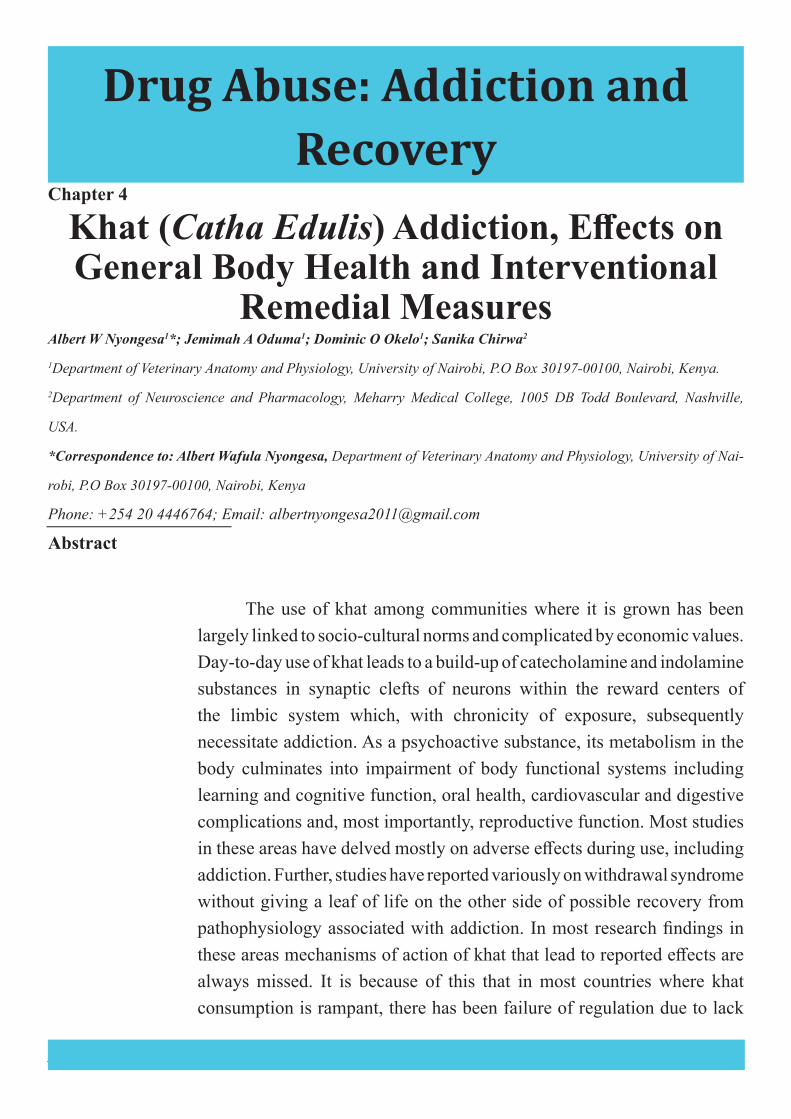
Serotonin (5-HT) is a neuromodulator whose properties are much more mysterious than those of dopamine although it is implicated in a wealth of important phenomena, ranging from analgesia [32], hallucinations [33] to a variety of mood disorders such as anxiety and depression [34]. Virtually all parts of the central nervous system receive innervation from serotonergic fibers arising from cell bodies located in two trunks of the midbrain serotonergic nuclei: the dorsal raphe nuclei (DRN) and the median raphe nuclei (MRN) [35]. Serotonin-containing bodies of the raphe nuclei project to dopaminergic cells in the VTA and SN, as well as nucleus accumbens, prefrontal cortex and striatum [36] (Figure 1). There are also serotonergic projections from the raphe to the peri-acqueductal gray involved in control of defensive and aversively motivational behaviours [37].
At electron microscopy there is presence of synaptic contacts of 3H5-HT-labelled terminals with both dopaminergic and non-dopaminergic dendrites in all sub-nuclei of the VTA, and in the SNc and SNr [35]. There is differential distribution of 5-HT receptor subtypes within the dopaminergic systems [39] that have led to the insight of dopamine-serotonin systems interaction in the brain (Figure 2)
Figure 1: Schematic representation of serotonin–dopamine interaction in the meso-corticolimbic and nigrostriatal dopaminergic system. Serotonin-containing cell bodies of raphe nuclei send projections to dopaminergic cells in both the ventral tegmental area (VTA, A10) and substantia nigra (SN, A9), and to their terminal fields in the nucleus accumbens, prefrontal cortex and striatum. [Adapted from Di Giovanni et al. (38)].
5
Drug Abuse: Addiction and Recovery
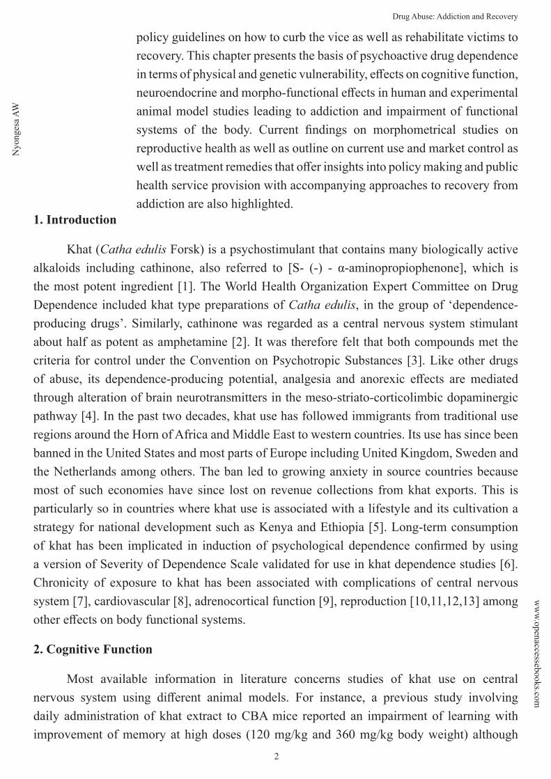
C. Noradrenaline
Noradrenaline, 3, 4-dihydroxyphenylethanolamine, is released from terminals of noradrenergic neurons in the brain from most postganglionic sympathetic neurons and from chromaffin cells in the adrenal medulla. The cell bodies of central noradrenergic neurons are all clustered within two bilateral groups of nuclei (A1 –A7) in the brain stem (Figure 3). These comprise the locus coeruleus (LC) complex and the lateral tegmental nuclei. The activity of noradrenergic neurons within locus coeruleus is governed by GABAergic projection from nucleus prepositus hyperglossi and glutamatergic input from the nucleus paragingantocellularis [40].
Synthesis of dopamine, adrenaline and noradrenaline share a common pathway. The amino acid L-tyrosine is a precursor substrate that undergoes hydroxylation in the presence of tyrosine hydroxylase (TH) to form L-dihydroxyphenylalanine (L-DOPA) followed by decarboxylation by DOPA decarboxylase to form dopamine. Dopamine is transported to the
Figure 2: Mid-saggital view of the rat brain with serotonin-immuno-reactive cell bodies. The blue and red ovals comprise two major subdivisions of the brain serotonergic system. Abbreviations: DRN, dorsal raphe nucleus; MRN, medial raphe nucleus; NRM; nucleus raphe magnus; NRO, nucleus raphe obscurus; SNc, substantia nigra pars compacta; VTA, ventral tegmental area. (Adapted from Di Giovanni et al. [38]).
Figure 3: Figure showing distribution of noradrenergic neurons in the brain. The cell bodies are clustered in nuclei (A1 – A7) in the pons/medulla regions of the brainstem and their axons project both rostrally and caudally to most regions of the neuraxis. The major nucleus is the locus coeruleus (A6). (Adapted from Stanford, [41]).
6
Drug Abuse: Addiction and Recovery
storage vesicles where dopamine β-hydroxylase converts it to noradrenaline. The process occurs in the cytoplasm of catecholamine-releasing neurons. Noradrenaline neurons influence arousal behaviours such as sleep/wakefulness, depression and anxiety [41]. The precise features of environmental stimuli that provoke increased noradrenergic transmission are unclear. Increased noradrenergic transmission in the brain mediates changes in selective attention. Another concept is that noradrenergic transmission influence emotional impact of a given stimulus. It is possible that the role and consequences of central noradrenergic transmission depends on type or severity of stimulus or individual differences in neurobiological coding behaviour.
3. Physical and Genetic Predisposition of Psychoactive Drug Dependence
Psychoactive drug dependence is a complex phenomenon characterized by interplay of several genes of an individual with respect to environmental factors associated with that individual. Individuals with genetic vulnerability to drugs of abuse experience a marked influence to drug dependence [42]. Indeed, the observed variance in behavior among substance abusers can be explained by differences in genetic make-up of the individuals. Therefore, in order to understand the genetic basis of addiction, there is need to identify genetic variation of that individual, although it only contributes partly to development of addiction.
Previous studies have reported genetic heritability of alcohol [43], opiates and cocaine [44]. The involvement of specific genes or complex of genes remains unclear. The challenge to this nature of studies is in the identification of genes that alter predisposition to drug dependence as well as in the understanding of how the function of genes interact with environmental factors influencing dependence of substance use. The difficulties in identifying genetic traits are associated with complexity of addiction as a trait. Genetic screening can help in identification of specific genes although in a general population, only probabilities rather than certainties shall be recorded. Opioid receptor genes for opioid dependence have reasonably been associated with opioid dependence [42]. In other substances of abuse, a complex of genes may be involved. For instance, alcohol heritability has been associated with genes involved in drug metabolism, alcohol receptor genes, and genes responsible for synthesis of GABA, serotonin and dopamine [45]. Heritability of alcoholism in individuals homozygous for ALDH2*2 allele encoding for less active variant of aldehyde dehydrogenase type 2 is rare [46]. Variants in different genes may contribute to addiction in different lineages thus necessitating a clear understanding of genetic and genomic variants that may possibly be implicated in development of drug addiction.
There is a direct link between an individual’s genotype with predisposing/risk factors which can either be environmental (availability of drugs, poverty, social change, cultural norms, peer influence, occupation) or individual (genetic disposition, personality disorders, social deprivation, depression) that can direct a certain response to drug dependence [47]. In view of this, it is prudent to incorporate behavioral assessment when investigating genetic
7
Drug Abuse: Addiction and Recovery
vulnerability of a certain individual in the understanding of development of drug addiction. The handicap, however, is the quantification of behavioral endpoints since they are more susceptible to environmental influence thus giving variance in behavioral manifestations. But this can be fine-tuned through allowing major focus on establishment of behavioral endpoints with same degree of sophistication and inter-rator reliability as earlier reported [48]. Environmental stimuli influences brain circuitry the same way as genetic involvement hence the need to combine specific phenotypes with genetic approaches in identification of development of addiction in humans and animals. In order to understand peculiarities of addiction, it is important to analyze factors that determine its development, involve use of animal model that reflects human situation as closely as possible and identify and verify loss of control and that of reversibility in a drug addicted animal.
In behavioral profiling, studies have shown that self-administration and conditioned reinforcement paradigms give more accurate behavioral tests mapping human addiction [49] with oral self-administration being more practical [50]. Other studies that have been done involve place-avoidance assay [51] and measurement of rate and degree of tolerance with opiate dependence [52]. The stability of behavioral abnormalities that characterize addiction is a clear indicator that gene expression may be involved in drug-induced behavioral changes [53]. Most studies in humans and experimental animals [54] have approached drug addiction cases from behavioral profiling. However, it should be understood that genetic vulnerability plays a key role to development of drug addiction. Whereas the genetic variations established in animal models may be different from those of humans, their identification can shed light on mechanisms underlying addiction process. No studies have shown genetic polymorphisms associated with khat dependence in humans or even animal models hence the need for researchers to offer particular focus in this area as a way of mapping out possible treatment in khat addictive cases. It should be noted that substance abuse has contributed markedly to global burden of disease in many parts of the world including Europe, Central and East Asia, North America and most parts of Africa. For instance, individuals living with infectious conditions such as HIV/AIDS inject themselves with psychoactive substances with sole aim of suppressing their stigma. The end result is contraction of other diseases such as hepatitis B and C through use of same needles. It goes without saying that various accidents, cases of suicide, assaults and even breakage of marriages are implicated in substance use and misuse. It is, therefore, of paramount importance to understand the in-depth of involvement of psychosocial, neurobiological and genetic factors influencing development of addiction and provide these insights to relevant policy enforcing agencies and public health service providers with a sole view of minimizing or eradicating the vice. This chapter highlights various adverse effects of khat use following long-term use and subsequent addiction in some cases. Studies done in humans and experimental animals have offered subjective and objective findings. Understanding of development of the effects on drug use and misuse offer a platform on which interventional scientific research initiatives are
8
Drug Abuse: Addiction and Recovery
undertaken with sole aim of looking for remedy of the cases to recovery.
4. Effects on Body Functional Systems at Chronic Exposure
Long-term khat use has been associated with behavioural changes [55], myocardial complications [56], oral lesions [57], gastro-intestinal and cardiovascular complications [8], adrenocortical and psychological complications [58], endocrine complications [9] and sexual function [10, 11] among an array of other central nervous system complications. This chapter focuses mostly on morpho-functional and molecular dimensions on reproductive function with long-term khat use since most controversial reports center around this subject. These effects stem from hypothalamic involvement down the pituitary to testicular function. The focus of the discussion is mostly centered on the male since khat chewing is more prevalent in males [59].
I. Reproductive Endocrine Effects
Numerous findings on khat and endocrine function have been reported in experimental animal and human case studies. In a cross-sectional study on Yemeni regular khat chewers, altered adrenocortical function was reported [58] similar to findings in baboons [60] and vervet monkeys [9, 61]. The effects on hormonal profiling seem to be bi-phasic where varying khat doses influence endocrine function differently. For instance, studies in rats [11] and mice [62] showed that low dose stimulates production of testosterone while high doses cause suppression. Chronicity of khat exposure impairs steroidogenic cell function [63, 13]. These reports have further been supported by findings of Al-Habori and Al-Mamary [64] showing reduction in cholesterol following feeding rabbits with khat over 6 month period. Cholesterol is synthesized in smooth endoplasmic reticulum [65], and is a precursor molecule in steroidogenesis [66].
II. Testicular Histomorphometric Effects
Khat use has been reported to have varying effects on the reproductive function in humans and experimental animals. Numerous reports, however, show contradictory findings on male reproductive function. Some reports indicate erectile dysfunction [67] and spermatorrhoea [3] and testicular cell degeneration [68] following khat and cathinone exposure. Recently Nyongesa et al. [10] reported cytotoxic effects of khat extracts on germinal epithelium (spermatogonia and primary spermatocytes) of male rabbits at sub-chronic exposure with no observable effects on Sertoli and Leydig cells. Other reports have implicated khat with aphrodisiac properties [69], a remedy for premature ejaculation [3] and sperm power booster [70]. The mechanisms underlying pharmacological action of khat extracts on testicular function with respect to aforementioned effects remains obscure. On this strength, authors here present testicular histomorphometric findings on 12 adult male vervet monkeys treated with 0.8, 3.2 and 6.4 mg/kg body weight of khat extracts on alternate days of the week for 4 months. At the end of
9
Drug Abuse: Addiction and Recovery
treatment period, testicular tissues of one animal from each group were harvested, following anaesthesia, for histomorphometric evaluations. The morphometric evaluation employed volume densities of mitochondria, sooth endoplasmic reticulum (SER), Golgi apparatus and lipid droplets of Leydig cells that were estimated from photomicrographs of central parts of testes photographed at x 6000 magnification. Point differential counting as described by Kavoi et al. [71] was used to evaluate volume density occupied by mitochondria, lipid droplets, Golgi apparatus, SER and rough endoplasmic reticulum (RER). Briefly, a transparent test grid with a square lattice of points was overlaid with random positioning on testicular electron micrographs projected on a computer monitor. The number of points hitting the mitochondria, SER, RER, lipid droplets and Golgi apparatus and those falling on projected field of testicular interstitium were counted. Volume density of component of interest [Vv (l)] was calculated as a percentage:
Vv (l) = (ΣNPl/ΣNPi) x 100,
Where, Vv (l) is volume density of cell component, ΣNPl is the number of test points hitting the image of the evaluated component of interest, and ΣNPi is the number of all points falling on the cell image (interstitium). By substituting in the equation above, volume densities of mitochondria, SER, RER, lipid droplets and Golgi apparatus were calculated using the following formulae, respectively:
Vv (m) = (ΣNPm/ΣNPi) x 100
Vv (ser) = (ΣNPser/ΣNPi) x 100
Vv (rer) = (ΣNPser/ΣNPi) x 100
Vv (l) = (ΣNPl/ΣNPi) x 100
Vv (ga) = (ΣNPga/ΣNPi) x 100
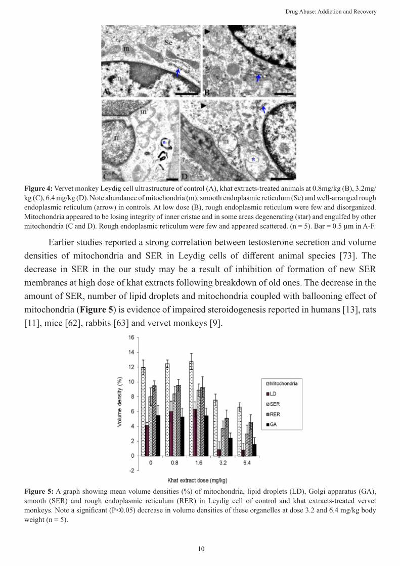
Results of this study showed that high dose of khat extracts at sub-chronic exposure to vervet monkeys resulted in alteration of sub-cellular organelles in Leydig cells (Fig 4C and D) compared to low dose (Figure 4B) and control group (Figure 4A). Of particular importance were SER, mitochondrial cristae and lipid droplets implicated in steroidogenesis [72].
10
Drug Abuse: Addiction and Recovery
Earlier studies reported a strong correlation between testosterone secretion and volume densities of mitochondria and SER in Leydig cells of different animal species [73]. The decrease in SER in the our study may be a result of inhibition of formation of new SER membranes at high dose of khat extracts following breakdown of old ones. The decrease in the amount of SER, number of lipid droplets and mitochondria coupled with ballooning effect of mitochondria (Figure 5) is evidence of impaired steroidogenesis reported in humans [13], rats [11], mice [62], rabbits [63] and vervet monkeys [9].
Figure 4: Vervet monkey Leydig cell ultrastructure of control (A), khat extracts-treated animals at 0.8mg/kg (B), 3.2mg/kg (C), 6.4 mg/kg (D). Note abundance of mitochondria (m), smooth endoplasmic reticulum (Se) and well-arranged rough endoplasmic reticulum (arrow) in controls. At low dose (B), rough endoplasmic reticulum were few and disorganized. Mitochondria appeared to be losing integrity of inner cristae and in some areas degenerating (star) and engulfed by other mitochondria (C and D). Rough endoplasmic reticulum were few and appeared scattered. (n = 5). Bar = 0.5 µm in A-F.
Figure 5: A graph showing mean volume densities (%) of mitochondria, lipid droplets (LD), Golgi apparatus (GA), smooth (SER) and rough endoplasmic reticulum (RER) in Leydig cell of control and khat extracts-treated vervet monkeys. Note a significant (P<0.05) decrease in volume densities of these organelles at dose 3.2 and 6.4 mg/kg body weight (n = 5).

11
Drug Abuse: Addiction and Recovery
The membrane-bound P450 cholesterol side chain cleavage enzyme (CYP11A) associated with mitochondria catalyzes conversion of cholesterol to pregnenolone through hydroxylation and cleavage of steroid substrates [74]. A large body of evidence suggests that SER, Golgi complex and lipid droplets are integral in steroidogenesis. Studies in humans showed that cytochrome P450 enzymes (CYP17, CYP19 and CYP21) are associated with SER [66]. Our findings on Leydig cell morphology are consistent with studies in humans [75] and birds [76] that indicated susceptibility of some sub-cellular elements to endocrine disrupters. Collectively, these findings point at adverse effects of khat extracts at high dose and long-term exposure on functions of steroidogenic organelles.
Sub-chronic exposure to high dose of khat extracts had adverse effects on developing germ cells in the seminiferous tubules, pointing towards impairment of spermatogenesis. The disorganized shape of spermatogonia as shown by cell membrane outline as well as vacuolation in spermatocytes, all point to degeneration in these cells (Figure 6). It is argued that immature germ cells are highly susceptible to noxious agents due to the abundance of histones in their chromatin material compared to mature forms that contain highly condensed chromatin due to argenine and cysteine-rich protamines [77].
Figure 6: Spermatocytes of vervet monkeys showing controls (A) and khat extracts-treated groups (B - D). Numerous cytoplasmic vacuolations (v) accompanied by disorganization of nuclear membrane in treated groups was observed. At high dose (6.4mg/kg) (C) numerous cytoplasmic vacuolations accompanied by disruption of nuclear membrane integrity was observed. Chromatin material in the nucleus appeared more condensed indicative of degenerative changes. (n = 5). Bar = 0.5 µm in A-D.
Vacuoles in spermatogenic cells are frequently encountered following impaired spermatogenesis. For instance, rabbits treated with 40.5g/kg of khat extract [10] showed vacuolation in spermatogenic cells. There was also evidence of impairment in nuclear function in our study as shown by irregular outline of nuclear membrane in spermatocytes.
12
Drug Abuse: Addiction and Recovery
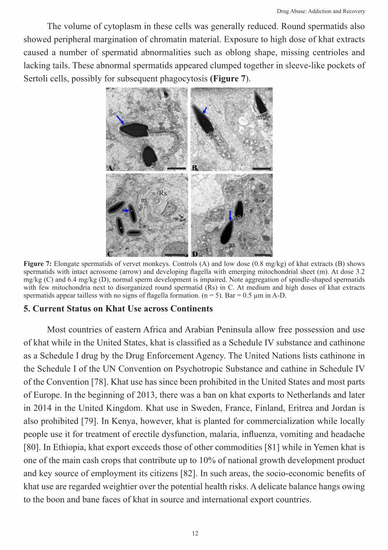
The volume of cytoplasm in these cells was generally reduced. Round spermatids also showed peripheral margination of chromatin material. Exposure to high dose of khat extracts caused a number of spermatid abnormalities such as oblong shape, missing centrioles and lacking tails. These abnormal spermatids appeared clumped together in sleeve-like pockets of Sertoli cells, possibly for subsequent phagocytosis (Figure 7).
5. Current Status on Khat Use across Continents
Most countries of eastern Africa and Arabian Peninsula allow free possession and use of khat while in the United States, khat is classified as a Schedule IV substance and cathinone as a Schedule I drug by the Drug Enforcement Agency. The United Nations lists cathinone in the Schedule I of the UN Convention on Psychotropic Substance and cathine in Schedule IV of the Convention [78]. Khat use has since been prohibited in the United States and most parts of Europe. In the beginning of 2013, there was a ban on khat exports to Netherlands and later in 2014 in the United Kingdom. Khat use in Sweden, France, Finland, Eritrea and Jordan is also prohibited [79]. In Kenya, however, khat is planted for commercialization while locally people use it for treatment of erectile dysfunction, malaria, influenza, vomiting and headache [80]. In Ethiopia, khat export exceeds those of other commodities [81] while in Yemen khat is one of the main cash crops that contribute up to 10% of national growth development product and key source of employment its citizens [82]. In such areas, the socio-economic benefits of khat use are regarded weightier over the potential health risks. A delicate balance hangs owing to the boon and bane faces of khat in source and international export countries.
Figure 7: Elongate spermatids of vervet monkeys. Controls (A) and low dose (0.8 mg/kg) of khat extracts (B) shows spermatids with intact acrosome (arrow) and developing flagella with emerging mitochondrial sheet (m). At dose 3.2 mg/kg (C) and 6.4 mg/kg (D), normal sperm development is impaired. Note aggregation of spindle-shaped spermatids with few mitochondria next to disorganized round spermatid (Rs) in C. At medium and high doses of khat extracts spermatids appear tailless with no signs of flagella formation. (n = 5). Bar = 0.5 µm in A-D.
13
Drug Abuse: Addiction and Recovery
6. Treatment Remedies of Khat Addiction
Like users of other drugs of addiction such as cocaine, amphetamine and morphine, drug abstinence and subjection to rehabilitation program has been used with various degrees of success. This varies among affected individuals: those with genetically inclined traits, social/peer as well as lifestyle habits. In the Kenyan context, abstinence followed by rehabilitation seems to be the available tool of treatment of khat and other drug addiction cases. Most of individuals incriminated in this exercise are victims of con-current users of other drugs such as tobacco, marijuana and even alcohol. There are accompanying challenges to the success of this approach since the vice in the affected individuals is occasioned by various factors as mentioned above and so a combination of medical and psychological counseling appears to be most appropriate intervention. There is very scanty information on medical intervention of khat addiction cases with one study [83] which reported use of bromocriptine although it is now a banned substance. Most available literature reports from areas of endemic khat use implicate psychological counseling as the preferred tool [79].
Successful patient rehabilitation or healing relies heavily on individual case approach. It should be understood that khat, like any other addiction drug, bears withdrawal syndrome to long-term use. These withdrawal symptoms are more traumatizing to deal with compared to the real effects of khat. A proper approach therefore should consider aspects of potential genetic involvement, lifestyle habits leading to anxiety and depression, as well as environmental/peer group influence. It is also important to establish whether or not the patient concomitantly uses khat with other psychostimulants. Involvement of family members and close associates ensures collective effort in rehabilitation process that in turn minimizes cases of some patients abandoning treatment due to lack of background knowledge.
Lack of satisfactory and convincing knowledge of wholesome adverse effects of khat on human health owing to improper experimental designs, use of wrong animals in studies modeling human functional systems, lack of funding, improper simulation of real-time khat use in the human context when designing experiments, and subjectivity in reporting results owing to political and economic inclinations have contributed to a pool of findings that are primarily contradictory and therefore do not add advisory value to policy making. Government agencies in such cases are faced with challenges of enforcing laws that can best govern regularization or illegalization of khat chewing habit as well as dealing with addiction cases. In most countries where khat use is a cultural norm and considered contributor to economies of scale, such as Kenya, Yemen, Ethiopia and many countries around the horn of Africa, cases of addiction treatment are not a priority. Such perceptions have compromised any meaningful efforts from researchers who report invaluable findings that could otherwise contribute immensely to intervention measures to curb the vice.

14
Drug Abuse: Addiction and Recovery
7. References
1. P. Kalix, O. Braenden, Pharmacological aspects of the chewing of khat leaves. Pharmacol. Rev. 1985; 149 – 164: 37.
2. World Health Organization, Expert Committee on Drug Dependence. Nineteenth Report, World Health Organization Technical Series, 1973; No. 526.
3. C. Pantelis, C.G. Hindler, J.C. Taylor, Use and abuse of khat distribution, pharmacology, side effects and description of psychosis attributed to khat chewing. Psychol.l Med. 1989; 657–668: 19.
4. B.A. Gosnell, J.M. Yracheta, S.M. Bell, K.E. Lane, Intravenous self-administration of cathinone by rats. Behav. Pharmacol. 1996; 526 – 531: 7.
5. M. Odenwald, N. Warfa, K. Bhui, T. Elbert, The stimulant khat- Another door in the wall? A call for overcoming the barriers. J. Ethnopharmacol. 2010; 615 – 619: 132.
6. S. Kassim, S. Islam, R. Croucher, Correlates of nicotine dependence in UK resident Yemeni khat chewers: A cross-sectional study. Nicotene Tob. Res. 2011; 1240 – 1249: 13.
7. P. Kalix, Cathinone: a natural amphetamine. Pharmacol. Toxicol. 1992; 77 – 86: 70.
8. A. Al-Motarreb, M. Al-Habori, K.J Broadley, Khat chewing, cardiovascular diseases and other internal medical problems: The current situation and directions for future research 2010; 540 – 548: 132.
9. A.W. Nyongesa, J.A. Oduma, M. al’Absi, S. Chirwa, Immunohistochemical localization of anterior pituitary cell types of vervet monkeys (Chlorocebus aethiops) following sub-chronic cathinone exposure. J. Ethnopharmacol. 2015; 168 – 177: 174.
10. A.W. Nyongesa, N.B. Patel, E.O. Wango, D.W. Onyango, High khat dose and long-term exposure impairs spermatogenesis: experimental study using rabbit model. J. Morphol. Sci. 2017; 156 – 16734.
11. A. Mohammed, E. Engidawork, Reproductive parameters are differentially altered following subchronic administration of Catha edulis Forsk (Khat) extract and cathinone in male rats. J. Ethnopharmacol. 2011; 977 – 983: 134.
12. M. Abdulwaheb, E. Makonnen, A. Debela, D. Abeba, Effect of Catha edulis Forsk (khat) extracts on male rat sexual behaviour. J. Ethnopharmacol. 2007; 250 – 256:110.
13. S.M. El-Shoura, M. Abdel Aziz, E.M. Elmalik, Deleterious effects of khat addiction on semen parameters and sperm ultrastructure. Hum. Reprod. 1995; 2295 – 2300: 9.
14. S.T. Kimani, A.W. Nyongesa, Effects of single daily khat (Catha edulis) extract on spatial learning and memory in CBA mice. Behav. Brain Res. 2008; 192 – 197: 195.
15. P. Nencini, A.M. Ahmed, Khat consumption: a pharmacological review. Drug Alcohol Depend. 1989; 19 – 29: 23.
16. N.A. Hassan, A.A. Gunaid, I.A. Murray-Lyon, Khat (Catha edulis) health aspects of khat chewing. East Mediterr Health J. 2007; 706 – 718: 13.
17. J. Zelger, H. Schorno, E. Carlin, Behavioural effects of cathinone: an amphetamine obtained from Catha edulis: comparisons with amphetamine, norpseudoephedrine, apomorphine and nomifensine. Bull. Narc. 1980; 67 – 81: 32.
18. P. Kalix, S. Geisshusler, R. Brenneisen, U. Koelbing, H.U. Fisch, Cathinone, a phenylapropylamine alkaloid from khat leaves that has amphetamine effects in humans. NIDA Res. Monogr. 1990; 289 – 290: 105.
19. S. Gough, I.B. Cookson, Khat-induced schizophreniform psychosis in the United Kingdom. Lancet 1984; 455: 1.
20. G. Yousef, Z. Huq, T. Lambert, Khat chewing as a cause of psychosis. Br. J. Hosp. Med. 1995; 322 – 326: 54.
15
Drug Abuse: Addiction and Recovery
21. R.J. Nielen, F.M. van der Heijden, S. Tuiner, W.M. Verhoeven, Khat and mushrooms associated with psychosis. World J. Biol. Psychiatry. 2004; 49 – 53: 5.
22. M. al’Absi, J. Grabowski, Concurrent use of tobacco and khat: Added burden on chronic disease epidemic. Addiction. 2012; 451 – 452: 107.
23. M.Y. Banjaw, W.J. Schmidt, Behavioural sensitization following repeated intermittent oral administration of Catha edulis in rats. Behav. Brain Res. 2005; 181 – 189: 156.
24. T. Steckler, F. Holsboer, Enhanced conditioned approach responses in transgenic mice with impaired glucocorticoid receptor function. Behav. Brain Res. 1999; 151 – 163: 102.
25. K.H. Karlsgodt, S. Lukas, I. Elman, Psychosocial stress and the duration of cocaine dependence. Am. J. Drug Alcohol Abuse., 2003; 539 – 551: 29.
26. N.K. Mello, J.H. Mendelson, Cocaine’s effects on neuroendocrine systems: clinical and preclinical studies. Pharmacol. Biochem. Behav. 1997; 571 – 599: 57.
27. M. Barrot, M. Marinelli, D.N. Abrous, F. Rouge-Pont, M. Le Moal, P.V. Piazza, The dopaminergic hyper-responsiveness of the shell of the nucleus accumbens is hormone-dependent. Eur. J. Neurosci. 2000; 973 – 979: 12.
28. M. Le Moal, H.Simon, Mesocorticolimbic dopaminergic network: functional and regulatory roles. Physiol. Rev. 1991; 155 – 234: 71.
29. G. Di Chiara, A. Imperato, Drugs abused by humans preferentially increase dopamine concentrations in the mesolimbic system of freely moving rats. Proc. Natl. Acad. Sci. USA1988; 5274 – 5278: 85.
30. E.A. Kiyatkin, Functional significance of mesolimbic dopamine. Neurosci. Biobehav. Rev. 1995; 573 – 598: 19.
31. A. Grace, B. Bunney, Dopamine. In: M.A. Rogawski, J.L. Barker, (Eds.). Neurotransmitter Action in the Vertebrate Nervous System, New York: Plenum Press., 1985: pp 285 – 319.
32. D. LeBars, Serotonin and pain. In: N.N. Osborne, M.Hamon, (Eds), Neuronal serotonin, 1988: pp 171 – 226.
33. G.K. Aghajanian, G.J. Marek, Serotonin and hallucinogens. Neuropsychopharmacology 1999; 165 – 235: 21.
34. H.G. Westernberg, J.A. den Boer, D.L. Murphy, (Eds), Advances in the Neurobiology of Anxiety Disorders. New York: Wiley. 1996.
35. H. Moukhles, O. Bosler, J.P. Bolam, A. Valleé, D. Umbriacco, M. Geffard, G. Doucet, Quantitative and morphometric data indicate precise cellular interactions between serotonin terminals and post-synaptic targets in rat substantia nigra. Neuroscience 1997; 1159 – 1171: 76.
36. E.J. Van Bockstaele, D.M. Cestari, V.M. Pickel, Synaptic structure and connectivity of serotonin terminals in the ventral tegmental area: Potential sites for modulation of mesolimbic dopamine neurons. Brain Res. 1994; 307 – 322: 647.
37. F.G. Graeff, On serotonin and experimental anxiety. Psychopharmacology 2003; 467–476: 163.
38. G. Di Giovanni, V. Di Matteo, M. Pierucci, E.Esposito, Serotonin-dopamine interaction: electrophysiological evidence. In: G. Di Giovannni, V. Di Matteo, E. Esposito, (Eds), Prog. Brain Res. 2008; 45 – 71: 172.
39. N.M. Barnes, T. Sharp, A review of central 5-HT receptors and their function. Neuropharmacology 1999; 1083 – 1152: 38.
40. G. Aston-Jones, M.T. Shipley, Chouvet Afferent regulation of locus coeruleus neurons: Anatomy, Physiology and Pharmacology. Prog. Brain Res. 1991; 47 – 73: 88.
16
Drug Abuse: Addiction and Recovery
41. S.C. Stanford, Noradrenaline. In: R.A. Webster (Ed), Neurotransmitters, drugs and brain function., John Willey and Sons Ltd. Baffins Lane, Chichester, West Sussex, UK, 2001: pp 163–185.
42. World Health Organization, Neuroscience of psychoactive substance use and dependence. 2004: pp 1 – 40.
43. K.S. Kendler, M.C. Neale, A.C. Heath, A twin family of alcoholism in women. Am. J. Psychiatry. 1994; 707 – 715: 151.
44. M.T. Tsuang, M.J. Lyons, JM. Meyer, T. Doyle, Co-occurrence of abuse of different drugs in men: the role of drug-specific and shared vulnerabilities. Arch. Gen. Psychiatry. 1998; 967 – 972: 55.
45. Y.C. Chen, R.B. Lu, G.S. Peng, M.F. Wang, Alcohol metabolism and cardiovascular response in an alcoholic patient homozygous for the ALDH2*2 variant gene allele. Alcohol. Clin. Exp. Res. 1999; 1853 – 1860: 23.
46. E.J. Nestler, Genes and Addiction. Nature Gen. 2000; 277 – 281: 26.
47. E.J. Nestler, M.T. Berhow, E.S. Brodkin, Molecular mechanisms of drug addiction: adaptations in signal transduction pathways. Mol. Psychiatry. 1996; 190 – 199: 1.
48. H. Würbel, Behaviour and the standardization fallacy. Nature Gen. 2000; 263: 26.
49. Y. Shaham, S. Erb, J. Stewart, Stress- induced relapse to heroin and cocaine seeking in rats: a review. Brain Res. Rev. 2000; 13 – 33: 33.
50. W.H. Berrettin, T.N. Ferraro, R.C. Alexander, A.M. Buchberg, W.H. Vogel, Quantitative trait loci mapping of three loci controlling morphine preference using inbred mouse strains. Nature Gen. 1994; 54 – 58: 7.
51. R. Maldonado, J.A. Blendy, E. Tzavara, P. Gass, Reduction of morphine abstinence in mice with a mutation in the gene encoding CREB. Science. 1996; 657 – 659: 273.
52. L.M. Bohn, R.T. Lefkowitz, R.R. Gainetdinov, Enhanced morphine analgesia in mice lacking b-arrestin 2. Science. 1999; 2495 – 2498: 268.
53. E.J. Nestler, G.K. Aghajanian, Molecular and cellular basis of addiction. Science. 1997; 58 – 63: 278.
54. A. Heyne, J. Wolffgramm, The development of addiction to d-amphetamine in an animal model: same principles for alcohol and opiate. Psychopharmacology. 1998; 510 – 518: 140.
55. A.W. Nyongesa, J.A. Oduma, M. Nakajima, H.O. Odongo, P.A. Adoyo, M. al’Absi, Acute and sub-chronic effects of purified cathinone from khat (Catha edulis) on behavioural profiles in vervet monkeys (Chlorocebus aethiops). Metab. Brain Dis. 2014a; 441 – 449: 29.
56. G. Cox, H. Rampes, Adverse effects of khat. A review: Adv. Psychiatry Treat. 2003; 456 – 463: 9.
57. O.M Lukandu, L.S Koech, P.N Kiarie, Oral lesions induced by chronic khat use consist essentially of thickened hyperkeratinized epithelium. International Journal of Dentistry 2015; 1 – 9: 2015.
58. M. al’Absi, N.S Khalil, M. Al-Habori, R. Hoffman, K. Fujiwara, L. Wittmers, Effects of chronic khat use on cardiovascular, adrenocortical and psychological responses to stress in men and women. The Am. J. Addict. 2013; 99 – 107: 22
59. A. Al-Motarreb, K. Baker, K.J. Broadley, Khat: Pharmacological and medical aspects and its social use in Yemen. Phytother. Res. 2002; 403 – 413: 16.
60. J.M. Mwenda, R.A. Owuor, C.M. Kyama, E.O. Wango, M.M. Arimi, D.K. Langat, Khat (Catha edulis) up-regulates testosterone and decreases prolactin and cortisol levels in the baboon. J. Ethnopharmacol. 2006; 379 – 384: 103.
61. A.W. Nyongesa, J.A. Oduma, M. Nakajima, H.O. Odongo, P.A. Adoyo, M. al’Absi, Dose-response inhibitory
17
Drug Abuse: Addiction and Recovery
effects of purified cathinone from khat (Catha edulis) on cortisol and prolactin release in vervet monkeys (Chlorocebus aethiops). Metab. Brain Dis. 2014b; 451 – 458: 29.
62. A.W. Nyongesa, N.B. Patel, D.W. Onyango, E.O. Wango, H.O. Odongo, In vitro study of the effects of khat (Catha edulis Forsk) extract on isolated mouse interstitial cells. J. Ethnopharmacol. 2007; 401 – 405: 110.
63. A.W. Nyongesa, N.B. Patel, D.W. Onyango, H.O. Odongo, E.O. Wango, Khat (Catha edulis) lowers plasma luteinizing hormone (LH) and testosterone secretion, but increases cortisol levels in male rabbits. J. Ethnopharmacol. 2008; 245 – 250: 116.
64. M. Al-Habori, M. Al-Mamary, Long term feeding effects of Catha edulis leaves on blood constituents in animals. Phytomedicine 2004; 639–644: 11.
65. A.K. Christensen, Leydig cells. In: R.O. Greep, E.B. Astwood, (Eds), Handbook of Physiology. Am. Physiol. Soc: Washington DC. 1975; 57: 5 Sect 7.
66. A.H. Payne, D.B. Hales, Overview of steroidogenic enzymes in the pathway from cholesterol to active steroid hormones. Endocrine Rev. 2004; 947 – 970: 25.
67. A.S. Elmi,. The chewing of khat in Somalia. J. Ethnopharmacol. 1983; 163 – 176: 8.
68. M.W. Islam, M. Tariq, A.M. Ageel, F.S. El-Feraly, I.A. Al-Meshal, I. Ashaf, An evaluation of the male reproductive toxicity of cathinone. Toxicology 1990; 223 – 234: 603.
69. A.D. Krikorian, Khat and its use: a historical perspective. J. Ethnopharmacol. 1984; 115: 12.
70. S.A. Adeoya-Osiguwa, L.R. Fraser, Cathine and norephedrine, both phenylpropanolamines, accelerate capacitation and then inhibit spontaneous acrosome loss. Hum.Reprod. 2005; 198 – 207: 20.
71. B.M. Kavoi, A.N. Makanya, J. Plendl, S.G. Kiama, Morphofunctional adaptations of the olfactory mucosa in postnatally developing rabbits. The Anat. Rec. 2012; 1352 – 1363: 295.
72. G. Mazzocchi, C. Robba, P. Rebuffat, G. Gottardo, G.G. Nussdorfer,. Effects of a chronic treatment with testosterone on the morphology of the interstitial cells of rat testis: an ultrastructural stereological study. Int. J. Androl. 1982; 130 – 136: 5.
73. B.R. Zirkin, L.L. Ewing, N. Kromann, R.C. Cohran, Testosterone secretion by rat, rabbit, guinea pig, dog and hamster testes perfused in vitro: correlation with Leydig cell ultrastructure. Endocrinology. 1980; 1867 – 1874: 107.
74. G.S. Boyd, E.R. Simpson, Studies on the conversion of cholesterol to pregnenolone in bovine adrenal mitochondria. In: McKern, K.W. (Ed). Functions of adrenal cortex. Appteton-Century Crofts: New York. 1968; 49 – 76: 1.
75. A. Marques-Pinto, D. Carvalho, Human infertility: are endocrine disruptors to blame? Endocrine Connect. 2013; R15 – R29: 2.
76. U.M. Bello, M.C. Madekurozwa, H.B. Groenewald, T.A. Aire, A. Arukwe, The effects on steroidogenesis and histopathology of adult male Japanese quails (Coturnix coturnix japonica) testis following pre-pubertal exposure to di (n-butyl) phthalate (DBP). Comp. Biochem. Physiol. Part C: Toxicol. Pharmacol. 2014; 24 – 33: 166.
77. B.M. Carlson,. Human embryology and developmental biology, (2nd edn.). Mosby, Chicago, New York, London, Toronto, Sydney, Milan, Tokyo. 1999
78. WHO Expert Committee on Drug Dependence. Thirty-fourth report. Geneva: World Health Organization; 2006:36 (Technical Report Series No. 942).
79. M. Odenwald, M. al’Absi, Khat use and related addiction, mental health and physical disorders: The need to address a growing risk. Eastern Mediterr. Health J. 2017; 236 – 244: 23.
18
Drug Abuse: Addiction and Recovery
80. A.W. Nyongesa, D.W. Onyango, Khat: A Boon or Bane to Humanity: In A.S Awaad, V.K Singh, J.N. Govil (Eds), Recent Proceedings in Medicinal Plants: Studium Press LLC, USA, 2010; 173 – 193: 28.
81. D. Hailu,. Should Khat be Banned? The Development Impact. IPC One Pager, 2007; 40. International Poverty Centre, Brazilia.
82. World Bank, Yemen Economic Update;. The World Bank Group, Sana’a. Spring 2005 (Accessed: February 3, 2006).
83. A.J. Giannini, N.S. Miller, C.E. Turner, Treatment of khat addiction. J Subst Abuse Treat. 1992; 9: 379–82.