drug delivery, entry and intracellular trafficking of
TRANSCRIPT

Drug delivery, entry and intracellular
trafficking of polymeric nanoparticles
DOCTORAL THESIS
DANIEL HOFMANN
MAX PLANCK INSTITUTE FOR POLYMER RESEARCH, MAINZ


Drug delivery, entry and intracellular
trafficking of polymeric nanoparticles
DOCTORAL THESIS
In fulfillment of the requirements for the degree of
Doktor rerum naturalium
(Dr. rer. nat.)
Submitted to
THE FACULTY OF BIOLOGY AT THE
JOHANNES GUTENBERG UNIVERSITÄT, MAINZ
This doctoral thesis has been carried out
AT THE MAX-PLANCK INSTITUTE FOR POLYMER RESEARCH, MAINZ
Daniel Hofmann
M.Sc. Cell and Molecular Biology
Born in Aschaffenburg, Germany
Submission: August 2014

Reviewers of the thesis committee
Supervisor
Prof. Dr. Katharina Landfester
Physical Chemistry of Polymers; Max-Planck Institute for Polymer Research, Mainz
Second reviewer of the thesis committee
Prof. Dr. Jacqueline Trotter
Department for Molecular Cell Biology; Johannes Gutenberg University, Mainz

Affidavit
The doctoral thesis was carried out from May 1st, 2011 to May 31
th, 2014 at the Max-Planck Institute
for Polymer Research under the supervision of PD Dr. Volker Mailänder and Prof. Dr. Katharina
Landfester.
I herewith declare that my doctoral thesis “Drug delivery, entry and intracellular trafficking of
polymeric nanoparticles“ has been written independently with no other sources and aids than quoted
in the text. The thesis has never been submitted to other faculties.
Hereby, I accept the examination regulations of the Faculty of Biology (Johannes Gutenberg
Universität, Mainz).
Date, Daniel Hofmann

Publications during this thesis
Hofmann, D. & Mailander, V. Pharmacology of nanocarriers on the microscale: importance of
uptake mechanisms and intracellular trafficking for efficient drug delivery. Nanomedicine
(Lond) 8, 321-323, doi:10.2217/nnm.13.2 (2013).
Baumann D., Hofmann D., Nullmeier S., Panther P., Dietze C., Musyanovych A., Ritz S.,
Landfester K. & Mailänder V. Complex encounters: nanoparticles in whole blood and their
uptake into different types of white blood cells. Nanomedicine (Lond) 8, 699-713,
doi:10.2217/nnm.12.111 (2013).
Hofmann, D., Messerschmidt, C., Bannwarth, M. B., Landfester, K. & Mailander, V. Drug
delivery without nanoparticle uptake: delivery by a kiss-and-run mechanism on the cell
membrane. Chemical Communications 50, 1369-1371, doi:10.1039/c3cc48130a (2014).
Hofmann, D., Tenzer, S., Bannwarth, M. B., Messerschmidt, C., Glaser S-F., Schild H.,
Landfester, K. & Mailander, V. Mass Spectrometry and Imaging Analysis of Nanoparticle-
Containing Vesicles Provide a Mechanistic Insight into Cellular Trafficking, ACS nano, ASAP,
doi: 10.1021/nn502754c.
Messerschmidt, C, Hofmann D., Kroeger-Brinkmann A., Landfester K., Mailänder V. &
Lieberwirth I. Cellular Uptake of Small Nanoparticles: Ultrastructure reveals size dependent
membrane morphologies; Major revision.
Other publications
Dühren-von Minden M., Übelhart R., Schneider D., Wossning T., Bach M., Buchner M.,
Hofmann D., Surova E., Follo M., Köhler F., Wardemann H., Zirlik K., Veelken H., Jumaa H.
Chronic lymphocytic leukaemia is driven by antigen-independent cell-autonomous signaling.
Nature 489, 309-312, doi:10.1038/nature1130.


Contents
I
I. Contents
I. Contents.......................................................................................................................................................... I
II. List of Figures .............................................................................................................................................. VI
III. List of Tables .............................................................................................................................................. VIII
IV. Abbreviations ............................................................................................................................................... IX
V. Acknowledgements .................................................................................................................................... XIII
VI. Abstract ..................................................................................................................................................... XIV
VII. Deutsche Zusammenfassung ....................................................................................................................... XV
1. Introduction to nanobiotechnology ............................................................................................................. 1
1.1 Nanomaterials – Synthesis of polymeric nanoparticles ............................................................................ 1
1.1.1 Forces inside the nanodroplet ....................................................................................................... 3
1.1.2 Stabilization of nanoparticles in biological fluids ......................................................................... 3
1.1.3 Biodegradable poly-L-lactide nanoparticles ................................................................................. 5
1.1.4 Superparamagnetic iron oxide nanoparticles ................................................................................ 5
1.1.5 Nanoparticle-membrane interactions ............................................................................................ 6
1.1.6 Polymeric nanoparticle based drug delivery ................................................................................. 7
1.1.6.1 Passive and active targeting of nanoparticles ............................................................................ 7
1.1.6.2 Non-targeted nanoparticle uptake and controlled cargo release ................................................ 8
1.2 Uptake mechanisms of nanoparticulate systems ...................................................................................... 9
1.2.1 Clathrin- and caveolae-mediated endocytosis .............................................................................. 9
1.2.2 Dynamin in clathrin- and caveolae-mediated endocytosis .......................................................... 10
1.2.3 Clathrin and caveolae-independent mechanisms ........................................................................ 11
1.2.3.1 RhoA-mediated uptake ............................................................................................................ 11
1.2.3.2 CLIC/GEEC pathway .............................................................................................................. 11
1.2.3.3 Flotillin-mediated endocytosis ................................................................................................. 12
1.2.4 Macropinocytosis ........................................................................................................................ 12
1.3 From early endocytic compartments to lysosomes – intracellular trafficking of nanomaterials ............ 14
1.3.1 Early endocytic compartments.................................................................................................... 15
1.3.2 Late endosomes – Multivesicular bodies .................................................................................... 16

Contents
II
1.3.3 Lysosomes .................................................................................................................................. 17
1.3.4 Fusion events and protein transport inside the endocytic system ............................................... 18
1.3.5 Intracellular trafficking of nanomaterials ................................................................................... 18
1.4 Aims of the study ................................................................................................................................... 20
2. Materials and Methods .............................................................................................................................. 21
2.1 Materials ................................................................................................................................................. 21
2.1.1 Nanoparticles .............................................................................................................................. 21
2.1.2 Chemicals ................................................................................................................................... 22
2.1.3 Instruments and Consumables .................................................................................................... 23
2.1.4 Small molecule inhibitors ........................................................................................................... 25
2.1.5 Cell lines and primary cells ........................................................................................................ 25
2.1.6 Dyes ............................................................................................................................................ 26
2.1.7 Antibodies and proteins .............................................................................................................. 26
2.1.8 Plasmids ...................................................................................................................................... 27
2.1.9 Self-designed plasmids ............................................................................................................... 28
2.1.10 siRNA/esiRNA ......................................................................................................................... 28
2.1.11 Primer ....................................................................................................................................... 29
2.1.12 Enzymes ................................................................................................................................... 30
2.1.13 Bacteria ..................................................................................................................................... 30
2.1.14 Buffers, solutions and markers ................................................................................................. 30
2.1.15 Media and sera .......................................................................................................................... 31
2.1.16 Commercial kits ........................................................................................................................ 31
2.1.17 Software and bioinformatics ..................................................................................................... 32
2.2 Methods .................................................................................................................................................. 33
2.2.1 Preparation of giant unilamellar vesicles .................................................................................... 33
2.2.2 Molecular biology and protein biochemistry .............................................................................. 33
2.2.2.1 Transformation of E. coli ......................................................................................................... 33
2.2.2.2 Plasmid DNA isolation from E. coli ........................................................................................ 33
2.2.2.3 DNA concentration measurements .......................................................................................... 34
2.2.2.4 SDS-PAGE .............................................................................................................................. 34
2.2.2.5 Western blotting ...................................................................................................................... 34
2.2.2.6 RNA isolation and cDNA synthesis ........................................................................................ 34

Contents
III
2.2.2.7 Quantitative real time polymerase chain reaction .................................................................... 34
2.2.3 Cell culture ................................................................................................................................. 35
2.2.3.1 Nanoparticle treatment of cells ................................................................................................ 35
2.2.3.2 Feeding of lipid droplets and nanoparticle cargo release ......................................................... 35
2.2.3.3 Modification of endocytosis and cell signaling by small inhibitor molecules ......................... 35
2.2.3.4 Nucleofection of siRNA and transfection of plasmid DNA .................................................... 36
2.2.4 Magnetic separation of intracellular vesicles .............................................................................. 36
2.2.5 Mouse experiments and in vitro cell sorting ............................................................................... 37
2.2.6 Immunocytochemistry ................................................................................................................ 37
2.2.6.1 Immunofluorescence staining .................................................................................................. 37
2.2.6.2 Filipin staining, CellMask Orange staining and LysoTracker staining .................................... 37
2.2.7 Microscopy ................................................................................................................................. 37
2.2.7.1 Fluorescence microscopy ......................................................................................................... 37
2.2.7.2 Confocal microscopy ............................................................................................................... 38
2.2.7.3 Cryo high pressure freezing electron microscopy .................................................................... 38
2.2.8 Flow cytometry ........................................................................................................................... 38
2.2.9 Fluorescence spectroscopy ......................................................................................................... 38
2.2.10 TOP3-based label free quantitative mass spectrometry ............................................................ 39
2.2.11 DAVID ontology analysis ........................................................................................................ 39
2.2.12 Statistical analysis ..................................................................................................................... 39
3. Results ......................................................................................................................................................... 40
3.1 Nanoparticles deliver cargo to cells via a “kiss-and-run” mechanism.................................................... 40
3.1.1 Hydrophobic nanoparticulate cargo is rapidly delivered into highly diffractive organelles of
Jurkat and HeLa cells .......................................................................................................................... 41
3.1.2 Hydrophobic PMI accumulates inside lipid droplets .................................................................. 43
3.1.3 Nanoparticulate PMI release is triggered by the contact with hydrophobic media and via the
temporary surface interactions with giant unilamellar vesicles ........................................................... 45
3.1.4 Covalent-bonding of cargo to the polymeric matrix leads to the retention of the cargo molecules
inside nanoparticles ............................................................................................................................. 46
3.2 SPIOPSN are endocytosed by a macropinocytic-like mechanism ......................................................... 48
3.2.1 SPIOPSN are cointernalized with dextran-488 and entry is dependent on F-actin ..................... 48
3.2.2 SPIOPSN entry is accompanied by the small GTPases Rac1 and cdc42 in the initial stages of
uptake .................................................................................................................................................. 49

Contents
IV
3.2.3 SPIOPSN entry is dependent on Na+/H
+ exchangers, phosphoinositide 3-kinase and p21-
activated kinase 1 but not on phosphokinase C or phospholipase C .................................................... 52
3.2.4 The macropinocytic-like uptake of SPIOPSN is dependent on dynamin II ................................ 54
3.2.5 Stimulation of classical macropinocytic pathways inhibits the uptake of SPIOPSN .................. 55
3.2.6 SPIOPSN are not taken up by a clathrin- and caveolae-dependent mechanisms ........................ 56
3.2.7 Other factors that orchestrate SPIOPSN entry: RNAi screening identifies flotillin-1 and the
tetraspanin CD81 as important factors during the uptake process of SPIOPSN .................................. 59
3.2.8 Changes in cholesterol levels influence the endocytosis of SPIOPSN ....................................... 60
3.2.9 ARF1 is an important factor in SPIOPSN uptake ....................................................................... 62
3.2.10 Deacidification of the endolysosomal vesicles affects uptake of SPIOPSN ............................. 63
3.3 Reconstruction of intracellular nanoparticle trafficking ......................................................................... 65
3.3.1 SPIOPSN are primarily transported via macropinosome-like organelles ................................... 65
3.3.2 SPIOPSN traffic along Rab7+ and Rab9
+ late endosomes .......................................................... 68
3.3.3 The R-SNARE protein VAMP7 is present on SPIOPSN-containing vesicles ............................ 69
3.3.4 SPIOPSN are transported inside vesicles that are positive for markers of intraluminal vesicles 70
3.3.5 SPIOPSN are transported via multivesicular bodies to multilamellar lysosomes ....................... 71
3.3.6 SPIOPSN are neither exocytosed nor transported by autophagosomes ...................................... 74
3.3.7 In vivo distribution of SPIOPSN in mice .................................................................................... 76
3.3.8 Shaping a picture of endocytosis: Reconstruction of intracellular nanoparticle trafficking ....... 78
3.3.9 DAVID protein ontology analysis of the vesicular fraction reveals an association of
endolysosomal proteins with SPIOPSN-containing vesicles ............................................................... 79
3.3.10 Reconstruction of intracellular nanoparticle trafficking ........................................................... 80
3.3.11 Reconstruction of the intravesicular lysosomal matrix ............................................................. 82
3.3.12 The nanoparticle protein corona is degraded inside Lamp1+/Lamp2
+ lysosomes..................... 83
4. Discussion .................................................................................................................................................... 86
4.1 Interaction of polymeric nanoparticles with biological systems can trigger a release of different
nanoparticulate components ......................................................................................................................... 86
4.2 PLLA and polystyrene nanoparticles release hydrophobic cargo on hydrophobic surfaces by “kiss-and-
run” ............................................................................................................................................................... 88
4.3 Polymeric superparamagnetic nanoparticles are internalized by a macropinocytic-like mechanism ..... 91
4.3.1 The entry of SPIOPSN is dependent on several factors of macropinocytosis ............................ 91
4.3.2 SPIOPSN internalization is accompanied by the small GTPases Rac1 and cdc42 ..................... 92
4.3.3 SPIOPSN entry is triggered by an atypical type of macropinocytosis ........................................ 93

Contents
V
4.2.4 Changes in cholesterol homeostasis suppress the uptake of SPIOPSN ...................................... 94
4.2.5 Flotillin-1 and CD81 knockdown as well as the overexpression of dominant negative ARF1
suppresses SPIOPSN entry .................................................................................................................. 96
4.3 Intracellular trafficking of nanoparticles ................................................................................................ 98
4.3.1 Relevance of SPIOPSN trafficking in vivo – Studying nanoparticulate biodistribution ........... 101
4.3.2 Reconstruction of intracellular nanoparticle trafficking displays a detailed picture of the
proteomic environment of SPIOPSN ................................................................................................. 102
5 Summary and conclusions ....................................................................................................................... 106
6 Bibliography ............................................................................................................................................. 108
7 Short CV.................................................................................................................................................... 123
8 Supplementary data ................................................................................................................................. 124

List of Figures
VI
II. List of Figures
Figure 1: Radical miniemulsion polymerization ..................................................................................... 2
Figure 2: Interaction forces between two encountering nanoparticles over a short distance .................. 4
Figure 3: Overview of the major endocytic pathways in mammalian cells ............................................ 9
Figure 4: Major stages in receptor-mediated macropinocytosis. ........................................................... 12
Figure 5: Overview of the general macropinocytic signaling pathways ............................................... 13
Figure 6: Potential routes of intracellular nanoparticles trafficking ...................................................... 15
Figure 7: The nanoparticulate cargo PMI is rapidly delivered into a subcellular compartment with a
high diffractive index ............................................................................................................ 42
Figure 8: Significant absence of free dye molecules in nanoparticulate supernatants reveals that PLLA-
Fe-PMI nanoparticles directly deliver PMI to the cells ......................................................... 43
Figure 9: The nanoparticulate cargo PMI accumulates inside hydrophobic lipid droplets ................... 44
Figure 10: PMI is released from the nanoparticle by hydrophobic interactions. ................................... 46
Figure 11: Covalent bonding of cargo molecules to the polymeric matrix inhibits dye release by the
“kiss-and-run” mechanism. ................................................................................................... 47
Figure 12: SPIOPSN are cointernalized with the fluid phase marker dextran-488 and entry is
dependent on F-actin polymerization ................................................................................... 49
Figure 13: The initial stage of SPIOPSN entry is mediated by the small GTPases Rac1. .................... 50
Figure 14: The initial stage of SPIOPSN entry is accompanied by the small GTPases cdc42 ............. 51
Figure 15: Na+/H
+ exchangers, PI3K and PAK1 control the uptake of SPIOPSN ................................ 53
Figure 16: Dynamin II is required for the internalization of SPIOPSN ................................................ 54
Figure 17: Stimulation of classical macropinocytic pathways suppress SPIOPSN uptake ................... 56
Figure 18: SPIOPSN are not internalized via a clathrin-mediated endocytic mechanism .................... 57
Figure 19: Caveolin-1 is not significantly required during the internalization process of SPIOPSN. .. 58
Figure 20: Flotillin-1 and CD81 partially control the internalization of SPIOPSN .............................. 60
Figure 21: Deregulation of the cholesterol homeostasis decreases SPIOPSN uptake ........................... 61
Figure 22: ARF1 has a crucial function during the uptake of SPIOPSN .............................................. 63
Figure 23: Deacidification of the endolysosomal system inhibits the uptake of SPIOPSN .................. 64
Figure 24: SPIOPSN colocalize with Rab5A in the early stage of nanoparticle trafficking ................. 66
Figure 25: RhoB is associated with SPIOPSN-containing macropinosomes ........................................ 67
Figure 26: SPIOPSN are transported along Rab7 and Rab9+ late endosomes ...................................... 68
Figure 27: VAMP7 is associated with SPIOPSN-containing vesicles .................................................. 69
Figure 28: SPIOPSN are transported inside vesicles that are positive for the intraluminal vesicle
marker Pmel17 and CD63 .................................................................................................... 70
Figure 29: SPIOPSN are transported inside morphologically distinct vesicles along the endolysosomal
pathway (HeLa) .................................................................................................................... 72

List of Figures
VII
Figure 30: SPIOPSN are transported inside morphologically distinct vesicles along the endolysosomal
pathway (hMSCs) ................................................................................................................ 73
Figure 31: SPIOPSN are finally stored inside Lamp1/2+ and Cathepsin D
+ lysosomes ....................... 74
Figure 32: SPIOPSN are neither exocytosed by Rab11+ recycling endosomes nor transported via
LC3B+ autophagosomes ...................................................................................................... 75
Figure 33: In vivo distribution of SPIOPSN .......................................................................................... 77
Figure 34: Scheme for the isolation of intracellular magnetic vesicles and their analysis by quantitative
peptide mass spectrometry. ................................................................................................. 78
Figure 35: Reconstruction of intracellular SPIOPSN trafficking .......................................................... 81
Figure 36: The proteomic dissection of a nanoparticle-containing lysosome ....................................... 83
Figure 37: Schematic illustration of the system that was used to study the degradation of the protein
corona inside the lysosomes ................................................................................................. 84
Figure 38: The protein corona is cointernalized with nanoparticles and then degraded inside the
lysosome. ............................................................................................................................. 85
Figure 39: Schematic illustration of the kiss-and-run mechanism ........................................................ 89

List of Tables
VIII
III. List of Tables
Table 1: Nanoparticles ........................................................................................................................... 21
Table 2: Chemicals ................................................................................................................................ 22
Table 3: Instruments .............................................................................................................................. 23
Table 4: Consumables ........................................................................................................................... 24
Table 5: Small molecule inhibitors of uptake ........................................................................................ 25
Table 6: Cell lines.................................................................................................................................. 25
Table 7: Dyes......................................................................................................................................... 26
Table 8: Primary, secondary antibodies and fluorescent tracers ........................................................... 26
Table 9: Published or purchasable plasmids ......................................................................................... 27
Table 10: DNA oligonucleotides cloned in pRNAi-H1-green and pRNAi-H1-neo. ............................. 28
Table 11: siRNAs and esiRNAs ............................................................................................................ 28
Table 12: Primer .................................................................................................................................... 29
Table 13: Enzymes ................................................................................................................................ 30
Table 14: Buffers, solutions and markers .............................................................................................. 30
Table 15: Media, supplements and sera ................................................................................................ 31
Table 16: Commercial kits .................................................................................................................... 31
Table 17: Software and bioinformatic tools .......................................................................................... 32
Table 18: Calculation of the protein enrichment factors ....................................................................... 79
Table 19: DAVID protein ontology analysis ......................................................................................... 79
Table 20: Factors worth to test in classical macropinocytosis .............................................................. 92
Abbreviations
IX
IV. Abbreviations
AAV2 Adeno-associated virus 2
ADRP Adipose differentiation-related protein
AP-1/AP-2 Adaptor protein 1/ Adaptor protein 2
ARF1 ADP-ribosylation factor 1
BafA1 Bafilomycin A1
BSA Bovine serum albumin
CCP Clathrin coated pit
CD Cluster of differentiation
cdc42 Cell division control protein 42
CLIC/GEEC Clathrin-independent carriers and GPI-enriched endocytic compartments
CLSM Confocal laser scanning microscopy
CME Clathrin-mediated endocytosis
COP vesicles Coatprotein-coated vesicles
CPP Cell penetrating peptide
CtxB Cholera toxin subunit B
DAG Diacylglycerol
DC-SIGN Dendritic cell-specific intercellular adhesion molecule-3-grabbing non-integrin
DGAT Diglyceride acyltransferase
DIC microscopy Differential interference contrast microscopy
DOPC 1,2-Dioleoyl-sn-glycero-3-phosphocholine
EE Early endosome
EEA1 Early endosome antigen 1
EGF Epidermal growth factor
EPR Enhanced permeability and retention effect
Eps15 Epsin 15
ESCRT Endosomal sorting complexes required for transport
esiRNA Endoribonuclease-prepared siRNA
FCS Fetal calf serum
GAP GTPase activating protein

List of Abbreviations
X
GAPDH Glyceraldehyde 3-phosphate dehydrogenase
GEF Guanin triphosphate exchange factor
GFP Green fluorescent protein
GOTERM Gene ontology term
GPI Glycosylphosphatidylinositol
GRAF1 GTPase regulator associated with focal adhesion kinase-1
GUV Giant unilamellar vesicle
HA Hemagglutinin
HCV Hepatitis C virus
HIV-1 Human immunodeficiency virus 1
hMSCs Human mesenchymal stem cells
HOPS Homotypic fusion and vacuole protein sorting complex
HPV Human papilloma virus
IL2R Interleukin-2-receptor
ILV Intraluminal vesicle
INT Intracellular nanoparticle transport
Lamp Lysosomal-associated membrane protein
LC3 Microtubule-associated protein 1A/1B-light chain 3
LD Lipid droplet
LDS Lithium dodecyl sulfate
LE Late endosome
LNP Lipid nanoparticle
LY Lysosome
M6PR Mannose-6-phosphate-receptor
MACS Magnetic activated cell sorting
MEP Miniemulsion polymerization
MHC Major histocompatibility complex
MLB Multilamellar body
MVB Multivesicular body
n. a. not available
n. d. not determined

List of Abbreviations
XI
n. s. not significant
nm nanometer
NP Nanoparticle
NPC Niemann-Pick disease, type C
NSF N-ethylmaleimide-sensitive factor
NSG NOD scid gamma
OA-BSA Oleic acid coupled BSA
PDMS Polydimethylsiloxane
PEG Polyethylene glycol
PI3K Phosphatidylinositide 3-kinases
PKC Protein kinase C
PLA Polylactide
PLC Phospholipase C
PLGA Poly(lactic-co-glycolic)
PLLA Poly-L-Lactide
PM Plasma membrane
PMA Phorbol myristate acetate
Pmel17 Premelanosome protein 17
PMT Photomultiplier
PNP Polymeric nanoparticles
ppm Parts per million
PS Polystyrene
PTOX Podophyllotoxin
PVDF Polyvinylidene fluoride
qRT-PCR Quantitative real time PCR
Rab Ras-related in brain
Rac1 Ras-related C3 botulinum toxin substrate 1
RhoA Ras homolog family member A
RNAi RNA interference
RTK Receptor tyrosine kinase
SD Standard deviation

List of Abbreviations
XII
SDS Sodium dodecyl sulfate
SDS-PAGE SDS polyacrylamide gel electrophoresis
shRNA Small hairpin RNA
siRNA Small interfering RNA
SNAP Soluble NSF attachment protein
SNARE Soluble N-ethylmaleimide-sensitive-factor attachment receptor
SPIONs Superparamagnetic iron oxide nanoparticles
SPIOPSN Superparamagnetic iron oxide polystyrene nanoparticles
SWAP-70 Switch-associated protein 70
TEM Transmission electron microscopy
TGN Trans-Golgi network
TIP47 Tail-interacting protein 47
VAMP7 Vesicle-associated membrane protein 7
Vps34 Vacuolar protein sorting 34
wt Wild type
Acknowledgements
XIII
V. Acknowledgements
I deeply thank Prof. Dr. Katharina Landfester and PD Dr. Volker Mailänder for their great supervision
throughout my thesis. I gratefully acknowledge the members of my thesis committee for their support.
Furthermore, it is a pleasure for me to express my gratitude to all the collaborators who set the basis
for a successful thesis. First of all, I would like to thank Dr. Markus B. Bannwarth (MPI-P) for the
fruitful discussions and the nanoparticle synthesis. Furthermore, I would like to acknowledge Dr.
Stefan Tenzer (Institute of Immunology, Mainz) as a cooperative partner in respect of mass
spectrometry. My thanks go to Claudia Messerschmidt and Dr. Ingo Lieberwirth for their great support
in the matter of TEM measurements. Last but not least, I am much indebted to the whole (bio) group,
especially, Jens-Michael, Susanne, Anita, Mela, Manuel, Marleen, Melanie, Caro, Simone, Steffi,
Maria, Patricia, Sandra, Niklas, Birger, Laura, Katta, Biao and Saman for a great time at the institute
and in Mainz.
Last but not least, I would like to thank my family, Lisa, friends and musicians for their support and
the remarkable moments during the time.

Abstract
XIV
VI. Abstract
Polymeric nanoparticles are small objects that are promising candidates for the delivery of drugs to
subcellular compartments. Since nanomaterials contact biological systems in these biomedical
applications, it is absolutely necessary to study their interplay with cellular components. The previous
research on the nano-bio-interface already revealed a large number of diverse interactions (e. g.
nanotoxicity, drug delivery mechanisms). In terms of drug delivery applications it is so far well
accepted that a successful cellular delivery of drugs mainly depends on the nanoparticle uptake and a
subsequent endosomal release of the cargo. Therefore, we examine (1) the drug delivery mechanism of
biodegradable iron-containing poly-L-lactide nanoparticles (PLLA-Fe-PMI) and study (2) the uptake
mechanisms and the intracellular trafficking pathways of nondegradable superparamagnetic iron oxide
polystyrene nanoparticles (SPIOPSN).
In this study, we identify an unknown and non-invasive drug delivery mechanism. We show that the
successful subcellular delivery of nanoparticulate cargo does not necessarily depend on the
internalization of nanomedicines. Our findings indicate that the release of nanoparticulate cargo is
simply triggered by the physicochemical interaction of hydrophobic poly-L-lactide nanoparticles with
a hydrophobic surface. In vitro, the membrane-mediated release of nanoparticulate cargo results in its
subsequent transport into TIP47+ and ADRP
+ lipid droplets. The release mechanism (“kiss-and-run”)
can be blocked by the covalent attachment of the nanoparticulate cargo molecule to the polymer,
highlighting the importance of material properties in drug delivery applications.
Further on, long-term studies reveal that an atypical macropinocytic mechanism mediates the uptake
of PLLA-Fe-PMI and SPIOPSN. We characterize this pathway and identify several factors that
influence the uptake of SPIOPSN. These include the small GTPases Rac1 and ARF1. Based on the
gained knowledge about the portal of entry, we investigate the intracellular trafficking of the
nanoparticles in more detail. Therefore, we dissect the intravesicular endolysosomal milieu of
magnetically isolated SPIOPSN-containing vesicles by mass spectrometry. Intensive research on this
project identifies markers of early endosomes, late endosomes/multivesicular bodies, Rab11+
endosomes, flotillin vesicles, lysosomes and COP vesicles. Finally, we analyze the effect of the
lysosomal milieu on the nanoparticulate protein corona. Here, it is shown that the nanoparticulate
protein corona is cointernalized with the nanoparticle and subsequently degraded after reaching
Lamp1+/Lamp2
+ lysosomes.
These findings indicate that one has to reconsider the classical strategy of the invasive nanoparticulate
drug delivery. Further on, the data show that polymeric nanoparticles underlie a macropinocytic-like
uptake mechanism. This results in an intracellular trafficking of the investigated nanoparticles from
macropinosomes via multivesicular bodies to lysosomes. .

Zusammenfassung
XV
VII. Deutsche Zusammenfassung
Polymere Nanopartikel sind kleine Teilchen, die vielseitige Einsatzmöglichkeiten für den Transport
von Wirkstoffen bieten. Da Nanomaterialien in diesen biomedizinischen Anwendungen oft mit
biologischen Systemen in Berührung kommen, erfordert das eine genaue Untersuchung ihrer
gegenseitigen Wechselwirkungen. In diesem speziellen Forschungsgebiet, welches sich auf die
Interaktionen von Nanomaterialien mit biologischen Komponenten konzentriert, wurde bereits eine
Vielzahl verschiedener Nanopartikel-Zell-Interaktionen (z. B. Nanotoxizität, Wirkstofftransport-
mechanismen) analysiert. Bezüglich der Untersuchungen zu nanopartikulären Wirkstofftransport-
mechanismen ist es im Allgemeinen akzeptiert, dass ein erfolgreicher zellulärer Transport
hauptsächlich von der Aufnahme des Nanotransporters abhängt. Deshalb analysieren wir in dieser
Arbeit (1) den Wirkstofftransportmechanismus für biologisch-abbaubare eisenhaltige Poly-L-
Milchsäure Nanopartikel (PLLA-Fe-PMI) sowie (2) die Aufnahmemechanismen und die
intrazellulären Transportwege von nicht-abbaubaren superparamagnetischen Polystyrolnanopartikeln
(SPIOPSN).
In dieser Arbeit identifizieren wir einen bisher unbekannten und nicht-invasiven Wirkstoff-
transportmechanismus. Dabei zeigt diese Studie, dass der subzelluläre Transport der nanopartikulärer
Fracht nicht unbedingt von einer Aufnahme der Nanotransporter abhängt. Der identifizierte
Arzneimitteltransportmechanismus basiert auf einem einfachen physikochemischen Kontakt des
hydrophoben Poly-L-Milchsäure-Nanopartikels mit einer hydrophoben Oberfläche, wodurch die
Freisetzung der nanopartikulären Fracht ausgelöst wird. In Zellexperimenten führt die
membranvermittelte Freisetzung der nanopartikulären Fracht zu ihrem sofortigen Transport in TIP47+-
und ADRP+- Lipidtröpfchen. Der Freisetzungsmechanismus („kiss-and-run") kann durch die kovalente
Einbindung des Frachtmoleküls in das Polymer des Nanopartikels blockiert werden.
Weiterhin wird in Langzeitversuchen gezeigt, dass die Aufnahme der untersuchten polymeren
Nanopartikel von einem Makropinozytose-ähnlichen Mechanismus gesteuert wird. Im Laufe dieser
Arbeit werden mehrere Faktoren identifiziert, die in diesem Aufnahmemechanismus eine Rolle
spielen. Darunter fallen unter anderem die kleinen GTPasen Rac1 und ARF1, die die Aufnahme von
SPIOPSN beeinflussen. Darauffolgend werden die intrazellulären Transportwege der Nanopartikel
untersucht. Mit Hilfe eines neuartigen Massenspektrometrieansatzes wird der intrazelluläre Transport
von nanopartikelhaltigen endozytotischen Vesikeln rekonstruiert. Intensive Untersuchungen
identifizieren Marker von frühen Endosomen, späten Endosomen/ multivesikulären Körpern, Rab11+-
Endosomen, Flotillin-Vesikeln, Lysosomen und COP-Vesikeln. Schließlich wird der Einfluss des
lysosomalen Milieus auf die Proteinhülle der Nanopartikel untersucht. Hier wird gezeigt, dass die
adsorbierte Proteinhülle auf den Nanopartikeln in die Zelle transportiert wird und anschließend im
Lysosom abgebaut wird.

Zusammenfassung
XVI
Insgesamt verdeutlicht diese Arbeit, dass die klassische Strategie des nanopartikulären und invasiven
Wirkstofftransportmechanismuses überdacht werden muss. Weiterhin lässt sich aus den Daten
schlussfolgern, dass polymere Nanopartikel einem atypischen Makropinozytose-ähnlichen
Aufnahmemechanismus unterliegen. Dies resultiert in einem intrazellulären Transport der
Nanopartikel von Makropinosomen über multivesikuläre Körperchen zu Lysosomen.

XVII
"Imagination is more important than knowledge."
Albert Einstein

Theoretical background
1
1. Introduction to nanobiotechnology
Nanobiotechnology is a rapidly growing field with a large number of newly synthesized materials that
have already been implemented in our daily life. The omnipresence of these nanomaterials raises
several issues about nanosafety and the likelihood of interactions with biological systems (1).
Especially, their administration in biomedical applications places high demands on the quality and
safety of such nanomaterials. Fortunately, there is a way to achieve these high requirements. The
improvement of nanoparticles mainly profits from the huge variety of possible synthesis protocols that
can be applied to properly adapt nanomaterials for the desired applications. The tremendous number of
synthesis routes provides a huge freedom to vary fundamental features such as drug release efficiency,
blood circulation half-life or the size of the nanomaterial (1). However, the manifoldness of the diverse
materials challenges life scientists to obtain a detailed view about nano-bio-interactions (1). To
guarantee safe nanoproducts in the future, it is therefore absolutely necessary to gain knowledge about
nano-bio-interactions. These include i. a. nanoparticulate cytotoxicity, cellular uptake mechanisms,
their environmental disposition and nanoparticle-membrane interactions.
One of the primary goals in biomedical nanotechnology is to understand these interactions and to
exploit them also for the development of novel drug delivery systems. A major hallmark of these
systems is that the nanoparticulate matrix serves as a protective carrier for the encapsulated
hydrophobic or hydrophilic effector molecules shielding them from environmental degradation (2).
The first attempts to design drug delivery systems range back into the 1980s, where scientists already
synthesized pH-responsive liposomes for the delivery of drugs (3). Nowadays, these applications are
highly customized for in vivo studies even applying lipid nanoparticles for a tissue-specific silencing
of proteins (4). Besides, also non-lipidic formulations are in the spotlight. At least since the FDA
approval of the albumin-based nanocarrier Abraxane® for breast cancer treatment, one has to realize
the huge financial interest behind drug delivery systems (5). However, not only the industry but also
the scientific community starts to gain deep knowledge about interaction of nanoparticles with cellular
systems and even with complete organisms (6). Especially in the field of nanoparticle-membrane
interactions and intracellular trafficking mechanisms of nanoparticles, a large progress has been made.
The scientific world starts to realize that the understanding of these interactions is a major prerequisite
for an efficient design of nanocarriers for future applications. Taking all of this into account, it is
realistic to assume that nanocarriers someday might play an indispensable role in biomedical
applications.
1.1 Nanomaterials – Synthesis of polymeric nanoparticles
Polymeric nanospheres are frequently defined as colloidal particles in a size range of 10-1000 nm (7,
8). They are subgrouped into two major classes termed as nanocapsules and nanoparticles.

Theoretical background
2
Nanocapsules are hollow-body spheres with a solid material shell that allow the encapsulation of
distinct hydrophobic or hydrophilic molecules inside the lumen (9, 10). In contrast, polymeric
nanoparticles (PNPs) are solid full-body particles. Distinct desired molecules may be adsorbed to their
surface or encapsulated within the polymeric matrix (6). Depending on the synthesized particle, PNPs
are either generated from presynthesized polymers or by direct polymerization (8). To obtain particles
by direct polymerization, techniques such as miniemulsion or microemulsion are utilized (11).
Miniemulsion polymerization (MEP) is a popular technique to synthesize a large number of stable and
monodisperse nanospheres (12). MEP provides a broad platform to generate nanospheres with
different reaction types (e. g. polyaddition, polycondensation, anionic/cationic polymerization,
catalytic polymerization) and empowers to control parameters like size, surface functionalization or
cargo loading (11).
Radical miniemulsion polymerization
During the process of radical MEP, a system of at least two immiscible phases is combined (Figure
1A). In oil-and-water (o/w) miniemulsion, the continuous phase consists of demineralized water with a
stabilizing ionic or steric surfactant. The dispersed phase contains the monomer or the dissolved
polymer, the polymerization initiator and an ultrahydrophobic substance (13). In the first step,
mechanical stirring of the two immiscible phases generates a temporary macroemulsion. To create a
stable miniemulsion with smaller and homogeneously size-distributed droplets, the dispersion is
exposed to high shear forces using ultrasonication. The terminal step of the radical MEP is the
polymerization of the monomer. A chain reaction is initiated polymerizing the monomeric core of the
nanodroplet. This results in the formation of solid and surfactant-stabilized nanoparticles (11).
Figure 1: Radical miniemulsion polymerization. (A) Formulation of two immiscible phases. (B) Generation of a stable
miniemulsion by mechanical stirring and ultrasound. (C) Radical polymerization of monomers or solvent evaporation for
preformed polymers results in the formation of solid nanoparticles. The synthesis of particles inside an emulsion is critical,
since particle formation predominantly depends on multiple factors like the surfactant concentration or the applied
monomer/polymer.

Theoretical background
3
Other routes of synthesis are performed for nanoparticles consisting of preformed polymers (e. g. PLA
nanoparticles) (14). The synthesis of those nanoparticles utilizes the solvent evaporation technique to
form particles. In this particular case, the polymer is first dissolved in the dispersed phase. Evaporation
of the solvent leads to the precipitation of the polymer inside the droplet. Finally, solid and tenside-
stabilized nanoparticles are obtained (11).
1.1.1 Forces inside the nanodroplet
In a nonpolymerized miniemulsion, mechanisms such as Ostwald ripening and coalescence
extensively influence the stability of the dispersion (15). Both phenomenon are ascribed to the
imbalance between the two major droplet forces – the external LaPlace pressure (pLaPlace) and the
internal osmotic pressure (posm) (16). pLaPlace is calculated from the Young-LaPlace equation and
describes the pressure difference between the inner and outer interface of the droplet. Here it is to
mention that a decrease of the droplet size results in an increased pLaPlace (17). During droplet
formation, pLaPlace drives the monomer transdiffusion through the aqueous phase (“Ostwald ripening”)
(15). Ostwald ripening occurs in polydisperse emulsions and describes a process, where the total
surface free energy of an emulsion is decreased owing to the reorganization of monomers from smaller
to larger droplets. Notably, the process of Ostwald ripening can be suppressed by the increase of posm.
This is achieved by the incorporation of low-water-soluble monomers (solubility < 10-5
g L-1
) or by the
addition of an ultrahydrophobe (e. g. hexadecane) into the droplets. Nonetheless, not only Ostwald
ripening alters the size distribution of nanoparticle dispersions. Also collisions between two
encountering droplets (“coalescence”) affect the average size distribution of nanoparticle dispersions.
Coalescence can be suppressed by surfactants on the surface of the nanodroplet.
1.1.2 Stabilization of nanoparticles in biological fluids
Efficient stabilization of particles is necessary to avoid their aggregation in biological experiments.
The colloidal stability of nanoparticles is thereby either obtained by ionic factors or steric stabilizers
(18, 19). Steric stabilization can be achieved by the assembly of small particles on larger particles
called pickering emulsion (18). Other approaches use long polymer chains that are adsorbed or
covalently linked to the particle surface (18). In this particular case, the stability of the particle is
mediated by the strong interaction of the chains with the continuous phase. With the proper solvent,
the entropy of the polymer chains is reduced due to away-stretching of the chains from the particle by
solvent influx into the chain matrix. Thereby, chain movements are restricted to a minimum resulting
in a repulsion of the particle from others (20). The higher the density of stabilizing polymer chains is,
the higher is the conformational order and the longer is the range of repulsion (21). Though, steric
stabilization is not always convertible to all nanoparticle systems. Therefore, it has to be noticed that
simple ionic surface charges are frequently applied to stabilize nanoparticles by repulsive forces.

Theoretical background
4
Stability of nanoparticles in biological media
In general, colloidal stability is calculated by the total interaction potential of the sum of the repulsive
electrostatic interaction energy (VE) and the attractive van der Waals energy (VA) (22). Electrostatic
interactions decrease with an increased distance between the encountering nanoparticles while van der
Waals forces act proportional to VE (Figure 2). Consequently, the total interaction energy depicts two
minima and a single maximum. The maximum is the energy barrier vmax that is needed to prevent
coagulation (22). Irreversible aggregation occurs, when the repulsive forces are too weak to preserve
vmax. Especially in vitro and in vivo, the ionic strength of the media has a major effect on the
aggregation of nanoparticles. Biological media often have an ionic strength of above 150 mM, which
results in the screening of the nanoparticulate repulsive forces. Moreover, the impact of van der Waals
forces are diminished (1). It is also worth to mention that the interplay of particles with proteins are
crucial for their steric stability inside biological media (23).
Figure 2: Interaction forces between two encountering nanoparticles over a short distance. The secondary minimum
describes the temporarily more powerful van der Waals forces in a long distance range between nanoparticles. In this distance
range, the attractive forces between the particles are too weak for a permanent aggregation. The secondary minimum defines
the state, where vmax was overcome. Irreversible particle aggregation occurs (basic figure obtained from C. Weiß, MPI-P,
Mainz).
After the addition of nanoparticles into serum, the nanoparticle surface is covered by over one hundred
adsorbed serum proteins (24, 25). Nanoparticulate surface molecules are screened by proteins that
massively influence several features in respect of nanoparticulate surface charge, size and colloidal
stability (26). However, different studies suggest that adsorbed proteins can also positively act as steric
stabilization for nanoparticles preventing their aggregation (23). Technically, the amount of protein
adsorption on nanoparticles is well controllable by the application of steric stabilizers on the
nanoparticle. Previous studies reveal that the modification by polyethyleneglycol (PEG) is also known

Theoretical background
5
as a promising candidate to suppress the formation of a protein corona (20, 27). In summary, this
shows that the stabilization of nanoparticles is an important topic for biomedical applications.
Colloidal stability needs to be guaranteed to perform reliable studies in vitro and in vivo with
nonaggregated nanoparticles.
1.1.3 Biodegradable poly-L-lactide nanoparticles
Several investigations demonstrate the synthesis of nanoparticles from preformed polymers like
polycyanoacrylate or poly-L-lactide (PLLA) by solvent evaporation (6, 28, 29). Polylactide (PLA) is
indispensable in tissue engineering and drug delivery applications owing to their high biocompatibility
(30). The ester bonds inside PLLA are hydrolytically cleavable by deesterification (31). Enzymes
inside the acidic environment of the endolysosomal system degrade PLLA and generate the non-toxic
product lactic acid (31). Subsequently, lactic acid can be easily removed by the organism (e. g. by
citric acid cycle). However, it is worth to mention that the efficient degradation of the particles
depends on several parameters including the molecular structure of the polymer, environmental
conditions, crystallinity, chain orientation or the presence of co-polymers (32). A study of Gonzalez et
al. showed that degradation of low molecular weight PLA microspheres takes more than eight month
under in vitro conditions displaying the robust features of PLA (33). In spite of this, the
biocompatibility and the high efficiency for cargo loading predestinates PLA nanoparticles as a
serious candidate for drug delivery (6). In this work, we use the L enantiomer of the saturated poly-α-
hydroxy ester to form iron oxide loaded PLLA-Fe-PMI nanoparticles.
1.1.4 Superparamagnetic iron oxide nanoparticles
For biological applications, superparamagnetic iron oxide nanoparticles (SPIONs) are frequently
composed of biocompatible magnetite or its oxidized form maghemite (34). These SPIONs can be
embedded in a polystyrene or a poly-L-lactide matrix using a multistep protocol (6, 35, 36). In the first
step of synthesis, magnetic nanoparticles are formed in a dispersion of oleic acid (35). In a second
miniemulsion process, the stabilized magnetic nanoparticles are used as seeds for the polymeric matrix
to form hybrid particles (37). Notably, these nanoparticles behave superparamagnetic (37). When a
magnetic field is applied to the nanoparticles, the magnetic moment of all unpaired electrons is
oriented in a single direction while the accuracy of orientation increases with the strength of the
external magnetic field (38, 39). Owing to their superparamagnetic features, neighboring nanoparticles
interact with each other and self-assemble in a single orientation (37). When the magnetic field is
removed, the unpaired electrons disorient again, which results in the loss of their magnetic properties.
The applications for these SPIONs are diverse. SPIONs can be used for cell separation, sample
enrichment (e. g. organelle isolation) or as contrast agents in magnetic resonance imaging (40, 41).
Other fields of applications deal with magnetic drug targeting, where ligands or effectors (e. g. viruses,
Theoretical background
6
siRNA, Doxorubicin) are coupled to a particle (42). Nanoparticles are then intravenously applied to
the organism. Accumulation of nanoparticles is achieved by placing a magnetic field besides the area
of interest (43). However, the fate of such SPIONs is not clarified in detail. Especially, the interaction
with membranes after their application remains less investigated.
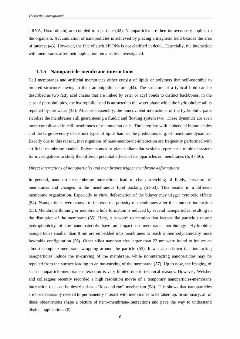
1.1.5 Nanoparticle-membrane interactions
Cell membranes and artificial membranes either consist of lipids or polymers that self-assemble to
ordered structures owing to their amphiphilic nature (44). The structure of a typical lipid can be
described as two fatty acid chains that are linked by ester or acyl bonds to distinct backbones. In the
case of phospholipids, the hydrophilic head is attracted to the water phase while the hydrophobic tail is
repelled by the water (45). After self-assembly, the noncovalent interactions of the hydrophilic parts
stabilize the membranes still guaranteeing a fluidic and floating system (46). These dynamics are even
more complicated in cell membranes of mammalian cells. The interplay with embedded biomolecules
and the large diversity of distinct types of lipids hamper the predictions e. g. of membrane dynamics.
Exactly due to this reason, investigations of nano-membrane-interaction are frequently performed with
artificial membrane models. Polymersomes or giant unilamellar vesicles represent a minimal system
for investigations to study the different potential effects of nanoparticles on membranes (6, 47-50).
Direct interactions of nanoparticles and membranes trigger membrane deformations
In general, nanoparticle-membrane interactions lead to chain stretching of lipids, curvature of
membranes and changes in the membranous lipid packing (51-53). This results in a different
membrane organization. Especially in vitro, deformation of the bilayer may trigger cytotoxic effects
(54). Nanoparticles were shown to increase the porosity of membranes after their intense interaction
(55). Membrane thinning or membrane hole formation is induced by several nanoparticles resulting in
the disruption of the membrane (55). Here, it is worth to mention that factors like particle size and
hydrophobicity of the nanomaterials have an impact on membrane morphology. Hydrophilic
nanoparticles smaller than 8 nm are embedded into membranes to reach a thermodynamically more
favorable configuration (56). Other silica nanoparticles larger than 22 nm were found to induce an
almost complete membrane wrapping around the particle (55). It was also shown that interacting
nanoparticles induce the in-curving of the membrane, while noninteracting nanoparticles may be
repelled from the surface leading to an out-curving of the membrane (57). Up to now, the imaging of
such nanoparticle-membrane interaction is very limited due to technical reasons. However, Welsher
and colleagues recently recorded a high resolution movie of a temporary nanoparticles-membrane
interaction that can be described as a “kiss-and-run” mechanism (58). This shows that nanoparticles
are not necessarily needed to permanently interact with membranes to be taken up. In summary, all of
these observations shape a picture of nano-membrane-interactions and pave the way to understand
distinct applications (6).

Theoretical background
7
1.1.6 Polymeric nanoparticle based drug delivery
Since the regular application of drugs is commonly systemic, the transport of effectors to the site of
action is often less than ideal. The cargo might degrade by hydrolysis, undergo unspecific interactions
with healthy tissues or can be excreted via the biliar system (59). As a promising alternative,
nanoparticle-based drug delivery could circumvent these issues. The proper design of nanoparticles
can protect the cargo from degradation and assists in an efficient drug delivery due to their targeting
properties. In these particular applications, micelles, dendrimers, liposomal formulations and a large
number of different nanoparticles serve as carriers to deliver e. g. siRNA or anti-cancer drugs to the
area of interest (60). Active targeting mechanisms (e. g. “enhanced permeation and retention effect”)
additionally provide the more effective accumulation of nanospheres inside e. g. solid tumors (61).
1.1.6.1 Passive and active targeting of nanoparticles
A fundamental advantage in the treatment of solid tumors is the defective vascular architecture created
by the rapid and uncontrolled growth of blood vessels inside the abnormal tissue (62). Combined with
a poor lymphatic drainage, nanomaterials passively accumulate inside the cancer tissue in consequence
of the EPR effect (63). In addition, an antigen-dependent targeting can be achieved by the specific
binding of nanoparticles to pathological surface proteins. It is worth to mention that cancer cells
overexpress particular antigens on their cell membrane. With the selection of the proper binding
partner, nanoparticle accumulation can be increased by their specific targeting to the cell of interest
(64). This predestines nanoparticles as ideal candidates for the targeting of cancer cells, if the target
molecule is not significantly expressed in other parts of the body (65).
In the past, several investigations confirmed the functionality of active targeting concepts. Dinauer et
al. investigated an antigen-dependent targeting of T cells by anti-CD3-conjugated nanoparticles (66).
They could show that these antibody-functionalized nanoparticles specifically bind to the CD3
receptor, which induces an internalization of the nanospheres (66). Other approaches functionalize
nanoparticles with DC-SIGN antibodies that bind this receptor on dendritic cells (67). Besides
biomolecules, also synthetic molecules can serve for cell specific targeting applications (68). PLGA
nanoparticles can be functionalized with prostate-specific-membrane-antigen-targeting aptamers (69).
The authors demonstrate an efficient and cell specific delivery of the antitumor agent cisplatin to
human prostate cancer cells (69).
However, the design of nanoparticles for active targeting is challenging. The major prerequisites for
these applications are that the targeted antigen needs to be well accessible to the nanoparticles and
displays a high endocytic internalization rate. Unspecific uptake via bulk endocytosis has to be
minimized to prevent unspecific delivery to other types of cells (70). Finally, the interaction of serum
proteins with nanoparticles may change their specific binding properties to the target molecule (25).
Previous reports suggest that the functionality of transferrin-functionalized nanoparticles is screened

Theoretical background
8
by the protein corona, thereby losing their target-specificity (26, 71). A solution for this could be
provided by novel innovative synthesis methods. Here, the targeting molecule is attached via a spacer
to the nanoparticle to circumvent the shielding effects of the protein corona (72). This approach could
set the basis for a successful targeted delivery.
1.1.6.2 Non-targeted nanoparticle uptake and controlled cargo release
A large number of studies investigate the nonspecific uptake mechanisms of nanoparticles (73, 74).
These studies mainly examine nontargeting nanoparticles with an encapsulated cargo to examine the
intracellular drug release in cell culture models (75, 76). Notably, these release mechanisms are highly
diverse (2). Cargo release either occurs by simple drug leakage from the particle matrix or by a
disassembly of the nanomaterial (76). However, after cargo release, the effector molecules are often
trapped inside endosomes while the target location is frequently found in the cytoplasm. To deliver the
cargo more efficiently to the proper subcellular compartment, an efficient endosomal release
mechanism is required.
Endosomal-to-cytoplasmic release either occurs via the membranous diffusion of the payload into the
cytosol or by the destruction/deformation of the endosomal membrane. It was demonstrated that the
decoration of nanoparticles with cell-penetrating peptides (CPP) results in an endosomal-to-
cytoplasmic transfer of the nanocarriers (77). GALA peptides and even other peptides induce a
membrane destabilization exclusively inside the acidic environment of the endolysosome (2, 78). In
accordance to the approach to exploit endolysosomal pH changes for cargo release, also other
applications have been demonstrated (79). Endosomolytic polymers with primary and/or tertiary
amines destabilize the endolysosomal milieu by proton-binding (“proton sponge effect”) (80). These
amines become protonated at an acidic pH (e. g. in the lysosome), which induce a vesicular influx of
protons. Thereby, osmotic swelling and disruption of the endosome occurs (81).
Notably, not only intracellular triggers but also extracellular stimuli are utilized for the controlled
nanoparticulate cargo release into the cytosol. Bräuchle and colleagues demonstrate a photochemical
rupture of the endosomes. Afterwards, a disulfide-bound dye molecule is activated inside the reducing
conditions of the cytoplasm displaying an exposure of the nanoparticulate cargo to the cytosolic milieu
(75). Further investigations reveal a near-infrared light-mediated release of photocaged siRNA from
silica-coated upconversion nanoparticles into the cytosol (82). Taking all of this into account, a
successful passive targeting and cargo release mainly depends on the design of the nanomaterial and
the strategy of release. Further on, uptake routes and the intracellular trafficking of cargo can
orchestrate the effective delivery of nanoparticle (4, 83, 84).

Theoretical background
9
1.2 Uptake mechanisms of nanoparticulate systems
Cells use endocytosis for a vast number of central processes. Uptake of extracellular nutrients and
macromolecules as well as the surface expression of receptors/lipids is orchestrated by the endocytic
system. Endocytosis is even indispensable for processes of cell adhesion and signal transduction (85).
Moreover, these mechanisms serve as entry portals for pathogens or particles to gain access to the
interior of a cell. Thereby, they exploit a large number of different pathways (86) (Figure 3). Here,
clathrin-mediated endocytosis (CME) is undoubtedly the best studied mechanism of endocytosis (87).
Other clathrin-independent pathways are caveolae-dependent endocytosis, RhoA-mediated
endocytosis, Arf6-mediated entry, flotillin-orchestrated uptake as well as the CLIC/GEEC pathway
(clathrin-independent carriers and GPI-enriched endocytic compartments) (85, 88-93). The bulk
uptake of large volumes of extracellular fluids and particles is mainly mediated by (macro)pinocytic
and phagocytic mechanisms (94, 95).
Figure 3: Overview of the major endocytic pathways in mammalian cells. Modified from (85, 93, 96).
1.2.1 Clathrin- and caveolae-mediated endocytosis
Clathrin-mediated endocytosis (CME) is required for the internalization of surface molecules such as
transferrin receptors, receptor tyrosine kinases or G-protein coupled receptors (97). Further on, cargoes
such as cholera toxin B subunit or nanoparticles have been shown to utilize clathrin-mediated
pathways (98, 99). The initial process of CME is accompanied by the assembly of cytoplasmic clathrin
at regions of the plasma membrane (PM) called clathrin-coated pits (CCPs) (100). After the
invagination of CCPs, the vesicles are pinched off from the PM to form clathrin-coated vesicles
(CCVs) (101). The molecular signaling on CCVs is mainly mediated by proteins of the adaptor
complex (102). These adaptor proteins interact with a variety of other regulatory proteins (e. g. AP180
and epsin) that influence pit assembly, vesicle budding and cytoskeleton interactions (87, 103).
One regulatory protein that is involved in clathrin lattice rearrangement is epsin 15, which interacts
with clathrin via the adaptor protein 2 (104, 105). In general, epsins act as scaffolding proteins that

Theoretical background
10
generate a curved and three dimensional amphiphatic helix for the assembly of clathrin baskets (87).
Therefore, it is not surprising that the overexpression of the dominant negative mutant epsin 15 (DIII)
suppresses the formation of clathrin-coated pits (104, 106). This shows that the associated regulatory
proteins seem to have an important effect on CME. This also affects the regulatory protein AP180
(103). Zhao and colleagues found that the overexpression of AP180 reduces clathrin accumulations on
the plasma membrane. This provides information that AP180 is an important organizer in CME (103).
Altogether, it is worth to mention that CME is a well understood endocytic pathway. CME as well as
caveolae-mediated endocytosis are potential entry pathways of nanoparticles that necessarily have to
be studied in detail.
Caveolae-mediated endocytosis
Caveolae are 50 - 80 nm large plasma membrane invaginations that orchestrate a clathrin-independent
endocytic pathway (107). On a structural level, caveolae are composed of cholesterol, several
sphingolipids, GPI-anchored proteins and the integral membrane proteins caveolin-1 and caveolin-2
(108, 109). Caveolin-1 acts as the major organizing unit into which caveolin-2 is migrating (110, 111).
This was shown with two experiments. The first experiment reveals that the stable expression of
caveolin-1 in caveolae-deficient K562 cells induces the upregulation and recruitment of caveolin-2.
Then, Parolini and colleagues showed a direct binding of both proteins to each other providing
information about two necessary markers for the detection of caveolae at the PM (111). Originally,
caveolae-mediated endocytosis was described as an entry mechanism of SV40 viruses (112).
However, also other small particles with a size of ≤ 100 nm have been shown to utilize caveolae-
mediated pathways (113, 114). The terminal uptake of such nanoparticles is then conducted by the
pinch off of the vesicles from the PM. The scission of vesicles in caveolae- as well as in clathrin-
mediated uptake is regulated by dynamin.
1.2.2 Dynamin in clathrin- and caveolae-mediated endocytosis
Dynamin is a GTPase that is recruited to CCPs and caveolae during the endocytic process (115, 116).
The protein assists in the scission of vesicles from the plasma membrane after its slow accumulation
around the growing pit (117). Originally, the relevance of dynamin for endocytosis was shown in
experiments, where the overexpression of dominant negative Dyn2-K44A results in the inhibition of
clathrin- and caveolae-mediated endocytosis (118). Morphologically, dynamin inhibition arrests
endocytosis at a stage, where vesicles are trapped as tubular structures at the PM (116, 119). This
results in an accumulation of non-budded vesicles (116). Altogether, this reveals a clear role for
dynamin inside the two best characterized endocytic pathways. However, it is worth to mention that
dynamin is also located inside membrane ruffles or podosomes (120, 121). These observations revive
the discussions, whether dynamin exclusively acts in CME and caveolae-mediated endocytosis or also
plays a role in other endocytic mechanisms (122).

Theoretical background
11
1.2.3 Clathrin and caveolae-independent mechanisms
Nowadays, lively discussions raise issues about the presence of other less characterized endocytic
pathways. In this research field, the literature provides a wide range of clathrin- and caveolae-
independent mechanisms (85). Though, the strict discrimination between these pathways is sometimes
challenging since functional overlaps have partially been observed (88, 90). This rigorous
classification is also hampered by the large number of investigated cargoes including viruses, toxins,
GPI-linked proteins, interleukin receptors or other cargoes. However, the commonly accepted clathrin-
, caveolae- and macropinocytosis-independent pathways are RhoA-mediated uptake, CLIC/GEEC
pathways and flotillin-mediated endocytosis.
1.2.3.1 RhoA-mediated uptake
One clathrin-independent but dynamin-dependent pathway is specifically regulated by the small
GTPase RhoA (91, 107, 123). This pathway was first discovered in a study that investigated the
internalization of the β-chain of the interleukin-2 receptor (IL-2Rβ) (107, 124). It was shown that
neither dominant negative epsin 15 nor AP180 overexpression affected the uptake of IL-2Rβ (124).
Clathrin-independency was further supported by the criterion that IL-2Rβ uptake is orchestrated by
Rac1, PAK1 and PAK2 (123). These factors are not functional in CME. However, observations have
shown that the overexpression of constitutively active RhoA can inhibit CME (124). Despite of this, a
direct interaction of CME and RhoA is unlikely (85). Altogether, RhoA acts in a clathrin-independent
pathway by the modulation of the actin cytoskeleton, thereby probably regulating a large number of
other processes (125).
1.2.3.2 CLIC/GEEC pathway
A further clathrin- and caveolae-independent mechanism is represented by the CLIC/GEEC pathway
(90). This entry mechanism orchestrates the internalization of glycosylphosphatidylinositol-anchored
proteins, extracellular fluids and certain bacterial exotoxins (90). Morphologically, the CLIC/GEEC
pathway is characterized by its ~ 40 nm wide tubular invaginations that are decorated with GTPase
regulator associated with focal adhesion kinase-1 (GRAF1) and phosphatidylinositol-4,5-bisphosphate
3-kinase (126). These membrane carriers are relatively devoid of caveolin-1 and flotillin-1 but are
regulated by cdc42 and ARF1 (90, 95, 126-128). Interestingly, the internalized GPI-linked proteins
can bypass conventional Rab5+ endocytic compartments raising questions of novel early endocytic
compartments (85). Indeed, GPI-anchored proteins are directly transported to early endocytic vesicles
termed as GPI-AP-enriched early endosomal compartment (GEEC). Altogether, these observations
provide information about a pathway of fluid phase endocytosis that has to be taken into account,
when investigating nanoparticle uptake.

Theoretical background
12
1.2.3.3 Flotillin-mediated endocytosis
Flotillins are membrane proteins with a high homology to caveolin-1 that cluster as organized domains
inside the PM (129). In 2006, flotillin-1 was originally described as a protein that mediates the entry of
ctxB and the GPI-anchored protein CD59 (88). The authors show that flotillin domains pinch off from
the PM in a clathrin-independent manner. After internalization, flotillins are present inside
multivesicular bodies and even show a close association to other endosomal processes (130, 131).
Flotillins have been suggested to be necessary for the Niemann-Pick-C1-like 1-mediated cholesterol
uptake inside endosomes (130). It was shown that the knockdown of flotillins significantly attenuates
the internalization of cholesterol and the endocytosis of NPC1L1 (130). These observations could
bridge the gap for flotillin in nanoparticle uptake. Indeed, previous studies with polyplexes and silica
nanoparticles showed a participation of flotillin in nanoparticle internalization (132, 133). All of these
points propose a versatile but important role for flotillin in endocytic processes that have to be
considered in the analysis of nanoparticle entry and trafficking.
1.2.4 Macropinocytosis
Classical macropinocytosis is a clathrin-, caveolae- and dynamin-independent process that occurs
spontaneously or can be initiated by e. g. a transient activation of receptor tyrosine kinases (Figure 4
and Figure 5) (134). During macropinocytosis, large volumes of fluids and extracellular objects are
internalized via macropinosomes (135, 136). Morphologically, macropinocytosis can be characterized
by ruffle formation and the internalization of classical fluid phase markers (e. g. HRP, dextran) (137).
This process of internalization is orchestrated by several small GTPases such as Ras, Cdc42 and Rac1
(138, 139). The early steps in macropinocytic signaling depend on the Ras/Cdc42/Rac1- or PI3K-
mediated activation of different kinases such as p21-activated kinase 1 (PAK1) (136). Active PAK1
induces actin polymerization and lamellipodia formation (137). In the terminal step of
macropinocytosis, PAK1 facilitates the closure of the macropinosomes by activating the carboxy-
terminal-binding protein-1/brefeldin A-ADP ribosylated substrate (CtBP-1/BARS) (135, 140).
Figure 4: Major stages in receptor-mediated macropinocytosis.

Theoretical background
13
Other factors that influence classical macropinocytosis
Considering the interactions of small GTPases on actin remodeling, it is not surprising that Rah/Rab34
has been associated with macropinosome formation (141). Other studies describe the impact of Rab5
and Rabankyrin-5 on the macropinocytic activity of fibroblasts and epithelial cells (142). While
overexpression of Rabankyrin-5 increases the macropinocytic activity of the cells, an RNAi-mediated
downregulation of Rabankyrin-5 diminishes the internalization rate of fluid phase markers and lipid
nanoparticles (4, 142). The diversity of action mechanisms is also highlighted by the studies that
implicate the kinases PLCγ, PKC and Src as factors for membrane ruffling (140, 143, 144). Besides,
the ADP-ribosylation factor Arf6 was shown to alter membrane protrusions (145). Altogether, these
studies connect a large number of factors to macropinocytic signaling events. With regards to actin
rearrangements, small GTPases and several kinases seem to have major impacts on the macropinocytic
activity of a cell and are the major targets during the investigations of nanoparticle uptake (Figure 5).
Figure 5: Overview of the general macropinocytic signaling pathways. Macropinocytosis occurs spontaneously or is
triggered by a growth factor-mediated activation of receptor tyrosine kinases (RTKs). Several small GTPases and kinases
facilitate the signalling cascade resulting in actin modulation. Actin reorganization induces the formation of the
macropinocytic cup. Modified from (140, 146).
Macropinocytosis-like mechanisms
Classical macropinocytosis is primarily reliant on the previously described criteria. However, it is
worth to mention that also atypical macropinocytic-like mechanisms have been observed in many
studies (140, 147, 148). Macropinocytic-like mechanisms depend on parameters that are normally not

Theoretical background
14
considered in fluid phase endocytosis or lack in comparison to classical macropinocytosis (140). In the
field of virology, a large number of different atypical macropinocytic types have been well studied
(140). Mulherkar et al. investigated a dynamin2-dependent macropinocytic entry of Ebola virus
glycoprotein. Since dynamin has never been implicated in classical macropinocytosis so far, this
mechanism can be considered as a macropinocytic-like pathway (148). Other groups reported an F-
actin and cholesterol-dependent, but an atypical pH-independent uptake of human cytomegalovirus
into dendritic cells (149). Another case of macropinocytic-like entry was published for human
papilloma virus type 16 (150). Entry and infection relies on the major hallmarks of classical
macropinocytosis but does not depend on Rho GTPases which are major key players in
macropinocytosis. In the field of nanotechnology, macropinocytic-like uptake mechanisms have
hardly been investigated. However, a study of Iversen et al. focused on several criteria of classical
macropinocytosis during quantum dot uptake. They found that ricin-conjugated quantum dots are
internalized via a dynamin-dependent macropinocytic-like pathway that was not described before
(147). Taken together, atypical macropinocytic-like uptake mechanisms are not only relevant in
virology but can also be an issue in the investigation of nanoparticulate entry and intracellular
nanoparticle trafficking.
1.3 From early endocytic compartments to lysosomes – intracellular
trafficking of nanomaterials
As described in the former chapters, nanoparticles utilize different endocytic mechanisms for their
cellular entry (93, 96). After their internalization, nanoparticles are transported along a specific
endocytic route (83, 151, 152). The process of intracellular nanoparticle trafficking (INT) is very
dynamic in record to the proteomic environment on the endocytic vesicles. During INT, important
adaptor and effector proteins are recruited or exchanged on the vesicles´ surface (153). It is important
to mention that homotypic/heterotypic fusion of vesicles and vesicular maturation processes can alter
the protein composition on a nanoparticle-containing vesicle (153, 154). This already happens in the
initial stages of intracellular trafficking. Early endosomes and early macropinosomes merge with each
other to form a novel vesicle (140). Moreover, endocytic interactions with recycling endosomes as
well as with vesicles of the trans-Golgi network induce the additional exchange of nonendocytic
proteins with the endolysosomal system (154). Also the fusion of lysosomes with autophagosomes
introduces components of e. g. defective organelles into the endocytic system (155, 156). Figure 6
shows the fusion events and participating vesicles that may be associated with intracellular
nanoparticle transport.

Theoretical background
15
Figure 6: Potential routes of intracellular nanoparticles trafficking. The intravesicular pH values of the endolysosomal
vesicles are depicted as distinctly colored vesicles. Abbreviations: EE: Early endosomes; RE: Recycling endosome; LE: Late
endosome/multivesicular body; LY: Lysosome; TGN: Trans-Golgi-Network. Arrows indicate endocytic fusion events
between the different compartments. Dotted lines indicate the possible macropinocytic interplay with the endocytic system.
Adapted from (140, 157, 158).
1.3.1 Early endocytic compartments
Early endocytic vesicles are a heterogeneous class of vesicular carriers during the initial stage of
intracellular trafficking (142, 159). Classical early endosomes can be identified by distinct markers
such as EEA1 or Rab5 and coexist in different subpopulations (159, 160). A previous report identified
at least two distinct classes of EE that display different maturation and mobility kinetics (161). The
authors detected a first population of Rab5+ endosomes that rapidly accumulated Rab7 within a short
time frame. The second population matures on a much slower time scale without accumulating Rab7
(161). According to these data, the mobility of vesicles on microtubules can be correlated with their
maturation state (161). While slowly maturating endosomes move to a less extent, rapidly maturing
vesicles show a higher motility rate. If this is connected to the distinct Rab domains on early
endosomes is not yet clarified.

Theoretical background
16
However, it is worth to mention that early endosomes contain separated surface patches of Rab4, Rab5
and Rab11 (162). Especially, Rab4 and Rab11 have been shown to control recycling and endosomal
fusion (162, 163). According to the study of Sönnichsen et al. it was proposed that the different
domains on early endosomes promote the pinch off of early endosomal subvesicles that are introduced
into the recycling pathway (Figure 6) (162). This shows that early endosomal compartments are
closely related precursors of the cellular recycling pathways. However, these classical endosomes
seem to be functionally different from early macropinosomes.
Early macropinosomes
Early macropinosomes are budded vesicles that are formed after macropinocytosis and partially bear
markers of early endosomes (142). Classical macropinosomes are characterized by the surface markers
Rab34, Rab5 and its effector Rabankyrin-5 (141, 142). Other studies highlight the transient association
of switch-associated protein 70 (SWAP-70) with macropinosomes in dendritic cells (164). Further
well accepted markers of growth factor induced macropinosomes are proteins of the sorting nexin
family as well as Rac1 (137, 165). The biogenesis of macropinosomes can be triggered by EGF
stimulation or via the overexpression of several sorting nexins such as SNX1 or SNX18 (166). After
macropinosomes formation, vesicles can fuse with other endocytic compartments or mature to Rab7+
late macropinosomes (140, 165, 167).
1.3.2 Late endosomes – Multivesicular bodies
Late endosomes are also termed as multivesicular bodies (MVBs) (168). These vesicles frequently
originate from Rab5+ early endosomes and are considered to be control centers for different processes
such as cell signaling or exocytosis (153, 168). Morphologically, MVBs are 400 – 1000 nm large
organelles that contain small intraluminal vesicles (ILVs) in a size range of typically ~ 50 nm (169).
When MVBs fuse with the plasma membrane, ILVs are externalized as exosomes in a Rab27b-
dependent manner (170, 171). All of these processes are well studied. However, the definite
identification of MVBs remains challenging. Unique marker proteins are relatively few since MVB
biogenesis mainly occurs transiently (168). Nowadays, characterization of MVBs is therefore
performed by the detection of mannose 6-phosphate receptor (M6PR), Lamp1/Lamp2, distinct Rab
proteins (Rab5, Rab7, Rab27, Rab35) as well as several tetraspanins (172). Here, it is to mention that
the tetraspanins CD82, CD9, CD81 and CD63 are abundant on MVBs (171, 173). Also the
intraluminal vesicles can be directly detected by the tetraspanin CD63 (174). In previous reports it was
shown that CD63 transports the melanocytic protein Pmel17 on ILVs in an ESCRT (endosomal
sorting complexes required for transport)-independent process (174, 175).
Contrary to the sorting of Pmel17 onto ILVs, the biogenesis of MVBs as well as the degradation of
proteins inside the endocytic system is mediated by the ESCRT machinery (176). For MVB

Theoretical background
17
biogenesis, Wollert and Hurley showed that the four components of ESCRT (0/I/II/III) induce the
formation of intraluminal vesicles inside giant unilamellar vesicles (176). Thereby, they recorded the
internalization of fluorescently-labeled ubiquitin. Inside cells, ubiquitinylation is an important
modification for the degradation of membrane proteins via ESCRT. Monoubiquitinylated or
polyubiquitinylated membrane proteins bind the ubiquitin-binding domain of ESCRT components,
which results in the degradation of the proteins (177). Altogether, the functional relevance of
functional ESCRTs has been well characterized. This was also shown in biomedical publications.
Deregulation of the ESCRT machinery contributes significantly to many human diseases such as
cancer or neurological diseases (178).
However, it is worth to mention that not only the late endosomal deregulation of ESCRT can mediate
severe diseases. In a particular case, a mutation of Niemann-Pick-disease C1 protein (NPC1) leads to a
pathological cholesterol accumulation inside late endosomes (83, 179). Consequently, cholesterol
homeostasis and intracellular trafficking pathways are disturbed. In this context, Sahay and colleagues
showed that NPC1 deficient cells longer retain nanotransporters inside the endosomal system than
healthy cells (83). This implies an important function of NPC1-dependent cholesterol homeostasis in
intracellular trafficking pathways of nanoparticles (180).
1.3.3 Lysosomes
Lysosomes are seen as terminal organelles in endocytic and autophagocytic pathways (158). These
vesicles are the final destination for a large number of cargos and mediate numerous functions such as
the downregulation of surface receptors, inactivation of pathogens or MHC-peptide loading (151,
156). Though, not only the functions of lysosomes are diverse. Electron microscopy reveals that the
morphology and size of lysosomes is strikingly heterogeneous (181). They frequently contain
electron-dense material and multilamellar membrane whorls (181). This material partially originates
from the lysosomal fusion with autophagocytic vesicles resulting in the formation of a multilamellar-
body-like structure (182).
Moreover, their acidic interior (pH 4.6-5) is maintained by several vacuolar H+ ATPases (183). This is
especially important for hydrolytic enzymes with an acidic pH optimum (184). Lysosomes supply a
protective membrane-shielded lumen for more than 50 different acid hydrolases (e. g. peptidases,
proteinases, phosphatases, nucleases, glycosidases, sulphatases, lipases) (156). The transport of these
proteins towards prelysosomal vesicles is frequently orchestrated by two independent M6P-receptors
that recognize the M6P tag on the potential lysosomal proteins (185). After protein binding, the
M6PR-protein-complex is shuttled from the trans-Golgi-network to endosomal vesicles, where the
cargo dissociates from M6PRs due to the acidic environment (186). As a consequence, lysosomes can
be distinguished from prelysosomal organelles by the lack of M6PR and the presence of Lamp1 and
Lamp2 (186).

Theoretical background
18
1.3.4 Fusion events and protein transport inside the endocytic system
In a protein-free system, the fusion of vesicles is mainly based on the interaction of different forces
between the two vesicular surfaces (187). Attractive forces like hydrophobicity and van der Waals
forces have to overcome several repulsive forces (e. g. electrostatic) for a successful adhesion of the
vesicles to each other (188). However, it is believed that the fusion of membranes cannot be induced
by simple adhesion, but by factors (e. g. surface charges) that alter the steric configuration of
individual lipids (189). The destabilization of single lipid domains facilitates the hydrophobic
interplay between lipids on opposing membranes resulting in their fusion (190, 191). In cellular
systems, fusion of organelles is additionally supported by two preliminary steps that are mediated by
protein complexes. In the first step, a tethering step loosely connects the two merging vesicles to each
other. In the subsequent step of docking, an irreversible binding of the vesicles is achieved that results
in the terminal fusion of the organelles (181).
The tethering of two early endosomes is mediated by the class C core vacuole/endosome tethering
complex (CORVET) while late endosomes and lysosomes are connected via the vacuole protein
sorting complex (HOPS) (192). In late endosomes/lysosome fusion processes, Rab7 is utilized as an
adaptor protein for the effectors of the HOPS complex (181). Subsequently after formation of the
tethering complex, the vesicular fusion is induced by the assembly of a trans-SNARE complex (181).
SNARE (soluble N-ethylmaleimide-sensitive factor attachment protein receptor) proteins are
characterized by a stretch of 60-70 amino acids that are arranged in heptad repeats (193). When a
SNARE protein encounters other SNARE motifs, an alpha-helical structure is formed. In heterotypic
late endosome/lysosome fusion, the SNARE complex consists of the R-SNARE VAMP7, Syntaxin-7,
Syntaxin-8 and VTI1B (181). In homotypic endosome/endosome fusion, VAMP8 is utilized instead of
VAMP7. It is believed that the formation of the SNARE complex finally results in the induction of
vesicular fusion (194). Even in macropinosomes/lysosome fusion and in autophagosome/lysosome
fusion a participation of SNARE proteins was observed (181). This gives rise to investigate SNARE
proteins in intracellular nanoparticle trafficking.
1.3.5 Intracellular trafficking of nanomaterials
The first approaches that studied the intracellular trafficking of nanoparticles have already been made.
Via the use of confocal spinning disc microscopy, Sandin et al. showed that polystyrene nanoparticles
are transported via Rab7+ endosomes to their terminal lysosomal destination (151). Another study
revealed similar results with polyplexes that actively moved through the cell inside endolysosomal
structures (132). Both of these studies describe less colocalization of nanomaterials with recycling
pathways and autophagocytic routes. However, nanoparticles can also exploit several secretory and
endocytic pathways. Sahay and colleagues observed a transport of lipid nanoparticles into Rab11+
endosomes implicating that LNPs are trafficked via the recycling pathways of the cell (83). Other

Theoretical background
19
reports detected α-Al2O3 nanoparticles inside LC3+ autophagosomes (152). All of these studies used
well accepted markers for their investigations. However, these examinations are biased by the present
knowledge about the recently known proteins in intracellular trafficking pathways of other objects
such as viruses or membrane proteins. Therefore, one primary aim of this thesis is to provide novel
information about the intracellular trafficking pathways in an unbiased approach.

Aims of the study
20
1.4 Aims of the study
This study was carried out to analyze the drug delivery abilities, the entry mechanisms and the
intracellular trafficking of several polymeric nanoparticles. Thereby, the analysis of nano-bio
interactions could reveal novel insights in terms of prospective applications for nanoparticles. In the
first part of this study, we wanted to investigate whether biodegradable poly-L-lactide nanoparticles
are a suitable candidate for a non-invasive drug delivery system in vitro. To analyze this, nanoparticle-
membrane interaction were preliminary studied with distinct cellular and cell-free systems. The
primary goal was to investigate the release mechanism and the subcellular transport of the
nanoparticulate cargo during nanoparticle-membrane interaction.
Secondly, we were interested in the mechanisms by which polymeric nanoparticles enter the cell. In
this project, we focused on the interaction of nanoparticles with the macropinocytic system. The
internalization of nanoparticles could rely on the classical factors of macropinocytic signaling.
Consequently, we intensively studied the influences of small GTPases and kinases on the
internalization mechanism of nanoparticles. To validate the functionality of these GTPases in
nanoparticle entry, we studied whether constitutively active or dominant negative forms of these
effector proteins have an impact on nanoparticle entry. Additionally, a set of small inhibitory
molecules and siRNAs was tested to inhibit several pathways of the endocytic system. We
investigated, whether this had an effect on nanoparticle entry.
In the third part of this thesis, we examined the intracellular trafficking of nanoparticles (INT). To
identify novel proteins of INT, we wanted to establish an innovative and unbiased method that enables
us to identify novel proteins in intracellular nanoparticle trafficking. The goal of this approach was to
dissect the INT by label free quantitative mass spectrometry. To test the specificity of this method, we
investigated promising proteins by confocal live cell imaging and other methods that could influence
the nanoparticle trafficking. We finally examined whether the lysosomal milieu had an impact on the
degradation of the nanoparticulate protein corona.
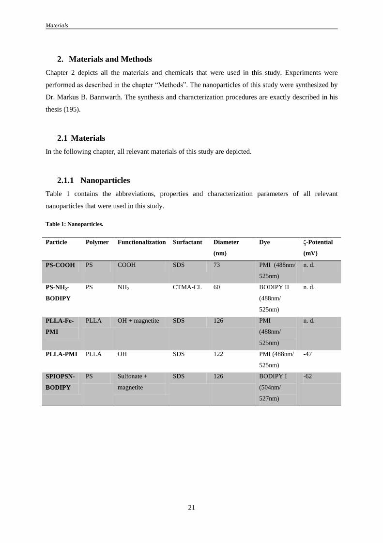
Materials
21
2. Materials and Methods
Chapter 2 depicts all the materials and chemicals that were used in this study. Experiments were
performed as described in the chapter “Methods”. The nanoparticles of this study were synthesized by
Dr. Markus B. Bannwarth. The synthesis and characterization procedures are exactly described in his
thesis (195).
2.1 Materials
In the following chapter, all relevant materials of this study are depicted.
2.1.1 Nanoparticles
Table 1 contains the abbreviations, properties and characterization parameters of all relevant
nanoparticles that were used in this study.
Table 1: Nanoparticles.
Particle Polymer Functionalization Surfactant Diameter
(nm)
Dye ζ-Potential
(mV)
PS-COOH PS COOH SDS 73 PMI (488nm/
525nm)
n. d.
PS-NH2-
BODIPY
PS NH2 CTMA-CL 60 BODIPY II
(488nm/
525nm)
n. d.
PLLA-Fe-
PMI
PLLA OH + magnetite SDS 126 PMI
(488nm/
525nm)
n. d.
PLLA-PMI PLLA OH SDS 122 PMI (488nm/
525nm)
-47
SPIOPSN-
BODIPY
PS Sulfonate +
magnetite
SDS 126 BODIPY I
(504nm/
527nm)
-62

Materials
22
2.1.2 Chemicals
Chemicals used in this study are listed in Table 2.
Table 2: Chemicals.
Chemical Supplier
1,2-Dioleoyl-sn-glycero-3-phosphocholine Avanti Polar Lipids; U. S. A.
2-[4-(2-hydroxyethyl)piperazin-1-yl]ethanesulfonic acid Sigma Aldrich; U. S. A.
2-Propanol Sigma Aldrich; U. S. A.
3-[(3-Cholamidopropyl)dimethylammonio]-1-propanesulfonate
hydrate
Serva,Germany
4-(2-hydroxyethyl)-1-piperazineethanesulfonic acid Sigma Aldrich; U. S. A.
Agar Sigma Aldrich; U. S. A.
Agarose Standard Roth; Germany
Ampicillin Sigma Aldrich; U. S. A.
BODIPY I/ BODIPY II Synthesized as described (196)
Bovine serum albumin Sigma Aldrich; U. S. A.
Calcium chloride Sigma Aldrich; U. S. A.
cOmplete protease inhibitor cocktail Roche; Switzerland
D(+)-Saccharose Roth; Germany
Dimethylsulfoxid Sigma Aldrich; U. S. A.
Dipotassium phosphate Sigma Aldrich; U. S. A.
DL-Dithiothreitol Sigma Aldrich; U. S. A.
Ethanol Sigma Aldrich; U. S. A.
Ethylene glycol-bis(2-aminoethylether)- tetraacetic acid Sigma Aldrich; U. S. A.
Ethylenediaminetetraacetic acid Roth; Germany
Fetal bovine serum Invitrogen; U. S. A.
Gentamicin Invitrogen; U. S. A.
Gluthatione Sigma Aldrich; U. S. A.
Glycine Sigma Aldrich; U. S. A.
Immersion oil for microscopy Leica; Germany
Magnesium chloride Sigma Aldrich; U. S. A.
Methanol Fisher Scientific; U. S. A.
N-(2,6-diisopropylphenyl)-perylene-3,4-dicarbonacid-imide (PMI) BASF; Germany
Nile Red Sigma Aldrich; U. S. A.
Nuclease-free water Qiagen; U. S. A.
Oil Red O Sigma Aldrich; U. S. A.
Paraformaldehyde Sigma Aldrich; U. S. A.
Penicillin/Streptomycin Invitrogen; U. S. A.
Peptone Sigma Aldrich; U. S. A.
Ponceau S Sigma Aldrich; U. S. A.

Materials
23
Potassium chloride Sigma Aldrich; U. S. A.
Pyruvate Life technologies; U. S. A.
Saponin Sigma Aldrich; U. S. A.
Sodium chloride Sigma Aldrich; U. S. A.
Sodium hydroxide Sigma Aldrich; U. S. A.
Thio-Urea Serva, Germany
Triton X-100 Sigma Aldrich; U. S. A.
Trizma Base Sigma Aldrich; U. S. A.
Trypan blue solution (0.4%) Sigma Aldrich; U. S. A.
Tryptone Sigma Aldrich; U. S. A.
Urea Serva, Germany
Yeast extract Sigma Aldrich; U. S. A.
2.1.3 Instruments and Consumables
Table 3 shows the instruments that were used in this work.
Table 3: Instruments.
Instrument Supplier
4D Nucleofector Lonza; Switzerland
Automated cell counter TC10 Bio-Rad; U. S. A.
Bacteria incubator UM200 Memmert; Germany
Cell culture flow c-(Max Pro)3-130 Berner; Germany
Cell culture incubator C200 Labotec; Germany
Centrifuge 5810R and 5430 Eppendorf; Germany
Confocal laser scanning microscope Leica SP5 II with CW-
STED
Leica; Germany
Dry blotting system iBlot® Invitrogen; U. S. A.
Flow cytometer CyFlow ML Partec; Germany
Fluorescence imager LAS-3000 Fujifilm, Japan
Fluorescence microscope IX81-ZDC Olympus; Germany
Freezer -20° Liebherr; Germany
Freezer -80°C Hera Freeze Top Thermo Scientific; Germany
Hamilton pipette Hamilton Company; U. S. A.
Incubator Innova 44 New Brunswick Scientific; U. S. A.
Inverted microscope CKX41 Olympus; Germany
Microcentrifuge Minispin plus Eppendorf; Germany
Microscale AE100 Mettler Toledo; Switzerland
Nitrogen tank LS6000 Taylor-Wharton; Germany
PCR Mastercycler EP Gradient S Eppendorf; Germany
Pipettes Eppendorf; Germany

Materials
24
Pipetting aid Accujet Pro Brand; Germany
Platereader Infinite M1000 Tecan; Germany
Power supply 250-EX Life technologies; U. S. A.
Real time system CFX96 Bio-Rad; Germany
SDS-PAGE/Western Blot chamber Mini/Maxi Biometra; Germany
Thermo mixer HL by Ditabis; Germany
Ultracentrifuge OptimaTM
MAX Beckman Coulter; U. S. A.
UV-Vis photospectrometer ND-8000 Thermo Scientific; Germany
Vacuum pump Vacusafe comfort IBS Integra Bioscience; Germany
Vortexer Reax Control Heidolph; Germany
Water bath Memmert; Germany
Table 4 displays the used consumables of this work.
Table 4: Consumables.
Consumables Supplier
27G needle BD Biosciences; U. S. A.
Adhesive PCR film Bio-Rad; Germany
Cell culture flasks Greiner; Germany
Celltrics sterile filters Partec, Germany
FACS tubes Partec; Germany
Falcon tubes (10 ml; 50 ml) Greiner; Germany
Ibidi itreat µ-dishes IBIDI; Germany
iBlot® anode stack Life technologies, U. S. A.
iBlot® cathode stack Life technologies, U. S. A.
Multiwell plates Corning Inc.; U. S. A.
Nucleofector cuvettes (100 µl) Lonza, Switzerland
NuPAGE 10% bis-tris gel Life technologies; U. S. A.
Parafilm Pechiney Plastic Packaging, U. S. A.
PCR tubes/PCR plates Bio-Rad; Germany
Pipette tips Greiner, Germany
Pipettes (5 ml; 10 ml; 25 ml; 50 ml) Greiner, Germany
Protran®
nitrocellulose membrane Sigma Aldrich; Germany
PVDF membrane Millipore, U. S. A.
QIA shredder columns Qiagen; U. S. A.
Single-use syringes Henke-Sass-Wolf; Germany
Sterile filter (0.2 µm; 0.45 µm) Millipore, U. S. A.

Materials
25
2.1.4 Small molecule inhibitors
Table 5 depicts the inhibitors with their known targets/mechanisms that were used for the suppression
of different endocytic mechanisms. Inhibitors were dissolved and stored according to manufacturer´s
recommendations.
Table 5: Small molecule inhibitors of uptake.
Inhibitor Supplier
5-(N-Ethyl-N-isopropyl) amiloride (lowering of submembraneous
pH)
Sigma Aldrich; U. S. A.
Bafilomycin A1 (Inhibition of v-type ATPases) Sigma Aldrich; U. S. A.
Blebbistatin (Inhibition of Myosin) Sigma Aldrich; U. S. A.
Brefeldin A (Inhibition of ARF1) Sigma Aldrich; U. S. A.
CA-074 methyl ester (Inhibition of cathepsins) Sigma Aldrich; U. S. A.
Chlorpromazine (Inhibition of CME) Sigma Aldrich; U. S. A.
Cytochalasin D (Inhibition of F-Actin polymerization) Sigma Aldrich; U. S. A.
Dynasore® (Inhibition of Dynamin I/II) Sigma Aldrich; U. S. A.
IPA-3 (Inhibition of PAK1) Tocris; U. S. A.
Ly294002 (Inhibition of PI3K) Tocris; U. S. A.
Methyl-β-cyclodextrin (Cholesterol depletion) Sigma Aldrich; U. S. A.
Nocodazole (Inhibition of microtubule polymerization) Sigma Aldrich; U. S. A.
Ro 31-8220 (Inhibition of PKC) Sigma Aldrich; U. S. A.
U18666A (Cholesterol synthesis inhibitor) Merck; Germany
U73122 (Inhibition of PLC) Sigma Aldrich; U. S. A.
Wortmannin (Inhibition of PI3K) Sigma Aldrich; U. S. A.
2.1.5 Cell lines and primary cells
Cell lines used in this study are listed in Table 6.
Table 6: Cell lines.
Name Cell type Supplier
Jurkat Immortalized CD3+ T lymphocytes DSMZ; Germany
HeLa Cervical carcinoma DSMZ; Germany
Multipotent human
stroma cells
(hMSCs)
Human bone-marrow derived stem cells+ for
CD29, CD73, CD44, CD90, CD105, CD146,
CD166 and MHC class I; negative for CD1a,
CD3, CD14, CD16, CD19, CD34, CD45,
CD133, MHC class II and SSEA2
University of Ulm; Germany
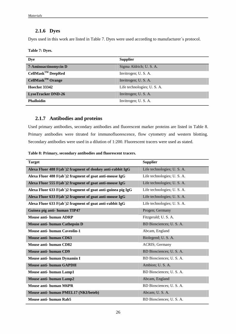
Materials
26
2.1.6 Dyes
Dyes used in this work are listed in Table 7. Dyes were used according to manufacturer´s protocol.
Table 7: Dyes.
Dye Supplier
7-Aminoactinomycin D Sigma Aldrich; U. S. A.
CellMaskTM
DeepRed Invitrogen; U. S. A.
CellMaskTM
Orange Invitrogen; U. S. A.
Hoechst 33342 Life technologies; U. S. A.
LysoTracker DND-26 Invitrogen; U. S. A.
Phalloidin Invitrogen; U. S. A.
2.1.7 Antibodies and proteins
Used primary antibodies, secondary antibodies and fluorescent marker proteins are listed in Table 8.
Primary antibodies were titrated for immunofluorescence, flow cytometry and western blotting.
Secondary antibodies were used in a dilution of 1:200. Fluorescent tracers were used as stated.
Table 8: Primary, secondary antibodies and fluorescent tracers.
Target Supplier
Alexa Fluor 488 F(ab´)2 fragment of donkey anti-rabbit IgG Life technologies; U. S. A.
Alexa Fluor 488 F(ab´)2 fragment of goat anti-mouse IgG Life technologies; U. S. A.
Alexa Fluor 555 F(ab´)2 fragment of goat anti-mouse IgG Life technologies; U. S. A.
Alexa Fluor 633 F(ab´)2 fragment of goat anti-guinea pig IgG Life technologies; U. S. A.
Alexa Fluor 633 F(ab´)2 fragment of goat anti-mouse IgG Life technologies; U. S. A.
Alexa Fluor 633 F(ab´)2 fragment of goat anti-rabbit IgG Life technologies; U. S. A.
Guinea pig anti- human TIP47 Progen; Germany
Mouse anti- human ADRP Fitzgerald; U. S. A.
Mouse anti- human Cathepsin D BD Biosciences; U. S. A.
Mouse anti- human Caveolin-1 Abcam, England
Mouse anti- human CD63 Biolegend; U. S. A.
Mouse anti- human CD82 ACRIS; Germany
Mouse anti- human CD9 BD Biosciences; U. S. A.
Mouse anti- human Dynamin I BD Biosciences; U. S. A.
Mouse anti- human GAPDH Ambion; U. S. A.
Mouse anti- human Lamp1 BD Biosciences; U. S. A.
Mouse anti- human Lamp2 Abcam, England
Mouse anti- human M6PR BD Biosciences; U. S. A.
Mouse anti- human PMEL17 (NKI/beteb) Abcam; U. S. A.
Mouse anti- human Rab5 BD Biosciences; U. S. A.

Materials
27
Mouse anti- human Ubiquitin Biolegend; U. S. A.
Mouse anti-CD81 (5A6) Santa Cruz Biotechnology; U. S. A.
Mouse anti-human Clathrin heavy chain BD Biosciences; U. S. A.
Mouse anti-human Flotillin-1 BD Biosciences; U. S. A.
Rabbit anti- human LC3B Life technologies; U. S. A.
Rabbit anti- human NPC1 Abcam, England
Rabbit anti- human Rab11 Life technologies; U. S. A.
Bovine serum albumin Alexa Fluor 488 Life technologies; U. S. A.
Cholera Toxin Subunit B Life technologies; U. S. A.
Transferrin-Alexa Fluor 488 Life technologies; U. S. A.
2.1.8 Plasmids
Plasmids obtained from other laboratories/companies are listed in Table 9.
Table 9: Published or purchasable plasmids.
Plasmid Addgene ID number Source
Alpha 5 integrin-GFP 15238 Addgene; U. S. A.
Arf4-GFP 39556 Addgene; U. S. A.
EGFP-Dyn2 K44A n. a. Dr. Sandra Ritz, MPI-P
GFP-Rab11-wt 12674 Addgene; U. S. A.
GFP-Rab5DN (S34N) 35141 Addgene; U. S. A.
GFP-RhoB 23225 Addgene; U. S. A.
pcDNA3 HA ARF1 DN-T31N 10833 Addgene, U. S. A.
pcDNA3 HA Arf6 10834 Addgene; U. S. A.
pcDNA3 HA Arf6 DN-T27N 10831 Addgene; U. S. A.
pcDNA3-EGFP-Cdc42-Q61L 12600 Addgene; U. S. A.
pcDNA3-EGFP-Cdc42-T17N 12976 Addgene; U. S. A.
pcDNA3-EGFP-RhoA-T19N 12967 Addgene; U. S. A.
pCMV6-AC-GFP-PMEL (NM_006928) n. a. Origene; U. S. A.
pCMV6-AC-GFP-SWAP70 (NM_015055) n. a. Origene; U. S. A.
pEGFP VAMP7 (1-220) 42316 Addgene; U. S. A.
pEGFP-Rab5A-wt n. a. Dr. Sandra Ritz, MPI-P
pEGFP-Tubulin 30487 Addgene; U. S. A.
pEGFP-β-Actin n. a. Dr. Sandra Ritz, MPI-P
pmaxGFPTM
n. a. Lonza; Switzerland
pRNAi-H1-green and pRNAi-H1-neo n. a. Biosettia; U. S. A.

Materials
28
2.1.9 Self-designed plasmids
shRNA constructs were generated by cloning of DNA oligonucleotides into pRNAi-H1-green and
pRNAi-H1-neo vectors. Cloning was conducted according to the manufacturer´s protocol (Biosettia;
U. S. A.). shRNA constructs were used to obtain a knockdown of human CD9, CD81, CD82, CD63,
flotillin-1, clathrin heavy chain, caveolin-1, NPC1 and dynamin-1. The used oligonucleotides were
ordered from Biosettia and are listed in Table 10. Simone-Franziska Glaser performed the cloning of
the constructs into pRNAi-H1-green.
Table 10: DNA oligonucleotides cloned in pRNAi-H1-green and pRNAi-H1-neo.
Accession no. Name Target sequence DNA nucleotide sequence
NM_0011728
95.1
Caveolin-1 GGTCAAGATTGACTTTGAA AAAAGGTCAAGATTGACTTTGAATTG
GATCCAATTCAAAGTCAATCTTGACC
NM_0012573
89.1
CD63 GCTGCTAACTACACAGATT AAAAGCTGCTAACTACACAGATTTTG
GATCCAAAATCTGTGTAGTTAGCAGC
NM_004356.
3
CD81 GGATGTGAAGCAGTTCTAT AAAAGGATGTGAAGCAGTTCTATTTG
GATCCAAATAGAACTGCTTCACATCC
NM_0010248
44.1
CD82
GCTGGGTCAGCTTCTACAA AAAAGCTGGGTCAGCTTCTACAATTG
GATCCAATTGTAGAAGCTGACCCAGC
NM_001769.
3
CD9 GCTTCCTCTTGGTGATATT AAAAGCTTCCTCTTGGTGATATTTTGG
ATCCAAAATATCACCAAGAGGAAGC
NM_004859.
3
Clathrin
heavy
chain
GCTTGATGCTCTGAAGAAT AAAAGCTTGATGCTCTGAAGAATTTG
GATCCAAATTCTTCAGAGCATCAAGC
NM_0010053
36.1
Dynamin-
1
GCAACCAGATGAACAAGAA AAAAGCAACCAGATGAACAAGAATT
GGATCCAATTCTTGTTCATCTGGTTGC
NM_005803 Flotillin-1 GGCAGAACAAGGAGATGTT AAAAGGCAGAACAAGGAGATGTTTTG
GATCCAAAACATCTCCTTGTTCTGCC
NM_000271 NPC1 GCCTCTCTGAATGATACAA AAAAGCCTCTCTGAATGATACAATTG
GATCCAATTGTATCATTCAGAGAGGC
2.1.10 siRNA/esiRNA
Table 11 shows the siRNAs/esiRNAs that were used to downregulate the listed target proteins.
Table 11: siRNAs and esiRNAs.
Target Sequence/Ref. number Supplier
ATG7-AF555 GGTCAAAGGACGAAGATAATT Qiagen; U. S. A.
Caveolin-1 GCCGUGUCUAUUCCAUCUATT Ambion; U. S. A.
CD63 UAUGGUCUGACUCAGGACAAGCUGUTT MWG, Germany

Materials
29
CD81 UGAUGUUCGUUGGCUUCGUTT MWG, Germany
CD82 GCCCUCAAGGGUGUGUAUATT Ambion; U. S. A.
CD9 GGAGUCUAUAUUCUGAUCGTT Ambion; U. S. A.
Cdc42-esiRNA EHU117241 Sigma Aldrich; U. S. A.
Clathrin heavy chain UAAUCCAAUUCGAAGACCAAUTT MWG, Germany
Dynamin I/II sc-43736 Santa Cruz Biotechnology; U. S. A.
Flotillin-1 sc-35391 Santa Cruz Biotechnology; U. S. A.
NPC1 s9669 Life technologies; U. S. A.
Rab5A S11678 Life technologies; U. S. A.
Rabankyrin-5-
esiRNA
EHU089471 Sigma Aldrich; U. S. A.
Rac1 S11711 Life technologies; U. S. A.
2.1.11 Primer
All primers for quantitative real time PCR were purchased from Eurofins MWG Operon (Germany)
and are listed in Table 12.
Table 12: Primer.
Target Oligonucleotide sequence
Caveolin-1 5´ GCGACCCTAAACACCTGAAC 3´
3´ TGTGTGTCAAAAGTGCCGTA 5´
CD63 5´ CGAAAAACAACCACACTGCT 3´
3´ TTTAGGGAAGGTACAGCTTC 5´
CD81 5´ CTCCAGCACACTGACTGCTT 3´
3´ TCGTTGGAGAAGTTCCTCCT 5´
CD82 5´ ACTGGACAGACAACGCTGAG 3´
3´ GTTGTCGGAAAGACACTCCT 5´
CD9 5´ GCATGCTGGGACTGTTCTTT 3´
3´ TAAGGGTGTTCCTACTCCAC 5´
cdc42 5´ TTGATACTGCAGGGCAAGAG 3´
3´ ATCTCAGGCACCCACTTTTC 5´
Clathrin heavy chain 5´ GAGCCTCTTGCTGACATCAC 3´
3´ TTATTAGCGGGTAGACTTCC 5´
DMT1 5´ TGAATGCCACAATACGAAGG 3´
3´ ATAAAGCCACAGCCGATGA 5´
Dynamin-1 5´ CGATATCGAGCTGGCTTACA 3´
3´ AGTCCCTTGGTCCTACTCTA 5´
Flotillin-1 5´ GCATTGCCCAGGTAAAAATC 3´
3´ CTTCTGCCTCCGACTCTAAC 5´
GAPDH 5´ AAGGTGAACGTCGGAGTCAA 3´

Materials
30
3´ GGTAGTTACTGGGGAAGTAA 5´
NPC1 5´ AAGGGGACGACTTCTTTGTG 3´
3´ ACACTGGTCCACCAAACGTA 5´
Rac1 5´ CCCGTGAAGAAGAGGAAGAG 3´
3´ TTGAGCAAAGCGTACAAAGG 5´
β-Actin 5´ TCGTGCGTGACATTAAGGGG 3´
3´ GTACTTGCGCTCAGGAGGAG 5´
2.1.12 Enzymes
Enzymes for cloning, qPCR and tryptic digest are listed in Table 13.
Table 13: Enzymes.
Enzyme Supplier
BamHI NEB; Germany
PacI NEB; Germany
Reverse transcriptase Bio-Rad; Germany
T4 ligase NEB; Germany
Trypsin Invitrogen; Germany
XhoI NEB; Germany
2.1.13 Bacteria
One Shot® TOP10 chemically competent E. coli (Invitrogen; U. S. A.) were used for the
transformation of plasmids and the growth of cultures for plasmid preparations. Bacteria were stored
at – 80°C.
2.1.14 Buffers, solutions and markers
Table 14 depicts commercially available buffers, solutions and markers utilized in this study.
Table 14: Buffers, solutions and markers.
Buffer Supplier
10x Hybridization buffer Biosettia; U. S. A.
10x Ligation buffer NEB; Germany
10x Restriction enzyme buffer 3 NEB; Germany
10x T4 Ligase buffer NEB; Germany
1x Dulbecco´s phosphate buffered saline Invitrogen; U. S. A.
1x Tris-acetate-EDTA buffer Sigma Aldrich; U. S. A.
2x LiSD-buffer NuPAGE Invitrogen; U. S. A.
6x DNA loading buffer Fermentas; U. S. A.

Materials
31
GeneRulerTM
50 bp/100bp Ladder Fermentas; U. S. A.
Hypochloride solution for flow systems Partec; Germany
Novex®
AP Chemiluminescent substrate Invitrogen; U. S. A.
NuPAGE®
Reducing agent Invitrogen; U. S. A.
NuPAGE® SDS Sample buffer Invitrogen; U. S. A.
SeeBlue® Plus2 prestained standard Life technologies; U. S. A.
Sheath Fluid for flow systems Partec; Germany
SYBR®
Safe DNA gel stain Invitrogen; U. S. A.
2.1.15 Media and sera
Table 15 lists the media, supplements and sera for bacterial culture and mammalian cell culture.
Table 15: Media, supplements and sera.
Medium Supplier
Ciprofloxacin Sigma Aldrich; U. S. A.
Dulbecco´s Modified Eagle Medium (DMEM) Invitrogen; U. S. A.
Fetal calf serum (FCS) Lonza; Switzerland
GlutaMAXTM
supplement 100x Life technologies; U. S. A.
ImMediaTM
Amp Agar Invitrogen; U. S. A.
ImMediaTM
Amp Liquid Invitrogen; U. S. A.
ImMediaTM
Kan Liquid Invitrogen; U. S. A.
ImMediaTM
Kan Liquid Invitrogen; U. S. A.
Non-essential amino acids 100x PAA; Austria
Penicillin/Streptomycin (Pen/Strep) 100x Invitrogen; U. S. A.
RPMI-1640 Invitrogen; U. S. A.
S. O. C. medium Invitrogen; U. S. A.
Self-made nucleofection buffer According to (197)
α-MEM Lonza; Switzerland
0.25% Trypsin-EDTA Life technologies; U. S. A.
2.5% Trypsin Life technologies; U. S. A.
2.1.16 Commercial kits
Table 16 depicts the used commercial kits.
Table 16: Commercial kits.
Kit Supplier
Amaxa-SE cell line 4D-nucleofector X Kit L Lonza; Switzerland
CellTiter-Glo® Luminescent cell viability assay Promega; U. S. A.
Fugene HD Promega; U. S. A.

Materials
32
iQ SYBR®
Green supermix Bio-Rad; U. S. A.
iScriptTM
DNA synthesis kit Bio-Rad; U. S. A.
Lipofectamin 2000 Invitrogen; U. S. A.
Novex®
Western breeze Invitrogen; U. S. A.
Qiagen plasmid midi kit Qiagen; U. S. A.
Qiagen plasmid mini kit Qiagen; U. S. A.
RNeasy mini kit Qiagen; U. S. A.
2.1.17 Software and bioinformatics
The used software and online tools are depicted in Table 17.
Table 17: Software and bioinformatic tools.
Software Supplier/Source
Bio-Rad CFX manager Bio-Rad; Germany
DAVID ontology analysis http://david.abcc.ncifcrf.gov/
FCS Express V4.0 DeNovo Software; U. S. A.
FlowMax 3 Partec; Germany
Graph Pad Prism 5 Graphpad software inc; U. S. A.
i-control 1.6.19.0 Tecan; Germany
ImageJ (Fiji) http://rsbweb.nih.gov/ij/
LASAF confocal software Leica; Germany
ND-8000 V2.0 Thermo Scientific; Germany
Primer3 http://bioinfo.ut.ee/primer3-0.4.0/
shRNA Designer http://biosettia.com/support/shrna-designer
Velocity PerkinElmer; U. S. A.

Methods
33
2.2 Methods
In this segment, a description of all used methods is provided.
2.2.1 Preparation of giant unilamellar vesicles
For release studies of nanoparticulate cargo, 1,2-dioleoyl-sn-glycero-3-phosphocholine (DOPC) giant
vesicles were prepared by electroformation. 10 mM DOPC (Avanti lipids, U. S. A.) were dissolved in
chloroform. 25 µl dissolved DOPC was introduced dropwise on indium tin oxide (ITO) coated glass
electrodes (5 x 5 cm2, 20 nm ITO) and dried under vacuum for 2 h. The two electrodes were clamped
together and separated by a PDMS spacer. The gap was filled with 400 µl MilliQ water and the
electrodes were connected to an AC volt generator (20 MHz programmable function generator, Series
8200, Kontron Messtechnik, Germany) under the following conditions: 1 V at 10 Hz for 2 h. Vesicles
were diluted 1:10 in water and release experiments were started with the exposure of GUVs to 150 µg
ml-1
nanoparticles for 0 to 180 min (198).
2.2.2 Molecular biology and protein biochemistry
In the following section, methods of plasmid cloning and techniques of protein biochemistry are
described.
2.2.2.1 Transformation of E. coli
For the amplification of plasmid DNA in bacteria, chemically competent OneShot® TOP10 E. coli
(Life technologies; U. S. A.) were used. For the transformation, bacteria were thawed on ice for 5 min.
30-300 ng of plasmid was added to the bacteria and incubated on ice for another 15 - 20 min. Heat-
shock was performed at 42 °C for 30 sec. After chilling on ice for 2 min, the transformed bacteria
were incubated in 1.5 ml S. O. C. medium for 1 h at 37 °C under agitation. The culture was plated on
antibiotic LB agar and incubated for 12 - 16 h at 37 °C.
2.2.2.2 Plasmid DNA isolation from E. coli
To amplify plasmid DNA at small or medium scales, a picked E. coli colony was cultured in 50 - 100
ml antibiotic LB medium overnight at 37°C. Cells were harvested at 6000 x g for 15 min at 4 °C.
Plasmid DNA was isolated with the Qiagen Mini/Midi preparation kits according to manufacturer´s
instructions. The DNA pellet was dissolved in an appropriate volume of nuclease-free water and
stored at -20 °C.

Methods
34
2.2.2.3 DNA concentration measurements
Concentrations of plasmid DNA and RNA were measured by determining the absorbance (A260) using
a NanoDrop 8000 (Thermo Scientific). Protein impurities were routinely checked by determining the
A260/A280 ratio. Samples with an absorbance ratio A260/A280 of 1.8-2.0 were used for further
experiments.
2.2.2.4 SDS-PAGE
To separate complex protein mixtures and cell lysates according to their size, a SDS-PAGE was
performed. The protein sample was mixed with LDS sample buffer, heated up for 5 min at 95 °C and
was then loaded on a 10% NuPAGE bis-tris gel. As a protein marker, the SeeBlue Plus2 prestained
marker (Life technologies) was used. Proteins were separated at 100 V for 1.5 h and then forwarded to
western blotting or coomassie staining. Coomassie staining was performed after the protocol of
Candiano et al.(199).
2.2.2.5 Western blotting
Transfer of proteins to PVDF membranes was performed with the iBlot dry-blotting system according
to the manufacturer´s protocol (Life technologies). Development of blots was conducted using the
western breeze immunodetection kits (Life technologies). The blots were recorded inside a LAS3000
imager (Fujifilm, Japan).
2.2.2.6 RNA isolation and cDNA synthesis
cDNA synthesis was performed after RNA isolation (Qiagen RNeasy kit) from treated or untreated
cells utilizing the iScript cDNA synthesis kit (Bio-Rad). cDNA synthesis was performed as stated in
the manufacturer´s protocol (Bio-Rad). Concentrations of RNA solutions were determined by
Nanodrop measurements. Quality of RNA was investigated on a 3 wt% agarose gel, which was loaded
with SYBR green.
2.2.2.7 Quantitative real time polymerase chain reaction
Quantitative real time PCR was used to analyze the expression levels of mRNA after siRNA-mediated
mRNA knockdown. Briefly, SYBR green insertion into specifically amplified PCR products was
measured. Gene expression was evaluated by the comparative ∆∆Ct-method (200). Normalization of
data was based on the expression levels of the two reference genes GAPDH and β-actin. Calculations
of normalized expression levels were conducted with the software CFX manager (Bio-Rad).

Methods
35
2.2.3 Cell culture
All experiments were aseptically carried out with HeLa, Jurkat and multipotent mesenchymal stroma
cells (hMSCs) according to security level S1 safety issues. HeLa cells (DSMZ, Germany) were
cultivated in DMEM (Life Technologies) that was supplemented with 10% fetal calf serum
(Invitrogen, U.S.A.), 100 units penicillin, 100 µg ml-1
streptomycin and 1 mM pyruvate (all Life
technologies). Jurkat cells were cultivated in RPMI-1640 supplemented with 10% fetal calf serum
(Invitrogen), 100 units penicillin, 100 µg ml-1
streptomycin and 1 mM pyruvate. Multipotent
mesenchymal stroma cells were cultured in α-MEM with 20% FCS, 100 units penicillin, 100 µg ml-1
,
1 mM pyruvate and 2 µg ml-1
ciprofloxacin. Cells were passaged until a confluency of ~80% was
reached. All cells were incubated in a humidified incubator with 5% CO2 at 37 °C (Labotec,
Germany).
2.2.3.1 Nanoparticle treatment of cells
Nanoparticle studies were performed with the standard concentrations of 75 µg ml-1
or 150 µg ml-1
(solid content). For this, cells were seeded at a concentration of 15.000 – 20.000 cells per cm-2
.
Nanoparticles were dispersed in full growth medium, added to cells and incubated for the indicated
time. Afterwards, nanoparticles were removed by washing and cells were forwarded to the indicated
analysis.
2.2.3.2 Feeding of lipid droplets and nanoparticle cargo release
For nanoparticle cargo release studies, HeLa cells were fed with oleic acid. For this, the complexation
of fatty acid free BSA was performed with oleic acid. 100 mM oleic acid was dissolved in 10 ml of
100 mM NaOH at 50 °C. In parallel, 150 mg fatty acid free BSA was dissolved in 1.15 ml PBS under
shaking at 50 °C. The molar ratio of OA/BSA was set to 6:1 (pH 7.6). For the feeding of lipid droplets,
HeLa cells were incubated with 25 µM OA-BSA overnight. The BSA-carrier served as a control. For
cargo release experiments, 150 µl ml-1
nanoparticles were added for 30 min and fluorescence images
were acquired using an Olympus XI81 fluorescence microscope.
2.2.3.3 Modification of endocytosis and cell signaling by small inhibitor
molecules
Inhibition of different endocytosis and signaling pathways was conducted utilizing freely permeable
small inhibitor molecules (Table 5). Inhibitors were dissolved according to manufacturer´s
recommendations – most frequently in DMSO. Depending on the accepted concentrations in literature
and the observed cytotoxic effects, the preincubation of the inhibitors was performed for 30 - 60 min
in PBS at the indicated concentrations. For cytochalasin D and the reversible PI3K inhibitor
Ly294002, target suppression was performed in full growth medium. After inhibition, nanoparticles

Methods
36
and controls were added to the cells as long as indicated. Quantification of endocytosis was performed
by flow cytometry.
2.2.3.4 Nucleofection of siRNA and transfection of plasmid DNA
The transfection of HeLa cells with siRNA was performed by nucleofection. 5 x 10⁵ cells were
resuspended in 90 µl of SE cell line solution. 30 - 300 pmol of siRNA was added. Nucleofection was
carried out in a 100 µl nucleofector cuvette that fitted into the 4D nucleofector (Lonza, Switzerland).
A cell line-specific nucleofection program was applied. Nucleofected cells were transferred into 1 ml
of full growth medium. After 2 - 3 days of knockdown, mRNA and protein levels were evaluated by
qPCR and western blotting, respectively. Transfection of plasmid DNA was performed using Fugene
HD (Promega) according to manufacturer´s protocol. Evaluation of protein expression was checked 24
- 48 h after transfection.
2.2.4 Magnetic separation of intracellular vesicles
For the purification of intracellular vesicles from cells, HeLa cells were loaded with SPIOPSN and the
magnetic isolation of vesicles was performed. For this, 150 µg ml-1
nanoparticles were added to 1.8 x
107 HeLa cells growing in the exponential phase (DSMZ, Germany). Cells were exposed to
nanoparticles for 20 h at 37 °C and 5% CO2. Then, cells were thoroughly washed ten times with a total
of 1000 ml of PBS (Life technologies) to remove extracellular particles and debris. After the
neutralization of trypsin with cell culture medium, further washes were conducted. The last washing
fraction was centrifuged (20.000 x g, 30 min) and macroscopically checked for free nanoparticles.
Afterwards, cell disruption was performed using a 27 G needle (BD biosciences). Disruption was
microscopically monitored after exactly 35 strokes. This step was performed on ice and in 0.5 ml PBS
with cOmplete protease inhibitor (Roche). Cell debris was removed by cooled centrifugation (5 min at
453 x g). The supernatant was recentrifuged until no debris was detectable. The brownish cytosolic
supernatant was then injected into a custom-made magnetic sorting device with a strong neodymium
magnet (Article no. Q-60-30-15-N; Supermagnete; Germany). Magnetic adhesion was conducted over
12 h under cooling and in the presence of protease inhibitors in a strong magnetic field. The small
magnetic pellet was carefully washed five times with 3 ml PBS (with protease inhibitor). For quality
control, parts of the magnetic pellet were transferred to transmission electron microscopy and checked
for non-vesicular impurities. An SDS-PAGE was performed to observe first differences in protein
composition of the fractions. For label free quantitative LC-MS, nanoparticle associated proteins were
solubilized in 25 µl buffer (7 M urea, 2 M thiourea, 4% CHAPS). After solubilization, nanoparticles
were removed for 30 min at 20.000 x g and the supernatant was forwarded to LC-MS analysis. The
nonmagnetic fraction was treated comparably.

Methods
37
2.2.5 Mouse experiments and in vitro cell sorting
Investigations of the in vivo distribution of nanoparticles were performed in the lab of PD Dr. Ralph
Meyer at the University medical center in Mainz. For this, 2 mg of SPIOPSN were tail-vein-injected
into NSG mice. After 20 h of SPIOPSN exposure, peripheral blood, spleen, liver and kidney were
prepared. Single cell suspensions were obtained using a cell strainer. Single cells were magnetically
separated by MACS columns and forwarded to confocal microscopy for qualitative analysis.
2.2.6 Immunocytochemistry
The following section describes the methods used for the detection of intracellular proteins and the
staining of cell compartments.
2.2.6.1 Immunofluorescence staining
For immunostainings, 15.000 cells cm-2
were seeded in Ibidi iTreat µ-dishes. After nanoparticle
incubation, cells were fixed with 4% paraformaldehyde and 0.025% glutaraldehyde for 20 min (both
Sigma Aldrich). Cell permeabilization was conducted with 0.1% saponin (Sigma Aldrich) for 10 min
at RT. Blocking was performed by the incubation of the cells with 3% BSA for 10 min at 37 °C. The
primary antibodies were incubated for 60 min in 1% BSA at 37 °C. After washing, the respective
secondary antibody was applied for 30 min in 1% BSA at 37 °C. The sample was washed again and
then forwarded to confocal microscopy.
2.2.6.2 Filipin staining, CellMask Orange staining and LysoTracker staining
Cholesterol was stained with filipin (83). After washing of the cells with PBS, 50 µg ml-1
filipin in
PBS was applied to the cells for 15 min. Cells were washed two times with PBS and then analyzed by
fluorescence microscopy. Cell Mask Orange and LysoTracker stainings were performed according to
manufacturer´s instructions.
2.2.7 Microscopy
In the upcoming section, the used microscopic methods are described.
2.2.7.1 Fluorescence microscopy
An Olympus XI81 fluorescence microscope equipped with a Olympus U-RFL-T-Mercury lamp unit
was used for fluorescent microscopy to visualize GUVs and cells. An UPlanFI 100x/1.30 immersion
oil objective was used for the experiments. CellSense Dimension software (Olympus) was utilized for
image recording.

Methods
38
2.2.7.2 Confocal microscopy
Cells were imaged by confocal laser scanning microscopy using a commercial setup (LSM SP5 STED
Leica laser scanning confocal microscope, Leica Microsystems, Germany) consisting of an inverse
fluorescence microscope DMI 6000 CS. The microscope was equipped with a laser combination and
with five detectors operating in a range of 400 - 800 nm. A HCX PL APO CS 63x/1.4-0.6 oil objective
was used. Nanoparticles (pseudocolored red) were excited with an argon laser (20 mW; λ=514 nm)
and detected at 530 - 545 nm. Secondary antibodies were excited with a 488 nm HeNe laser (pseudo-
colored green) and detected at 510 - 525 nm in a sequential scanning mode. PMI was pseudocolored in
green. Image analysis was performed with LAS AF software (Leica, Germany). Recording of z-Stacks
was conducted in a stepsize of 300 nm. For live cell imaging, cells were incubated with nanoparticles,
washed with DMEM and PBS and analyzed by confocal microscopy using the XYT-mode recording a
single image each 3s. Images were evaluated with LAS AF 3000 software (Leica) or Velocity
(PerkinElmer).
2.2.7.3 Cryo high pressure freezing electron microscopy
Cryo high pressure freezing electron microscopy experiments were performed by Claudia
Messerschmidt as described in the published work (201).
2.2.8 Flow cytometry
Nanoparticle uptake was quantitatively measured by flow cytometry. Cells were seeded at 15 000 cells
cm-2
and incubated with 75 - 150 µg ml-1
nanoparticles for the indicated time. Then, cells were washed
with DPBS (Life Technologies), trypsinized and forwarded to flow cytometry. Flow cytometric
analysis was performed using a CyFlow ML cytometer (Partec) with a 488 nm laser for the excitation
of BODIPY and a 527 nm band pass filter for emission detection. Cytotoxicity was measured staining
the cells with 28.6 mg ml-1
7-aminoactinomycin D (7-AAD). Excitation of 7-AAD was conducted at
562 nm and emission was measured at 682 nm. Data analysis was performed using FCS Express V4
software (DeNovo Software, U.S.A.) by selecting the counted cells on a forward/sideward scatter plot,
thereby excluding cell debris. These events were further analyzed for uptake. Median intensity was
determined in triplicates.
2.2.9 Fluorescence spectroscopy
Fluorescence spectroscopy was performed by using a Tecan Infinite® M1000 PRO (Tecan, Germany).
Samples were prepared in a 96-well plate (Greiner). Absorbance or fluorescence measurements were
performed with the standardized settings of the software icontrol® (Tecan).

Methods
39
2.2.10 TOP3-based label free quantitative mass spectrometry
Samples were prepared for mass spectrometry as described in section 2.2.4. TOP3-based label free
quantitative mass spectrometry measurements were performed by Dr. Stefan Tenzer (Institute of
Immunology, University clinics Mainz) (24). Database alignment of the peptide fragments was
conducted by Dr. Stefan Tenzer.
2.2.11 DAVID ontology analysis
After the data evaluation of the MS spectra, protein enrichment factors were determined. All proteins
that were > 2-fold enriched inside the magnetic fraction in comparison to the nonmagnetic supernatant
were forwarded to DAVID protein ontology analysis (http://david.abcc.ncifcrf.gov/). Functional
annotation clustering was performed using DAVID ontology analysis with the GOTERM_CC_FAT.
High stringency analysis classified proteins in different annotation clusters. P-values and benjamini-
values were calculated. Reconstruction of intracellular nanoparticle trafficking is based on the
GOTERMs “vesicles” and “lysosome”.
2.2.12 Statistical analysis
Statistical analysis of the data was performed using MS Office Excel 2010 (Microsoft) and GraphPad
Prism 5.0 (GraphPad software, U. S. A.). For statistics, mean and standard deviation of the data sets
were calculated. For analytical statistics, treated groups were compared and analyzed with GraphPad
Prism 5.0. To compare two data sets, a Student´s t-test was performed. Data groups with a p-value less
than 0.05 were considered as significantly different.
Results
40
3. Results
The following chapter provides the major results of this work. The gained observations are subdivided
into three parts. The first section is based on the publication “Drug delivery without nanoparticle
uptake: delivery by a kiss-and-run mechanism on the cell membrane” describing the interaction of
nanoparticles with cell membranes and the non-invasive delivery of hydrophobic molecules to cells
(6). The second part reveals the results of the entry mechanisms of superparamagnetic iron oxide
polystyrene nanoparticles (2). Finally, the third section presents the findings of the intracellular
nanoparticle trafficking (Hofmann et al., major revision).
3.1 Nanoparticles deliver cargo to cells via a “kiss-and-run” mechanism
In the first part of this study, we have investigated the drug delivery potential of biodegradable poly-L-
lactide nanoparticles in terms of drug release kinetics, drug release mechanisms and subcellular
distribution of the nanoparticulate cargo. The initial idea was to demonstrate a novel and non-invasive
strategy for drug delivery, since nearly all concepts of nanoparticulate drug delivery devices rely on
intracellular nanoparticle uptake. One major drawback of invasive drug delivery systems is that the
nanoparticulate drug stays inside the endosomal lumen, while the target site is frequently located
inside the cytoplasm. Release can only be triggered by the disruption of the endosome or by the
disassembly of the nanomaterial through fairly unknown endolysosomal components of nanoparticle-
containing vesicles. From the site of material science, this strategy also requires a sophisticated
synthesis protocol to obtain such nanomaterials (6, 83, 202-204). To bypass complex endocytosis
mechanisms and ambitious synthesis routes, a direct way of cargo delivery right on the cell membrane
could avoid these hurdles.
One of the most important prerequisites for the demonstration of such a non-invasive drug delivery
mechanism is the rapid delivery of the cargo within several minutes. The likelihood of a regular
nanoparticulate endocytosis has to be minimized, since cargo release can then occur via the endosomal
membrane. After intensive research on this, we have identified a suitable and biodegradable
nanoparticulate system that was synthesized of poly-L-lactide (detailed description of nanomaterial
properties in Table 1). Additionally, the nanoparticles were loaded with the hydrophobic and
fluorescent model cargo N-(2,6-diisopropylphenyl)-perylene-3,4-dicarbonacid-imide (PMI, BASF) to
monitor cargo release. The magnetite load assisted in the detection of the nanoparticles by
transmission electron microscopy (TEM). To examine the nanoparticulate cargo release in more detail,
we studied the interaction of nanoparticles with cellular systems.

Results
41
3.1.1 Hydrophobic nanoparticulate cargo is rapidly delivered into highly
diffractive organelles of Jurkat and HeLa cells
To test whether PLLA-Fe-PMI nanoparticles have the potential to rapidly and non-invasively release
their cargo molecules, we studied the interactions with the human T lymphocyte cell line Jurkat.
Previous studies from our group and others showed that primary T lymphocytes and Jurkat cells lack
the uptake of polymeric nanoparticles (66, 205, 206). Therefore, Jurkat cells are a suitable system to
demonstrate the non-invasive delivery of the cargo molecules. Experimentally, Jurkat cells were
incubated with PLLA-Fe-PMI nanoparticles for 20 min as seen in the representative confocal images
in Figure 7. Confocal images were recorded every 2-3 min to track the intracellular cargo delivery.
After 5 min of incubation, a perfect colocalization of PMI with highly diffractive organelles was
observed (n = 200 cells; colocalization of PMI with highly diffractive organelles = 100%). Similar
observations were recorded for the PMI transport into the cervical cancer cell line HeLa (n = 120 cells;
100% colocalization of PMI with highly diffractive organelles).
Over time, the PMI signal accumulated inside the highly diffractive organelles. PMI accumulation was
dependent on the PLLA-Fe-PMI concentration as shown by flow cytometry (Figure 7C). Further
experiments with hMSCs (n = 10 cells; 100% colocalization with highly diffractive organelles)
additionally confirmed the results of a rapid cargo delivery in primary cells (data not shown). Several
other tested nanoparticles such as PLLA-PMI and PS-COOH-PMI revealed similar results in concerns
to the velocity of PMI delivery into highly diffractive organelles of HeLa cells as also shown in the
respective publication (6).

Results
42
Figure 7: The nanoparticulate cargo PMI is rapidly delivered into a subcellular compartment with a high diffractive
index. (A) Jurkat cells were exposed to 150 µg ml-1 PLLA-Fe-PMI nanoparticles. PMI accumulation was analyzed by
confocal live cell imaging. A low intracellular accumulation of PMI was visible after 5 min of incubation. PMI further
accumulated inside organelles that had a high diffractive index in DIC microscopy (white arrows). (B) HeLa cells were
exposed to 150 µg ml-1 PLLA-Fe-PMI nanoparticles. The accumulation of PMI inside highly diffractive organelles was
subsequently recorded by confocal microscopy after 15 min of nanoparticle incubation (white arrows). (C) HeLa cells were
exposed to 150 µg ml-1 and 300 µg ml-1 PLLA-Fe-PMI for 1 h, respectively. PMI delivery was quantified by flow cytometric.
Scale bars: 10 µm. Representative images are shown.
To assure that the staining of intracellular compartments originated from the nanoparticle and not from
the free dye, we analyzed if non-encapsulated dye molecules are present in our nanoparticulate
dispersion. For these experiments, the supernatants of PLLA-Fe-PMI were obtained by the
centrifugation of the nanoparticle stock solution. Subsequently, experiments with nanoparticle
supernatants were performed. To study the presence of free dye molecules, the same volume of PLLA-
Fe-PMI supernatant was added to HeLa cells that was equal to the used volumes in the nanoparticle-
containing experiments (Figure 8A). Also a three times higher amount of supernatant did not stain the
intracellular compartments. Moreover, we measured the fluorescence of the supernatants by
fluorescence spectroscopy (Figure 8B). In summary, the results showed that the nanoparticle
supernatants significantly lacked free dye molecules in in vitro experiments after two hours of
exposure to HeLa cells. Therefore, it was not surprising that only the treatment with PLLA-Fe-PMI
nanoparticles induced an accumulation of PMI inside the cells (Figure 8A). The analysis of the
supernatants by fluorescence spectroscopy confirmed that the investigated nanoparticle suspension
lacked significant amounts of free dye molecules (n = 3; student´s t test; *** indicates p<0.001).

Results
43
Figure 8: Significant absence of free dye molecules in nanoparticulate supernatants demonstrates that PLLA-Fe-PMI
nanoparticles directly deliver PMI to the cells. Supernatants of PLLA-Fe-PMI nanoparticles were tested for the presence
of free dye molecules in the continuous phase. (A) PLLA-Fe-PMI supernatants were obtained by the centrifugation of PLLA-
Fe-PMI nanoparticles (30 min; 20 000 x g). The appropriate volume of supernatant was added to HeLa cells for 2 h at 37°C
that equated to the volumes of the solid contents of 150 µg ml-1 and 450 µg ml-1, respectively. Water and pure PLLA-Fe-PMI
nanoparticles (150 µg ml-1) served as controls. (B) The fluorescence of PLLA-Fe-PMI supernatants was measured by
fluorescence spectroscopy. Water and PLLA-Fe-PMI nanoparticles (150 µg ml-1) served as a control. Values are given as
mean + SD. *** indicates p < 0.001. Scale bar: 10 µm. TL: Transmission light. FL: Fluorescence light. Representative
images are shown.
Taken together, it was shown that PLLA-Fe-PMI nanoparticles directly deliver PMI to Jurkat, HeLa
and multipotent human stroma cells in a rapid and efficient manner. In all cells, PMI accumulated
inside organelles with a highly diffractive index (Figure 9).
3.1.2 Hydrophobic PMI accumulates inside lipid droplets
By keeping the previous images of DIC microscopy in mind, we suspected that the highly diffractive
intracellular compartments could be lipid droplets (LDs) (207). Lipid droplets are intracellular
organelles that are present in most of the cells, thereby playing a fundamental role in storing neutral
lipids (e. g. triacylglycerols and sterol esters) (208). Lipid droplets are stabilized by a monolayer of
amphiphilic phospholipids, whereas integral proteins such as tail-interacting protein 47 (TIP47) or
adipose differentiation-related protein (ADRP) are present as important constituents of the LD
membrane (209, 210).

Results
44
To study the subcellular disposition of PMI more precisely, we started to artificially increase the
number of lipid droplets by the feeding of HeLa cells with BSA-coupled oleic acid (209). After 18 h
of oleic acid treatment, PLLA-Fe-PMI nanoparticles were exposed to the cells (Figure 9). After 5 - 15
min, PMI accumulated again inside these highly diffractive organelles that had increased in number
and size due to the oleic acid treatment. These results indicated that the total intracellular accumulation
of PMI is dependent on the mass of incorporated fatty acids inside LDs.
Figure 9: The nanoparticulate cargo PMI accumulates inside hydrophobic lipid droplets. HeLa cells were exposed to
PLLA-Fe-PMI nanoparticles. Cells were analyzed for the subcellular disposition of PMI. (A) HeLa cells were fed with pure
BSA carrier or with 25 µM BSA-oleic acid complexes for 18 h. Afterwards, cells were exposed to 150 µg ml-1 PLLA-Fe-
PMI nanoparticles for 15 min. The PMI signal was analyzed by fluorescence microscopy (white arrows). (B) 150 µg ml-1
PLLA-Fe-PMI nanoparticles were incubated for 15 min on HeLa cells. After fixation and permeabilization, cells were
immunostained for the lipid droplet markers TIP47 and ADRP. Boxes at the bottom right corner show a magnification of the
original images. (C) A correlative microscopy approach of a single cell combining transmission electron microscopy (1) and
confocal microscopy (2) assisted in the generation of an ultrastructure/fluorescence overlay image to study the disposition of
the fluorescent signal and the PLLA-Fe-PMI nanoparticle. Magnification displays PLLA-Fe-PMI nanoparticles (black
arrows) in close proximity to the cell membrane. Scale bar: 10 µm. N1/N2: Nucleus 1/ Nucleus 2 (TEM micrographs were
recorded by Claudia Messerschmidt). Representative images are shown.
In order to confirm these results further, PMI signals were analyzed for their colocalization with TIP47
and ADRP by immunofluorescence stainings. The PMI signals perfectly colocalized with TIP47 (n =
150 cells; visible ring-like-colocalization: 100%) and ADRP (n = 100 cells; visible ring-like

Results
45
colocalization: 100%) after nanoparticle exposure. Transmission light analysis identified a high
PMI/TIP47/ADRP colocalization with organelles of a high diffractive index. Moreover, a correlative
TEM/cLSM approach demonstrated a lack of nanoparticles after 30 min inside cells indicating that
PMI delivery is not mediated by an endosomal escape of the dye. Despite of extensive TEM studies,
PLLA-Fe-PMI nanoparticles were only detected in the extracellular regions close to the plasma
membrane. From these observations one could assume that the interactions between membrane and
nanoparticles are not permanent. Indeed, in all live cell imaging experiments, we detected only
temporary membrane interactions of PLLA-Fe-PMI nanoparticles that might mediate the delivery of
PMI into the cells. Taken together, we could conclude that the encapsulated molecule PMI is released
from the nanoparticles into the phospholipid membrane of the cell. This phenomenon was also noticed
in release experiments with giant unilamellar vesicles (Figure 10).
3.1.3 Nanoparticulate PMI release is triggered by the contact with
hydrophobic media and via the temporary surface interactions with
giant unilamellar vesicles
With the previous results, we demonstrated a rapid release and a specific transport of PMI into lipid
droplets. In the next experiments, we wanted to gain knowledge about the non-invasive release
mechanism of PMI from the PLLA-Fe-PMI nanoparticles. Therefore, we first examined the release of
PMI inside different hydrophobic and hydrophilic media (Figure 10A). The release was investigated
under permanent contact between the media and nanoparticles for 45 min at 37 °C. Afterwards, we
quantified the released dye in the supernatants by fluorescence spectroscopy. We showed that regular
growth medium and 1% DMSO failed to trigger the release of hydrophobic cargo into the continuous
phase, whereas glyceryl trioleate induced a significant release of PMI into the hydrophobic phase
(Figure 10). These results demonstrated that hydrophobic environments can induce the effective
release of hydrophobic molecules out of nanoparticles.
Then, we analyzed whether hydrophobic surfaces like artificial cell membranes can potentially trigger
the release of PMI. To solve this question, we performed cargo release experiments with giant
unilamellar vesicles to simulate a hydrophobic cell membrane a nanoparticle can interact with (Figure
10C). These experiments were performed in pure water and with PLLA-Fe-PMI nanoparticles. The
exposure of PLLA-Fe-PMI to DOPC vesicles led to an accumulation of PMI inside the membrane of
the GUVs. During this live vesicle experiment, PLLA-Fe-PMI nanoparticles were observed to briefly
interact with GUVs for 50 - 100 ms leaving the membrane after a short touching event (Figure 10C;
white arrows). We referred to this event as a brief “kiss”, leaving behind some stains on the touched
object. After its “kiss”, the particle detached and “runs” away (“kiss-and-run” mechanism). After 15 –
20 min, the DOPC membrane was extensively labeled by PMI.

Results
46
Figure 10: PMI is released from the nanoparticle by hydrophobic interactions with media and surfaces. (A)150 µg ml-1
PLLA-Fe-PMI nanoparticles were incubated in 1% DMSO, full growth medium and in glyceryl trioleate solution. The
samples were shortly mixed and stored for 45 min at 37°C. After centrifugation, the fluorescence intensity of the supernatant
was quantified by fluorescence spectroscopy (measurements were performed by Dr. Markus B. Bannwarth). (B) Giant
vesicles made of 1,2-dioleoyl-sn-glycero-3-phosphocholine were prepared by electroformation. PLLA-Fe-PMI nanoparticle
supernatant, BODIPY and PMI were tested for their features to stain membranes. (C) DOPC vesicles were exposed to 150 µg
ml-1 PLLA-Fe-PMI nanoparticles for 15 min. PMI accumulation was then recorded by live vesicle imaging to show the “kiss-
and-run” mechanism.
Hence, we concluded that the investigated PMI-containing nanoparticles briefly interacted with the
phospholipid bilayer of cells and GUVs, thereby releasing the hydrophobic cargo. In cell experiments,
PMI diffused through the membrane and accumulated inside the even more hydrophobic surrounding
of lipid droplets, while in the case of GUVs only the membrane was stained. In all cases, we observed
a “kiss-and-run” mechanism for nanoparticles with noncovalently bound cargo molecules. In contrary,
cargo release was suppressed by the covalent bonding of the cargo to the polymeric matrix (Figure
11).
3.1.4 Covalent-bonding of cargo to the polymeric matrix leads to the
retention of the cargo molecules inside nanoparticles
In summary, all nanoparticles (with non-covalently bound PMI) that have been tested in this study
revealed excellent drug delivery properties. To block the delivery of cargo by “kiss-and-run”,
polystyrene nanospheres with covalently-bound dyes were synthesized and analyzed by live vesicle
and live cell imaging. We identified a polystyrene nanoparticle (PS-NH2-BODIPY with a covalently-

Results
47
linked BODIPY dye) that strongly interacted with the giant vesicle. The nanoparticles dashed around
on the surface of the vesicle without releasing the dye molecules (Figure 11; white arrows). In
contrast, superparamagnetic nanoparticles (SPIOPSN-BODIPY) failed to permanently interact with
the surface of the giant vesicle. However, they also revealed a “kiss-and-run”-like mechanism lacking
dye release.
Figure 11: Covalent bonding of cargo molecules to the polymeric matrix inhibits dye release by the “kiss-and-run”
mechanism. (A) GUVs (DOPC) were exposed to 150 µg ml-1 PS-NH2-BODIPY nanoparticles for 15 min. Live vesicle
imaging was utilized to show “kiss-and-run”. (B) 150 µg ml-1 SPIOPSN-BODIPY were incubated on GUVs (DOPC) for 15
min. Live vesicle imaging was utilized to show “kiss-and-run”. (C) HeLa cells were exposed to 150 µg ml-1 SPIOPSN-
BODIPY for 20 h. The lipid droplet marker TIP47 was visualized by immunostainings. Representative images are shown.
(D) HeLa cells were exposed to SPIOPSN-BODIPY supernatants (volume equaled to the solid content volume of 150 µg
ml-1). 20 h later, cells were stained for TIP47 (green). (E) Quantification of free dye molecules was performed by
fluorescence spectroscopy.

Results
48
In cell experiments with SPIOPSN, the nanoparticles also displayed a “kiss-and-run” mechanism in
form of a temporary membrane interaction. However, the nanoparticles lacked the colocalization with
the lipid droplet marker TIP47 showing that no hydrophobic dye molecules was released from the
particles (Figure 11C). Further on, we investigated whether the nanoparticle solution contained free
dye molecules. The significant absence of free dye in the supernatant of the nanoparticle dispersion
was shown by in vitro measurements and fluorescence spectroscopy (Figure 11D/E).
These observations implied that a covalent bonding between dyes and the polymeric matrix of
nanoparticles blocked the dye release by “kiss-and-run”. In this case, SPIOPSN-BODIPY
nanoparticles are potentially attractive to study cellular entry mechanisms (see section 3.2).
3.2 SPIOPSN are endocytosed by a macropinocytic-like mechanism
Having investigated the drug delivery properties of several nanoparticles, a well-characterized
nanoparticle (SPIOPSN-BODIPY) was identified to analyze the entry mechanisms and intracellular
trafficking pathways of polymeric nanoparticles. SPIOPSN-BODIPY are negatively charged
superparamagnetic iron oxide polystyrene nanoparticles with an average size of 126 nm that showed
less aggregation, only temporarily-interacted with cell membranes and were efficiently taken up into
cells. This led us to the analysis of the uptake mechanism, which is utilized by these nanoparticles to
enter a cell.
3.2.1 SPIOPSN are cointernalized with dextran-488 and entry is dependent
on F-actin
To investigate the entry mechanisms of SPIOPSN, we first analyzed the fluid phase uptake in co-
internalization experiments with fluorescently labeled dextran-488. Therefore, HeLa cells were
coincubated with SPIOPSN and dextran-488. Colocalization was analyzed after 6 h of incubation by
confocal microscopy (Figure 12). Examinations showed that SPIOPSN were transported via dextran-
488-filled vesicles. These organelles were also visible by transmission light. Since dextran-488
internalization is generally accepted as a tracer of an F-actin-dependent process called
macropinocytosis, we pretreated HeLa cells with cytochalasin D to prevent actin polymerization. After
5 h of SPIOPSN exposure, we detected a significant drop of the internalization rate of nanoparticles in
cytochalasin D pretreated cells as compared to untreated cells (decrease to 36.84%, SD 5.17%, n = 6,
*** indicates p < 0.001, Student´s t test). Uptake of dextran-488 was inhibited to a comparable amount
(decrease to 52.27%, SD 1.17%, n = 6, *** indicates p < 0.001, Student´s t test).

Results
49
Figure 12: SPIOPSN are cointernalized with the fluid phase marker dextran-488 and entry is dependent on F-actin
polymerization. (A) HeLa cells were coincubated with 5 µg ml-1 10 kDa dextran-488 and SPIOPSN for 6 h. Colocalization
was then analyzed by confocal microscopy. Scale bar: 1 µm. (B) HeLa cells were preincubated with 150 µM cytochalasin D
for 45 min at 37°C. Afterwards, HeLa cells were exposed to 150 µg ml-1 SPIOPSN or dextran-488 for 5 h, respectively.
Uptake was quantified by flow cytometry. (C) 150 µg ml-1 SPIOPSN were incubated on HeLa for 20 h and then analyzed for
macropinocytic structures by cryo-transmission electron microscopy. (D) 1200 µg ml-1 PLLA-Fe-PMI nanoparticles were
incubated for 24 h on HeLa cells and then analyzed for macropinocytic structures (white box) by cryo-transmission electron
microscopy (TEM micrographs were taken by C. Messerschmidt). Scale bar: 100 nm. Representative images are shown.
These data pointed towards a macropinocytic uptake mechanism. This hypothesis was supported by
transmission electron microscopy, where potential macropinocytic-like structures were identified for
SPIOPSN-BODIPY and for PLLA-Fe-PMI nanoparticles. Therefore, we further asked whether small
GTPases, which are supposed to be involved in macropinocytosis, mediate the entry of SPIOPSN into
HeLa cells (Figure 13).
3.2.2 SPIOPSN entry is accompanied by the small GTPases Rac1 and cdc42
in the initial stages of uptake
The formation of macropinosomes is a complex process that is subdivided into cup initiation, cup
extension, cup closure and the separation of macropinosomes (Figure 4). These mechanisms are

Results
50
tightly regulated by several small GTPases (146). To focus on the early effectors of classical
macropinocytosis, we overexpressed constitutively active Rac1 Q61L, cdc42 Q61L and cdc42 T17N
as GFP fusion proteins in HeLa cells. 24 h after transfection, SPIOPSN were added to the cells for 6 h.
Colocalization of SPIOPSN with Rac1 was analyzed by confocal microscopy. To our knowledge,
nanoparticles were found for the first time in roundly shaped Rac1 Q61L+ macropinosomes (Figure
13). Nanoparticle tracking analysis revealed that nanoparticles moved inside a defined volume. No
direct movements of Rac1 Q61L vesicles were observed. This implied that these vesicles were not
budding off from the membrane displaying an early maturation stage of SPIOPSN-containing
macropinosomes.
Figure 13: The initial stage of SPIOPSN entry is mediated by the small GTPases Rac1. (A) HeLa cells were transfected
with Rac1 Q61L-GFP. 24 h later, cells were exposed to 150 µg ml-1 SPIOPSN for 6 h. SPIOPSN entry was analyzed by
confocal microscopy. Scale bar (left image): 10 µm. Scale bar (right image): 1 µm. (B) SPIOPSN movement inside Rac1
Q61L-GFP+ vesicles was analyzed by live cell imaging generating a motility profile of the nanoparticle on the left site. Scale
bar: 1 µm. Representative images are shown.
Similar images were recorded after the overexpression of cdc42 Q61L (Figure 14). After 6 h of
incubation, SPIOPSN were located inside roundly shaped premacropinosomes. The morphology of
these organelles was similar to the Rac1 Q61L phenotype. In contrary to this, the overexpression of
dominant negative cdc42 T17N resulted in a distinct phenotype, also displaying the colocalization
with SPIOPSN. Surprisingly, we did not observe a significantly reduced uptake of SPIOPSN in cdc42
T17N+ cells (211). Also the downregulation of cdc42 by esiRNA failed to suppress the number of
internalized SPIOPSN (Figure 14C). Here, flow cytometric quantification showed no significant
changes in SPIOPSN uptake after knockdown. Therefore, we concluded that cdc42 accompanied the
internalization of SPIOPSN but had no significant impact to inhibit the uptake.

Results
51
Figure 14: The initial stage of SPIOPSN entry is accompanied by the small GTPases cdc42. (A) HeLa cells were
transfected with cdc42 Q61L-GFP for 24 h. Afterwards, cells were exposed to 150 µg ml-1 SPIOPSN for 6 h. Colocalization
was analyzed by confocal microscopy. (B) HeLa cells were transfected with cdc42 T17N-GFP for 24 h. Subsequently, cells
were exposed to 150 µg ml-1 SPIOPSN for 6 h. Colocalization was analyzed by confocal microscopy. SPIOPSN movement
inside cdc42 T17N+ vesicles was recorded by live cell imaging. Representative images are shown. (C) HeLa cells were
nucleofected with cdc42 esiRNA. Knockdown efficiency was determined after 3 d by qRT-PCR. HeLa cells were exposed to
150 µg ml-1 SPIOPSN. 6 h later, uptake was quantified by flow cytometry.
Taken together, with cdc42 and Rac1, novel proteins have been identified that are associated with the
early SPIOPSN-containing macropinosomes. However, we could not show that the overexpression of
cdc42 T17N and cdc42 knockdown altered the number of internalized SPIOPSN. These observations
pointed towards an atypical macropinocytic-like mechanism. Further on, these investigations raised
issues concerning the macropinocytic signaling that might orchestrate SPIOPSN entry (Figure 15).

Results
52
3.2.3 SPIOPSN entry is dependent on Na+/H
+ exchangers, phosphoinositide
3-kinase and p21-activated kinase 1 but not on phosphokinase C or
phospholipase C
Next, we were interested in the signaling mechanisms, which mediated the endocytosis of SPIOPSN.
The previous observations led to the hypothesis that SPIOPSN are internalized by a macropinocytic-
like mechanism. To prove this, we preliminary investigated the generally accepted factors that
influence viral macropinocytosis (212). Therefore, we tested whether Na+/H
+ exchangers controlled
SPIOPSN entry (94). Other factors that were shown to regulate viral macropinocytosis are kinases.
Two of the most important kinases are p21-activated kinase (PAK1) and phosphoinositide 3-kinase
(PI3K) (136). Further regulatory kinases such as the protein kinase C (PKC) and phospholipase C have
been implicated to be necessary for a classical macropinocytosis (144, 213).
To analyze whether ethylisopropylamiloride (EIPA; inhibitor of Na+/H
+ exchangers) had an inhibitory
effect on SPIOPSN entry, we pretreated HeLa cells with a high, but nontoxic concentration of EIPA.
After 5 h of SPIOPSN incubation, uptake decreased to 75.50% (SD 0.23%; n = 4; *** indicates p <
0.001; Student´s t test) (Figure 15). The uptake of fluid-phase marker dextran-488 was reduced to
83.66% (n = 3; *** indicates p < 0.001, student´s t test) in comparison to nontreated cells.
Furthermore, we observed a suppressed uptake of SPIOPSN after PI3K inhibition in wortmannin-
pretreated HeLa cells (n = 6; *** indicates p < 0.001, student´t t test; 73.27% uptake in comparison to
non-treated cells; SD 2.01). Similar results were obtained after the pretreatment of cells with the PI3K
inhibitor Ly294002 (n = 6; *** indicates p < 0.001, student´t t test; reduction of uptake down to
61.24%; SD 2%). Inhibition of PAK1 by IPA3 diminished SPIOPSN uptake to 75.51% in comparison
to nontreated cells (n = 3; *** indicates p < 0.001, student´t t test; SD 1.23%). The control experiment
showed that PAK1 inhibition significantly suppressed the internalization rate of the fluid-phase marker
dextran-488 (n = 2; *** indicates p < 0.001, student´s t test; reduction to 83.33%) (Figure 15C).

Results
53
Figure 15: Na+/H+ exchangers, PI3K and PAK1 control the uptake of SPIOPSN. Flow cytometric quantification of
SPIOPSN uptake in HeLa cells. Cells were pretreated (30 - 45 min) with several inhibitors for Na+/H+ exchangers (EIPA),
PI3K (Wortmannin and Ly294002), PAK1 (IPA3), protein kinase C (Ro-31-8220) and phospholipase C (U73122). The
applied concentrations showed no morphological toxicity. After pretreatment, HeLa cells were exposed to 150 µg ml-1
SPIOPSN for 5 h. Subsequently, the uptake of SPIOPSN was quantified by flow cytometry. Reversible inhibitors were
coincubated with nanoparticles. DMSO served as a negative control. Dextran served as a control for fluid-phase endocytosis.
(A) Pretreatment of HeLa cells with 100 µM EIPA (n = 3 independent experiments; *** indicates p < 0.001, student´s t test).
(B) Pretreatment with 25 nM Wortmannin and with 50 µM of Ly294002 (n = 3 independent experiments; *** indicates p <
0.001, student´s t test), respectively. (C) Pretreatment with 25 µM IPA3 (n = 3 independent experiments; *** indicates p <
0.001, student´s t test). (D) Pretreatment with 7 mM Ro-31-8220 (n = 2 independent experiments). (E) Pretreatment with 10
µM U73122 (n = 2 independent experiments).
Then, we investigated the effects of phospholipase C (Inhibitor U73122) and phosphokinase C
(Inhibitor Ro-31-8220) on the internalization rate of SPIOPSN. Cells were exposed to high but non-
toxic concentrations of both inhibitors. Taken together, no significant changes in the uptake of
SPIOPSN were observed. This showed that PLC and PKC did not play are role during the entry of
SPIOPSN. Other factors that were tested included nocodazole. Inhibition of microtubule
polymerization by nocodazole reduced SPIOPSN internalization to 75.50% (n = 2; SD 3.55%; data not
shown). Though, this was not surprising since nocodazole inhibits a large number of distinct
mechanisms. Exactly due to this reason, we focused on a more specific mechanism and investigated
the effect of dynamin II on the entry rate of SPIOPSN. We found that the internalization of SPIOPSN
is dependent on dynamin II (Figure 16).

Results
54
3.2.4 The macropinocytic-like uptake of SPIOPSN is dependent on dynamin
II
After demonstrating that several kinases affected the endocytosis of SPIOPSN, we focused on the
GTPase dynamin II. Dynamin II has been extensively studied in CME and caveolae-dependent
endocytosis. However, the participation of dynamin II in macropinocytic processes has been
controversially discussed (214). In spite of this, we investigated the role of dynamin II during the entry
of SPIOPSN. Our experiments showed that the overexpression of dominant negative dynamin 2
K44A-GFP resulted in a significant reduction of SPIOPSN uptake in comparison to non-transfected
cells (white circles in Figure 16A). This was confirmed by the dynasore-mediated inhibition of
dynamin II (uptake in comparison the non-treated control: 68.68%, n = 3; *** indicates p < 0.001; SD
2.06%). Altogether, these data indicated that dynamin II played an important role during the entry of
SPIOPSN.
Figure 16: Dynamin II is required for the internalization of SPIOPSN. A qualitative and quantitative analysis of
SPIOPSN uptake in dependence on dynamin II manipulations was performed. (A) 24 h after transfection of Dyn2K44A-GFP,
HeLa cells were exposed to 150 µg ml-1 SPIOPSN for 6 h. The uptake of SPIOPSN was qualitatively analyzed by confocal
microscopy. White borders indicate GFP-negative cells. Representative images are shown. (B) HeLa cells were pretreated
with 100 µM Dynasore for 40 min. Dynasore-treated and nontreated cells were exposed to 150 µg ml-1 SPIOPSN for 5 h.
40 µg ml-1 Alexa Fluor-488 coupled transferrin served as a control. Uptake was quantified by flow cytometry (p < 0.001; n
=3 independent experiments). In all samples, uptake was quantified as the median of the cellular fluorescence intensity.
This chapter and the previous sections showed that several inhibitors were tested to explore the factors
that mediated SPIOPSN uptake. Contrary to the inhibitor experiments, we investigated whether
several stimulatory effects have an impact on the uptake of SPIOPSN. Surprisingly, the data revealed
that the stimulation of macropinocytosis via DAG and EGF resulted in a reduced uptake of SPIOPSN
(Figure 17).

Results
55
3.2.5 Stimulation of classical macropinocytic pathways inhibits the uptake of
SPIOPSN
Phorbol-12-myristate-13-acetate is a phorbol ester that is known to activate phosphokinase C. Thus,
downstream pathways such as macropinocytosis and others can be triggered (215). Further on, growth
factor mediated macropinocytosis can be induced by epidermal growth factor (EGF) stimulation (134,
216).
To approach the question, if classical stimulators of macropinocytosis can boost the uptake of
SPIOPSN, we pretreated HeLa cells with PMA or EGF for 45 min. Subsequently, we removed the
supernatant and incubated SPIOPSN for 5 h. The incubation with dextran-488 served as a control of
fluid-phase endocytosis. After incubation, SPIOPSN uptake was quantified by flow cytometry. Upon
PMA stimulation, SPIOPSN uptake decreased to 75.5%; (SD 1.6%; n = 6; *** indicates p < 0.001;
student´s t test), whereas dextran uptake was stimulated to ~190%; (SD 0.8%; n = 4; *** indicates p <
0.001, student´s t test) in comparison to the untreated control (Figure 17). Moreover, the stimulation of
HeLa cells with EGF decreased the uptake of SPIOPSNs, too. This was also qualitatively confirmed
by the imaging of EGF-stimulated and nonstimulated HeLa cells. Dextran uptake was not affected by
the EGF stimulation (Figure 17B).
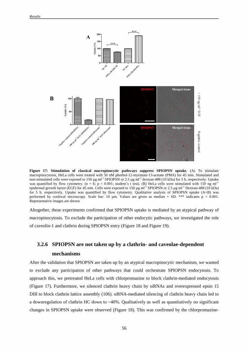
Results
56
Figure 17: Stimulation of classical macropinocytic pathways suppress SPIOPSN uptake. (A) To stimulate
macropinocytosis, HeLa cells were treated with 50 nM phorbol-12-myristate-13-acetate (PMA) for 45 min. Stimulated and
non-stimulated cells were exposed to 150 µg ml-1 SPIOPSN or 2.5 µg ml-1 dextran-488 (10 kDa) for 5 h, respectively. Uptake
was quantified by flow cytometry. (n = 6; p < 0.001; student´s t test). (B) HeLa cells were stimulated with 150 ng ml-1
epidermal growth factor (EGF) for 45 min. Cells were exposed to 150 µg ml-1 SPIOPSN or 2.5 µg ml-1 Dextran-488 (10 kDa)
for 5 h, respectively. Uptake was quantified by flow cytometry. Qualitative analysis of SPIOPSN uptake (A+B) was
performed by confocal microscopy. Scale bar: 10 µm. Values are given as median + SD. *** indicates p < 0.001.
Representative images are shown.
Altogether, these experiments confirmed that SPIOPSN uptake is mediated by an atypical pathway of
macropinocytosis. To exclude the participation of other endocytic pathways, we investigated the role
of caveolin-1 and clathrin during SPIOPSN entry (Figure 18 and Figure 19).
3.2.6 SPIOPSN are not taken up by a clathrin- and caveolae-dependent
mechanisms
After the validation that SPIOPSN are taken up by an atypical macropinocytic mechanism, we wanted
to exclude any participation of other pathways that could orchestrate SPIOPSN endocytosis. To
approach this, we pretreated HeLa cells with chlorpromazine to block clathrin-mediated endocytosis
(Figure 17). Furthermore, we silenced clathrin heavy chain by siRNAs and overexpressed epsin 15
DIII to block clathrin lattice assembly (106). siRNA-mediated silencing of clathrin heavy chain led to
a downregulation of clathrin HC down to ~40%. Qualitatively as well as quantitatively no significant
changes in SPIOPSN uptake were observed (Figure 18). This was confirmed by the chlorpromazine-

Results
57
mediated inhibition of CME, where also no significant changes in the uptake of SPIOPSN were
observed.
Figure 18: SPIOPSN are not internalized via a clathrin-mediated endocytic mechanism. HeLa cells were transfected
with siRNA to silence the clathrin heavy chains. Additionally, CME was inhibited by chlorpromazine treatment and via the
overexpression of epsin 15 DIII-GFP. Subsequently, SPIOPSN uptake was qualitatively and quantitatively analyzed. (A)
Three days after the nucleofection of siRNA, downregulation of clathrin heavy chain was quantified by western blotting. (B)
Mock treated and siRNA treated HeLa cells were exposed to 150 µg ml-1 SPIOPSN for 6 h. Immunofluorescence stainings
for clathrin heavy chains were performed. Uptake was qualitatively examined by confocal imaging. (C) Uptake of SPIOPSN
in siRNA- and mock-treated cells was quantified by flow cytometry (n = 4). (D) HeLa cells were pretreated with 25 µM
chlorpromazine and then exposed to 150 µg ml-1 SPIOPSN for 5 h. Quantification of uptake was performed via flow
cytometric analysis (n = 4). (E) HeLa cells were transfected with epsin 15-DIII-GFP. 24 h after transfection, cells were
exposed to 150 µg ml-1 SPIOPSN for 6 h. Uptake was qualitatively studied by confocal microscopy. Scale bars: 10 µm.
Representative images are shown.
Moreover, also the overexpression of dominant-negative epsin 15 (DIII) did not display any impact on
the internalization rate of SPIOPSN. Taking all of this into account, we concluded that CME is not
significantly utilized during the entry of SPIOPSN.

Results
58
Next, we investigated the impact of caveolae-mediated endocytosis on the uptake rate of SPIOPSN.
Three days after nucleofection, siRNA-mediated silencing suppressed the levels of caveolin-1 to
~ 32% (Figure 19A). Qualitatively and quantitatively, no differences in SPIOPSN uptake were
observed. As expected, overexpression of caveolin-1 did not significantly alter the amount of
SPIOPSN that were internalized (Figure 19D). Therefore, we concluded that SPIOPSN are not
internalized by a caveolin-1 dependent mechanism.
Figure 19: Caveolin-1 is not significantly required during the internalization process of SPIOPSN. HeLa cells were
transfected with siRNA to silence caveolin-1. Caveolae-mediated endocytosis was disturbed by the overexpression of
caveolin-1-GFP. The uptake of SPIOPSN was qualitatively and quantitatively analyzed. (A) Three days after nucleofection,
downregulation of caveolin-1 was quantified by western blotting (B) Immunofluorescence stainings were performed for
caveolin-1. Uptake was qualitatively determined by confocal imaging. (C) Mock- and siRNA treated cells were exposed to
150 µg ml-1 SPIOPSN for 6 h. Uptake was quantified by flow cytometry. (D) HeLa cells were transfected with caveolin-1-
GFP. 24 h after transfection, cells were exposed to 150 µg ml-1 SPIOPSN for 6 h. Uptake was qualitatively studied by
confocal microscopy. Scale bars: 10 µm. Representative images are shown.

Results
59
Other pathways that might have an impact on the internalization rate of SPIOPSN were also
investigated. Among others, we focused on the CLIC/GEEC-mediated endocytosis. To investigate
whether this pathway mediates the uptake of SPIOPSN, we performed cointernalization experiments
with the CLIC/GEEC marker cholera toxin subunit B to analyze the colocalization with SPIOPSN
(data not shown). In this particular case, no colocalization of SPIOPSN with ctxB was observed.
Therefore, we concluded that SPIOPSN are not taken up via the CLIC/GEEC pathway. This was
confirmed by TEM studies were no tubular CLIC/GEEC-like invaginations were observed.
Further on, we checked whether the small GTPase RhoA had an impact on the uptake of SPIOPSN
(data not shown). The overexpression of the dominant negative RhoA had no effect on the
internalization rate of SPIOPSN. Therefore, we concluded that SPIOPSN did not exploit a RhoA-
mediated endocytosis.
3.2.7 Other factors that orchestrate SPIOPSN entry: RNAi screening
identifies flotillin-1 and the tetraspanin CD81 as important factors
during the uptake process of SPIOPSN
As shown in the previous sections, the uptake of SPIOPSN is partially dependent on factors of a
macropinocytic-like mechanism. Still, it is equally important to mention that uptake seemed not to be
affected by CME; caveolae-mediated endocytosis, RhoA-mediated endocytosis or the CLIC/GEEC
pathway. Also the ADP-ribosylation factor Arf6 does not seem to play a role in the uptake of
SPIOPSN (Figure 25). However, the literature also provides several proteins that have been described
to act independently from the commonly accepted endocytic pathways (88, 217). Therefore, we
investigated the influences of some proteins that were assumed to interact with SPIOPSN.
Since tetraspanins and flotillin-1 were earlier described as part of the plasma membrane, of
intracellular vesicles and as proteins that control the endocytotic machinery, we focused on these
factors. We studied the internalization rates of SPIOPSNs after RNAi-mediated silencing of flotillin-1
and the tetraspanins CD82, CD81, CD63 and CD9. The reason for the selection of these distinct
proteins is based on an experiment, where we magnetically isolated SPIOPSN-containing vesicles
(shown in Figure 35). The previously mentioned vesicular proteins were present inside this fraction as
detected by peptide mass spectrometry. These data implicated that SPIOPSN-containing vesicles
contained significant amounts of flotillin-1 and the previously mentioned tetraspanins.
Knockdown was verified by the quantitative evaluation of the western blots except for the expression
levels of CD82. The expression levels of CD82 knockdown was validated by qRT-PCR. We observed
a silencing rate of ~15% (remaining CD82 mRNA; data not shown). Subsequently after gene
silencing, the uptake rates of SPIOPSN were quantified. Flow cytometric analysis revealed a
significant reduction of uptake in cells that were silenced for flotillin-1 (n = 4; p < 0.01, student´s t

Results
60
test) and CD81 (n = 4; p < 0.001, student´s t test). The knockdown of the tetraspanins CD82, CD63
and CD9 did not impair SPIOPSN uptake (Figure 20).
Figure 20: Flotillin-1 and CD81 partially control the internalization of SPIOPSN. To silence flotillin-1 and CD81, HeLa
cells were nucleofected with 60 pmol of target-specific siRNA. (A) 3 d after nucleofection, protein expression levels were
determined by western blotting (all samples: n = 3; p < 0.001; student´s t test). Knockdown of CD82 was quantified by q-RT-
PCR (mRNA silencing down to ~15%; data not shown). (B) 3 d after nucleofection, HeLa cells were exposed to 150 µg ml-1
SPIOPSN for 6 h. The uptake of nanoparticles was quantified by flow cytometry. Significant changes in uptake were
observed in flotillin-1 and CD81 silenced samples in comparison to the mock control. No significant changes in SPIOPSN
uptake were detected in the samples that were silenced for CD82, CD63 and CD9.
Altogether, it was shown that CD81 and flotillin-1 had an impact on the internalization mechanisms of
the nanoparticles. Since the membrane organization of CD81 and flotillin-1 was previously shown to
depend on cholesterol, we investigated the impact of cholesterol depletion on the uptake of SPIOPSN
(Figure 21) (131, 218). Moreover, it is worth to mention that cholesterol was also suggested to have a
major impact on macropinocytic events.
3.2.8 Changes in cholesterol levels influence the endocytosis of SPIOPSN
Since the depletion of cholesterol often deregulates macropinocytic processes, we tested whether
changes in cholesterol homeostasis have an impact on the internalization rate of SPIOPSNs.
Therefore, we depleted the membranous cholesterol by the treatment of methyl-β-cyclodextrin (Figure
21). After cholesterol depletion and nanoparticle exposure, we observed a significant drop of the
amount of internalized nanoparticles to 75.24% (n = 6; ** indicates p < 0.01; SD 2.57%). We further
disturbed the intracellular cholesterol homeostasis by the cholesterol synthesis inhibitor U18666A.
This inhibitor induced a Niemann-Pick-disease-like phenotype (Figure 21B). These NPC-like HeLa
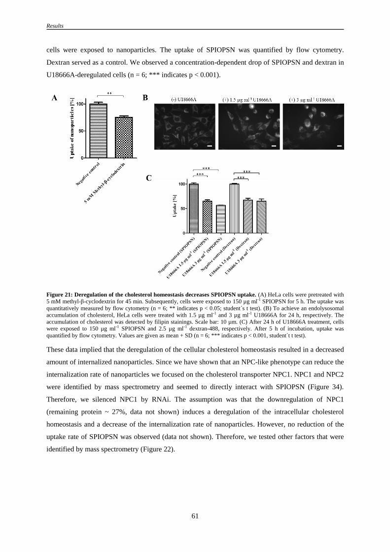
Results
61
cells were exposed to nanoparticles. The uptake of SPIOPSN was quantified by flow cytometry.
Dextran served as a control. We observed a concentration-dependent drop of SPIOPSN and dextran in
U18666A-deregulated cells (n = 6; *** indicates p < 0.001).
Figure 21: Deregulation of the cholesterol homeostasis decreases SPIOPSN uptake. (A) HeLa cells were pretreated with
5 mM methyl-β-cyclodextrin for 45 min. Subsequently, cells were exposed to 150 µg ml-1 SPIOPSN for 5 h. The uptake was
quantitatively measured by flow cytometry (n = 6; ** indicates p < 0.05; student´s t test). (B) To achieve an endolysosomal
accumulation of cholesterol, HeLa cells were treated with 1.5 µg ml-1 and 3 µg ml-1 U18666A for 24 h, respectively. The
accumulation of cholesterol was detected by filipin stainings. Scale bar: 10 µm. (C) After 24 h of U18666A treatment, cells
were exposed to 150 µg ml-1 SPIOPSN and 2.5 µg ml-1 dextran-488, respectively. After 5 h of incubation, uptake was
quantified by flow cytometry. Values are given as mean + SD (n = 6; *** indicates p < 0.001, student´t t test).
These data implied that the deregulation of the cellular cholesterol homeostasis resulted in a decreased
amount of internalized nanoparticles. Since we have shown that an NPC-like phenotype can reduce the
internalization rate of nanoparticles we focused on the cholesterol transporter NPC1. NPC1 and NPC2
were identified by mass spectrometry and seemed to directly interact with SPIOPSN (Figure 34).
Therefore, we silenced NPC1 by RNAi. The assumption was that the downregulation of NPC1
(remaining protein ~ 27%, data not shown) induces a deregulation of the intracellular cholesterol
homeostasis and a decrease of the internalization rate of nanoparticles. However, no reduction of the
uptake rate of SPIOPSN was observed (data not shown). Therefore, we tested other factors that were
identified by mass spectrometry (Figure 22).

Results
62
3.2.9 ARF1 is an important factor in SPIOPSN uptake
ADP-ribosylation factor 1 (ARF1) is a small GTP-binding protein that has a crucial function during
the process of COP vesicle formation (219). Furthermore, it was also shown that ARF1 regulates
macropinocytic events (220). Experimentally, ARF1 can be inhibited by brefeldin A or by the
overexpression of dominant negative mutants (221).
To investigate the role of ARF1 during the uptake of SPIOPSN, we firstly blocked ARF1 by a
pharmacological treatment of HeLa cells with brefeldin A. Subsequently, the pretreated cells were
exposed to nanoparticles and nanoparticulate uptake was quantified (Figure 22). Brefeldin A treated
cells showed a significantly decreased internalization rate of SPIOPSN (71.7%; n = 6; * indicates p <
0.05; SD 10,6%). Dextran uptake was not affected by the brefeldin A treatment. Owing to this result,
we now asked whether SPIOPSN are directly associated with ARF1. Since we have identified ARF1
as a SPIOPSN-associated protein by mass spectrometry, we overexpressed a dominant negative
mutant of ARF1 in HeLa cells and measured the colocalization with SPIOPSN. Here, rare events of
colocalization were observed. This low rate of colocalization was also linked to the observation that
less nanoparticles were taken up from ARF1 T31N+ cells. Refering to this we concluded that the
nonfunctional ARF1 had a massive effect on the uptake of SPIOPSN. Therefore, we quantified the
number of endocytic vesicles of ARF1 T31N transfected and nontransfected cells by analyzing the
confocal z-stacks. By comparing GFP+ and GFP
- cells, it was shown that the number of endocytosed
SPIOPSN was significantly decreased in the ARF1 T31N+ fraction (n = 31; *** indicates p < 0.001)
(Figure 22).

Results
63
Figure 22: ARF1 has a crucial function during the uptake of SPIOPSN. SPIOPSN uptake was investigated upon ARF1
inhibition. (A) HeLa cells were pretreated with 10 µg ml-1 brefeldin A to inhibit ARF1. Pretreated and nontreated HeLa cells
were exposed to 150 µg ml-1 SPIOPSN for 6 h. Uptake of SPIOPSN was quantified by flow cytometry (n = 6; * indicates p <
0.05, student´s t test). (B) Colocalization studies with the dominant negative mutant ARF1 T31N-GFP revealed rare
colocalization events with SPIOPSN after 6 h of nanoparticle exposure. Scale bar: 10 µm. ROI: Scale bar: 1 µm. (C) HeLa
cells were transfected with ARF1 T31N-GFP. 24 h later, cells were exposed to 150 µg ml-1 SPIOPSN for 6 h. Qualitative
confocal images revealed a massively reduced amount of internalized nanoparticles in ARF1 T31N-GFP+ cells. (D) z-stacks
of ARF1 T31N-GFP+ and GFP- cells were recorded to manually quantify the number of SPIOPSN-containing vesicles after 6
h of nanoparticle exposure. A significant reduction of SPIOPSN-containing vesicles in ARF1 T31N-GFP+ cells was observed
(p < 0.001; student´s t test). Representative images are shown.
Altogether, it was shown that ARF1 is a crucial factor during the uptake of SPIOPSN. Whether ARF1
is also deregulated after the deacidification of the endocytic system is unknown. However, this is not
the major question we wanted to solve. In the next section, we showed that the deacidification of the
endocytic system resulted in a drop of the nanoparticulate uptake rate (Figure 23).
3.2.10 Deacidification of the endolysosomal vesicles affects uptake of
SPIOPSN
The acidification of the endolysosomal system is facilitated by the endolysosomal v-type ATPase H+
pumps. To approach the question, if deacidification of the endolysosomal system affects the uptake of
SPIOPSN, v-type ATPase H+ pumps were inhibited by bafilomycin A1. The efficiency of inhibition
was confirmed by LysoTracker staining (Figure 23). If our hypothesis holds true, HeLa cells should
reduce their ability to internalize SPIOPSN. Therefore, we pretreated HeLa cells with bafilomycin A1
for 45 min and then exposed them to SPIOPSN or dextran. Deacidification of the endolysosomal
system significantly reduced the internalization of SPIOPSN and dextran. The analysis showed that
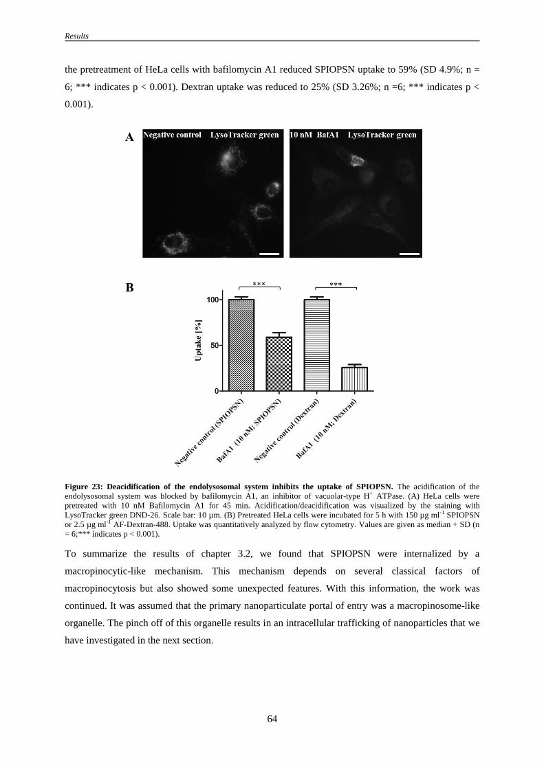
Results
64
the pretreatment of HeLa cells with bafilomycin A1 reduced SPIOPSN uptake to 59% (SD 4.9%; n =
6; *** indicates p < 0.001). Dextran uptake was reduced to 25% (SD 3.26%; n =6; *** indicates p <
0.001).
Figure 23: Deacidification of the endolysosomal system inhibits the uptake of SPIOPSN. The acidification of the
endolysosomal system was blocked by bafilomycin A1, an inhibitor of vacuolar-type H+ ATPase. (A) HeLa cells were
pretreated with 10 nM Bafilomycin A1 for 45 min. Acidification/deacidification was visualized by the staining with
LysoTracker green DND-26. Scale bar: 10 µm. (B) Pretreated HeLa cells were incubated for 5 h with 150 µg ml-1 SPIOPSN
or 2.5 µg ml-1 AF-Dextran-488. Uptake was quantitatively analyzed by flow cytometry. Values are given as median + SD (n
= 6;*** indicates p < 0.001).
To summarize the results of chapter 3.2, we found that SPIOPSN were internalized by a
macropinocytic-like mechanism. This mechanism depends on several classical factors of
macropinocytosis but also showed some unexpected features. With this information, the work was
continued. It was assumed that the primary nanoparticulate portal of entry was a macropinosome-like
organelle. The pinch off of this organelle results in an intracellular trafficking of nanoparticles that we
have investigated in the next section.

Results
65
3.3 Reconstruction of intracellular nanoparticle trafficking
The previous chapters showed that SPIOPSN have been internalized via a macropinocytic-like
mechanism. The identified mechanism is dependent on several factors that have never been associated
with nanoparticulate entry. This newly gained knowledge raised interesting issues that primarily
focused on the fate of the nanoparticles after their entry. Indeed, the intracellular nanoparticle
trafficking after macropinocytic-like entry is less investigated. Therefore, we asked how SPIOPSN
traffic through the cell when the primary compartment is a macropinosome-like organelle.
3.3.1 SPIOPSN are primarily transported via macropinosome-like organelles
Preliminary, we analyzed several classical markers of early endosomes in the initial stage of
endocytosis. Therefore, we focused on the Ras-related protein 5A (Rab5A), which is known to be
present on classical macropinosomes and early endosomes (142, 222). In colocalization experiments
with overexpressed Rab5A wt, only low amounts of colocalization with nanoparticles was observed.
However, we found some SPIOPSN inside Rab5A+ vesicles after 6 h of exposure (Figure 24A/B).
These Rab5A+ vesicles were visible under transmission light and resembled fluid-filled vesicles
(Figure 24B). Since Rab5 was described as an important protein in the biogenesis of endosomes and
macropinosomes, we analyzed whether the downregulation of Rab5A had an impact on the
internalization rate of SPIOPSN. Surprisingly, no reduction in the endocytosis rate of SPIOPSN was
observed (Figure 24D). Other proteins that have been described as markers of early endosomes are
early endosome antigen 1 (EEA1). In this case, no colocalization of EEA1 with SPIOPSN was found
in immunofluorescence staining. This was confirmed by the MS data, where EEA1 was not enriched
(data not shown).
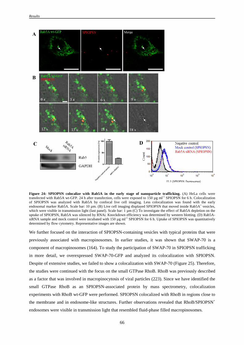
Results
66
Figure 24: SPIOPSN colocalize with Rab5A in the early stage of nanoparticle trafficking. (A) HeLa cells were
transfected with Rab5A wt-GFP. 24 h after transfection, cells were exposed to 150 µg ml-1 SPIOPSN for 6 h. Colocalization
of SPIOPSN was analyzed with Rab5A by confocal live cell imaging. Less colocalization was found with the early
endosomal marker Rab5A. Scale bar: 10 µm. (B) Live cell imaging displayed SPIOPSN that moved inside Rab5A+ vesicles,
which were visible in transmission light (last panel). Scale bar: 1 µm (C) To investigate the effect of Rab5A depletion on the
uptake of SPIOPSN, Rab5A was silenced by RNAi. Knockdown efficiency was determined by western blotting. (D) Rab5A-
siRNA sample and mock control were incubated with 150 µg ml-1 SPIOPSN for 6 h. Uptake of SPIOPSN was quantitatively
determined by flow cytometry. Representative images are shown.
We further focused on the interaction of SPIOPSN-containing vesicles with typical proteins that were
previously associated with macropinosomes. In earlier studies, it was shown that SWAP-70 is a
component of macropinosomes (164). To study the participation of SWAP-70 in SPIOPSN trafficking
in more detail, we overexpressed SWAP-70-GFP and analyzed its colocalization with SPIOPSN.
Despite of extensive studies, we failed to show a colocalization with SWAP-70 (Figure 25). Therefore,
the studies were continued with the focus on the small GTPase RhoB. RhoB was previously described
as a factor that was involved in macropinocytosis of viral particles (223). Since we have identified the
small GTPase RhoB as an SPIOPSN-associated protein by mass spectrometry, colocalization
experiments with RhoB wt-GFP were performed. SPIOPSN colocalized with RhoB in regions close to
the membrane and in endosome-like structures. Further observations revealed that RhoB/SPIOPSN+
endosomes were visible in transmission light that resembled fluid-phase filled macropinosomes.

Results
67
Figure 25: RhoB is associated with SPIOPSN-containing macropinosomes. To investigate specific markers of
macropinosomes, several proteins were overexpressed. SWAP-70 is a protein that was shown to be transiently present on
macropinosomes. RhoB is a small GTPase that was associated with macropinocytosis. ARF6-GFP is a small GTPase that is
known to transiently mediate classical macropinocytosis. After 24 h of transfection, HeLa cells were exposed to 150 µg ml-1
SPIOPSN for 6 h. Subsequently, colocalization was analyzed by confocal live cell imaging. (A) SWAP-70 displayed no
colocalization with SPIOPSN. (B) Colocalization of SPIOPSN with RhoB was found in close proximity to the cell membrane
(B) and in the perinuclear regions of the cell. (C) Live cell imaging showed that SPIOPSN moved within fluid-phase filled
RhoB+ vesicles through the cell. (D) Less colocalization was observed with α5-Integrin-GFP (E) No colocalization was found
with ARF6 DN-HA. Detection of ARF6 DN-HA was performed by immunofluorescence. Qualitatively, the expression of
dominant negative ARF6 did not alter the internalization rate of SPIOPSN in comparison to nontransfected cells.
Representative images are shown.
Next, overexpression of α5-Integrin-GFP resulted in rare colocalization events with SPIOPSN. α5-
Integrins were previously identified as important proteins during the early macropinocytic stages of
adenoviruses (224). Moreover, we investigated the influences of ARF6 on SPIOPSN trafficking.
ADP-ribosylation factor 6 is a GTP-binding protein that was identified as a modulator of Rac-
GTPases. Previous work showed that ARF6 can control the actin structure and colocalized with an
early phagosomal mechanisms (92). To get a closer insight if ARF6 has an impact on SPIOPSN
uptake, we overexpressed the dominant negative form ARF6 T27N-HA. Afterwards, cells were
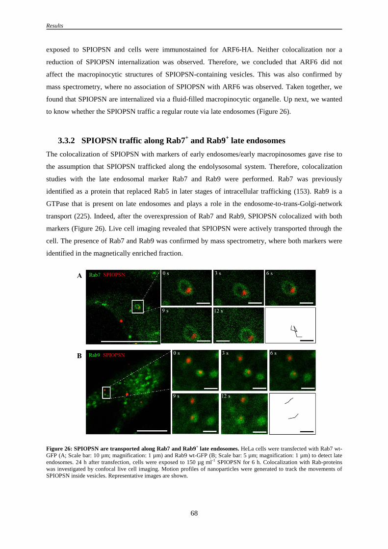
Results
68
exposed to SPIOPSN and cells were immunostained for ARF6-HA. Neither colocalization nor a
reduction of SPIOPSN internalization was observed. Therefore, we concluded that ARF6 did not
affect the macropinocytic structures of SPIOPSN-containing vesicles. This was also confirmed by
mass spectrometry, where no association of SPIOPSN with ARF6 was observed. Taken together, we
found that SPIOPSN are internalized via a fluid-filled macropinocytic organelle. Up next, we wanted
to know whether the SPIOPSN traffic a regular route via late endosomes (Figure 26).
3.3.2 SPIOPSN traffic along Rab7+ and Rab9
+ late endosomes
The colocalization of SPIOPSN with markers of early endosomes/early macropinosomes gave rise to
the assumption that SPIOPSN trafficked along the endolysosomal system. Therefore, colocalization
studies with the late endosomal marker Rab7 and Rab9 were performed. Rab7 was previously
identified as a protein that replaced Rab5 in later stages of intracellular trafficking (153). Rab9 is a
GTPase that is present on late endosomes and plays a role in the endosome-to-trans-Golgi-network
transport (225). Indeed, after the overexpression of Rab7 and Rab9, SPIOPSN colocalized with both
markers (Figure 26). Live cell imaging revealed that SPIOPSN were actively transported through the
cell. The presence of Rab7 and Rab9 was confirmed by mass spectrometry, where both markers were
identified in the magnetically enriched fraction.
Figure 26: SPIOPSN are transported along Rab7 and Rab9+ late endosomes. HeLa cells were transfected with Rab7 wt-
GFP (A; Scale bar: 10 µm; magnification: 1 µm) and Rab9 wt-GFP (B; Scale bar: 5 µm; magnification: 1 µm) to detect late
endosomes. 24 h after transfection, cells were exposed to 150 µg ml-1 SPIOPSN for 6 h. Colocalization with Rab-proteins
was investigated by confocal live cell imaging. Motion profiles of nanoparticles were generated to track the movements of
SPIOPSN inside vesicles. Representative images are shown.

Results
69
3.3.3 The R-SNARE protein VAMP7 is present on SPIOPSN-containing
vesicles
After the detection of SPIOPSN inside Rab7+ and Rab9
+ vesicles it was assumed that SPIOPSN were
transported via late endosomes. To investigate whether these late endosomes fuse with lysosomes, we
investigated the colocalization of SPIOPSN with the vesicle-associated membrane protein 7
(VAMP7). VAMP7 is a membrane protein of the SNARE family that mediates the heterotypic fusion
of late endosomes and lysosomes (191). To investigate the participation of VAMP7 in SPIOPSN
trafficking, full-length VAMP7-GFP was overexpressed and SPIOPSN trafficking was investigated by
confocal live cell imaging. A colocalization of SPIOPSN with VAMP7 was observed. The tracked
nanoparticles were actively transported inside VAMP7+
vesicles through the cell (Figure 27). An
association of VAMP7 with SPIOPSN was also shown by mass spectrometry. Here, we identified
VAMP7 as an enriched protein of the magnetic vesicular fraction.
Figure 27: VAMP7 is associated with SPIOPSN-containing vesicles. To investigate the presence of VAMP7 on
SPIOPSN-containing vesicles, we overexpressed VAMP7-GFP. (A) HeLa cells were transfected with VAMP7-GFP. 24 h
later, cells were exposed to 150 µg ml-1 SPIOPSN for 6 h. Subsequently, colocalization of SPIOPSN with VAMP7 was
analyzed by confocal live cell imaging (Scale bar: 10 µm). (B) Confocal live cell imaging displayed an active transport of
SPIOPSN inside VAMP7+ vesicles. Representative images are shown.

Results
70
3.3.4 SPIOPSN are transported inside vesicles that are positive for markers
of intraluminal vesicles
Early and late endosomes accumulate intraluminal vesicles (ILVs) during the processes of vesicular
maturation (171). ILVs have been shown to carry several markers such as the melanocytic protein
Pmel17. In previous reports it was demonstrated that Pmel17 is sorted via an ubiquitin-/ ESCRT-
independent mechanism into ILVs of MVBs (174, 226). Furthermore, it was shown that the
tetraspanin CD63 decorates the ILVs (227). Therefore, Pmel17 and CD63 were used to investigate the
association of SPIOPSN with multivesicular bodies. As expected, we have identified no native Pmel17
in HeLa cells (Figure 28B). Therefore, we overexpressed Pmel17 in HeLa cells. 24 h after
transfection, cells were exposed to SPIOPSN. The detection of Pmel17 was performed by
immunofluorescence. Notably, a clear colocalization of Pmel17 with nanoparticles was observed.
Next, we incubated HeLa cells with SPIOPSN and detected CD63 by immunofluorescence staining.
Nanoparticles displayed a high rate of colocalization with CD63 (Figure 28).
Figure 28: SPIOPSN are transported inside vesicles that are positive for the intraluminal vesicle marker Pmel17 and
CD63. (A) HeLa cells were transfected with Pmel17. 24 h later, cells were exposed to 150 µg ml-1 SPIOPSN for 6 h. Pmel17
was detected by immunostaining. Colocalization was analyzed by confocal microscopy. Scale bar: 10 µm. (B) Staining of
nontransfected HeLa cells with NKI/Beteb antibodies showed that HeLa cells lack the expression of Pmel17. Scale bar 10
µm. (C) HeLa cells were incubated with 150 µg ml-1 SPIOPSN for 20 h and then stained for CD63. Scale bar: 10 µm. Spots
of colocalization were marked with white arrows (n = 5; representative images are shown).
Since previous reports suggested that Pmel17 is sorted via an ESCRT-independent mechanism into
MVBs, we hypothesized that SPIOPSN-containing vesicles contained a low amount of ubiquitin.
Indeed, we were not able to detect ubiquitin by immunofluorescence in any SPIOPSN-containing
vesicle (data not shown). In spite of this, the morphology of SPIOPSN-containing vesicles revealed
that the nanoparticles are transported via classical MVBs (Figure 29).

Results
71
3.3.5 SPIOPSN are transported via multivesicular bodies to multilamellar
lysosomes
The data of the previous sections showed that SPIOPSN were transported via early endocytic vesicles
to multivesicular bodies. To confirm this on an ultrastructural level, cryo-freezed sections of
nanoparticle-exposed HeLa cells were prepared. After recording the TEM micrographs, SPIOPSN-
containing-vesicles were subclassified owing to distinct morphological criteria. Vesicles that lacked
intraluminal vesicles were classified as early endocytic vesicles. Vesicles, which contained ILVs were
categorized as multivesicular bodies. Multilamellar lysosomes are vesicles that stored membrane
whorls of an onion-like structure. Further on, we recorded hybrid structures of multivesicular bodies
and multilamellar lysosomes/multilamellar bodies (MLL or MLB). These vesicles contained
intraluminal vesicles and membrane whorls.
The analysis of the TEM micrographs confirmed the previous experiments of confocal imaging.
SPIOPSN were transported along early endocytic vesicles, multivesicular bodies, MVB-MLL-hybrid
organelles and multilamellar lysosomes (Figure 29). To analyze the distribution of SPIOPSN inside
the distinct types of vesicles more precisely, a semiquantitative classification was performed. The
morphological subcategorization of the vesicles was based on the formerly described criteria.
Quantification was performed with the data of four independent experiments, counting the identified
SPIOPSN-containing vesicles on the prepared slices inside more than 25 cells. Taken together, most of
the SPIOPSN were found inside MVB/MLL-hybrid organelles and in multilamellar lysosomes after 20
h of nanoparticle exposure.

Results
72
Figure 29: SPIOPSN are transported inside morphologically distinct vesicles along the endolysosomal pathway
(HeLa). Cryo-TEM micrographs revealed the different types of vesicles that contained electron dense SPIOPSN (dark areas).
Scale bar: 200 nm. (A) HeLa cells were exposed to 150 µg ml-1 SPIOPSN for 20 h. Cells were cryo-freezed and prepared for
TEM. SPIOPSN were found in vesicles with a size range of 0.5 – 1 µm in diameter. Vesicles without ILVs were identified
(early endocytic vesicles). Multivesicular bodies contained SPIOPSN and intraluminal vesicles (size ~ 50 nm). Multilamellar
lysosomes were identified. MVB/MLB-hybrid organelles were found. (B) Semiquantitative analysis of TEM micrographs
revealed that SPIOPSN were mainly present inside multilamellar bodies after 20 h of incubation (n = 4 independent
experiments). Vesicles were categorized due to their morphological appearance as shown in the scheme in (A). (Sample
preparation and TEM micrographs were prepared by C. Messerschmidt).
To assure that MLBs are the final organelle in SPIOPSN trafficking, HeLa cells were pulsed for 6 h
with SPIOPSN. This procedure was followed by a postincubation of 14 h without nanoparticles in the
growth medium. Notably, almost all of the SPIOPSN accumulated inside MLB-like-structures (data
not shown).
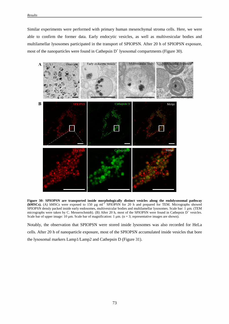
Results
73
Similar experiments were performed with primary human mesenchymal stroma cells. Here, we were
able to confirm the former data. Early endocytic vesicles, as well as multivesicular bodies and
multilamellar lysosomes participated in the transport of SPIOPSN. After 20 h of SPIOPSN exposure,
most of the nanoparticles were found in Cathepsin D+ lysosomal compartments (Figure 30).
Figure 30: SPIOPSN are transported inside morphologically distinct vesicles along the endolysosomal pathway
(hMSCs). (A) hMSCs were exposed to 150 µg ml-1 SPIOPSN for 20 h and prepared for TEM. Micrographs showed
SPIOPSN densly packed inside early endosomes, multivesicular bodies and multilamellar lysosomes. Scale bar: 1 µm. (TEM
micrographs were taken by C. Messerschmidt). (B) After 20 h, most of the SPIOPSN were found in Cathepsin D+ vesicles.
Scale bar of upper image: 10 µm. Scale bar of magnification: 1 µm. (n = 3; representative images are shown).
Notably, the observation that SPIOPSN were stored inside lysosomes was also recorded for HeLa
cells. After 20 h of nanoparticle exposure, most of the SPIOPSN accumulated inside vesicles that bore
the lysosomal markers Lamp1/Lamp2 and Cathepsin D (Figure 31).

Results
74
Figure 31: SPIOPSN are finally stored inside Lamp1/2+ and Cathepsin D+ lysosomes. HeLa cells were exposed to
150 µg ml-1 SPIOPSN for 20 h, fixed, permeabilized and double-stained for the lysosomal markers Lamp1/Lamp2. Another
sample was stained for the lysosomal marker cathepsin D. SPIOPSN showed colocalization with both markers (n = 8).
Representative images are shown
3.3.6 SPIOPSN are neither exocytosed nor transported by autophagosomes
Next, we analyzed whether intracellular nanoparticle trafficking interfered with recycling pathways or
autophagosomal trafficking. Since we have identified Rab11A and Rab11B by mass spectrometry, we
asked the question whether SPIOPSN are generally exocytosed by recycling endosomes. To quantify
this process, HeLa cells were pulsed for 4 h with SPIOPSN and postincubated without nanoparticles
for another 16 h. The mean fluorescence intensity of the cells was measured over time. Notably, no
significant drop of the fluorescent nanoparticle signal was observed (Figure 32A). We concluded that
SPIOPSN were not exocytosed by the cell. This observation was confirmed by the following set of
experiments. Intensive research on this project confirmed that SPIOPSN did not colocalize with Rab11
wt-GFP and Rab11 that was visualized by immunofluorescence stainings (Figure 32B). Consequently,
we suggested that SPIOPSN were not significantly externalized by the recycling pathways.
To investigate the interplay with autophagosomes, HeLa cells were incubated with SPIOPSN and
immunostained for the autophagosomal marker LC3. Also here, SPIOPSN did not colocalize with the
LC3. This indicated that SPIOPSN trafficking was not directly linked to the autophagosomal system.

Results
75
Figure 32: SPIOPSN are neither exocytosed by Rab11+ recycling endosomes nor transported via LC3B+
autophagosomes. (A) To test whether SPIOPSN are exocytosed, we pulsed HeLa cells for 4 h with 150 µg ml-1 SPIOPSN.
After a washing step, we postincubated the cells without nanoparticles for another 16 h. The mean fluorescence intensity of
the nanoparticles was recorded over time. Full-time exposed cells served as a control. No significant exocytosis of SPIOPSN
was observed after 20 h of total exposure. (B) HeLa cells were transfected with Rab11 wt-GFP. 24 h later, cells were exposed
to 150 µg ml-1 SPIOPSN for 20 h. Live cell imaging displayed no clear colocalization of Rab11 with SPIOPSN.
(C/Magnification: D) HeLa cells were exposed to 150 µg ml-1 SPIOPSN and then immunostained for Rab11 after 20 h of
nanoparticle exposure. SPIOPSN displayed very low amounts of colocalization with Rab11+ endosomes. (E) To study the
crosstalk of SPIOPSN trafficking with the autophagosomal pathway, we incubated HeLa cells with 150 µg ml-1 SPIOPSN. 20
h later, autophagosomes were immunostained for LC3. Cells were examined by confocal imaging. No clear colocalization of
SPIOPSN with LC3 was observed. (B/C/E): Scale bar: 10 µm. (D/F): Scale bar: 1 µm. Representative images are shown.

Results
76
3.3.7 In vivo distribution of SPIOPSN in mice
The former chapters described the intracellular trafficking routes of nanoparticles. In the upcoming
sections, we exploited the formerly gained knowledge and the superparamagnetic features of the
nanoparticles to further investigate the intracellular trafficking pathways and the biodistribution of
nanoparticles.
To study whether superparamagnetic nanoparticles were an appropriate tool to analyze the
biodistribution in mice, we tail-vain injected SPIOPSN into NSG mice. 24 h later, peripheral blood,
bladder fluid, spleen, liver and kidney were isolated. Single cell suspensions were obtained from
spleen, liver and kidney. SPIOPSN-containing cells were magnetically isolated and examined via
confocal microscopy. In summary, a magnetic enrichment of cells from the peripheral blood failed.
Therefore, we concluded that nanoparticles were efficiently cleared from the blood stream without the
uptake by the phagocytic system of the blood. In this context, it is worth to mention that NSG mice are
immunodeficient mice with less or no phagocytic cells (228).
Also kidney cells and the bladder fluids lacked SPIOPSN. In contrary, massive amounts of SPIOPSN
accumulated inside liver and spleen. Most of the SPIOPSN displayed endocytic structures that
resembled the previously identified intracellular vesicles (Figure 33).

Results
77
Figure 33: In vivo distribution of SPIOPSN. 2 mg of SPIOPSN were tail-vain injected into NSG mice (n = 4). 24 h later,
spleen, liver, kidney, peripheral blood and bladder were isolated. Single-cell suspensions were generated. Tissue fractions
were magnetically sorted by MACS. Uptake was qualitatively analyzed by confocal microscopy. (A) Splenic cells
internalized SPIOPSN via endocytic structures. The left image shows a randomly recorded overview about the isolated
fraction. The right image displays a randomly chosen magnification of SPIOPSN-containing cells (B) Liver cells internalized
SPIOPSN via endocytic-like vesicles. The left image presents an overview about the isolated fraction. The right image
displays a randomly chosen magnification of SPIOPSN-containing cells. (C) No SPIOPSN were detected inside the
magnetically enriched fraction of the kidney. Scale bar: 10 µm. Tail-vain injection and animal preparation was performed by
Patricia Okwieka, University Medical Center, Mainz.
Taking all of the results into account, we resumed that SPIOPSN are applicable to study their
biodistribution (in vivo) after magnetic cell separation. We could show that SPIOPSN are taken up into
endocytic-like vesicles in vivo. These vesicles resembled the identified structures in vitro. Therefore,
we thought about an approach to exploit the magnetic features of the SPIOPSN on a subcellular level.
The aim of the next project was the magnetic isolation of intracellular SPIOPSN-containing vesicles.
With this application, we wanted to dissect the intracellular nanoparticle trafficking on a proteomic
level (Figure 34).

Results
78
3.3.8 Shaping a picture of endocytosis: Reconstruction of intracellular
nanoparticle trafficking
As stated in the previous chapters, we found that SPIOPSN are trafficked from macropinosomes via
multivesicular bodies to multilamellar lysosomes. Therefore, we asked whether we could characterize
the SPIOPSN-containing vesicles in more detail.
To obtain a proteomic snapshot of SPIOPSN-containing vesicles, HeLa cells were incubated with
SPIOPSN for 20 h (Figure 34). Subsequently, noninternalized SPIOPSN were removed by extensive
washing. Cells were mechanically disrupted and cell debris was removed by multistep centrifugation.
SPIOPSN-containing vesicles were magnetically separated in a magnetic field for 12 h under cooling
and with the addition of protease inhibitors. The magnetic fraction was carefully washed with PBS and
then solubilized inside urea buffer. The nonmagnetic fraction was also solubilized with urea buffer.
Quality controls were performed by the analysis of the magnetic/nonmagnetic fraction by TEM and
SDS-PAGE. Subsequently, the fractions were forwarded to Dr. Stefan Tenzer, who performed the
mass spectrometry measurements (Institute for Immunology, University Medical Center, Mainz).
Figure 34: Scheme for the isolation of intracellular magnetic vesicles and their analysis by quantitative peptide mass
spectrometry. 1.8 x 10⁷ HeLa cells were exposed to 150 µg ml-1 SPIOPSN for 20 h. After incubation, non-
internalized SPIOPSN were extensively removed by washing. Cells were trypsinized, centrifuged and extensively washed
with PBS. The terminal washing fraction was centrifuged at 20 000 x g for 30 min. No free SPIOPSN were detected. To
obtain the cytosol with SPIOPSN-containing vesicles, cells were mechanically disrupted. Intact cells and debris were
removed by centrifugation. This step was repeated until no pellet was visible anymore. The cytosolic supernatant with the
magnetic endosomes were injected into a custom-made magnetic device. Magnetic separation of SPIOPSN-containing
vesicles was performed for 12 h under cooling and the inhibition of proteases. The magnetic pellet was carefully washed with
PBS. Proteins of the magnetic and the nonmagnetic fraction were solubilized in urea buffer. Peptides were generated by a
tryptic digest. Samples were quantitatively measured by a TOP3-dependent MS approach and peptide fragments were
identified by a database-related search. Enrichment factors are based on the determined ppm values and were calculated
comparing the magnetic vs. the nonmagnetic fraction. Enriched proteins were forwarded to DAVID ontology analysis and
classified by their subcellular localization. Intracellular nanoparticle trafficking was reconstructed based on the results of
DAVID protein ontology analysis.

Results
79
After mass spectrometry measurements and data evaluation, the amount of proteins between the
magnetic and the nonmagnetic fraction was compared. Based on the averaged ppm values of the
quantified proteins, protein enrichment factors were calculated (example in Table 18).
Table 18: Calculation of the protein enrichment factors (example). Ppm values of the proteins in the magnetic fraction
were compared with the ppm values of the proteins of the supernatant. Based on the averaged ppm values, enrichment factors
were calculated for each protein. The calculation of the enrichment factor of the protein Rab5C is shown as an example.
Quantification of proteins was performed in triplicates. Averaged ppm values were calculated.
Uniprot ID Magnetic pellet
(ppm value)
Supernatant
(ppm value)
Enrichment
factor
RAB5C_HUMAN 34285 6339 5,41
3.3.9 DAVID protein ontology analysis of the vesicular fraction reveals an
association of endolysosomal proteins with SPIOPSN-containing
vesicles
Overall, we were able to identify and quantify 1492 proteins in the magnetically enriched fraction,
whereas 884 were significantly enriched by a factor of at least 2 in comparison to the nonmagnetic
fraction. We then applied the online tool “DAVID protein ontology” to analyze the intracellular
nanoparticle trafficking (INT) in silico (229). Proteins were grouped into annotation clusters owing to
their known subcellular localization in literature (Table 19).
Table 19: DAVID protein ontology analysis (GOTERM: GOTERM_CC_FAT). Proteins that were magnetically enriched
> 2-fold in comparison to the nonmagnetic supernatant have been considered in DAVID ontology analysis. The ontology
analysis clustered the different proteins owing to their known subcellular localization. The enrichment scores for the distinct
subgroups are based on the EASE score. In the presented table, the starting five annotation clusters are shown with
enrichment scores larger than 9. The full analysis is provided in the supplementary information of this thesis. Counts show
the proteins that are classified into the individual terms. We mainly identified proteins of the endolysosomal system and from
mitochondria. P-value: modified fisher exact p-value. The smaller the p-value, the higher enriched the proteins inside the
respective class. The proteins of the red-marked GOTERMs are listed in the supplementary information of this work.
Annotation Cluster Enrichment Score/ Term Protein count p-value
Annotation Cluster 1 Enrichment Score: 42.07
envelope 145 3.6E-45
organelle envelope 144 1.2E-44
mitochondrial envelope 115 7.1E-43
mitochondrial membrane 111 1.6E-42
mitochondrial inner
membrane
96 5.3E-41
organelle inner membrane 99 1.5E-40
Annotation Cluster 2 Enrichment Score: 29.76
lysosome 69 1.3E-30
lytic vacuole 69 1.3E-30
vacuole 75 2.9E-30

Results
80
Annotation Cluster 3 Enrichment Score: 16.73
vesicle 105 3.8E-18
membrane-bounded
vesicle
93 2.5E-17
cytoplasmic membrane-
bounded vesicle
91 2.9E-17
cytoplasmic vesicle 100 4.2E-17
Annotation Cluster 4 Enrichment Score: 9.24
membrane fraction 99 2.9E-10
insoluble fraction 101 4.7E-10
cell fraction 120 1.4E-9
3.3.10 Reconstruction of intracellular nanoparticle trafficking
The dissection of intracellular nanoparticle trafficking is based on the identified proteins that were
categorized into the GOTERMs lysosome and vesicle (Table 19, for a detailed protein list, see
supplementary information). We used the simplest endocytic model for the reconstruction of INT
(Figure 35). In our approach, we identified several enriched endolysosomal proteins in the magnetic
fraction. Early endosomes can be recognized by the markers RAB5A and RAB5C (230). Further
adaptor proteins such as proteins from the AP-1/AP-2 family (e. g. AP1A1, AP1B1, AP2B2) have
been described as key players in endocytic trafficking (231, 232). Proteins such as RhoB and ARF1
have been associated with the (macro)pinocytic machinery (90, 233).
RAB7A and RAB9 are important markers of multivesicular bodies (234). MVBs have been associated
with several tetraspanins (e. g. CD9, CD63, CD81, CD82) and HLA molecules (e. g. HLA MHC class
I antigen A*68) (173, 174, 235-237). Further proteins that are present on MVBs are Niemann Pick
disease C1 (NPC1) and Niemann Pick disease C2 (NPC2) (238). Flotillin 1 (FLOT1) and flotillin 2
(FLOT2) was also identified by MS. Proteins such as LAMP1 and LAMP2, Mannose-6-P-receptor-
CD (MPRD) and Mannose-6-P-receptor-CI (MPRI) have been shown to associate with the
endolysosomal pathway (239, 240). LTOR1, LTOR2, LTOR3 are present on lysosomes and mediate
several events of intracellular trafficking (241). We were also able to identify several v-type proton
ATPase pumps that obtain the lysosomal pH (e. g. VATG1, VATC1). In case of recycling endosomes,
RAB11A and RAB11B were identified. RAB11 is mainly associated with recycling endosomes, but
also fulfills different tasks inside the cell, for instance on MVBs (162, 242). Additionally, we
identified proteins that were previously shown to be with COP vesicles (e. g. SC23A, SAR1A, COPE)
(243).

Results
81
Fig
ure
3
5:
Rec
on
stru
ctio
n
of
intr
ace
llu
lar
SP
IOP
SN
tr
aff
ick
ing
. In
trac
ellu
lar
nan
op
arti
cle
traf
fick
ing
was
d
isse
cted
. IN
T
is
bas
ed
on
the
GO
TE
RM
s ves
icle
an
d l
yso
som
e (s
ee r
ed m
ark
ed t
erm
s in
Tab
le 1
9).
Pro
tein
nam
es a
re d
epic
ted
by t
hei
r U
nip
rot_
IDs.
Th
e fu
ll n
ames
are
pro
vid
ed
by t
he
sup
ple
men
tary
par
t.

Results
82
Altogether, we have identified a large number of novel marker proteins that were associated with
SPIOPSN-containing vesicles. These proteins enable us to create a snapshot of the participating
vesicles during nanoparticle trafficking after 20 h of exposure. Since we have detected the majority of
nanoparticles inside lysosome-like organelles after 20 h, it was not surprising that we have identified a
large number of enriched lysosomal matrix proteins. Therefore, we reconstructed the intravesicular
milieu of a nanoparticle in more detail (Figure 36).
3.3.11 Reconstruction of the intravesicular lysosomal matrix
The hydrolases and cofactors of the lysosomal matrix have been extensively studied in the proteomic
field (156). Many of the previous investigations performed density gradient fractionation or affinity
chromatography to obtain the vesicles of interest. Our approach utilized the magnetic isolation of
SPIOPSN-containing vesicles.
In our study, we have identified acetylgalactosamine-6-sulphatase N (GALNS) and N-
acetylglucosamine-6-sulphatase (GNS) as hydrolases for the degradation of chondroitin sulphate and
keratin sulphate. Acid ceramidase (ASAH1) is known to hydrolyze ceramide into sphingosine and free
fatty acids. Arylsulphatase A (ARSA) and arylsulphatase B (ARSB) hydrolyzes cerebroside sulphates.
Cathepsin B/D/Z/L1/C are enzymes for unspecific proteolysis. The CLN5 protein was also identified
as a lysosomal matrix protein. Other identified proteins are hexosaminidase A (HEXA) and
hexosaminidase B (HEXB). These proteins have the potential to degrade GM2 gangliosides. β-
mannosidase (MANBA) cleaves mannosyl residues. Palmitoyl-protein thioesterase 1 and palmitoyl-
protein thioesterase 2 removes thioester-linked fatty acyl groups from proteins and peptides. Proteins
that are not mentioned in the text are listed as Uniprot IDs in Figure 36.

Results
83
Figure 36: The proteomic dissection of a nanoparticle-containing lysosome. The data of mass spectrometry revealed that
several lysosomal proteins were enriched inside the magnetic fraction. Protein abbreviations are given as Uniprot_ID. Full
names are provided by the supplementary information.
The reconstruction of the lysosomal milieu of nanoparticles becomes especially important for the
design of biodegradable nanocarriers. Frequently, the nanoparticle synthesis is often not tailormade for
the endolysosomal system. Consequently, the intravesicular degradation of the nanocarriers fails.
Since we used a non-biodegradable nanoparticle, we were not able to show the degradation of the
particle itself. However, we tested whether the components of the lysosome can degrade the
nanoparticulate protein corona (Figure 37 and Figure 38).
3.3.12 The nanoparticle protein corona is degraded inside Lamp1+/Lamp2
+
lysosomes
Based on the gained knowledge about the reconstructed lysosomal matrix, we next asked the question
whether the enzymes of the lysosomal matrix have the potential to degrade the protein corona of
SPIOPSN. The nanoparticulate protein corona is formed, when nanoparticles enter a biological fluid
with proteins (244). To investigate whether the protein corona is degraded inside the lysosome,
SPIOPSN were incubated with Alexa Fluor 647 labelled bovine serum albumin to adsorb a
fluorescently labeled protein on the surface of the nanoparticles.

Results
84
Figure 37: Schematic illustration of the system that was used to study the degradation of the protein corona inside the
lysosomes. Nanoparticles were preincubated in pure BSA647 for 60 min. Afterwards, nanoparticles were diluted with media
and then added to the cells. After incubation, cells were immunostained for Lamp1/2. SPIOPSN and BSA647 were analyzed
for colocalization with the lysosomal markers by confocal microscopy.
The coincubation of SPIOPSN with BSA647 (without preincubation) led to the internalization of both
cargos into different vesicles only showing a minor rate of colocalization (Figure 38). This shows that
BSA-AF647 was not significantly adsorbed on the surface of nanoparticles, which were already
covered with proteins. Furthermore, we concluded that SPIOPSN and BSA647 exploited distinct
internalization pathways. Taking this into account for the following experiments, we resumed that we
only could detect preadsorbed proteins on the nanoparticles. Indeed, the incubation of BSA647-
preincubated SPIOPSN revealed strong signals of colocalization between SPIOPSN and BSA647.
Therefore, it was concluded that the protein corona is cointernalized with the nanoparticle into
endocytic vesicles. Next, we investigated, whether BSA647 is still present inside lysosomes.
Interestingly, no BSA647 was observed inside Lamp1/Lamp2+ vesicles. In contrary, vesicles negative
for Lamp1/2 still contained BSA647. Our interpretation for this was that BSA647 is degraded inside
Lamp1/Lamp2+ organelles. The investigation of BSA647-coated SPIOPSN revealed that the
nanoparticles inside Lamp1/Lamp2+ vesicles lack BSA647 after 20 h of incubation (Figure 38). Owing
to this, we concluded that parts of the protein corona were degraded by the environment of the
lysosomes.

Results
85
Figure 38: The protein corona is cointernalized with nanoparticles and then degraded inside the lysosome. (A) HeLa
cells were coexposed to 150 µg ml-1 SPIOPSN and 0.5 mg ml-1 BSA647 for 8 h. Almost no colocalization was observed. (B)
150 µg ml-1 SPIOPSN were preincubated in 0.5 mg ml-1 BSA647 for 60 min and then added for 6 h to HeLa cells. A large
number of colocalization spots were observed (white arrows). (C) HeLa cells were incubated with BSA647 for 20 h. BSA647
was not detectable in Lamp1/2+ lysosomes. (D) Cells were exposed to BSA647-preincubated SPIOPSN (150 µg ml-1) for 20
h. No BSA647 was present on SPIOPSN. Minor amounts of excess BSA647 accumulated inside Lamp1/Lamp2- cells after
20 h of incubation. (E) Magnification of Lamp1/2+ lysosomes containing SPIOPSN. Less to no BSA647 was detected inside
lysosomes that harbored BSA647-precoated SPIOPSN.

Discussion
86
4. Discussion
This study aimed to elucidate the interaction of polymeric nanoparticles with biological systems in
terms of drug delivery, entry and intracellular trafficking pathways of nanomaterials. In summary, we
could show that biodegradable PLLA nanoparticles are potentially suitable for a rapid and effective
delivery of drugs by a novel mechanism we referred to as “kiss-and-run”. For the first time, we
provide evidence for a non-invasive mechanism transporting nanoparticulate cargo over the cell
membrane into lipid droplets. The nanoparticulate cargo release occurs during several milliseconds
owing to a physicochemical interaction between the nanoparticle and the plasma membrane.
We further show that these polymeric nanoparticles are taken up by cells after their long-term
exposure. Therefore, issues about the uptake mechanisms and intracellular trafficking pathways of
polymeric nanoparticles were raised. For this project, we changed the polymer of the nanoparticle to
polystyrene. We show that ~ 126 nm large SPIOPSN are internalized by a macropinocytic-like entry
mechanism. In a series of experiments we demonstrate that their entry mainly depends on distinct
small GTPases, kinases and on cholesterol.
Further on, we established a novel method for the unbiased dissection of intracellular nanoparticle
trafficking. This method is based on the identification of proteins, which are located on nanoparticle-
containing vesicles. Under native conditions, nanoparticles are transported from early endocytic
vesicles via multivesicular bodies into the terminal multilamellar lysosomes. This dissection of INT
may serve as a novel fundament to describe nanoparticle trafficking in more detail. Taking all of these
data together, we propose a novel drug delivery mechanism for polymeric nanoparticles and
investigated a so far unknown mechanism of the entry and trafficking of polymeric nanoparticles.
4.1 Interaction of polymeric nanoparticles with biological systems can trigger
a release of different nanoparticulate components
Polymeric nanoparticles are a versatile tool to shield drugs from environmental influences (2, 6). The
primary goal of these drug delivery vehicles is the transport of the loaded cargo to the location of
interest (43, 245). However, less is known about the release of nanoparticulate components that
coexist inside the nanoparticle and should primarily not be delivered. These factors include polymers,
stabilizing surfactants or dye molecules (246). Notably, the circumstance that these factors sometimes
are released is no disadvantage. Moreover, this observation was utilized to study the mechanism of
nanoparticulate drug delivery in more detail. To track the interaction of nanoparticles with cells, we
chose a nanoparticulate model system with a noncovalently-bound dye. This enabled us to
fluorescently track the cargo release during the interplay with different cell types.

Discussion
87
The release kinetics of PMI during the exposure of HeLa cells or other cell types to PLLA-Fe-PMI
nanoparticles reproducibly revealed a fast accumulation of the model dye into lipid droplets (Figure 7,
Figure 8 and Figure 9). To our knowledge, no comparable study exists that could show an uptake of
nanoparticles within the first five minutes of exposure. Such fast rates of internalization are only
known for biomacromolecules (e. g. EGF) or for freely diffusing small molecules (247, 248).
Therefore, we assumed and also demonstrated that the encapsulated dye molecules were independently
transported from the polymeric matrix into the cells (Figure 9). Interestingly, this phenomenon was
afterwards also recognized for other nanoparticles (e. g. PLLA-PMI or PS-COOH) (6). Tomcin and
colleagues used these observations and demonstrated that also other components of nanoparticles (e. g.
surfactants) are released during nano-bio-interactions (249). They microscopically showed a release of
block-co-polymers from nanoparticles before the nanoparticle itself enters the cell.
Another interesting observation was made during the investigation of the interaction of cells with
SPIOPSN. Here an undesired delivery of a nanoparticulate component was observed. After
nanoparticle exposure, it was obvious that the number of intracellular TIP47+ lipid droplets had more
than doubled (data not shown). It seemed that this effect is triggered by a surfactant-like component of
the nanoparticle. During miniemulsion polymerization, SPIOs are generated inside oleic acid droplets
that are probably not completely removed from the nanoparticle after their magnetic purification (35,
37). In biological experiments it is likely that oleic acid molecules are released from the particle and
are subsequently stored inside lipid droplets. In vitro, this is artifically achieved by the feeding of cells
with a BSA carrier, which is complexed with oleic acid (209). In the context of lipid droplets and
nanoparticles, it is worth to mention that other studies also observed a increase of LDs after
nanoparticle exposure (250, 251). Here the authors assumed that the increase of LDs is triggered by
the quantum dot-mediated induction of reactive oxygen species. After the exposure of PC12 cell to
CdTe nanoparticles, the amount of ROS was significantly increased in the culture (250). We also
considered this and measured ROS levels and the mRNA levels of hypoxia-inducible transcription
factor-1α (data not shown). However, no significant elevation of these factors was observed (data not
shown). Therefore, it was concluded that the increased number of LDs is based on the import and the
incorporation of nanoparticulate oleic acid into LDs.
Taken together, the observation of a membrane-nanoparticle-interaction-triggered release of
nanoparticulate components is exciting. Though, this newly gained knowledge also raises issues about
the relevance of these data for other studies. Here it should be noted that a large number of distinct
nanoparticles are synthesized and tested for their interaction with biological systems (252). Thereby, a
variety of studies already used noncovalently bound fluorescent tracers in nanoparticles that might be
released during biological experiments (253). Most notably in uptake studies of nanoparticles, the
release of such fluorescent dyes or the presence of nonencapsulated dye molecules can result in the
false positive interpretation of the data (254, 255). This thesis should motivate nanoscientists to

Discussion
88
recognize that the deep characterization of the nanomaterial is a major prerequisite for their
application in biological studies (256). Furthermore, the scientific community has to raise issues about
the fate of such distinct hydrophobic molecules (e. g. dyes or drugs) that are released from
nanoparticles and partially accumulate inside lipid droplets (208). Since the biology of LDs is so far
less understood, one can only hardly predict the biological consequences after the disposition of
distinct hydrophobic drugs. Especially, the question whether the hydrophobic molecule can be
released after LD disassembly is a point that needs to be clarified in the future.
Prospective applications that could utilize the non-invasive “kiss-and-run” mechanism
In general, non-invasive nanoparticle-based drug delivery mechanisms can be utilized to gain access to
cells with a low rate of endocytosis. In the human body, several cells are predestined for a rapid and
high uptake of extracellular material due to their high phagocytic potential (257). Conversely, other
cell types such as lymphocytes naturally lack high endocytic rates (206). These cells are hardly
accessible via invasive drug delivery mechanisms (205). However, in several cases an intracellular
delivery of drugs is mandatory. For such applications, one can exploit the “kiss-and-run” mechanism.
Moreover, the accumulation of hydrophobic substances inside lipid droplets bears the potential to
develop novel applications. One example could be the treatment of dyslipidemic patients (258). So far,
the metabolism of LDs is less understood, but it is known that the fatty acid synthesis depends on the
enzymes DGAT1 and DGAT2 (259). In dyslipidemic patients, an excess of lipid storage is observed
(260). To counteract this, the nanoparticulate delivery of a niacin-conjugate to LDs seems to be
promising for a therapeutic approach (258). Others reported the importance of LDs in the life cycle of
hepatitis C viruses (HCV) (261). It was shown that the replication of HCV depends on LDs. Someday
this could give the motivation to establish a nanoparticle-based drug delivery approach for the
targeting of LDs to inhibit the life cycle of HCV.
4.2 PLLA and polystyrene nanoparticles release hydrophobic cargo on
hydrophobic surfaces by “kiss-and-run”
Before PMI accumulates inside lipid droplets, PMI is released by a “kiss-and-run” mechanism (Figure
10). This mechanism can be described as a physico-chemical and temporary interaction of the
nanoparticle with a hydrophobic membrane (Figure 39). But what are the parameters that drive the
release of PMI out of the polymeric matrix? Probably one of the most crucial factors is the partition
coefficient of the hydrophobic substance (262). This coefficient describes the ratio of distribution of a
compound in two imminiscible phases (263, 264). In pharmacokinetics of nanoparticulate systems it is
desirable that the compound is hydrophobic enough to stay in the nanoparticle. However, the cargo
should also be able to pass the plasma membrane without getting stuck. After the diffusion through the
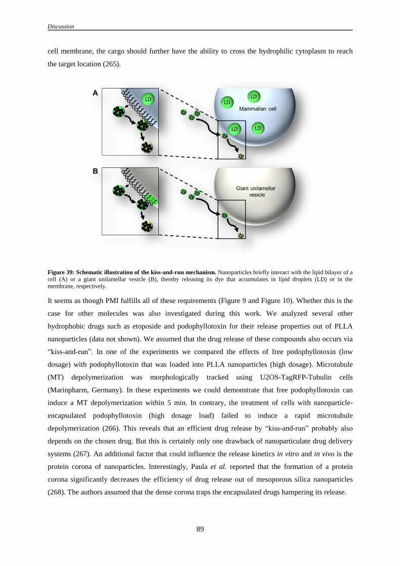
Discussion
89
cell membrane, the cargo should further have the ability to cross the hydrophilic cytoplasm to reach
the target location (265).
Figure 39: Schematic illustration of the kiss-and-run mechanism. Nanoparticles briefly interact with the lipid bilayer of a
cell (A) or a giant unilamellar vesicle (B), thereby releasing its dye that accumulates in lipid droplets (LD) or in the
membrane, respectively.
It seems as though PMI fulfills all of these requirements (Figure 9 and Figure 10). Whether this is the
case for other molecules was also investigated during this work. We analyzed several other
hydrophobic drugs such as etoposide and podophyllotoxin for their release properties out of PLLA
nanoparticles (data not shown). We assumed that the drug release of these compounds also occurs via
“kiss-and-run”. In one of the experiments we compared the effects of free podophyllotoxin (low
dosage) with podophyllotoxin that was loaded into PLLA nanoparticles (high dosage). Microtubule
(MT) depolymerization was morphologically tracked using U2OS-TagRFP-Tubulin cells
(Marinpharm, Germany). In these experiments we could demonstrate that free podophyllotoxin can
induce a MT depolymerization within 5 min. In contrary, the treatment of cells with nanoparticle-
encapsulated podophyllotoxin (high dosage load) failed to induce a rapid microtubule
depolymerization (266). This reveals that an efficient drug release by “kiss-and-run” probably also
depends on the chosen drug. But this is certainly only one drawback of nanoparticulate drug delivery
systems (267). An additional factor that could influence the release kinetics in vitro and in vivo is the
protein corona of nanoparticles. Interestingly, Paula et al. reported that the formation of a protein
corona significantly decreases the efficiency of drug release out of mesoporous silica nanoparticles
(268). The authors assumed that the dense corona traps the encapsulated drugs hampering its release.

Discussion
90
On the interface of membranes and nanoparticles, distinct mechanisms could drive the release of
hydrophobic substances out of nanoparticles (55). In general it is known that surfactants have the
potential to disrupt the membrane organization by changing the lipid conformation (269). Since
nanoparticles are stabilized with surfactants such as sodiumdodecyl sulfate (SDS) it would not be
surprising that lipids are reorganized after nanoparticle-membrane interaction (35). Exactly, this
interaction could favor the release of the cargo out of the nanoparticle. Other factors that could have an
impact on the release mechanism of drugs on the membrane are the hydrophobicity of the
nanomaterial and the strength of interaction between nanoparticles and cellular membranes (55). In
this study, we analyzed different nanoparticles on their interactions with giant unilamellar vesicles
(Figure 10 and Figure 11). As an example, PS-NH2-BODIPY (NH2 functionalized) as well as PLLA-
Fe-PMI (COOH functionalized) and SPIOPSN-BODIPY (Sulfonate functionalized) displayed two
completely different motifs of interaction on the giant unilamellar vesicles. The positively charged PS-
NH2-BODIPY nanoparticles have been observed to permanently interact with the GUVs by dashing
around onto the vesicles surface (Figure 11A). Interestingly, after some time of interaction, the
nanoparticles disrupted the vesicles implying a strong interaction between both partners. In contrary,
the negatively charged nanoparticles PLLA-Fe-PMI and SPIOPSN-BODIPY only temporarily
interacted with the GUV without destroying it (Figure 11B).
If the strength of interaction leads to an increased efficiency of cargo release is not clear from our
observations since we have used covalently-bound dyes for these experiments. However, we
demonstrated that the disruption of the membrane was only induced by strongly interacting and
aminofunctionalized nanoparticles (own observation). Lui et al. supported this hypothesis (270). They
showed that the surface functionalization of silica nanoparticles has an important impact on the
membrane integrity of tethered bilayers (270). The authors compared four types of surface
functionalizations (COOH, NH2, bare, PEG). According to their data they observed a distinct order for
the surface functionalization regarding their potential to disrupt membranes (amine > carboxyl ~ bare
> PEG). In other studies, the mechanism of disruption leads to a hole formation after membrane-
nanoparticle interaction (271). Further on, computer simulations calculated that nanoparticle-
membrane interactions are also able to induce a thinning of the contacted membrane (272). Over time,
this could also result in the collapse of the membrane (55).
To come back to the release mechanism of PMI, one needs also to discuss the effect of hydrophobicity
on the interactions of nanoparticles. As previously mentioned, it was demonstrated that PLLA-Fe-PMI
nanoparticles release their hydrophobic cargo PMI in hydrophobic media and on hydrophobic surfaces
of GUVs (Figure 10). This shows that hydrophobicity might be a crucial factor that can drive the
interactions between nanoparticles and lipid membranes. In the past, several simulations calculated the
impact of hydrophobic nanoparticles on membranes. Yang and colleagues modeled the simulation of a
hydrophobic and a semihydrophobic nanoparticle with a bilayer (273). While the interaction of

Discussion
91
hydrophobic nanoparticles with membranes can result in their inclusion into the bilayer, the
semihydrophobic nanoparticle is only found to adsorb into the membrane. Also a morphological
reorganization of the membrane lipids strongly depends on the hydrophobicity of nanoparticles (51).
All of these calculations referring to the impact of hydrophobicity on the nanoparticle-membrane
interactions are in compliance with the provided data of this thesis. Based on the release experiments
that have been performed, we propose that the release for PMI is mainly triggered by hydrophobic
interactions between the membrane and the nanoparticles. This hydrophobic interaction could
temporarily provide a more favorable hydrophobic environment for the PMI inside the membrane than
inside the nanoparticle.
4.3 Polymeric superparamagnetic nanoparticles are internalized by a
macropinocytic-like mechanism
After the long-term exposure of PLLA-FE-PMI nanoparticles, we observed their uptake into cells.
Morphologically, the endocytic structures that mediated the nanoparticle uptake resembled
macropinocytic ruffles (Figure 12). However, we previously showed that PLLA-Fe-PMI nanoparticles
are not suitable to investigate nanoparticle uptake mechanisms (6). To reliably study the entry
mechanisms of polymeric nanoparticles, we therefore used well-characterized superparamagnetic iron
oxide polystyrene nanoparticles (SPIOPSN) with a fluorescently bound fluorochrome (BODIPY). The
SPIOPSN fulfilled all requirements that were necessary to conduct these kinds of studies (6, 37). Since
we have shown that SPIOPSN only temporarily interact with cell membranes, we demonstrated an
uptake of SPIOPSN via the fluid phase endocytosis (Figure 12).
4.3.1 The entry of SPIOPSN is dependent on several factors of
macropinocytosis
To examine the entry routes of SPIOPSN, several endocytic pathways have been investigated by the
application of different freely permeable inhibitor molecules (Table 5). The set of inhibitors covered a
large number of different endocytic pathways. After intensive research on this project, we mainly
focused on inhibitors of macropinocytosis (Figure 12 and Figure 15). Since no standards for the
investigation of nanoparticulate macropinocytosis are defined yet, we checked several criteria that
have been considered as crucial in viral macropinocytic mechanisms (140). These include the
cointernalization of SPIOPSN with fluid-phase tracers. Furthermore, it is necessary to examine the
uptake after the modulation of the actin cytoskeleton. Here Rho GTPases as well as Na+/H
+
exchangers play a major role (94). Last but not least, the influences of several kinases, dynamin,
myosin II and microtubule dynamics have to be checked (212). We have summarized some of the
major factors that have been tested in this study (Table 20).

Discussion
92
Table 20: Factors worth to test in classical macropinocytosis (212).
Factors Effect
F-actin cytoskeleton Inhibition influences ruffling and blebbing.
Fluid-phase uptake Macropinocytic cargoes often colocalize with fluid
phase markers (e. g. HRP, Dextran).
Rho GTPases Rac1 or Cdc42 mediates macropinocytosis.
Na+/ H
+exchangers Inhibition by amiloride blocks macropinocytosis by
the lowering of submembraneous pH.
Kinases Inhibition of signaling kinases should suppress
macropinocytosis.
Our images show the co-uptake of SPIOPSN with fluorescently-labeled dextran, an important marker
of fluid phase endocytosis (Figure 12). As previously demonstrated, dextran is utilized for the tracking
of macropinosomes (274). Morphologically, we could show the internalization of nanoparticles via
heterogeneously sized vesicles that are also visible by DIC microscopy (Figure 12). Therefore, we
concluded from our experiments that SPIOPSN are internalized via a mechanism of fluid-phase
endocytosis (212). Further experiments with an inhibitor of F-actin polymerization allowed to assume
that actin reorganization plays an important role during the entry of SPIOPSN and dextran (Figure 12).
Previous reports for several cargo molecules like peptides, viruses and fluid phase markers already
showed a major participation of actin during their cellular entry (275, 276). Also on an ultrastructural
level, we partially observe the classical macropinocytic ruffles and protrusions that embraced
SPIOPSN and PLLA-Fe-PMI nanoparticles (Figure 12) (277).
4.3.2 SPIOPSN internalization is accompanied by the small GTPases Rac1
and cdc42
As a consequence of this pathway, the entry of SPIOPSN might depend on some small GTPases that
are known to induce the reorganization of the actin cytoskeleton (127, 278). To our knowledge, no one
has shown the participation of small GTPases during the uptake of nanoparticles. Therefore, we
characterized the possible effects of Rac1 as a small GTPase that is known to be located on the inside
of macropinocytic ruffles (136, 137). It was demonstrated that the overexpression of the permanently
active mutant of Rac1 Q61L induced the formation of roundly-shaped vesicles (Figure 13). These
organelles were previously described as nonbudded premacropinosomes (137). The overexpression of
this mutant drastically increased the probability to track SPIOPSN during the early stages of uptake
since the terminal vesicular budding was not completed yet (137). And indeed, SPIOPSN were found
in roundly-shaped premacropinosomes that seemed to be trapped in the early stages of fluid phase
uptake (Figure 13). The same phenotype has been observed after the overexpression of constitutively
active cdc42 Q61L (Figure 14). In spite of intensive research in the literature, we were not able to find

Discussion
93
a comparable confocal close-up image of this cdc42 Q61L-induced phenotype. However, it is
generally accepted that the small GTPase cdc42 is tightly associated with Rac1 implying similar
effects after overexpression (279, 280). Further on, it is known that both proteins act in the preliminary
stages of macropinocytic ruffle formation (280). Based on this argumentation, it was not so surprising
that SPIOPSN were found in roundly-shaped cdc42 Q61L+ and Rac1 Q61L
+ premacropinosomes.
Moreover, it is also worth to mention that dominant negative cdc42 T17N-GFP colocalized with
SPIOPSN. In this particular case, the vesicles were not static as seen after the overexpression of the
constitutively active mutants (Figure 14). All cdc42 T17N
+ vesicles directly moved through the cell
resembling budded cdc42+-macropinocytic vesicle (Figure 14). The colocalization of cdc42 with
nanoparticle-containing vesicles is in accordance with the data of the mass spectrometric analysis,
where we identified cdc42 as a protein of the SPIOPSN-associated fraction (Supplementary Table S4).
However, no quantitative and qualitative reduction of the internalization rate of SPIOPSN was
observed after esiRNA-mediated knockdown of cdc42 and the overexpression of cdc42 T17N (Figure
14). The impact of cdc42 knockdown was previously shown for the macropinocytic uptake of the
ebola virus (211). In this study the authors reported a reduction of viral uptake after the siRNA-
mediated downregulation of cdc42. Taking all of these data into account, we concluded that in spite of
the colocalization, cdc42 is not prominent enough to significantly control the uptake rate of SPIOPSN.
Consequently, we proposed that the nanoparticles are taken up by an atypical macropinocytic
mechanism (150).
4.3.3 SPIOPSN entry is triggered by an atypical type of macropinocytosis
As demonstrated by confocal imaging, SPIOPSN are internalized via macropinocytic structures
(Figure 12, Figure 13 and Figure 14). These specific membrane domains are ordered structures (212).
Their formation is tightly regulated by several kinases (140, 146). Most of the kinases were shown to
modulate the actin cytoskeleton via several routes (281, 282). Thereby, the PI3K signaling is one of
the major pathways, which mediate macropinocytosis (283). Combinations of different experiments
allow concluding that PI3K signaling acts in a first order followed by the signaling of PLC (144, 284).
Stable transfections as well as pharmacological inhibition of PI3K and PLC demonstrated that both
enzymes are essential factors in macropinocytic signaling (285). Taking these observations into
account, we pharmacologically inhibited PI3K by wortmannin and Ly294002 (Figure 15). Notably, we
observed a significant drop of internalized SPIOPSN by analyzing PI3K-inhibited cells.
Subsequently, the PI3K downstream signaling proteins PKC and PLC were inhibited. Both factors are
important actin modulators (144, 286). Therefore, it was even more than surprising that even high
concentrations of specific inhibitors for PKC and PLC did not alter the internalization rate of
SPIOPSN. It is important to mention that this observation is consistent with a case in the field of
virology, where a PKC-independent uptake was already reported for HIV-1 (140). This shows that a

Discussion
94
uniform picture of macropinocytosis can hardly be shaped. Since the exact mechanisms are still
elusive and seem to mainly depend on the investigated cell type and cargo it is challenging to set a
standard (140). Also the multiple options of actin reorganization make it hard to draw a clear picture
of macropinocytic mechanisms. This also includes the kinase PAK1. PAK1 binds the GTP-bound
form of cdc42/Rac1 and modulates the actin cytoskeleton (287). Indeed, we showed that the
pharmacological inhibition of PAK1 suppressed the entry of SPIOPSN in the same manner compared
to PI3K inhibition (Figure 15).
Cup closure or the scission of macropinosomes
Besides the modulation of the actin cytoskeleton, the closure of the macropinosome depends on PAK1
and requires the phosphorylation of CtBP1/BARS in growth factor triggered macropinocytosis and
during echovirus-1 entry (135, 288). This process seems to be independent from dynamin (135).
Contrary to this, rare cases of a dynamin-dependent macropinocytosis have been reported (147, 148,
289). This was also observed in our studies. Dynasore treatment as well as the overexpression of
dominant negative dynamin II drastically suppressed the number of internalized nanoparticles (Figure
16). On a subcellular level, it is known that dynamin participates in the formation of macropinocytic
protrusions (214). Schlunck and colleagues showed that the disruption of dynamin II leads to an
altered Rac localization from the cell edges into abnormal dorsal ruffles (214). The consequences of
this are inhibited cell spreading and the decrease of lamellipodia formation. Taken together, dynamin
is so far mainly seen as a factor of clathrin and caveolae-mediated endocytosis (Figure 18 and Figure
19). However, it is more than important to consider the action of dynamin II in macropinocytic
studies.
Stimulation of classical macropinocytosis results in a diminished uptake of nanoparticles
In agreement with the previous results, we concluded that nanoparticles are taken up by an atypical
macropinocytic pathway. This was underlined by experiments inducing classical macropinocytic
pathways by EGF or PMA (Figure 17). Both types of stimulation had no boosting effect on the rate of
internalization. Instead, stimulation of HeLa cells with EGF or PMA resulted in a massive drop of
internalized SPIOPSN. We postulated that the uptake of nanoparticles after macropinocytic
stimulation could be limited by their own size or owing to the diameter of the pinocytic ruffle (Figure
17). During this process, nanomaterials could be excluded from the macropinocytic ruffle. Normally,
EGF and PMA are known to trigger classical signaling cascades of macropinocytosis resulting in an
increase of endogenous fluid phase material as we have shown with dextran (Figure 17) (94, 290).
4.2.4 Changes in cholesterol homeostasis suppress the uptake of SPIOPSN
So far we discussed how the internalization of SPIOPSN depends on different kinases and small
GTPases. Hitherto, we did not discuss the role of cholesterol during nanoparticle entry. We observed

Discussion
95
that the depletion of cholesterol by mβCD suppressed the uptake ratio of SPIOPSN (Figure 21). In
general, cholesterol is an important membrane organizer in mammalian cells (291). The disturbance of
cholesterol homeostasis can have profound effects on cellular functions resulting in lipid-associated
disorders such as Niemann-Pick disease type C or Tangier disease (292). It further causes the
deregulation of macropinocytic signaling (138, 293, 294). So far it is known that several membrane
proteins prefer to interact with distinct lipids forming highly organized lipid rafts (295). Especially, in
macropinocytic signaling, the subcellular localization of important membrane proteins such as Rac1
depends on an intact cholesterol homeostasis. Grimmer and colleagues demonstrated that the depletion
of cholesterol prevents the localization of Rac1 to the plasma membrane in A431 cells (138). Further
on, the authors showed that the reorganization of the actin cytoskeleton and consequently the
membrane ruffling can be influenced by membraneous cholesterol (138, 296). In a particular study
with human embryonic kidney cells it has been demonstrated that the reduction of membranous
cholesterol decreases the length of actin-dependent protrusions (297).
Disturbance of the endolysosomal cholesterol homeostasis influences the uptake of nanoparticles
During this study, we further demonstrated that not only the depletion of membraneous cholesterol but
also the disturbance of endolysosomal cholesterol homeostasis had an effect on the uptake of
SPIOPSN (Figure 21). U18666A triggered a Niemann-Pick disease type C by accumulating
cholesterol and other lipids in the membrane of the endolysosomal system (Figure 21) (298). Inside
these NPC-like HeLa cells, the uptake rate of dextran and SPIOPSN was drastically decreased
assuming that NPC proteins might be associated with SPIOPSN-containing vesicles. Indeed, NPC1
and NPC2 were identified by mass spectrometry in the magnetically enriched fraction (Supplementary
Table S2). NPC1 and NPC2 are necessary proteins for the efflux of cholesterol and other lipids from
late endosomal compartments (298). When these proteins are nonfunctional, lipids accumulate inside
the endolysosomal system. Therefore, we investigated whether a siRNA-mediated knockdown of
NPC1 could induce such a phenotype. In previous studies, it was observed that siRNA-mediated
knockdown of NPC1 is sufficient for a deregulation of cholesterol inside the endolysosomal system
(299). Despite of comparable knockdown rates, we did not observe any significant effects on the
nanoparticle uptake rate after NPC1 downregulation. Therefore, we concluded that this method is
insufficient to study the effect of NPC1 on the uptake of nanoparticles. Notabily, other reports used a
more efficient technique to study the impact of NPC1 on the intracellular transport of nanoparticles
(83). The authors used NPC1-deficient MEFs and demonstrated that cells with nonfunctional NPC1
have a disturbed rate of exosome formation resulting in an increased retention of the nanomaterial
inside the endolysosomal system (83). This study shows the relevance of cholesterol and its
transporters in the endocytic mechanisms of nanoparticles.

Discussion
96
4.2.5 Flotillin-1 and CD81 knockdown as well as the overexpression of dominant
negative ARF1 suppresses SPIOPSN entry
Flotillins are organized as punctuate membrane domains enriched in cholesterol and sphingomyelin
(300, 301). The subcellular localization of flotillins depends on cholesterol (131). Furthermore,
flotillin microdomains have been implied to modulate the actin cytoskeleton (302). It was shown that
flotillin microdomains specifically interact with the cytoskeletal myosin IIa and spectrin (303).
Experiments with flotillin-1 knockout mice revealed that flotillin-1 is compromising myosin IIa
activity, thereby implicating a role for flotillins in the modulation of the actin cytoskeleton (303).
Taking the cholesterol dependence of flotillin and the role in actin modulation into account, one could
assume a direct link between flotillins and macropinocytic uptake mechanisms (304). To our
knowledge, this has not been observed in detail. However, Glebov et al. published a role for flotillin in
a clathrin-independent pathway showing that flotillin-1 mediates the transport of the GPI-anchored
CD59 and cholera toxin B subunit (88). Considering the investigated cargo molecules, this pathway
resembles the CLIC/GEEC pathway, which is also known to mediate the uptake of fluids (90).
In this study we detected a significant drop of internalized nanoparticles in flotillin-1-siRNA-treated
cells but observed no colocalization with ctxB (Figure 20; data not shown). Together, this implies an
indirect association of flotillin (88) with the uptake mechanism of the investigated nanoparticles.
Notably, we identified flotillin-1, flotillin-2 and the GPI-anchored protein CD59 in the magnetic
fraction by the mass spectrometry analysis of SPIOPSN-containing vesicles (Supplementary Table S2,
Supplementary Table S4). We propose an accumulation of flotillin-1, flotillin-2 and CD59 after the
fusion of other endocytic vesicles with SPIOPSN-containing vesicles (305, 306). Furthermore, we
assume that the uptake of nanoparticles is indirectly influenced by flotillin-1, since we did not observe
a colocalization with ctxB. Altogether, this shows that endocytic proteins and pathways are not strictly
separated from each other and could overlap in special cases.
Tetraspanins
Besides flotillin microdomains, tetraspanin-enriched microdomains (TEM) were investigated to study
their impact on the uptake of SPIOPSN (Figure 20). In previous works, tetraspanins have been
considered as proteins that laterally organize cellular membranes via their interactions with integrins
(307). These TEMs consist of the tetraspanins CD9, CD81, CD82 and CD63 which partially interact
with each other on the plasma membrane of HeLa cells (307). It has been shown that
TEMs/tetraspanins are important receptors for the infection process of HIV-1 and HCV (307, 308).
Spoden and colleagues showed that the downregulation or inhibition of several tetraspanins lead to a
diminished infection rate of HPV16 (308). We also observed an association of tetraspanins with
SPIOPSN-containing vesicles by mass spectrometry (Supplementary Table S2). Therefore, we
assumed that TEMs are cointernalized with the nanoparticles or even influence their uptake. To study

Discussion
97
this in more detail, we suppressed CD9, CD81, CD82 and CD63 by a siRNA-mediated approach
(Figure 20). We observed no influence on SPIOPSN uptake after the knockdown of CD9, CD82 and
CD63 but detected a suppressed rate of uptake after CD81 knockdown. Interestingly, CD81 was
previously identified as a binding partner for the small macropinocytic GTPase Rac (309). Tejera et al.
showed that the knockdown of CD81 resulted in significant alterations of focal adhesion and Rac
activation (309). In dendritic cells, CD81 influences the formation of lamellipodia indicating an
impact on the actin cytoskeleton (310). All of these data highlight a potential association of CD81 with
the macropinocytic uptake of SPIOPSN and lays the foundation for further investigations to study the
effects of tetraspanins on nanoparticle entry.
The inhibition of ARF1 results in a decreased uptake of nanoparticles
Previously, Kumari et al. demonstrated that ARF1 is directly associated with the internalization of
ctxB and dextran (90). Consequently, they defined ARF1 as a player of the CLIC/GEEC pathway (90).
A second study of Hasegawa and colleagues showed that a mutation of ARF1 causes a significant
decrease of internalized dextran during a PDGF-triggered macropinocytosis in NIH3T3 cells (220).
The overexpression of dominant negative ARF1 T31N was shown to induce an incomplete closure of
the circular dorsal ruffles mediated by actin (220). The authors resumed that the macropinocytic
uptake mechanism is blocked by ARF1 (220). Taken together, both groups reported different functions
of ARF1 in two distinct pathways.
In this study, we identified four signs indicating a pivotal role of ARF1 in the entry mechanism of
SPIOPSN (Figure 22). The first evidence was supplied by the detection of ARF1 in the vesicular
fraction of the magnetically purified pellet (Supplementary Table S3). Secondy, we observed a direct
interaction between nanoparticles and ARF1 T31N by confocal microscopy (Figure 22). We have
demonstrated that the overexpression of ARF1 T31N resulted in a massive decrease of internalized
SPIOPSN highlighting the significant effect of ARF1 on nanoparticle endocytosis (Figure 22). We
further found that pharmacological inhibiton of ARF1 suppressed the uptake of SPIOPSN (Figure 22).
Therefore, we concluded that ARF1 is directly involved in a macropinocytic-like uptake of
nanoparticles. By following the definition of Kumari et al., one could describe this entry mechanism
as a dynamin-independent CLIC/GEEC pathway (90). However, since we have identified dynamin as
an important factor during SPIOPSN entry (Figure 16), we considered this uptake mechanism as a
macropinocytic-like one. This was also supported by cryo-TEM images, where we did not identify any
tubular endosomal structures that are characteristic for CLIC/GEEC endocytosis (Figure 12). Anyway,
the classical structures of the CLIC/GEEC pathway have a diameter of about 40 nm, which are to
small for the investigated nanoparticles (126).

Discussion
98
Crosstalk between distinct endocytic pathways
It seems that several endocytic mechanisms have significant functional and morphological overlaps
hampering the classification of a single pathway. For instance, the CLIC/GEEC pathway is seen as a
clathrin-independent pathway (96). However, a crosstalk between both pathways has been observed
(85). Disturbance of the CLIC/GEEC pathway triggers a compensation of GPI-linked protein uptake
by clathrin-mediated endocytosis (128). Another example of a crosstalk was published by Naslavsky
et al. (311). The authors showed that the GPI-linked protein CD59 and MHCI are internalized by a
non-CLIC-GEEC pathway. This pathway is clathrin-and dynamin independent and utilizes ARF6-
positive endosomes (311). As previously mentioned, also the flotillin-mediated pathway and the
CLIC/GEEC pathway was reported to mediate the internalization of CD59 (88). The CLIC/GEEC
pathway also resembles macropinocytic-like pathways as shown for adeno-associated virus 2 (AAV2)
(95). Internalization of AAV2 depends on factors such as cdc42, ARF1, GRAF1 and EIPA but is
insensitive to Rac1 or other macropinocytic inhibitors (95). The resume of the authors was that AAV2
is internalized via a pleiomorphic CLIC/GEEC pathway. Taking all of this into account, it is
challenging to identify model cargoes that can be specifically assigned to a single pathway. This
shows the close relationship between several entry mechanisms massively depending on cell type and
investigated cargo.
4.3 Intracellular trafficking of nanoparticles
So far we discussed the mechanisms how cells internalize superparamagnetic iron oxide polystyrene
nanoparticles. We have demonstrated that the entry of SPIOPSN is mediated by an atypical type of
macropinocytosis mainly depending on cholesterol, kinases and typical small GTPases of several
closely related endocytic mechanisms. To understand the intracellular trafficking of nanoparticles after
macropinocytic uptake, we investigated the intracellular trafficking routes of these SPIOPSN (Figure
24-37). This question becomes especially exciting since several endocytic membrane proteins are also
located inside the endolysosomal system (307). The reason for this originates in the functionality of
the endolysosomal system. Notably, it is responsible for the renewal and recycling of plasma
membrane proteins and membrane lipids by internalizing them (312).
As a result, the investigation of intracellular trafficking is a challenging field due to the high turnover
rates of the participating proteins (312). Vesicles that bud off from the membrane are consequently
able to fuse with other endocytic vesicles afterwards forming a complete novel vesicle with a distinct
protein composition (181). Further on, it is well accepted that e. g. the maturation of early endosomes
to late endosomes is accompanied by a rapid exchange of important markers (e. g. Rab proteins). This
impedes the detection of high rates of colocalization with nanoparticles especially under the
asynchronous uptake of nanoparticles (Figure 32) (153).

Discussion
99
Early endocytic markers
Since we have proposed an uptake of SPIOPSN via macropinosome-like vesicles, we first checked for
the early endocytic marker Rab5 (Figure 24) (142). For epithelial cells and fibroblasts it has been
demonstrated that Rab5 and its effector Rabankyrin-5 are important proteins with a cooperative
function for the biogenesis of macropinosomes and the endocytic system (142, 230). The authors
showed that knockdown of Rab5 and Rabankyrin-5 decreases fluid phase uptake in NIH 3T3 cells.
They found that Rab5 and Rabankyrin-5 colocalize with fluid phase-filled macropinosomes that are
negative for EEA1 and transferrin (142). In our study, we also observed nanoparticles in fluid phase
filled macropinosomes (Figure 25). In spite of extensive studies, no significant association of
SPIOPSN with the classical early endosomal marker EEA1 was observed by confocal imaging (data
not shown) and by the proteomic analysis of SPIOPSN-containing vesicles. Though, we detected
SPIOPSN in Rab5+ vesicles that were detectable by DIC microscopy (Figure 24). Surprisingly, the
knockdown of Rab5A (Figure 24) and Rabankyrin-5 (data not shown) did not significantly suppress
the uptake of SPIOPSN as contrarily demonstrated by other studies (142). From this, one can infer that
other factors might drive the internalization of nanoparticles in HeLa cells. This might also be in
accordance with the low rate of colocalization between nanoparticles and Rab5, in spite of the
significant detection of Rab5A and Rab5C by mass spectrometry. The low number of colocalization of
Rab5 with nanoparticles was also observed by previous studies, where the authors analyzed the
colocalization of nanoparticles in the early stages of uptake (151).
Contrary to the low rate of colocalization of Rab5A with SPIOPSN, we found a higher number of
SPIOPSN inside RhoB+ vesicles. We detected SPIOPSN inside RhoB
+ vesicles that actively moved
through the cell (Figure 25). These vesicles were visible by DIC microscopy and resembled fluid
phase-filled macropinosomes. Besides the vesicular localization of RhoB-GFP, the protein also
localized to the plasma membrane implying that RhoB plays a role in the early stages of endocytosis
(313). Indeed, RhoB was detected by mass spectrometry as a protein of the membrane fraction
(Supplementary Table S4). From literature it is known that RhoB is a small GTPase acting in the
actin-mediated transport of endocytic vesicles (314). Studies using confocal microscopy detected
RhoB mainly in early endosomes (315). In this context, RhoB was further shown to influence the entry
of virions (223). Overexpression of RhoB significantly increased the endogenous levels of ebola virus
glycoprotein and VSVG pseudotyped vector transduction. No effect was observed for GP pseudotyped
HIV or adeno-associated virus 2 vector implying a specific role for RhoB during the uptake of viruses
(223). However, we concluded that SPIOPSN were internalized by RhoB+ vesicles resembling early
macropinosomes.

Discussion
100
Late markers of endocytosis
During the life span of an endosome, several fusion and maturation events change the proteomic
composition of the vesicle (153). It is well accepted that early endosome to late endosome maturation
can be measured by the steady conversion of Rab5 to Rab7 on the surface of endosomes (153). In our
experiments, a colocalization of Rab7+ vesicles with SPIOPSN has been recorded (Figure 26;
Supplementary Table S2). These late endosomes contained intraluminal vesicles and are probably
linked to anterograde and retrograde TGN-transport mechanisms (Figure 28, Figure 29 and Figure 30)
(154). TGN-to-LE transport delivers lysosomal hydrolases and membranes to the inside of late
endosomes providing proteins for the hydrolytic milieu of the lysosome (156). Lysosome-associated
membrane proteins are directly transported by two major pathways. The most prominent pathway is
orchestrated by the mannose-6-phosphate-receptor (316). Both types of the M6PR, MPRD and MRPI,
were associated with SPIOPSN-containing vesicles (Supplementary Table S2). A so far unknown
pathway is M6PR-independent and is mediated by VAMP7 and hVps41 (154). The authors suggest a
role for VAMP7 in the transport of lysosome-associated membrane protein carriers showing that
VAMP7 is directly involved in cargo transport from TGN-to-LE (154). VAMP7 is also well
characterized as an R-SNARE mediating the fusion of late endosomes to lysosomes (192). Notably,
we detected the R-SNARE VAMP7 on SPIOPSN-containing vesicles in colocalization experiments
and by mass spectrometry (Figure 27; Supplementary Table S2). We proposed that those vesicles,
which are SPIOPSN/VAMP7+
, were recorded during a heterotypic fusion event between late
endosomes and lysosomes. This assumption was based on the observation that several SPIOPSN
accumulated inside these organelles. We resumed that such a large number of SPIOPSN was so far
only observed in lysosomal organelles.
Another prominent protein on late endosomes is the small GTPase Rab9, which also colocalized with
SPIOPSN (Figure 26, Supplementary Table S2) (317). The important role of Rab9 on endosomes
becomes apparent after Rab9 downregulation. The knockdown of Rab9 results in a size increase of
late endosomes (318). Furthermore, it was demonstrated that Rab9 stimulates the backtransport of
M6PR from late endosomes to the TGN in a cell free system (317). This is in compliance with a study
that microscopically demonstrated the fusion of Rab9+ late endosomes with the TGN (225). The
authors assume that the endocytic system uses a vesicle intermediate to communicate between the
endocytic system and the TGN-network, thereby implying the strong interaction between both systems
(225).
Pmel17 and tetraspanins in the trafficking pathways of SPIOPSN
We know from previous studies that the endocytic uptake of viruses and nanoparticles can depend on
tetraspanin-enriched microdomains (217, 307). Tetraspanins can act as receptors for HPV16 entry or
assist in the ESCRT-dependent or independent sorting of other proteins into intraluminal vesicles of

Discussion
101
multivesicular bodies (174, 217). In this particular case, the well-studied tetraspanin CD63 facilitates
the transport of the melanosomal protein Pmel17 into the ILVs of multivesicular bodies in an
ubiquitin-independent manner (174, 226, 227). To study the association of ubiquitin, CD63 and
Pmel17 with SPIOPSN-containing vesicles, their colocalization was studied (Figure 28). Interestingly,
SPIOPSN-containing vesicles lacked the colocalization with ubiquitin (data not shown). Instead, we
detected a colocalization of CD63 with SPIOPSN (Figure 28). Therefore, we asked the question,
whether Pmel17 colocalizes with SPIOPSN-containing vesicles. And indeed, we observed a high rate
of colocalization of the ILV marker Pmel17 with SPIOPSN inside HeLa cells (Figure 28). This is in
accordance with our TEM pictures displaying the close proximity of SPIOPSN to ILVs (Figure 29 and
Figure 30). Taking the colocalization of SPIOPSN with CD63 and Pmel17 and the non-colocalization
with ubiquitin into account, it is important to raise questions about different MVB subtypes (319). For
early endosomes, the existence of subpopulations has already been shown (161). However, if this is
also true for multivesicular body has to be investigated in the future.
Multilamellar lysosomes are the terminal organelle for SPIOPSN
The disassembly of nanomaterials is a major prerequisite for the release of encapsulated effector
molecules in drug delivery applications (2). Therefore, it is of great interest to identify the terminal
subcellular localization of a nanoparticle to gain knowledge about the endolysosomal enzymes that
could potentially degrade nanomaterials (Figure 36). In pulse experiments with SPIOPSN using
transmission electron microscopy, we found almost all SPIOPSN after 20 h inside multilamellar
lysosomes (data not shown). These vesicles contained several multilamellar membranes whorls
(Figure 29 and Figure 30) (182). In the past, several groups proposed that multilamellar bodies evolve
from the fusion with autophagosomes (182). Indeed, it is well known that lysosomes fuse with LC3+
autophagosomes (320). However, LC3-II is degraded after autolysosomal fusion challenging the
detection of a direct interaction between lysosome and autophagosome by LC3-II (321). In our
experiments, immunostainings for LC3-II revealed no colocalization with SPIOPSN signals indicating
that nanoparticles are not transported via the autophagosomal pathway (Figure 32). However, we
found a large number of mitochondrial proteins inside the magnetically enriched fraction
(Supplementary Table S5). Further on, TEM images showed multilamellar membrane whorls inside
multilamellar lysosomes. Altogether, this implies that SPIOPSN are stored inside lysosomes, which
fused with autophagosomes.
4.3.1 Relevance of SPIOPSN trafficking in vivo – Studying nanoparticulate
biodistribution
Especially, metal containing particles such as SPIOs or gold nanoparticles raise concerns about
biodistribution and toxicity while they can be rapidly cleared from the blood stream entering organs
such as liver and spleen (322, 323). The distribution of nanoparticles inside in vivo systems majorly
Discussion
102
depends on their surface modification and size (324, 325). In vitro, we tested our SPIOPSN for
aggregation in blood and found less morphologically visible aggregation in blood serum. Therefore,
we considered this system as stable and injected SPIOPSN into NSG mice (Figure 33). NSG mice are
immunodeficient and lack functional immune cells such as dendritic cells or macrophages. Therefore,
it is a proper system to study the biodistribution of nanoparticles independently from the immune
system. After injection, we magnetically isolated the nanoparticle-containing cells from complete
tissues. We observed an endolysosomal like disposition of SPIOPSN after 24 h mainly in spleen and
liver cells, while the peripheral blood, kidney and bladder lacked nanoparticles. This is in partial
compliance with other studies, mainly detecting nanoparticles in liver and kidney (324). With this
experiment, we could show the endolysosomal SPIOPSN uptake in vivo. Moreover, SPIOPSN were
proven to be sufficient for the magnetic isolation of cells. This magnetic potential of SPIOPSN was
subsequently used to study the intracellular trafficking of nanoparticles in more detail (Figure 34).
4.3.2 Reconstruction of intracellular nanoparticle trafficking displays a detailed
picture of the proteomic environment of SPIOPSN
So far we have discussed how nanoparticles traffic along the endocytic route. To understand these
trafficking pathways in more detail, one has to identify novel factors that are associated with the
transport of nanoparticles (Figure 35 and Figure 36). Hitherto, these studies of intracellular
nanoparticle trafficking were always based on the manipulation of the cellular systems (132, 151).
This could result in drastical changes of the intracellular trafficking machinery. Overexpression or
knockdown of membrane proteins may significantly alter the trafficking behavior of cells (153, 318).
Moreover, the recent methods are biased by other cargoes such as viruses and can only analyze a low
number of proteins at the same time. During this study, we achieved to circumvent such pitfalls by
generating an unbiased snapshot of intracellular nanoparticle trafficking using mass spectrometry
(Figure 35). Another advantage of this untampered approach is the option to characterize the
lysosomal matrix milieu of enzymes (Figure 36). This could provide information for the prospective
design of nanocarriers achieving a more specific endolysosomal degradation for an efficient release of
the effector molecules.
Identification of proteins heavily depends on the method of subcellular fractionation
Normally, the purification of subcellular compartments is based on the size and the density of the
organelle (156). This results in a biased analysis of the isolated vesicles, since only physical
parameters are considered. Most of the researchers use differential centrifugation in a sucrose or
percoll gradient for these kind of purifications (156, 326, 327). Sometimes, the distribution of distinct
organelles (e.g. lysosomes and mitochondria) significantly overlap in hyperosmolar sucrose preventing
an appropriate separation of distinct compartments (156). Using our protocol for the magnetic
isolation of vesicles, one is only obtaining vesicles, which have engulfed the nanoparticle (Figure 34).

Discussion
103
This approach excludes the isolation of endosomes that are not participating in the trafficking of
magnetic nanoparticles.
Another great pitfall of all subcellular fractionation methods are unavoidable impurities inside the
sample. Especially, in case of sensitive MS methods a large number of unwanted proteins are
detectable (156). In conclusion: The more sensitive the method, the more challenging the subcellular
fractionation. The proteomic analysis of the endolysosomal system is even more complicated since the
cell uses the endocytic pathway for the degradation of non-endolysosomal constituents (328).
Especially, the fusion of autophagosomes with lysosomes may insert impurities of defect organelles
such as mitochondria or other damaged vesicles (328). To reliably distinguish between positive and
false positive results, one has to focus on the enrichment of proteins in the purified fraction in
comparison to the remaining fraction (Table 18). However, potentially positive proteins still can be
screened by low copy numbers or degradative processes. Here the major drawbacks include the
detection of low copy number proteins as well as small proteins (e. g. subunits), hydrophobic insoluble
transmembrane proteins, proteins with posttranslational modifications and loosely attached proteins
(156). Further on, endogenous fusion events e. g. between different endocytic proteins impedes the
clear interpretation and the identification of direct effector proteins in the trafficking of SPIOPSN
(156). Altogether, these factors hamper the interpretation of the data. Nevertheless, the analysis of
vesicular proteins by MS is a valuable tool to screen for new proteins. However, the relevance of these
data has to be confirmed by other methods.
Reconstruction of intracellular trafficking identifies novel promising candidates for prospective
studies
The dissection of intracellular nanoparticle trafficking identified a large number of novel proteins that
have never been associated with nanoparticles before (Figure 35). These include major markers of
different endocytic compartments such as early endocytic compartmens, multivesicular bodies,
lysosomes, COP vesicles and lysosomes (Figure 35). Interesting candidates for future applications are
primarily represented by proteins of the tetraspanin family. The targeting of tetraspanin-enriched
microdomains could be a novel approach for targeted delivery applications towards the endosomal
system since these domains are easily accessible to extracellular particles (307).
However, the identification of several nanoparticle associated proteins gives also room for
speculations. Especially, the identification of COP proteins raises questions, whether COP vesicles
directly interact with the endolysosomal vesicles of a nanoparticle. In this context, it is important to
mention that our mass spectrometry data is in compliance with the study of Aniento et al., where the
authors identified epsilon COP and beta COP on endosomal membranes (329). Whether this originates
from a fusion event of endosomes with COP vesicles needs to be elucidated with other methods,
which seems to be challenging. The identification of rare events in SPIOPSN trafficking is a major

Discussion
104
problem when using standardized methods such as confocal microscopy. This was also shown by
Sandin et al. (151). Despite of using spinning disc confocal microscopy, only a low number of
colocalization events were detectable between nanoparticles and the early endosomal marker Rab1A,
while no colocalization was observed with the COPI coat complex (151). The limitations often
originate from the high dynamics of intracellular trafficking. Also low expression levels of the target
protein or limitations in the detection by confocal microscopy might play a major role. During this
study, we observed that especially the microscopic imaging of magnetic nanoparticles bears
limitations. The detection of nanoparticles by PMTs rapidly gives rise to false positive signals on a
very low signaling intensity owing to the high light scattering properties of the nanomaterial. This
restricts the sensitive detection of markers by immunofluorescence or overexpression of GFP fusion
proteins to a minimum. Therefore, our mass spectrometry approach is the more sensitive one, but data
interpretation needs to be carefully performed.
Identification of lysosomal matrix proteins and their possible effects on the nanoparticulate protein
corona
In the future, it will be necessary to study the degradation of internalized nanomaterial in more detail.
Already a large number of studies consider nanomaterials as biodegradable structures but only less
provide information about the long-term stability inside the vesicular environment. Even potentially
biodegradable nanoparticles remain for a long time inside the endolysosomal system without getting
significantly degraded (Lieberwirth, unpublished data) (33). After the deposition of nanoparticles
inside lysosomal compartments, the protein corona and the nanomaterial underlies a harsh vesicular
milieu of several hydrolases, proteases and other degradative enzymes. It has recently been reported
that parts of the nanoparticulate protein corona are cointernalized with the nanoparticle (330).
Regularly, ubiquitinylated proteins that are marked for degradation are transported to the lysosome.
Therefore, it is possible that proteins on the surface of nanoparticles are marked with ubiquitin
considering them for the degradative lysosomal pathway. Since we have not found ubiquitin in
nanoparticle containing vesicles at all, we proposed that the transport of nanoparticles to lysosomes is
a passively directed process. Anyway, the lysosomal milieu seems to degrade the nanoparticulate
protein corona very efficiently shown in an example using a fluorescently labeled albumin (Figure 39).
Especially, for albumin it is known that it is degraded into the lysosome of cells (331). A major
candidate for the degradation of albumin is cathepsin D and cathepsin B (332). Since we observed a
colocalization of cathepsin D with nanoparticles (Figure 31) and identified cathepsin D/B by mass
spectrometry, it was proposed that the protein corona of nanoparticles is degraded inside SPIOPSN
containing lysosomes after a short time. This data is in compliance with the data of Wang et al., where
protein corona degradation experiments were performed with fluorescently labeled serum showing a
degradation of the protein corona inside lysosomal structures (330). Taking this into account, one has
to consider that not only the protein corona but also the intravesicular milieu may drastically change

Discussion
105
the surface modification and material properties of nanomaterials. We have to keep in mind that this
might have a crucial impact on the intracellular drug delivery abilities of nanoparticles.

Summary and conclusions
106
5 Summary and conclusions
This work investigated the drug delivery abilities, the entry mechanisms and the intracellular
trafficking pathways of different polymeric nanoparticles. We learned that the different types of
interaction between nanoparticles and cells mainly depend on the used materials. To exploit intelligent
drug delivery mechanisms, nanoparticulate systems need to be improved and their interaction with
biological components need to be understood.
In the first part of the study, we wanted to know whether we can transport an encapsulated
hydrophobic molecule over a membrane barrier. This is particularly important in drug delivery
approaches for hardly accessible cells such as B or T lymphocytes. To approach this question, we
studied nanoparticle-membrane interaction and a subsequent cargo release of the hydrophobic dye
PMI on the membrane of HeLa cells, hMSCs and Jurkat cells. We observed a rapid staining of
intracellular lipid droplets and reproduced this delivery mechanism in all three cell lines. We found
that the transport of the membrane to the interior of the cell mainly depends on the nanoparticulate
carrier but not on free dye molecules. Finally, we hypothesize that PMI release is largely triggered by
a temporary nanoparticle-membrane contact. Hydrophobic surfaces of giant vesicles and of plasma
membranes can trigger nanoparticle cargo release in a typical “kiss-and-run” dependent manner.
Next, we showed that the uptake of the well-characterized polymeric nanoparticle SPIOPSN is
mediated by a macropinocytic-like mechanism. The treatment of cells with typical macropinocytic
inhibitors significantly decreased the uptake of SPIOPSN into cells. However, inhibition of classical
macropinocytic kinases such as PKC and PLC did not affect the entry of SPIOPSN. Also the
stimulation of classical macropinocytosis by PMA and EGF decreased the uptake of SPIOPSN. We
further investigated several proteins such as ARF1, Rac1 and cdc42 and showed their important role
during the uptake of nanoparticles. Also the knockdown of flotillin-1 as well as the suppression of the
tetraspanin CD81 decreased the levels of endogenous nanoparticles. Altogether, this implies an
atypical type of macropinocytic uptake the investigated nanoparticles utilized.
Finally, we analyzed the intracellular trafficking pathways of a nanoparticle after a macropinocytic-
like entry in HeLa cells. By the proteomic dissection, the intracellular nanoparticle trafficking (INT)
was reconstructed based on a simple endocytic model. The analysis of SPIOPSN-containing vesicles
resulted in the detection of proteins that have never been linked to INT before. To confirm the data of
mass spectrometry, we analyzed several identified key players of intracellular trafficking by confocal
live cell imaging. By this approach, SPIOPSN were found to be actively transported via
macropinosomes, late endosomes and lysosomes. We were able to reproduce the colocalization of
nanoparticles with lysosomal proteins in hMSCs. Strikingly, we identified several lysosomal matrix
proteins in the enriched magnetic fraction of the nanoparticle-containing vesicles. It was assumed that
the properties of the lysosomal compartment mediate the degradation of the nanoparticulate protein

Summary and conclusions
107
corona. The degradation of protein corona may give access to the bare surface and the polymeric
matrix of nanoparticles opening the way for the degradation of the nanomaterial.
In conclusion, we present a detailed picture of a so far unknown drug delivery mechanism that
depends on temporary nanoparticle-membrane interactions. This could remarkably shift the paradigm
of drug delivery mechanisms in the field of nano-bio-interactions. Furthermore, we provide evidence
that polymeric nanoparticles are mainly internalized via an unspecific atypical macropinocytosis
mechanism followed by a subsequent transport of nanoparticles to multilamellar lysosomes. This may
set the basis for prospective nanoparticle studies and might help to understand nano-bio-interactions in
more detail.

Bibliography
108
6 Bibliography
1. Nel AE, et al. (2009) Understanding
biophysicochemical interactions at the
nano-bio interface. Nat Mater
8(7):543-557.
2. Hofmann D & Mailander V (2013)
Pharmacology of nanocarriers on the
microscale: importance of uptake
mechanisms and intracellular
trafficking for efficient drug delivery.
Nanomedicine (Lond) 8(3):321-323.
3. Yatvin MB, Kreutz W, Horwitz BA, &
Shinitzky M (1980) pH-sensitive
liposomes: possible clinical
implications. Science 210(4475):1253-
1255.
4. Gilleron J, et al. (2013) Image-based
analysis of lipid nanoparticle-mediated
siRNA delivery, intracellular
trafficking and endosomal escape. Nat
Biotechnol 31(7):638-646.
5. Cecco S, Aliberti M, Baldo P,
Giacomin E, & Leone R (2014) Safety
and efficacy evaluation of albumin-
bound paclitaxel. Expert Opin Drug
Saf 13(4):511-520.
6. Hofmann D, Messerschmidt C,
Bannwarth MB, Landfester K, &
Mailander V (2014) Drug delivery
without nanoparticle uptake: delivery
by a kiss-and-run mechanism on the
cell membrane. Chemical
Communications 50(11):1369-1371.
7. Soppimath KS, Aminabhavi TM,
Kulkarni AR, & Rudzinski WE (2001)
Biodegradable polymeric nanoparticles
as drug delivery devices. J Control
Release 70(1-2):1-20.
8. Rao JP & Geckeler KE (2011)
Polymer nanoparticles: Preparation
techniques and size-control
parameters. Prog Polym Sci 36(7):887-
913.
9. Siebert JM, Baier G, Musyanovych A,
& Landfester K (2012) Towards
copper-free nanocapsules obtained by
orthogonal interfacial "click"
polymerization in miniemulsion. Chem
Commun (Camb) 48(44):5470-5472.
10. Tiarks F, Landfester K, & Antonietti
M (2001) Preparation of polymeric
nanocapsules by miniemulsion
polymerization. Langmuir 17(3):908-
918.
11. Landfester K (2009) Miniemulsion
Polymerization and the Structure of
Polymer and Hybrid Nanoparticles.
Angew Chem Int Edit 48(25):4488-
4507.
12. Ugelstad J, Elaasser MS, &
Vanderho.Jw (1973) Emulsion
Polymerization - Initiation of
Polymerization in Monomer Droplets.
J Polym Sci Pol Lett 11(8):503-513.
13. Landfester K & Mailander V (2013)
Nanocapsules with specific targeting
and release properties using
miniemulsion polymerization. Expert
Opin Drug Deliv 10(5):593-609.
14. Hocherl A, Dass M, Landfester K,
Mailander V, & Musyanovych A
(2012) Competitive cellular uptake of
nanoparticles made from polystyrene,
poly(methyl methacrylate), and
polylactide. Macromol Biosci
12(4):454-464.
15. Baldan A (2002) Review Progress in
Ostwald ripening theories and their
applications to nickel-base superalloys
- Part I: Ostwald ripening theories. J
Mater Sci 37(11):2171-2202.
16. Bibette J, Morse DC, Witten TA, &
Weitz DA (1992) Stability-Criteria for
Emulsions. Phys Rev Lett
69(16):2439-2442.
17. Thiam AR, Farese RV, & Walther TC
(2013) The biophysics and cell biology
of lipid droplets. Nat Rev Mol Cell Bio
14(12):775-786.
18. Cao Z, Schrade A, Landfester K, &
Ziener U (2011) Synthesis of
raspberry-like organic–inorganic
hybrid nanocapsules via pickering
miniemulsion polymerization:
Colloidal stability and morphology.
Journal of Polymer Science Part A:
Polymer Chemistry 49(11):2382-2394.
19. Musyanovych A, Rossmanith R,
Tontsch C, & Landfester K (2007)
Effect of Hydrophilic Comonomer and
Surfactant Type on the Colloidal
Stability and Size Distribution of
Carboxyl- and Amino-Functionalized
Polystyrene Particles Prepared by
Miniemulsion Polymerization.
Langmuir 23(10):5367-5376.

Bibliography
109
20. Gref R, et al. (2000) 'Stealth' corona-
core nanoparticles surface modified by
polyethylene glycol (PEG): influences
of the corona (PEG chain length and
surface density) and of the core
composition on phagocytic uptake and
plasma protein adsorption. Colloids
Surf B Biointerfaces 18(3-4):301-313.
21. Fritz G, Schadler V, Willenbacher N,
& Wagner NJ (2002) Electrosteric
stabilization of colloidal dispersions.
Langmuir 18(16):6381-6390.
22. MolinaBolivar JA, GalisteoGonzalez
F, & HidalgoAlvarez R (1997)
Colloidal stability of protein-polymer
systems: A possible explanation by
hydration forces. Phys Rev E
55(4):4522-4530.
23. Wells MA, Abid A, Kennedy IM, &
Barakat AI (2012) Serum proteins
prevent aggregation of Fe2O3 and
ZnO nanoparticles. Nanotoxicology
6:837-846.
24. Tenzer S, et al. (2013) Rapid
formation of plasma protein corona
critically affects nanoparticle
pathophysiology. Nat Nanotechnol
8(10):772-781.
25. Monopoli MP, Aberg C, Salvati A, &
Dawson KA (2012) Biomolecular
coronas provide the biological identity
of nanosized materials. Nat
Nanotechnol 7(12):779-786.
26. Salvati A, et al. (2013) Transferrin-
functionalized nanoparticles lose their
targeting capabilities when a
biomolecule corona adsorbs on the
surface. Nat Nanotechnol 8(2):137-
143.
27. Natte K, et al. (2013) Impact of
polymer shell on the formation and
time evolution of nanoparticle-protein
corona. Colloids Surf B Biointerfaces
104:213-220.
28. Murakami H, Kobayashi M, Takeuchi
H, & Kawashima Y (1999) Preparation
of poly(DL-lactide-co-glycolide)
nanoparticles by modified spontaneous
emulsification solvent diffusion
method. Int J Pharm 187(2):143-152.
29. Peracchia MT, et al. (1999) Stealth
PEGylated polycyanoacrylate
nanoparticles for intravenous
administration and splenic targeting. J
Control Release 60(1):121-128.
30. Athanasiou KA, Agrawal CM, Barber
FA, & Burkhart SS (1998)
Orthopaedic applications for PLA-
PGA biodegradable polymers.
Arthroscopy 14(7):726-737.
31. Höcherl A (2012) Qualitative and
absolute quantitative studies of the
cell-nanoparticle interaction.
(Universitätsbibliothek Mainz).
32. Rezwan K, Chen QZ, Blaker JJ, &
Boccaccini AR (2006) Biodegradable
and bioactive porous
polymer/inorganic composite scaffolds
for bone tissue engineering.
Biomaterials 27(18):3413-3431.
33. Gonzalez MF, Ruseckaite RA, &
Cuadrado TR (1999) Structural
changes of polylactic-acid (PLA)
microspheres under hydrolytic
degradation. J Appl Polym Sci
71(8):1223-1230.
34. Tartaj P, Morales MD, Veintemillas-
Verdaguer S, Gonzalez-Carreno T, &
Serna CJ (2003) The preparation of
magnetic nanoparticles for
applications in biomedicine. J Phys D
Appl Phys 36(13):R182-R197.
35. Ramirez LP & Landfester K (2003)
Magnetic polystyrene nanoparticles
with a high magnetite content obtained
by miniemulsion processes. Macromol
Chem Physic 204(1):22-31.
36. Schmidtke-Schrezenmeier G, et al.
(2011) Labeling of mesenchymal
stromal cells with iron oxide-poly(L-
lactide) nanoparticles for magnetic
resonance imaging: uptake,
persistence, effects on cellular function
and magnetic resonance imaging
properties. Cytotherapy 13(8):962-975.
37. Bannwarth MB, et al. (2013) Well-
defined nanofibers with tunable
morphology from spherical colloidal
building blocks. Angew Chem Int Ed
Engl 52(38):10107-10111.
38. Bonnemain B (1998)
Superparamagnetic agents in magnetic
resonance imaging: Physicochemical
characteristics and clinical applications
- A review. J Drug Target 6(3):167-
174.
39. Krishnan KM, et al. (2006)
Nanomagnetism and spin electronics:
materials, microstructure and novel
properties. J Mater Sci 41(3):793-815.

Bibliography
110
40. Franko A, et al. (2013) Efficient
Isolation of Pure and Functional
Mitochondria from Mouse Tissues
Using Automated Tissue Disruption
and Enrichment with Anti-TOM22
Magnetic Beads. Plos One
8(12):e82392.
41. Antonelli A, et al. (2013) New
strategies to prolong the in vivo life
span of iron-based contrast agents for
MRI. Plos One 8(10):e78542.
42. Alexiou C, et al. (2011) Cancer
therapy with drug loaded magnetic
nanoparticles-magnetic drug targeting.
J Magn Magn Mater 323(10):1404-
1407.
43. Bao G, Mitragotri S, & Tong S (2013)
Multifunctional nanoparticles for drug
delivery and molecular imaging. Annu
Rev Biomed Eng 15:253-282.
44. Monnard PA & Deamer DW (2002)
Membrane self-assembly processes:
steps toward the first cellular life. Anat
Rec 268(3):196-207.
45. Chandler D (2005) Interfaces and the
driving force of hydrophobic
assembly. Nature 437(7059):640-647.
46. Nicolson GL (2013) The Fluid-Mosaic
Model of Membrane Structure: Still
relevant to understanding the structure,
function and dynamics of biological
membranes after more than 40years.
Biochim Biophys Acta.
47. Jaskiewicz K, et al. (2012) Probing
Bioinspired Transport of Nanoparticles
into Polymersomes. Angew Chem Int
Edit 51(19):4613-4617.
48. Lesniak A, et al. (2013) Nanoparticle
adhesion to the cell membrane and its
effect on nanoparticle uptake
efficiency. J Am Chem Soc
135(4):1438-1444.
49. Lesniak A, et al. (2012) Effects of the
presence or absence of a protein
corona on silica nanoparticle uptake
and impact on cells. ACS Nano
6(7):5845-5857.
50. Tatur S, Maccarini M, Barker R,
Nelson A, & Fragneto G (2013) Effect
of functionalized gold nanoparticles on
floating lipid bilayers. Langmuir
29(22):6606-6614.
51. Schulz M, Olubummo A, & Binder
WH (2012) Beyond the lipid-bilayer:
interaction of polymers and
nanoparticles with membranes. Soft
Matter 8(18):4849-4864.
52. Aranda-Espinoza H, Berman A, Dan
N, Pincus P, & Safran S (1996)
Interaction between inclusions
embedded in membranes. Biophys J
71(2):648-656.
53. Dan N, Berman A, Pincus P, & Safran
SA (1994) Membrane-Induced
Interactions between Inclusions. J
Phys Ii 4(10):1713-1725.
54. Li Y, Chen X, & Gu N (2008)
Computational investigation of
interaction between nanoparticles and
membranes: hydrophobic/hydrophilic
effect. J Phys Chem B 112(51):16647-
16653.
55. Roiter Y, et al. (2008) Interaction of
nanoparticles with lipid membrane.
Nano Lett 8(3):941-944.
56. Gopalakrishnan G, et al. (2006)
Multifunctional lipid/quantum dot
hybrid nanocontainers for controlled
targeting of live cells. Angew Chem Int
Edit 45(33):5478-5483.
57. Lipowsky R & Dobereiner HG (1998)
Vesicles in contact with nanoparticles
and colloids. Europhys Lett 43(2):219-
225.
58. Welsher K & Yang H (2014) Multi-
resolution 3D visualization of the early
stages of cellular uptake of peptide-
coated nanoparticles. Nat Nanotechnol
9(3):198-203.
59. Simon BH, Ando HY, & Gupta PK
(1995) Circulation time and body
distribution of 14C-labeled amino-
modified polystyrene nanoparticles in
mice. J Pharm Sci 84(10):1249-1253.
60. Ambade AV, Savariar EN, &
Thayumanavan S (2005) Dendrimeric
micelles for controlled drug release
and targeted delivery. Mol Pharm
2(4):264-272.
61. Fang J, Nakamura H, & Maeda H
(2011) The EPR effect: Unique
features of tumor blood vessels for
drug delivery, factors involved, and
limitations and augmentation of the
effect. Adv Drug Deliv Rev 63(3):136-
151.
62. Maeda H (2013) The link between
infection and cancer: tumor
vasculature, free radicals, and drug
delivery to tumors via the EPR effect.
Cancer Sci 104(7):779-789.

Bibliography
111
63. Chari RV (1998) Targeted delivery of
chemotherapeutics: tumor-activated
prodrug therapy. Adv Drug Deliv Rev
31(1-2):89-104.
64. Welte Y, Davies C, Schafer R, &
Regenbrecht CR (2013) Patient
derived cell culture and isolation of
CD133(+) putative cancer stem cells
from melanoma. J Vis Exp
(73):e50200.
65. Brannon-Peppas L & Blanchette JO
(2004) Nanoparticle and targeted
systems for cancer therapy. Adv Drug
Deliv Rev 56(11):1649-1659.
66. Dinauer N, et al. (2005) Selective
targeting of antibody-conjugated
nanoparticles to leukemic cells and
primary T-lymphocytes. Biomaterials
26(29):5898-5906.
67. Cruz LJ, et al. (2011) Multimodal
imaging of nanovaccine carriers
targeted to human dendritic cells. Mol
Pharm 8(2):520-531.
68. Weissleder R, Kelly K, Sun EY,
Shtatland T, & Josephson L (2005)
Cell-specific targeting of nanoparticles
by multivalent attachment of small
molecules. Nat Biotechnol
23(11):1418-1423.
69. Dhar S, Gu FX, Langer R, Farokhzad
OC, & Lippard SJ (2008) Targeted
delivery of cisplatin to prostate cancer
cells by aptamer functionalized Pt(IV)
prodrug-PLGA-PEG nanoparticles.
Proc Natl Acad Sci U S A
105(45):17356-17361.
70. Baier G, et al. (2012) Suppressing
unspecific cell uptake for targeted
delivery using hydroxyethyl starch
nanocapsules. Biomacromolecules
13(9):2704-2715.
71. Mirshafiee V, Mahmoudi M, Lou KY,
Cheng JJ, & Kraft ML (2013) Protein
corona significantly reduces active
targeting yield. Chemical
Communications 49(25):2557-2559.
72. Aktas Y, et al. (2005) Development
and brain delivery of chitosan-PEG
nanoparticles functionalized with the
monoclonal antibody OX26.
Bioconjug Chem 16(6):1503-1511.
73. Davda J & Labhasetwar V (2002)
Characterization of nanoparticle
uptake by endothelial cells. Int J
Pharm 233(1-2):51-59.
74. Win KY & Feng SS (2005) Effects of
particle size and surface coating on
cellular uptake of polymeric
nanoparticles for oral delivery of
anticancer drugs. Biomaterials
26(15):2713-2722.
75. Sauer AM, et al. (2010) Role of
endosomal escape for disulfide-based
drug delivery from colloidal
mesoporous silica evaluated by live-
cell imaging. Nano Lett 10(9):3684-
3691.
76. Cauda V, et al. (2010) Colchicine-
Loaded Lipid Bilayer-Coated 50 nm
Mesoporous Nanoparticles Efficiently
Induce Microtubule Depolymerization
upon Cell Uptake. Nano Letters
10(7):2484-2492.
77. Liu BR, et al. (2013) Endocytic
Trafficking of Nanoparticles Delivered
by Cell-penetrating Peptides
Comprised of Nona-arginine and a
Penetration Accelerating Sequence.
Plos One 8(6).
78. Hatakeyama H, et al. (2009) A pH-
sensitive fusogenic peptide facilitates
endosomal escape and greatly
enhances the gene silencing of siRNA-
containing nanoparticles in vitro and in
vivo. J Control Release 139(2):127-
132.
79. Zhang K, et al. (2010) Structure-
activity relationships of cationic shell-
crosslinked knedel-like nanoparticles:
shell composition and transfection
efficiency/cytotoxicity. Biomaterials
31(7):1805-1813.
80. Zhang K, Fang H, Wang Z, Taylor JS,
& Wooley KL (2009) Cationic shell-
crosslinked knedel-like nanoparticles
for highly efficient gene and
oligonucleotide transfection of
mammalian cells. Biomaterials
30(5):968-977.
81. Elsabahy M & Wooley KL (2012)
Design of polymeric nanoparticles for
biomedical delivery applications.
Chem Soc Rev 41(7):2545-2561.
82. Yang Y, Liu F, Liu X, & Xing B
(2013) NIR light controlled
photorelease of siRNA and its targeted
intracellular delivery based on
upconversion nanoparticles. Nanoscale
5(1):231-238.
83. Sahay G, et al. (2013) Efficiency of
siRNA delivery by lipid nanoparticles

Bibliography
112
is limited by endocytic recycling. Nat
Biotechnol 31(7):653-658.
84. Wang Y & Huang L (2013) A window
onto siRNA delivery. Nat Biotechnol
31(7):611-612.
85. Doherty GJ & McMahon HT (2009)
Mechanisms of endocytosis. Annu Rev
Biochem 78:857-902.
86. Furthauer M & Smythe E (2014)
Systems dynamics in endocytosis.
Traffic 15(3):338-346.
87. Ford MG, et al. (2002) Curvature of
clathrin-coated pits driven by epsin.
Nature 419(6905):361-366.
88. Glebov OO, Bright NA, & Nichols BJ
(2006) Flotillin-1 defines a clathrin-
independent endocytic pathway in
mammalian cells. Nat Cell Biol
8(1):46-54.
89. Hayer A, et al. (2010) Caveolin-1 is
ubiquitinated and targeted to
intralumenal vesicles in
endolysosomes for degradation. J Cell
Biol 191(3):615-629.
90. Kumari S & Mayor S (2008) ARF1 is
directly involved in dynamin-
independent endocytosis. Nat Cell Biol
10(1):30-41.
91. Kim JG, et al. (2012) Ras-related
GTPases Rap1 and RhoA collectively
induce the phagocytosis of serum-
opsonized zymosan particles in
macrophages. J Biol Chem
287(7):5145-5155.
92. Donaldson JG (2003) Multiple roles
for Arf6: sorting, structuring, and
signaling at the plasma membrane. J
Biol Chem 278(43):41573-41576.
93. Iversen TG, Skotland T, & Sandvig K
(2011) Endocytosis and intracellular
transport of nanoparticles: Present
knowledge and need for future studies.
Nano Today 6(2):176-185.
94. Koivusalo M, et al. (2010) Amiloride
inhibits macropinocytosis by lowering
submembranous pH and preventing
Rac1 and Cdc42 signaling. J Cell Biol
188(4):547-563.
95. Nonnenmacher M & Weber T (2011)
Adeno-associated virus 2 infection
requires endocytosis through the
CLIC/GEEC pathway. Cell Host
Microbe 10(6):563-576.
96. Sahay G, Alakhova DY, & Kabanov
AV (2010) Endocytosis of
nanomedicines. J Control Release
145(3):182-195.
97. Sigismund S, et al. (2008) Clathrin-
mediated internalization is essential for
sustained EGFR signaling but
dispensable for degradation. Dev Cell
15(2):209-219.
98. Torgersen ML, Skretting G, van Deurs
B, & Sandvig K (2001) Internalization
of cholera toxin by different endocytic
mechanisms. J Cell Sci 114(Pt
20):3737-3747.
99. Smith PJ, et al. (2012) Cellular entry
of nanoparticles via serum sensitive
clathrin-mediated endocytosis, and
plasma membrane permeabilization.
Int J Nanomed 7:2045-2055.
100. Ferreira F, et al. (2012) Endocytosis of
G protein-coupled receptors is
regulated by clathrin light chain
phosphorylation. Curr Biol
22(15):1361-1370.
101. Merrifield CJ, Feldman ME, Wan L, &
Almers W (2002) Imaging actin and
dynamin recruitment during
invagination of single clathrin-coated
pits. Nat Cell Biol 4(9):691-698.
102. Conner SD & Schmid SL (2003)
Differential requirements for AP-2 in
clathrin-mediated endocytosis. J Cell
Biol 162(5):773-779.
103. Zhao X, et al. (2001) Expression of
auxilin or AP180 inhibits endocytosis
by mislocalizing clathrin: evidence for
formation of nascent pits containing
AP1 or AP2 but not clathrin. J Cell Sci
114(Pt 2):353-365.
104. Benmerah A, Bayrou M, Cerf-
Bensussan N, & Dautry-Varsat A
(1999) Inhibition of clathrin-coated pit
assembly by an Eps15 mutant. J Cell
Sci 112 ( Pt 9):1303-1311.
105. Chen H, et al. (1998) Epsin is an EH-
domain-binding protein implicated in
clathrin-mediated endocytosis. Nature
394(6695):793-797.
106. Benmerah A, Poupon V, Cerf-
Bensussan N, & Dautry-Varsat A
(2000) Mapping of Eps15 domains
involved in its targeting to clathrin-
coated pits. J Biol Chem 275(5):3288-
3295.
107. Mayor S & Pagano RE (2007)
Pathways of clathrin-independent
endocytosis. Nat Rev Mol Cell Biol
8(8):603-612.

Bibliography
113
108. Thomsen P, Roepstorff K, Stahlhut M,
& van Deurs B (2002) Caveolae are
highly immobile plasma membrane
microdomains, which are not involved
in constitutive endocytic trafficking.
Mol Biol Cell 13(1):238-250.
109. Simons K & Ikonen E (1997)
Functional rafts in cell membranes.
Nature 387(6633):569-572.
110. Fujimoto T, Kogo H, Nomura R, &
Une T (2000) Isoforms of caveolin-1
and caveolar structure. Journal of Cell
Science 113(19):3509-3517.
111. Parolini I, et al. (1999) Expression of
caveolin-1 is required for the transport
of caveolin-2 to the plasma membrane.
Retention of caveolin-2 at the level of
the golgi complex. J Biol Chem
274(36):25718-25725.
112. Pelkmans L, Kartenbeck J, & Helenius
A (2001) Caveolar endocytosis of
simian virus 40 reveals a new two-step
vesicular-transport pathway to the ER.
Nat Cell Biol 3(5):473-483.
113. Hao X, et al. (2012) Caveolae-
mediated endocytosis of biocompatible
gold nanoparticles in living Hela cells.
J Phys Condens Matter
24(16):164207.
114. Wang Z, Tiruppathi C, Minshall RD,
& Malik AB (2009) Size and dynamics
of caveolae studied using nanoparticles
in living endothelial cells. ACS Nano
3(12):4110-4116.
115. Oh P, McIntosh DP, & Schnitzer JE
(1998) Dynamin at the neck of
caveolae mediates their budding to
form transport vesicles by GTP-driven
fission from the plasma membrane of
endothelium. J Cell Biol 141(1):101-
114.
116. Damke H, Binns DD, Ueda H, Schmid
SL, & Baba T (2001) Dynamin
GTPase domain mutants block
endocytic vesicle formation at
morphologically distinct stages. Mol
Biol Cell 12(9):2578-2589.
117. Taylor MJ, Perrais D, & Merrifield CJ
(2011) A high precision survey of the
molecular dynamics of mammalian
clathrin-mediated endocytosis. PLoS
Biol 9(3):e1000604.
118. McNiven MA, et al. (2000) Regulated
interactions between dynamin and the
actin-binding protein cortactin
modulate cell shape. J Cell Biol
151(1):187-198.
119. Macia E, et al. (2006) Dynasore, a
cell-permeable inhibitor of dynamin.
Dev Cell 10(6):839-850.
120. Ochoa GC, et al. (2000) A functional
link between dynamin and the actin
cytoskeleton at podosomes. J Cell Biol
150(2):377-389.
121. McNiven MA, Baldassarre M, &
Buccione R (2004) The role of
dynamin in the assembly and function
of podosomes and invadopodia. Front
Biosci 9:1944-1953.
122. Cao H, Chen J, Awoniyi M, Henley
JR, & McNiven MA (2007) Dynamin
2 mediates fluid-phase
micropinocytosis in epithelial cells. J
Cell Sci 120(Pt 23):4167-4177.
123. Grassart A, Dujeancourt A, Lazarow
PB, Dautry-Varsat A, & Sauvonnet N
(2008) Clathrin-independent
endocytosis used by the IL-2 receptor
is regulated by Rac1, Pak1 and Pak2.
Embo Rep 9(4):356-362.
124. Lamaze C, et al. (2001) Interleukin 2
receptors and detergent-resistant
membrane domains define a clathrin-
independent endocytic pathway. Mol
Cell 7(3):661-671.
125. Ory S, Munari-Silem Y, Fort P, &
Jurdic P (2000) Rho and Rac exert
antagonistic functions on spreading of
macrophage-derived multinucleated
cells and are not required for actin
fiber formation. J Cell Sci 113 ( Pt
7):1177-1188.
126. Lundmark R, et al. (2008) The
GTPase-activating protein GRAF1
regulates the CLIC/GEEC endocytic
pathway. Curr Biol 18(22):1802-1808.
127. Chadda R, et al. (2007) Cholesterol-
sensitive Cdc42 activation regulates
actin polymerization for endocytosis
via the GEEC pathway. Traffic
8(6):702-717.
128. Sabharanjak S, Sharma P, Parton RG,
& Mayor S (2002) GPI-anchored
proteins are delivered to recycling
endosomes via a distinct cdc42-
regulated, clathrin-independent
pinocytic pathway. Dev Cell 2(4):411-
423.
129. Solis GP, et al. (2007) Reggie/flotillin
proteins are organized into stable

Bibliography
114
tetramers in membrane microdomains.
Biochem J 403(2):313-322.
130. Ge L, et al. (2011) Flotillins play an
essential role in Niemann-Pick C1-like
1-mediated cholesterol uptake. Proc
Natl Acad Sci U S A 108(2):551-556.
131. Strauss K, et al. (2010) Exosome
secretion ameliorates lysosomal
storage of cholesterol in Niemann-Pick
type C disease. J Biol Chem
285(34):26279-26288.
132. Vercauteren D, et al. (2011) Dynamic
colocalization microscopy to
characterize intracellular trafficking of
nanomedicines. ACS Nano 5(10):7874-
7884.
133. Kasper J, et al. (2013) Flotillin-
involved uptake of silica nanoparticles
and responses of an alveolar-capillary
barrier in vitro. Eur J Pharm
Biopharm 84(2):275-287.
134. Bryant DM, et al. (2007) EGF induces
macropinocytosis and SNX1-
modulated recycling of E-cadherin. J
Cell Sci 120(Pt 10):1818-1828.
135. Liberali P, et al. (2008) The closure of
Pak1-dependent macropinosomes
requires the phosphorylation of
CtBP1/BARS. EMBO J 27(7):970-
981.
136. Dharmawardhane S, et al. (2000)
Regulation of macropinocytosis by
p21-activated kinase-1. Mol Biol Cell
11(10):3341-3352.
137. Fujii M, Kawai K, Egami Y, & Araki
N (2013) Dissecting the roles of Rac1
activation and deactivation in
macropinocytosis using microscopic
photo-manipulation. Sci Rep 3:2385.
138. Grimmer S, van Deurs B, & Sandvig
K (2002) Membrane ruffling and
macropinocytosis in A431 cells require
cholesterol. J Cell Sci 115(Pt
14):2953-2962.
139. Mercer J & Helenius A (2008)
Vaccinia virus uses macropinocytosis
and apoptotic mimicry to enter host
cells. Science 320(5875):531-535.
140. Mercer J & Helenius A (2012)
Gulping rather than sipping:
macropinocytosis as a way of virus
entry. Curr Opin Microbiol 15(4):490-
499.
141. Sun P, et al. (2003) Small GTPase
Rah/Rab34 is associated with
membrane ruffles and
macropinosomes and promotes
macropinosome formation. J Biol
Chem 278(6):4063-4071.
142. Schnatwinkel C, et al. (2004) The
Rab5 effector Rabankyrin-5 regulates
and coordinates different endocytic
mechanisms. PLoS Biol 2(9):E261.
143. Veithen A, Cupers P, Baudhuin P, &
Courtoy PJ (1996) v-Src induces
constitutive macropinocytosis in rat
fibroblasts. J Cell Sci 109 ( Pt 8):2005-
2012.
144. Amyere M, et al. (2000) Constitutive
macropinocytosis in oncogene-
transformed fibroblasts depends on
sequential permanent activation of
phosphoinositide 3-kinase and
phospholipase C. Mol Biol Cell
11(10):3453-3467.
145. Radhakrishna H, Klausner RD, &
Donaldson JG (1996) Aluminum
fluoride stimulates surface protrusions
in cells overexpressing the ARF6
GTPase. J Cell Biol 134(4):935-947.
146. Swanson JA (2008) Shaping cups into
phagosomes and macropinosomes. Nat
Rev Mol Cell Biol 9(8):639-649.
147. Iversen TG, Frerker N, & Sandvig K
(2012) Uptake of ricinB-quantum dot
nanoparticles by a macropinocytosis-
like mechanism. J Nanobiotechnology
10:33.
148. Mulherkar N, Raaben M, de la Torre
JC, Whelan SP, & Chandran K (2011)
The Ebola virus glycoprotein mediates
entry via a non-classical dynamin-
dependent macropinocytic pathway.
Virology 419(2):72-83.
149. Haspot F, et al. (2012) Human
cytomegalovirus entry into dendritic
cells occurs via a macropinocytosis-
like pathway in a pH-independent and
cholesterol-dependent manner. Plos
One 7(4):e34795.
150. Schelhaas M, et al. (2012) Entry of
human papillomavirus type 16 by
actin-dependent, clathrin- and lipid
raft-independent endocytosis. PLoS
Pathog 8(4):e1002657.
151. Sandin P, Fitzpatrick LW, Simpson
JC, & Dawson KA (2012) High-speed
imaging of Rab family small GTPases
reveals rare events in nanoparticle
trafficking in living cells. ACS Nano
6(2):1513-1521.

Bibliography
115
152. Li H, Li Y, Jiao J, & Hu HM (2011)
Alpha-alumina nanoparticles induce
efficient autophagy-dependent cross-
presentation and potent antitumour
response. Nat Nanotechnol 6(10):645-
650.
153. Rink J, Ghigo E, Kalaidzidis Y, &
Zerial M (2005) Rab conversion as a
mechanism of progression from early
to late endosomes. Cell 122(5):735-
749.
154. Pols MS, et al. (2013) hVps41 and
VAMP7 function in direct TGN to late
endosome transport of lysosomal
membrane proteins. Nat Commun
4:1361.
155. Minton K (2013) Membrane
dynamics: How lysosomes SNARE
autophagosomes. Nat Rev Mol Cell
Biol 14(2):65.
156. Schroder BA, Wrocklage C, Hasilik A,
& Saftig P (2010) The proteome of
lysosomes. Proteomics 10(22):4053-
4076.
157. Nonnenmacher M & Weber T (2012)
Intracellular transport of recombinant
adeno-associated virus vectors. Gene
Ther 19(6):649-658.
158. Galvez T, Gilleron J, Zerial M, &
O'Sullivan GA (2012) SnapShot:
Mammalian Rab proteins in endocytic
trafficking. Cell 151(1):234-234 e232.
159. Spang A (2009) On the fate of early
endosomes. Biol Chem 390(8):753-
759.
160. Mu FT, et al. (1995) EEA1, an early
endosome-associated protein. EEA1 is
a conserved alpha-helical peripheral
membrane protein flanked by cysteine
"fingers" and contains a calmodulin-
binding IQ motif. J Biol Chem
270(22):13503-13511.
161. Lakadamyali M, Rust MJ, & Zhuang
X (2006) Ligands for clathrin-
mediated endocytosis are differentially
sorted into distinct populations of early
endosomes. Cell 124(5):997-1009.
162. Sonnichsen B, De Renzis S, Nielsen E,
Rietdorf J, & Zerial M (2000) Distinct
membrane domains on endosomes in
the recycling pathway visualized by
multicolor imaging of Rab4, Rab5, and
Rab11. J Cell Biol 149(4):901-914.
163. McCaffrey MW, et al. (2001) Rab4
affects both recycling and degradative
endosomal trafficking. FEBS Lett
495(1-2):21-30.
164. Oberbanscheidt P, Balkow S, Kuhnl J,
Grabbe S, & Bahler M (2007) SWAP-
70 associates transiently with
macropinosomes. Eur J Cell Biol
86(1):13-24.
165. Kerr MC, et al. (2006) Visualisation of
macropinosome maturation by the
recruitment of sorting nexins. J Cell
Sci 119(Pt 19):3967-3980.
166. Wang JT, et al. (2010) The SNX-PX-
BAR family in macropinocytosis: the
regulation of macropinosome
formation by SNX-PX-BAR proteins.
Plos One 5(10):e13763.
167. Racoosin EL & Swanson JA (1993)
Macropinosome maturation and fusion
with tubular lysosomes in
macrophages. J Cell Biol 121(5):1011-
1020.
168. Hanson PI & Cashikar A (2012)
Multivesicular body morphogenesis.
Annu Rev Cell Dev Biol 28:337-362.
169. Murk JL, et al. (2003) Influence of
aldehyde fixation on the morphology
of endosomes and lysosomes:
quantitative analysis and electron
tomography. J Microsc 212(Pt 1):81-
90.
170. Ostrowski M, et al. (2010) Rab27a and
Rab27b control different steps of the
exosome secretion pathway. Nat Cell
Biol 12(1):19-30; sup pp 11-13.
171. Denzer K, Kleijmeer MJ, Heijnen HF,
Stoorvogel W, & Geuze HJ (2000)
Exosome: from internal vesicle of the
multivesicular body to intercellular
signaling device. J Cell Sci 113 Pt
19:3365-3374.
172. Woodman PG & Futter CE (2008)
Multivesicular bodies: co-ordinated
progression to maturity. Curr Opin
Cell Biol 20(4):408-414.
173. Hoorn T, Paul P, Janssen L, Janssen H,
& Neefjes J (2012) Dynamics within
tetraspanin pairs affect MHC class II
expression. J Cell Sci 125(Pt 2):328-
339.
174. van Niel G, et al. (2011) The
tetraspanin CD63 regulates ESCRT-
independent and -dependent
endosomal sorting during
melanogenesis. Dev Cell 21(4):708-
721.
Bibliography
116
175. Theos AC, et al. (2006) A lumenal
domain-dependent pathway for sorting
to intralumenal vesicles of
multivesicular endosomes involved in
organelle morphogenesis. Dev Cell
10(3):343-354.
176. Wollert T & Hurley JH (2010)
Molecular mechanism of
multivesicular body biogenesis by
ESCRT complexes. Nature
464(7290):864-869.
177. Shields SB, et al. (2009) ESCRT
ubiquitin-binding domains function
cooperatively during MVB cargo
sorting. Journal of Cell Biology
185(2):213-224.
178. Lee JA, Beigneux A, Ahmad ST,
Young SG, & Gao FB (2007) ESCRT-
III dysfunction causes autophagosome
accumulation and neurodegeneration.
Curr Biol 17(18):1561-1567.
179. Liscum L (2007) A role for NPC1 and
NPC2 in intestinal cholesterol
absorption--the hypothesis gutted.
Biochem J 408(1):e1-3.
180. Cruz JC, Sugii S, Yu C, & Chang TY
(2000) Role of Niemann-Pick type C1
protein in intracellular trafficking of
low density lipoprotein-derived
cholesterol. J Biol Chem 275(6):4013-
4021.
181. Luzio JP, Pryor PR, & Bright NA
(2007) Lysosomes: fusion and
function. Nat Rev Mol Cell Biol
8(8):622-632.
182. Hariri M, et al. (2000) Biogenesis of
multilamellar bodies via autophagy.
Mol Biol Cell 11(1):255-268.
183. Mellman I, Fuchs R, & Helenius A
(1986) Acidification of the endocytic
and exocytic pathways. Annu Rev
Biochem 55:663-700.
184. Kagedal K, Johansson U, & Ollinger K
(2001) The lysosomal protease
cathepsin D mediates apoptosis
induced by oxidative stress. FASEB J
15(9):1592-1594.
185. Coutinho MF, Prata MJ, & Alves S
(2012) Mannose-6-phosphate pathway:
a review on its role in lysosomal
function and dysfunction. Mol Genet
Metab 105(4):542-550.
186. Eskelinen EL, Tanaka Y, & Saftig P
(2003) At the acidic edge: emerging
functions for lysosomal membrane
proteins. Trends Cell Biol 13(3):137-
145.
187. Helm CA, Israelachvili JN, &
McGuiggan PM (1992) Role of
hydrophobic forces in bilayer adhesion
and fusion. Biochemistry 31(6):1794-
1805.
188. Helm CA, Israelachvili JN, &
McGuiggan PM (1989) Molecular
mechanisms and forces involved in the
adhesion and fusion of amphiphilic
bilayers. Science 246(4932):919-922.
189. Tyauble H, Teubner M, Woolley P, &
Eibl H (1976) Electrostatic interactions
at charged lipid membranes. I. Effects
of pH and univalent cations on
membrane structure. Biophys Chem
4(4):319-342.
190. Noguchi H & Takasu M (2001) Fusion
pathways of vesicles: A Brownian
dynamics simulation. J Chem Phys
115(20):9547-9551.
191. Jahn R & Grubmuller H (2002)
Membrane fusion. Curr Opin Cell Biol
14(4):488-495.
192. Balderhaar HJ & Ungermann C (2013)
CORVET and HOPS tethering
complexes - coordinators of endosome
and lysosome fusion. J Cell Sci 126(Pt
6):1307-1316.
193. Cypionka A, et al. (2009)
Discrimination between docking and
fusion of liposomes reconstituted with
neuronal SNARE-proteins using FCS.
Proc Natl Acad Sci U S A
106(44):18575-18580.
194. Jahn R & Scheller RH (2006)
SNAREs--engines for membrane
fusion. Nat Rev Mol Cell Biol
7(9):631-643.
195. Bannwarth M (2014) Smart magnetic
dispersions-from switchable release to
well-defined hybrid nanofibers.
(Mainz, Univ., Diss., 2014).
196. Nikiforow I, et al. (2010) Self-
Stratification During Film Formation
from Latex Blends Driven by
Differences in Collective Diffusivity.
Langmuir 26(16):13162-13167.
197. van den Hoff MJ, Moorman AF, &
Lamers WH (1992) Electroporation in
'intracellular' buffer increases cell
survival. Nucleic Acids Res
20(11):2902.
198. Ritz S, et al. (2013) Cell-free
expression of a mammalian olfactory

Bibliography
117
receptor and unidirectional insertion
into small unilamellar vesicles
(SUVs). Biochimie 95(10):1909-1916.
199. Candiano G, et al. (2004) Blue silver:
a very sensitive colloidal Coomassie
G-250 staining for proteome analysis.
Electrophoresis 25(9):1327-1333.
200. Schefe JH, Lehmann KE, Buschmann
IR, Unger T, & Funke-Kaiser H (2006)
Quantitative real-time RT-PCR data
analysis: current concepts and the
novel "gene expression's CT
difference" formula. J Mol Med (Berl)
84(11):901-910.
201. Hofmann D, Messerschmidt C,
Bannwarth MB, Landfester K, &
Mailander V (2013) Drug delivery
without nanoparticle uptake: delivery
by a kiss-and-run mechanism on the
cell membrane. Chem Commun
(Camb).
202. Cauda V, et al. (2010) Colchicine-
loaded lipid bilayer-coated 50 nm
mesoporous nanoparticles efficiently
induce microtubule depolymerization
upon cell uptake. Nano Lett
10(7):2484-2492.
203. Yezhelyev MV, Qi L, O'Regan RM,
Nie S, & Gao X (2008) Proton-sponge
coated quantum dots for siRNA
delivery and intracellular imaging. J
Am Chem Soc 130(28):9006-9012.
204. Huschka R, et al. (2011) Light-
induced release of DNA from gold
nanoparticles: nanoshells and
nanorods. J Am Chem Soc
133(31):12247-12255.
205. Baumann D, et al. (2013) Complex
encounters: nanoparticles in whole
blood and their uptake into different
types of white blood cells.
Nanomedicine (Lond) 8(5):699-713.
206. Heiniger HJ, Sodergren A, & Marshall
JD (1981) Endocytosis in cytotoxic T-
lymphocytes. Cell Immunol 59(2):429-
434.
207. Farese RV, Jr. & Walther TC (2009)
Lipid droplets finally get a little R-E-
S-P-E-C-T. Cell 139(5):855-860.
208. Thiam AR, Farese RV, Jr., & Walther
TC (2013) The biophysics and cell
biology of lipid droplets. Nat Rev Mol
Cell Biol 14(12):775-786.
209. Bulankina AV, et al. (2009) TIP47
functions in the biogenesis of lipid
droplets. J Cell Biol 185(4):641-655.
210. Tauchi-Sato K, Ozeki S, Houjou T,
Taguchi R, & Fujimoto T (2002) The
surface of lipid droplets is a
phospholipid monolayer with a unique
Fatty Acid composition. J Biol Chem
277(46):44507-44512.
211. Nanbo A, et al. (2010) Ebolavirus Is
Internalized into Host Cells via
Macropinocytosis in a Viral
Glycoprotein-Dependent Manner. Plos
Pathogens 6(9).
212. Mercer J & Helenius A (2009) Virus
entry by macropinocytosis. Nat Cell
Biol 11(5):510-520.
213. Sarkar K, Kruhlak MJ, Erlandsen SL,
& Shaw S (2005) Selective inhibition
by rottlerin of macropinocytosis in
monocyte-derived dendritic cells.
Immunology 116(4):513-524.
214. Schlunck G, et al. (2004) Modulation
of Rac localization and function by
dynamin. Mol Biol Cell 15(1):256-267.
215. Tahara E, Kadara H, Lacroix L, Lotan
D, & Lotan R (2009) Activation of
protein kinase C by phorbol 12-
myristate 13-acetate suppresses the
growth of lung cancer cells through
KLF6 induction. Cancer Biol Ther
8(9):801-807.
216. Haigler HT, McKanna JA, & Cohen S
(1979) Rapid stimulation of
pinocytosis in human carcinoma cells
A-431 by epidermal growth factor. J
Cell Biol 83(1):82-90.
217. Spoden G, et al. (2008) Clathrin- and
caveolin-independent entry of human
papillomavirus type 16--involvement
of tetraspanin-enriched microdomains
(TEMs). Plos One 3(10):e3313.
218. Silvie O, et al. (2006) Cholesterol
contributes to the organization of
tetraspanin-enriched microdomains
and to CD81-dependent infection by
malaria sporozoites. J Cell Sci 119(Pt
10):1992-2002.
219. Yu X, Breitman M, & Goldberg J
(2012) A structure-based mechanism
for Arf1-dependent recruitment of
coatomer to membranes. Cell
148(3):530-542.
220. Hasegawa J, Tsujita K, Takenawa T,
& Itoh T (2012) ARAP1 regulates the
ring size of circular dorsal ruffles
through Arf1 and Arf5. Mol Biol Cell
23(13):2481-2489.

Bibliography
118
221. Donaldson JG, Finazzi D, & Klausner
RD (1992) Brefeldin A inhibits Golgi
membrane-catalysed exchange of
guanine nucleotide onto ARF protein.
Nature 360(6402):350-352.
222. Gorvel JP, Chavrier P, Zerial M, &
Gruenberg J (1991) rab5 controls early
endosome fusion in vitro. Cell
64(5):915-925.
223. Quinn K, et al. (2009) Rho GTPases
modulate entry of Ebola virus and
vesicular stomatitis virus pseudotyped
vectors. J Virol 83(19):10176-10186.
224. Meier O, et al. (2002) Adenovirus
triggers macropinocytosis and
endosomal leakage together with its
clathrin-mediated uptake. J Cell Biol
158(6):1119-1131.
225. Barbero P, Bittova L, & Pfeffer SR
(2002) Visualization of Rab9-mediated
vesicle transport from endosomes to
the trans-Golgi in living cells. J Cell
Biol 156(3):511-518.
226. Berson JF, Harper DC, Tenza D,
Raposo G, & Marks MS (2001)
Pmel17 initiates premelanosome
morphogenesis within multivesicular
bodies. Mol Biol Cell 12(11):3451-
3464.
227. Pols MS & Klumperman J (2009)
Trafficking and function of the
tetraspanin CD63. Exp Cell Res
315(9):1584-1592.
228. Shultz LD, et al. (1995) Multiple
defects in innate and adaptive
immunologic function in NOD/LtSz-
scid mice. J Immunol 154(1):180-191.
229. Ashburner M, et al. (2000) Gene
Ontology: tool for the unification of
biology. Nat Genet 25(1):25-29.
230. Zeigerer A, et al. (2012) Rab5 is
necessary for the biogenesis of the
endolysosomal system in vivo. Nature
485(7399):465-470.
231. Motley A, Bright NA, Seaman MN, &
Robinson MS (2003) Clathrin-
mediated endocytosis in AP-2-
depleted cells. J Cell Biol 162(5):909-
918.
232. Ren X, Farias GG, Canagarajah BJ,
Bonifacino JS, & Hurley JH (2013)
Structural basis for recruitment and
activation of the AP-1 clathrin adaptor
complex by Arf1. Cell 152(4):755-
767.
233. Sanchez EG, et al. (2012) African
swine fever virus uses
macropinocytosis to enter host cells.
PLoS Pathog 8(6):e1002754.
234. Vanlandingham PA & Ceresa BP
(2009) Rab7 regulates late endocytic
trafficking downstream of
multivesicular body biogenesis and
cargo sequestration. J Biol Chem
284(18):12110-12124.
235. Mazurov D, Barbashova L, & Filatov
A (2013) Tetraspanin protein CD9
interacts with metalloprotease CD10
and enhances its release via exosomes.
FEBS J.
236. Fritzsching B, et al. (2002) Release
and intercellular transfer of cell
surface CD81 via microparticles. J
Immunol 169(10):5531-5537.
237. Kleijmeer MJ, et al. (2001) Antigen
loading of MHC class I molecules in
the endocytic tract. Traffic 2(2):124-
137.
238. Frolov A, et al. (2001) Cholesterol
overload promotes morphogenesis of a
Niemann-Pick C (NPC)-like
compartment independent of inhibition
of NPC1 or HE1/NPC2 function. J
Biol Chem 276(49):46414-46421.
239. Cieutat AM, et al. (1998) Azurophilic
granules of human neutrophilic
leukocytes are deficient in lysosome-
associated membrane proteins but
retain the mannose 6-phosphate
recognition marker. Blood 91(3):1044-
1058.
240. van Deurs B, Holm PK, Kayser L,
Sandvig K, & Hansen SH (1993)
Multivesicular bodies in HEp-2 cells
are maturing endosomes. Eur J Cell
Biol 61(2):208-224.
241. Sancak Y, et al. (2010) Ragulator-Rag
complex targets mTORC1 to the
lysosomal surface and is necessary for
its activation by amino acids. Cell
141(2):290-303.
242. Fader CM & Colombo MI (2009)
Autophagy and multivesicular bodies:
two closely related partners. Cell
Death Differ 16(1):70-78.
243. Venditti R, et al. (2012) Sedlin
controls the ER export of procollagen
by regulating the Sar1 cycle. Science
337(6102):1668-1672.
244. Hirsh SL, et al. (2013) The Vroman
effect: competitive protein exchange

Bibliography
119
with dynamic multilayer protein
aggregates. Colloids Surf B
Biointerfaces 103:395-404.
245. Lux CD, et al. (2012) Biocompatible
Polymeric Nanoparticles Degrade and
Release Cargo in Response to
Biologically Relevant Levels of
Hydrogen Peroxide. Journal of the
American Chemical Society
134(38):15758-15764.
246. Kelsch A, et al. (2012) HPMA
copolymers as surfactants in the
preparation of biocompatible
nanoparticles for biomedical
application. Biomacromolecules
13(12):4179-4187.
247. Miaczynska M, et al. (2004) APPL
proteins link Rab5 to nuclear signal
transduction via an endosomal
compartment. Cell 116(3):445-456.
248. Mitra R, Goodman OB, & Le TT
(2014) Enhanced detection of
metastatic prostate cancer cells in
human plasma with lipid bodies
staining. BMC Cancer 14:91.
249. Tomcin S (2014) Analyse der
intrazellulären Freisetzung von
Wirkstoffmodellen via Konfokaler
Laser-Raster-Mikroskopie. (Mainz,
Univ., Diss., 2014).
250. Khatchadourian A & Maysinger D
(2009) Lipid Droplets: Their Role in
Nanoparticle-induced Oxidative
Stress. Mol Pharmaceut 6(4):1125-
1137.
251. Przybytkowski E, Behrendt M, Dubois
D, & Maysinger D (2009)
Nanoparticles can induce changes in
the intracellular metabolism of lipids
without compromising cellular
viability. FEBS J 276(21):6204-6217.
252. Mailander V & Landfester K (2009)
Interaction of nanoparticles with cells.
Biomacromolecules 10(9):2379-2400.
253. Tenuta T, et al. (2011) Elution of
Labile Fluorescent Dye from
Nanoparticles during Biological Use.
Plos One 6(10).
254. Xu P, et al. (2009) Intracellular drug
delivery by poly(lactic-co-glycolic
acid) nanoparticles, revisited. Mol
Pharm 6(1):190-201.
255. Sun X, Li F, Wang Y, & Liang W
(2010) Cellular uptake and elimination
of lipophilic drug delivered by
nanocarriers. Pharmazie 65(10):737-
742.
256. Dawson KA (2013) Leave the policing
to others. Nat Nanotechnol 8(2):73.
257. Savina A & Amigorena S (2007)
Phagocytosis and antigen presentation
in dendritic cells. Immunol Rev
219:143-156.
258. Sharma A, et al. (2011) Multivalent
niacin nanoconjugates for delivery to
cytoplasmic lipid droplets.
Biomaterials 32(5):1419-1429.
259. Harris CA, et al. (2011) DGAT
enzymes are required for
triacylglycerol synthesis and lipid
droplets in adipocytes. J Lipid Res
52(4):657-667.
260. Konige M, Wang H, & Sztalryd C
(2014) Role of adipose specific lipid
droplet proteins in maintaining whole
body energy homeostasis. Biochim
Biophys Acta 1842(3):393-401.
261. Miyanari Y, et al. (2007) The lipid
droplet is an important organelle for
hepatitis C virus production. Nat Cell
Biol 9(9):1089-1097.
262. Magalhaes LM, et al. (2010) High-
throughput microplate assay for the
determination of drug partition
coefficients. Nat Protoc 5(11):1823-
1830.
263. Burton PS, Goodwin JT, Vidmar TJ, &
Amore BM (2002) Predicting drug
absorption: How nature made it a
difficult problem. J Pharmacol Exp
Ther 303(3):889-895.
264. Lipinski CA, Lombardo F, Dominy
BW, & Feeney PJ (2012)
Experimental and computational
approaches to estimate solubility and
permeability in drug discovery and
development settings. Adv Drug
Deliver Rev 64:4-17.
265. Kubinyi H (1979) Nonlinear
dependence of biological activity on
hydrophobic character: the bilinear
model. Farmaco Sci 34(3):248-276.
266. Jordan MA, Thrower D, & Wilson L
(1992) Effects of vinblastine,
podophyllotoxin and nocodazole on
mitotic spindles. Implications for the
role of microtubule dynamics in
mitosis. J Cell Sci 102 ( Pt 3):401-416.
267. De Jong WH & Borm PJ (2008) Drug
delivery and nanoparticles:applications

Bibliography
120
and hazards. Int J Nanomedicine
3(2):133-149.
268. Paula AJ, et al. (2013) Influence of
Protein Corona on the Transport of
Molecules into Cells by Mesoporous
Silica Nanoparticles. Acs Appl Mater
Inter 5(17):8387-8393.
269. Heerklotz H (2008) Interactions of
surfactants with lipid membranes. Q
Rev Biophys 41(3-4):205-264.
270. Liu Y, Zhang Z, Zhang Q, Baker GL,
& Worden RM (2014) Biomembrane
disruption by silica-core nanoparticles:
effect of surface functional group
measured using a tethered bilayer lipid
membrane. Biochim Biophys Acta
1838(1 Pt B):429-437.
271. Leroueil PR, et al. (2007) Nanoparticle
interaction with biological membranes:
does nanotechnology present a Janus
face? Acc Chem Res 40(5):335-342.
272. Lin XB, Wang CL, Wang M, Fang K,
& Gu N (2012) Computer Simulation
of the Effects of Nanoparticles'
Adsorption on the Properties of
Supported Lipid Bilayer. J Phys Chem
C 116(33):17960-17968.
273. Li Y, Chen X, & Gu N (2008)
Computational Investigation of
Interaction between Nanoparticles and
Membranes: Hydrophobic/Hydrophilic
Effect. Journal of Physical Chemistry
B 112(51):16647-16653.
274. Krieger SE, Kim C, Zhang L,
Marjomaki V, & Bergelson JM (2013)
Echovirus 1 entry into polarized Caco-
2 cells depends on dynamin,
cholesterol, and cellular factors
associated with macropinocytosis. J
Virol 87(16):8884-8895.
275. Saeed MF, Kolokoltsov AA, Albrecht
T, & Davey RA (2010) Cellular Entry
of Ebola Virus Involves Uptake by a
Macropinocytosis-Like Mechanism
and Subsequent Trafficking through
Early and Late Endosomes. Plos
Pathogens 6(9).
276. Nakase I, et al. (2004) Cellular uptake
of arginine-rich peptides: roles for
macropinocytosis and actin
rearrangement. Mol Ther 10(6):1011-
1022.
277. Rossman JS, Leser GP, & Lamb RA
(2012) Filamentous influenza virus
enters cells via macropinocytosis. J
Virol 86(20):10950-10960.
278. Ridley AJ, Paterson HF, Johnston CL,
Diekmann D, & Hall A (1992) The
small GTP-binding protein rac
regulates growth factor-induced
membrane ruffling. Cell 70(3):401-
410.
279. Allen WE, Jones GE, Pollard JW, &
Ridley AJ (1997) Rho, Rac and Cdc42
regulate actin organization and cell
adhesion in macrophages. J Cell Sci
110 ( Pt 6):707-720.
280. Hoppe AD & Swanson JA (2004)
Cdc42, Rac1, and Rac2 display
distinct patterns of activation during
phagocytosis. Mol Biol Cell
15(8):3509-3519.
281. Martin SS, et al. (1996) Activated
phosphatidylinositol 3-kinase is
sufficient to mediate actin
rearrangement and GLUT4
translocation in 3T3-L1 adipocytes.
Journal of Biological Chemistry
271(30):17605-17608.
282. Jiménez C, et al. (2000) Role of the
PI3K regulatory subunit in the control
of actin organization and cell
migration. The Journal of cell biology
151(2):249-262.
283. Araki N, Johnson MT, & Swanson JA
(1996) A role for phosphoinositide 3-
kinase in the completion of
macropinocytosis and phagocytosis by
macrophages. J Cell Biol 135(5):1249-
1260.
284. Koyama N, Kashimata M, Sakashita
H, Sakagami H, & Gresik EW (2003)
EGF-stimulated signaling by means of
PI3K, PLCgamma1, and PKC
isozymes regulates branching
morphogenesis of the fetal mouse
submandibular gland. Dev Dyn
227(2):216-226.
285. Amyere M, et al. (2002) Origin,
originality, functions, subversions and
molecular signalling of
macropinocytosis. Int J Med Microbiol
291(6-7):487-494.
286. Larsson C (2006) Protein kinase C and
the regulation of the actin
cytoskeleton. Cell Signal 18(3):276-
284.
287. Shin YJ, Kim EH, Roy A, & Kim JH
(2013) Evidence for a novel
mechanism of the PAK1 interaction
with the Rho-GTPases Cdc42 and Rac.
Plos One 8(8):e71495.

Bibliography
121
288. Haga Y, Miwa N, Jahangeer S, Okada
T, & Nakamura S (2009)
CtBP1/BARS is an activator of
phospholipase D1 necessary for
agonist-induced macropinocytosis.
EMBO J 28(9):1197-1207.
289. Gold S, Monaghan P, Mertens P, &
Jackson T (2010) A Clathrin
Independent Macropinocytosis-Like
Entry Mechanism Used by Bluetongue
Virus-1 during Infection of BHK
Cells. Plos One 5(6).
290. Swanson JA (1989) Phorbol esters
stimulate macropinocytosis and solute
flow through macrophages. J Cell Sci
94 ( Pt 1):135-142.
291. Kraft ML (2013) Plasma membrane
organization and function: moving past
lipid rafts. Mol Biol Cell 24(18):2765-
2768.
292. Maxfield FR & Tabas I (2005) Role of
cholesterol and lipid organization in
disease. Nature 438(7068):612-621.
293. Puntoni M, Sbrana F, Bigazzi F, &
Sampietro T (2012) Tangier disease:
epidemiology, pathophysiology, and
management. Am J Cardiovasc Drugs
12(5):303-311.
294. Carstea ED, et al. (1997) Niemann-
Pick C1 disease gene: homology to
mediators of cholesterol homeostasis.
Science 277(5323):228-231.
295. Lingwood D & Simons K (2010) Lipid
Rafts As a Membrane-Organizing
Principle. Science 327(5961):46-50.
296. Manes S & Martinez C (2004)
Cholesterol domains regulate the actin
cytoskeleton at the leading edge of
moving cells. Trends in Cell Biology
14(6):275-278.
297. Khatibzadeh N, Spector AA, Brownell
WE, & Anvari B (2013) Effects of
Plasma Membrane Cholesterol Level
and Cytoskeleton F-Actin on Cell
Protrusion Mechanics. Plos One 8(2).
298. Subramanian K & Balch WE (2008)
NPC1/NPC2 function as a tag team
duo to mobilize cholesterol. P Natl
Acad Sci USA 105(40):15223-15224.
299. Ganley IG & Pfeffer SR (2006)
Cholesterol accumulation sequesters
Rab9 and disrupts late endosome
function in NPC1-deficient cells.
Journal of Biological Chemistry
281(26):17890-17899.
300. Simons K & Toomre D (2000) Lipid
rafts and signal transduction. Nat Rev
Mol Cell Biol 1(1):31-39.
301. Strauss K (2010) Exosomes Act as
Molecular Vehicles Contributing to
Cellular Cholesterol Efflux.
302. Langhorst MF, Solis GP, Hannbeck S,
Plattner H, & Stuermer CA (2007)
Linking membrane microdomains to
the cytoskeleton: regulation of the
lateral mobility of reggie-1/flotillin-2
by interaction with actin. FEBS Lett
581(24):4697-4703.
303. Ludwig A, et al. (2010) Flotillin
microdomains interact with the cortical
cytoskeleton to control uropod
formation and neutrophil recruitment.
J Cell Biol 191(4):771-781.
304. Solis GP, et al. (2012)
Reggies/flotillins regulate E-cadherin-
mediated cell contact formation by
affecting EGFR trafficking. Mol Biol
Cell 23(10):1812-1825.
305. Langhorst MF, et al. (2008)
Trafficking of the microdomain
scaffolding protein reggie-1/flotillin-2.
Eur J Cell Biol 87(4):211-226.
306. Cai B, Katafiasz D, Horejsi V, &
Naslavsky N (2011) Pre-sorting
endosomal transport of the GPI-
anchored protein, CD59, is regulated
by EHD1. Traffic 12(1):102-120.
307. Nydegger S, Khurana S, Krementsov
DN, Foti M, & Thali M (2006)
Mapping of tetraspanin-enriched
microdomains that can function as
gateways for HIV-1. J Cell Biol
173(5):795-807.
308. Spoden G, et al. (2008) Clathrin- and
Caveolin-Independent Entry of Human
Papillomavirus Type 16-Involvement
of Tetraspanin-Enriched
Microdomains (TEMs). Plos One
3(10).
309. Tejera E, et al. (2013) CD81 regulates
cell migration through its association
with Rac GTPase. Molecular Biology
of the Cell 24(3):261-273.
310. Quast T, et al. (2011) CD81 is
essential for the formation of
membrane protrusions and regulates
Rac1-activation in adhesion-dependent
immune cell migration. Blood
118(7):1818-1827.
311. Naslavsky N, Weigert R, & Donaldson
JG (2004) Characterization of a

Bibliography
122
nonclathrin endocytic pathway:
membrane cargo and lipid
requirements. Mol Biol Cell
15(8):3542-3552.
312. Grant BD & Donaldson JG (2009)
Pathways and mechanisms of
endocytic recycling. Nat Rev Mol Cell
Biol 10(9):597-608.
313. Prendergast GC (2001) Actin' up:
RhoB in cancer and apoptosis. Nat Rev
Cancer 1(2):162-168.
314. Fernandez-Borja M, Janssen L,
Verwoerd D, Hordijk P, & Neefjes J
(2005) RhoB regulates endosome
transport by promoting actin assembly
on endosomal membranes through
Dia1. Journal of Cell Science
118(12):2661-2670.
315. Rondanino C, et al. (2007) RhoB-
dependent modulation of
postendocytic traffic in polarized
Madin-Darby canine kidney cells.
Traffic 8(7):932-949.
316. Griffiths G, Hoflack B, Simons K,
Mellman I, & Kornfeld S (1988) The
mannose 6-phosphate receptor and the
biogenesis of lysosomes. Cell
52(3):329-341.
317. Lombardi D, et al. (1993) Rab9
functions in transport between late
endosomes and the trans Golgi
network. EMBO J 12(2):677-682.
318. Ganley IG, Carroll K, Bittova L, &
Pfeffer S (2004) Rab9 GTPase
regulates late endosome size and
requires effector interaction for its
stability. Mol Biol Cell 15(12):5420-
5430.
319. Heijnen HF, et al. (1998)
Multivesicular bodies are an
intermediate stage in the formation of
platelet alpha-granules. Blood
91(7):2313-2325.
320. Webb JL, Ravikumar B, &
Rubinsztein DC (2004) Microtubule
disruption inhibits autophagosome-
lysosome fusion: implications for
studying the roles of aggresomes in
polyglutamine diseases. Int J Biochem
Cell Biol 36(12):2541-2550.
321. Tanida I, Ueno T, & Kominami E
(2008) LC3 and Autophagy. Methods
Mol Biol 445:77-88.
322. Lee PW, et al. (2010) The
characteristics, biodistribution,
magnetic resonance imaging and
biodegradability of superparamagnetic
core-shell nanoparticles. Biomaterials
31(6):1316-1324.
323. Sonavane G, Tomoda K, & Makino K
(2008) Biodistribution of colloidal
gold nanoparticles after intravenous
administration: effect of particle size.
Colloids Surf B Biointerfaces
66(2):274-280.
324. Shah NB, et al. (2012) Blood-
nanoparticle interactions and in vivo
biodistribution: impact of surface PEG
and ligand properties. Mol Pharm
9(8):2146-2155.
325. Alexis F, Pridgen E, Molnar LK, &
Farokhzad OC (2008) Factors
affecting the clearance and
biodistribution of polymeric
nanoparticles. Mol Pharm 5(4):505-
515.
326. Taguchi T, et al. (2000) Isolation of
highly purified rat cerebral lysosomes
using percoll gradients with a variety
of calcium concentrations. Environ
Health Prev Med 4(4):217-220.
327. Shibko S & Tappel AL (1965) Rat-
Kidney Lysosomes: Isolation and
Properties. Biochem J 95:731-741.
328. Mizushima N (2007) Autophagy:
process and function. Genes Dev
21(22):2861-2873.
329. Aniento F, Gu F, Parton RG, &
Gruenberg J (1996) An endosomal
beta COP is involved in the pH-
dependent formation of transport
vesicles destined for late endosomes.
Journal of Cell Biology 133(1):29-41.
330. Wang F, et al. (2013) The
biomolecular corona is retained during
nanoparticle uptake and protects the
cells from the damage induced by
cationic nanoparticles until degraded
in the lysosomes. Nanomedicine
9(8):1159-1168.
331. Ohshita T & Hiroi Y (1998)
Degradation of serum albumin by rat
liver and kidney lysosomes. J Nutr Sci
Vitaminol (Tokyo) 44(5):641-653.
332. Mego JL (1984) Role of Thiols, Ph
and Cathepsin-D in the Lysosomal
Catabolism of Serum-Albumin.
Biochemical Journal 218(3):775-783.

Short CV
123
7 Short CV
Mr. Daniel Hofmann
Born June 13th 1986 in Aschaffenburg
Education
October 2009 - April 2011
Friedrich-Alexander University of Erlangen-Nürnberg;
Cell and Molecular Biology (M.Sc.).
September 2005 – February 2009
HS Furtwangen;
Biotechnology (B. Sc.).
September 2002 – June 2004
Professional eduction; Biotechnological lab assistant;
NTA/ FH Isny.
June 2011 - May 2014
Max Planck Institute for Polymer Research in Mainz;
Dissertation.
October 2010 - March 2011
Max Planck Institute for Immunobiology and Epigenetics in
Freiburg;
Master Thesis.
September 2008 – July 2009
Institute for Anatomy and Cell Biology at the University of
Heidelberg;
Bachelor Thesis.
September 2007 – February 2008
CellMed AG in Alzenau;
Industrial internship.
Practical experience
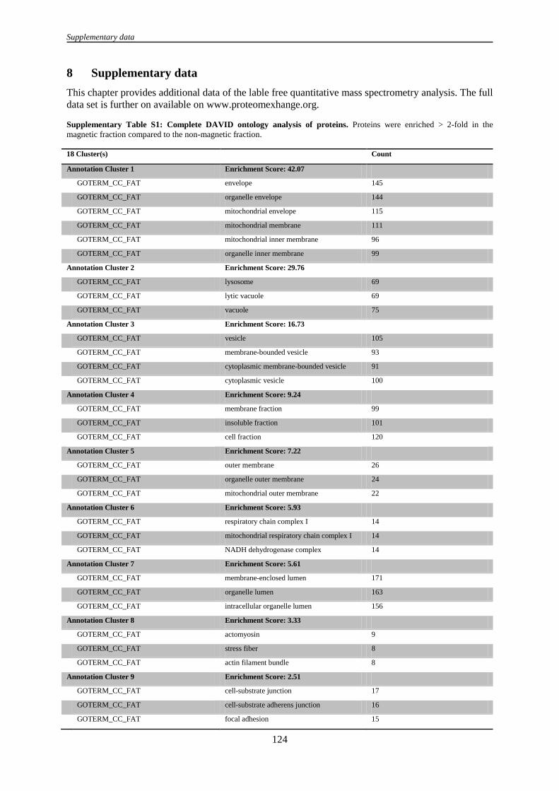
Supplementary data
124
8 Supplementary data
This chapter provides additional data of the lable free quantitative mass spectrometry analysis. The full
data set is further on available on www.proteomexhange.org.
Supplementary Table S1: Complete DAVID ontology analysis of proteins. Proteins were enriched > 2-fold in the
magnetic fraction compared to the non-magnetic fraction.
18 Cluster(s) Count
Annotation Cluster 1 Enrichment Score: 42.07
GOTERM_CC_FAT envelope 145
GOTERM_CC_FAT organelle envelope 144
GOTERM_CC_FAT mitochondrial envelope 115
GOTERM_CC_FAT mitochondrial membrane 111
GOTERM_CC_FAT mitochondrial inner membrane 96
GOTERM_CC_FAT organelle inner membrane 99
Annotation Cluster 2 Enrichment Score: 29.76
GOTERM_CC_FAT lysosome 69
GOTERM_CC_FAT lytic vacuole 69
GOTERM_CC_FAT vacuole 75
Annotation Cluster 3 Enrichment Score: 16.73
GOTERM_CC_FAT vesicle 105
GOTERM_CC_FAT membrane-bounded vesicle 93
GOTERM_CC_FAT cytoplasmic membrane-bounded vesicle 91
GOTERM_CC_FAT cytoplasmic vesicle 100
Annotation Cluster 4 Enrichment Score: 9.24
GOTERM_CC_FAT membrane fraction 99
GOTERM_CC_FAT insoluble fraction 101
GOTERM_CC_FAT cell fraction 120
Annotation Cluster 5 Enrichment Score: 7.22
GOTERM_CC_FAT outer membrane 26
GOTERM_CC_FAT organelle outer membrane 24
GOTERM_CC_FAT mitochondrial outer membrane 22
Annotation Cluster 6 Enrichment Score: 5.93
GOTERM_CC_FAT respiratory chain complex I 14
GOTERM_CC_FAT mitochondrial respiratory chain complex I 14
GOTERM_CC_FAT NADH dehydrogenase complex 14
Annotation Cluster 7 Enrichment Score: 5.61
GOTERM_CC_FAT membrane-enclosed lumen 171
GOTERM_CC_FAT organelle lumen 163
GOTERM_CC_FAT intracellular organelle lumen 156
Annotation Cluster 8 Enrichment Score: 3.33
GOTERM_CC_FAT actomyosin 9
GOTERM_CC_FAT stress fiber 8
GOTERM_CC_FAT actin filament bundle 8
Annotation Cluster 9 Enrichment Score: 2.51
GOTERM_CC_FAT cell-substrate junction 17
GOTERM_CC_FAT cell-substrate adherens junction 16
GOTERM_CC_FAT focal adhesion 15

Supplementary data
125
Annotation Cluster 10 Enrichment Score: 2.29
GOTERM_CC_FAT clathrin adaptor complex 8
GOTERM_CC_FAT AP-type membrane coat adaptor complex 8
GOTERM_CC_FAT clathrin coat 9
GOTERM_CC_FAT endocytic vesicle membrane 6
Annotation Cluster 11 Enrichment Score: 2.15
GOTERM_CC_FAT AP-2 adaptor complex 4
GOTERM_CC_FAT clathrin coat of coated pit 5
GOTERM_CC_FAT clathrin-coated endocytic vesicle 5
GOTERM_CC_FAT clathrin coat of endocytic vesicle 4
GOTERM_CC_FAT clathrin-coated endocytic vesicle membrane 4
GOTERM_CC_FAT clathrin vesicle coat 4
Annotation Cluster 12 Enrichment Score: 2.1
GOTERM_CC_FAT microbody part 9
GOTERM_CC_FAT peroxisomal part 9
GOTERM_CC_FAT microbody membrane 7
GOTERM_CC_FAT peroxisomal membrane 7
Annotation Cluster 13 Enrichment Score: 2.09
GOTERM_CC_FAT protein-lipid complex 8
GOTERM_CC_FAT plasma lipoprotein particle 8
GOTERM_CC_FAT triglyceride-rich lipoprotein particle 6
GOTERM_CC_FAT very-low-density lipoprotein particle 6
GOTERM_CC_FAT high-density lipoprotein particle 6
Annotation Cluster 14 Enrichment Score: 1.58
GOTERM_CC_FAT tricarboxylic acid cycle enzyme complex 4
GOTERM_CC_FAT dihydrolipoyl dehydrogenase complex 3
GOTERM_CC_FAT mitochondrial alpha-ketoglutarate
dehydrogenase complex
3
GOTERM_CC_FAT mitochondrial tricarboxylic acid cycle
enzyme complex
3
Annotation Cluster 15 Enrichment Score: 1.38
GOTERM_CC_FAT COPI vesicle coat 4
GOTERM_CC_FAT COPI coated vesicle membrane 4
GOTERM_CC_FAT COPI-coated vesicle 4
Annotation Cluster 16 Enrichment Score: 0.97
GOTERM_CC_FAT vesicle lumen 7
GOTERM_CC_FAT platelet alpha granule lumen 6
GOTERM_CC_FAT cytoplasmic membrane-bounded vesicle
lumen
6
Annotation Cluster 17 Enrichment Score: 0.58
GOTERM_CC_FAT contractile fiber part 11
GOTERM_CC_FAT contractile fiber 11
GOTERM_CC_FAT myofibril 9
Annotation Cluster 18 Enrichment Score: 0.44
GOTERM_CC_FAT spectrin 3
GOTERM_CC_FAT cortical actin cytoskeleton 3
GOTERM_CC_FAT cortical cytoskeleton 3

Supplementary data
126
Supplementary Table S2: Reconstruction of SPIOPSN trafficking is based on the GOTERM: Lysosome.
Uniprot_ID Full protein name
AGAL_HUMAN galactosidase, alpha
ANAG_HUMAN N-acetylglucosaminidase, alpha-
AP3M1_HUMAN adaptor-related protein complex 3, mu 1 subunit
ARL8A_HUMAN ADP-ribosylation factor-like 8A
ARL8B_HUMAN ADP-ribosylation factor-like 8B
ARSA_HUMAN arylsulfatase A
ARSB_HUMAN arylsulfatase B
ASAH1_HUMAN N-acylsphingosine amidohydrolase (acid ceramidase) 1
ASM_HUMAN sphingomyelin phosphodiesterase 1, acid lysosomal
BGAL_HUMAN galactosidase, beta 1
BGLR_HUMAN glucuronidase, beta
CATA_HUMAN catalase
CATB_HUMAN cathepsin B
CATC_HUMAN cathepsin C
CATD_HUMAN cathepsin D
CATL1_HUMAN cathepsin L1
CATZ_HUMAN cathepsin Z
CD63_HUMAN CD63 molecule
CLCN7_HUMAN chloride channel 7
CLN3_HUMAN ceroid-lipofuscinosis, neuronal 3
CLN5_HUMAN ceroid-lipofuscinosis, neuronal 5
DNS2A_HUMAN deoxyribonuclease II, lysosomal
DPP2_HUMAN dipeptidyl-peptidase 7
EPDR1_HUMAN ependymin related protein 1 (zebrafish)
GALNS_HUMAN galactosamine (N-acetyl)-6-sulfate sulfatase
GGH_HUMAN gamma-glutamyl hydrolase (conjugase, folylpolygammaglutamyl hydrolase)
GILT_HUMAN interferon, gamma-inducible protein 30
GLCM_HUMAN glucosidase, beta; acid (includes glucosylceramidase)
GNS_HUMAN glucosamine (N-acetyl)-6-sulfatase
HEXA_HUMAN hexosaminidase A (alpha polypeptide)
HEXB_HUMAN hexosaminidase B (beta polypeptide)
LAMP1_HUMAN lysosomal-associated membrane protein 1
LAMP2_HUMAN lysosomal-associated membrane protein 2
LGMN_HUMAN legumain
LICH_HUMAN lipase A, lysosomal acid, cholesterol esterase
LYAG_HUMAN glucosidase, alpha; acid
MA2B1_HUMAN mannosidase, alpha, class 2B, member 1
MANBA_HUMAN mannosidase, beta A, lysosomal
MCLN1_HUMAN mucolipin 1
MPRD_HUMAN mannose-6-phosphate receptor (cation dependent)
MPRI_HUMAN insulin-like growth factor 2 receptor
NAGAB_HUMAN N-acetylgalactosaminidase, alpha-
NCUG1_HUMAN chromosome 1 open reading frame 85
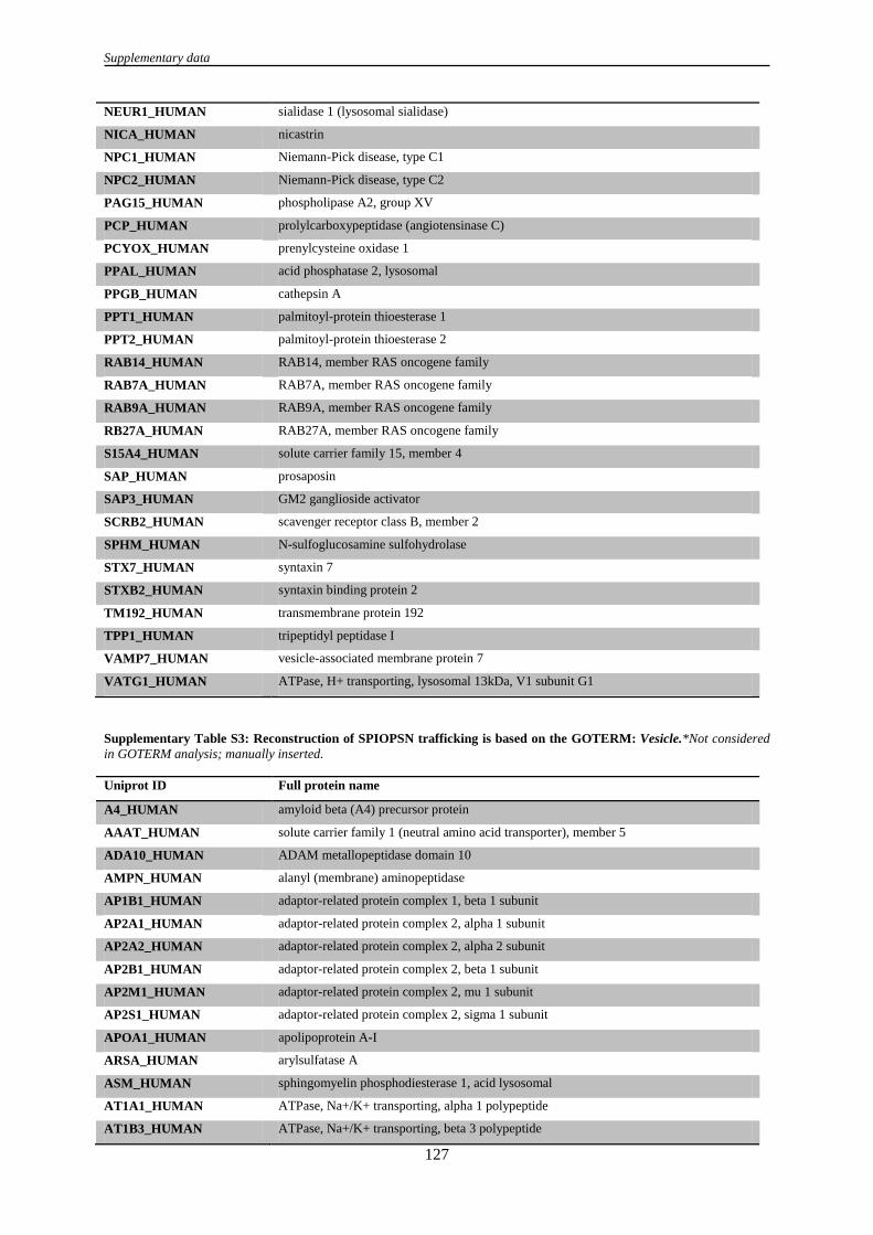
Supplementary data
127
NEUR1_HUMAN sialidase 1 (lysosomal sialidase)
NICA_HUMAN nicastrin
NPC1_HUMAN Niemann-Pick disease, type C1
NPC2_HUMAN Niemann-Pick disease, type C2
PAG15_HUMAN phospholipase A2, group XV
PCP_HUMAN prolylcarboxypeptidase (angiotensinase C)
PCYOX_HUMAN prenylcysteine oxidase 1
PPAL_HUMAN acid phosphatase 2, lysosomal
PPGB_HUMAN cathepsin A
PPT1_HUMAN palmitoyl-protein thioesterase 1
PPT2_HUMAN palmitoyl-protein thioesterase 2
RAB14_HUMAN RAB14, member RAS oncogene family
RAB7A_HUMAN RAB7A, member RAS oncogene family
RAB9A_HUMAN RAB9A, member RAS oncogene family
RB27A_HUMAN RAB27A, member RAS oncogene family
S15A4_HUMAN solute carrier family 15, member 4
SAP_HUMAN prosaposin
SAP3_HUMAN GM2 ganglioside activator
SCRB2_HUMAN scavenger receptor class B, member 2
SPHM_HUMAN N-sulfoglucosamine sulfohydrolase
STX7_HUMAN syntaxin 7
STXB2_HUMAN syntaxin binding protein 2
TM192_HUMAN transmembrane protein 192
TPP1_HUMAN tripeptidyl peptidase I
VAMP7_HUMAN vesicle-associated membrane protein 7
VATG1_HUMAN ATPase, H+ transporting, lysosomal 13kDa, V1 subunit G1
Supplementary Table S3: Reconstruction of SPIOPSN trafficking is based on the GOTERM: Vesicle.*Not considered
in GOTERM analysis; manually inserted.
Uniprot ID Full protein name
A4_HUMAN amyloid beta (A4) precursor protein
AAAT_HUMAN solute carrier family 1 (neutral amino acid transporter), member 5
ADA10_HUMAN ADAM metallopeptidase domain 10
AMPN_HUMAN alanyl (membrane) aminopeptidase
AP1B1_HUMAN adaptor-related protein complex 1, beta 1 subunit
AP2A1_HUMAN adaptor-related protein complex 2, alpha 1 subunit
AP2A2_HUMAN adaptor-related protein complex 2, alpha 2 subunit
AP2B1_HUMAN adaptor-related protein complex 2, beta 1 subunit
AP2M1_HUMAN adaptor-related protein complex 2, mu 1 subunit
AP2S1_HUMAN adaptor-related protein complex 2, sigma 1 subunit
APOA1_HUMAN apolipoprotein A-I
ARSA_HUMAN arylsulfatase A
ASM_HUMAN sphingomyelin phosphodiesterase 1, acid lysosomal
AT1A1_HUMAN ATPase, Na+/K+ transporting, alpha 1 polypeptide
AT1B3_HUMAN ATPase, Na+/K+ transporting, beta 3 polypeptide

Supplementary data
128
BASI_HUMAN basigin (Ok blood group)
CATB_HUMAN cathepsin B
CATD_HUMAN cathepsin D
CAV1_HUMAN caveolin 1, caveolae protein, 22kDa
CAV2_HUMAN caveolin 2
CD9_HUMAN CD9 molecule
CH60_HUMAN heat shock 60kDa protein 1 (chaperonin) pseudogene 5
CLIC4_HUMAN chloride intracellular channel 4
CLN3_HUMAN ceroid-lipofuscinosis, neuronal 3
CLUS_HUMAN clusterin
COPB_HUMAN coatomer protein complex, subunit beta 1
COPB2_HUMAN coatomer protein complex, subunit beta 2 (beta prime)
COPE_HUMAN coatomer protein complex, subunit epsilon
COPZ1_HUMAN coatomer protein complex, subunit zeta 1
DAB2_HUMAN disabled homolog 2, mitogen-responsive phosphoprotein (Drosophila)
DLDH_HUMAN dihydrolipoamide dehydrogenase
DNJC5_HUMAN DnaJ (Hsp40) homolog, subfamily C, member 5
DP13A_HUMAN adaptor protein, phosphotyrosine interaction, PH domain and leucine zipper containing 1
DPP2_HUMAN dipeptidyl-peptidase 7
ECE1_HUMAN endothelin converting enzyme 1
EGFR_HUMAN epidermal growth factor receptor
EHD1_HUMAN EH-domain containing 1
FIBA_HUMAN fibrinogen alpha chain
FIBB_HUMAN fibrinogen beta chain
FIBG_HUMAN fibrinogen gamma chain
FLOT1_HUMAN flotillin 1
GANAB_HUMAN glucosidase, alpha; neutral AB
GGH_HUMAN gamma-glutamyl hydrolase (conjugase, folylpolygammaglutamyl hydrolase)
GIPC1_HUMAN GIPC PDZ domain containing family, member 1
GNAI3_HUMAN guanine nucleotide binding protein (G protein), alpha inhibiting activity polypeptide 3
GNAS2_HUMAN GNAS complex locus
GPNMB_HUMAN glycoprotein (transmembrane) nmb
GTR1_HUMAN solute carrier family 2 (facilitated glucose transporter), member 1
HEXB_HUMAN hexosaminidase B (beta polypeptide)
ITA1_HUMAN integrin, alpha 1
ITB1_HUMAN integrin, beta 1 (fibronectin receptor, beta polypeptide)
LAMP1_HUMAN lysosomal-associated membrane protein 1
LAMP2_HUMAN lysosomal-associated membrane protein 2
MPRI_HUMAN insulin-like growth factor 2 receptor
MYH11_HUMAN myosin, heavy chain 11, smooth muscle
MYOF_HUMAN myoferlin
NEUR1_HUMAN sialidase 1 (lysosomal sialidase)
NICA_HUMAN nicastrin
P4K2A_HUMAN phosphatidylinositol 4-kinase type 2 alpha
PA24A_HUMAN phospholipase A2, group IVA (cytosolic, calcium-dependent)

Supplementary data
129
PICAL_HUMAN phosphatidylinositol binding clathrin assembly protein
PLD1_HUMAN phospholipase D1, phosphatidylcholine-specific
PPT1_HUMAN palmitoyl-protein thioesterase 1
PTN1_HUMAN protein tyrosine phosphatase, non-receptor type 1
RAB14_HUMAN RAB14, member RAS oncogene family
RAB21_HUMAN RAB21, member RAS oncogene family
RAB2A_HUMAN RAB2A, member RAS oncogene family
RAB35_HUMAN similar to hCG1778032; RAB35, member RAS oncogene family
RAB3A_HUMAN RAB3A, member RAS oncogene family
RAB3D_HUMAN RAB3D, member RAS oncogene family
RAB5A_HUMAN RAB5A, member RAS oncogene family
RAB5C_HUMAN RAB5C, member RAS oncogene family
RAB6B_HUMAN RAB6B, member RAS oncogene family
RAB7A_HUMAN RAB7A, member RAS oncogene family
RAI3_HUMAN G protein-coupled receptor, family C, group 5, member A
RB11A_HUMAN RAB11A, member RAS oncogene family
RB11B_HUMAN RAB11B, member RAS oncogene family
RB27A_HUMAN RAB27A, member RAS oncogene family
RPN1_HUMAN ribophorin I
SATT_HUMAN solute carrier family 1 (glutamate/neutral amino acid transporter), member 4
SC23A_HUMAN Sec23 homolog A (S. cerevisiae)
SCFD1_HUMAN sec1 family domain containing 1
SDCB1_HUMAN syndecan binding protein (syntenin)
SH3B4_HUMAN SH3-domain binding protein 4
SNTB2_HUMAN syntrophin, beta 2 (dystrophin-associated protein A1, 59kDa, basic component 2)
SPG21_HUMAN spastic paraplegia 21 (autosomal recessive, Mast syndrome)
STOM_HUMAN phosphatidylethanolamine binding protein 1
STOM_HUMAN stomatin
STX12_HUMAN syntaxin 12
STXB1_HUMAN syntaxin binding protein 1
STXB2_HUMAN syntaxin binding protein 2
STXB3_HUMAN syntaxin binding protein 3
SYPL1_HUMAN synaptophysin-like 1
TFR1_HUMAN transferrin receptor (p90, CD71)
TMED2_HUMAN transmembrane emp24 domain trafficking protein 2
TMEDA_HUMAN transmembrane emp24-like trafficking protein 10 (yeast)
TPP1_HUMAN tripeptidyl peptidase I
TSP1_HUMAN thrombospondin 1
VA0D1_HUMAN ATPase, H+ transporting, lysosomal 38kDa, V0 subunit d1
VAMP7_HUMAN vesicle-associated membrane protein 7
VAPA_HUMAN VAMP (vesicle-associated membrane protein)-associated protein A, 33kDa
VATB2_HUMAN ATPase, H+ transporting, lysosomal 56/58kDa, V1 subunit B2
VATC1_HUMAN ATPase, H+ transporting, lysosomal 42kDa, V1 subunit C1
VPP1_HUMAN ATPase, H+ transporting, lysosomal V0 subunit a1
YKT6_HUMAN YKT6 v-SNARE homolog (S. cerevisiae)

Supplementary data
130
Manually inserted: Uniprot
ID
Full protein name
1B55_HUMAN* HLA class I histocompatibility antigen, B-55 alpha chain
1C06_HUMAN* HLA class I histocompatibility antigen, Cw-6 alpha chain
ARF1_HUMAN* ADP-ribosylation factor 1
ARF4_HUMAN* ADP-ribosylation factor 4
COPG1_HUMAN* Coatomer subunit gamma-1
LTOR1_HUMAN* Ragulator complex protein LAMTOR1
LTOR2_HUMAN* Ragulator complex protein LAMTOR2
LTOR3_HUMAN* Ragulator complex protein LAMTOR3
RAB1A_HUMAN* Ras-related protein Rab-1A
RAP1A_HUMAN* Ras-related protein Rap-1A
RAP1B_HUMAN* Ras-related protein Rap-1b
Supplementary Table S4: GOTERM: Membrane fraction.
Uniprot ID Full protein name
1A68_HUMAN major histocompatibility complex, class I, A
1B55_HUMAN, major histocompatibility complex, class I, C; major histocompatibility complex, class I,
B
1C06_HUMAN major histocompatibility complex, class I, C; major histocompatibility complex, class I,
B
5NTD_HUMAN 5'-nucleotidase, ecto (CD73)
A4_HUMAN amyloid beta (A4) precursor protein
AAAT_HUMAN solute carrier family 1 (neutral amino acid transporter), member 5
ACSL4_HUMAN acyl-CoA synthetase long-chain family member 4
ADAM9_HUMAN ADAM metallopeptidase domain 9 (meltrin gamma)
AIP_HUMAN aryl hydrocarbon receptor interacting protein
ANKH_HUMAN ankylosis, progressive homolog (mouse)
ANPRC_HUMAN natriuretic peptide receptor C/guanylate cyclase C (atrionatriuretic peptide receptor C)
APOE_HUMAN hypothetical LOC100129500; apolipoprotein E
ARF6_HUMAN ADP-ribosylation factor 6
ARL1_HUMAN ADP-ribosylation factor-like 1
ARSA_HUMAN arylsulfatase A
AT12A_HUMAN ATPase, H+/K+ transporting, nongastric, alpha polypeptide
AT1A1_HUMAN ATPase, Na+/K+ transporting, alpha 1 polypeptide
AT1A2_HUMAN ATPase, Na+/K+ transporting, alpha 2 (+) polypeptide
AT1B1_HUMAN ATPase, Na+/K+ transporting, beta 1 polypeptide
AT2A1_HUMAN ATPase, Ca++ transporting, cardiac muscle, fast twitch 1
AT2B1_HUMAN ATPase, Ca++ transporting, plasma membrane 1
AT2B4_HUMAN ATPase, Ca++ transporting, plasma membrane 4
CADH2_HUMAN cadherin 2, type 1, N-cadherin (neuronal)
CAV1_HUMAN caveolin 1, caveolae protein, 22kDa
CAV2_HUMAN caveolin 2
CD166_HUMAN hypothetical protein LOC100133690; activated leukocyte cell adhesion molecule

Supplementary data
131
CD276_HUMAN CD276 molecule
CD59_HUMAN CD59 molecule, complement regulatory protein
CD81_HUMAN CD81 molecule
CD82_HUMAN CD82 molecule
CDC42_HUMAN cell division cycle 42 (GTP binding protein, 25kDa); cell division cycle 42 pseudogene 2
CERU_HUMAN ceruloplasmin (ferroxidase)
CKAP4_HUMAN cytoskeleton-associated protein 4
CLN3_HUMAN ceroid-lipofuscinosis, neuronal 3
CNN2_HUMAN calponin 2
COMT_HUMAN catechol-O-methyltransferase
CPT1A_HUMAN carnitine palmitoyltransferase 1A (liver)
CTNA1_HUMAN catenin (cadherin-associated protein), alpha 1, 102kDa
CTNA2_HUMAN catenin (cadherin-associated protein), alpha 2
CTNB1_HUMAN catenin (cadherin-associated protein), beta 1, 88kDa
CTND1_HUMAN catenin (cadherin-associated protein), delta 1
CTR1_HUMAN solute carrier family 7 (cationic amino acid transporter, y+ system), member 1
CXAR_HUMAN coxsackie virus and adenovirus receptor
CYB5B_HUMAN cytochrome b5 type B (outer mitochondrial membrane)
DAF_HUMAN CD55 molecule, decay accelerating factor for complement (Cromer blood group)
DD19A_HUMAN DEAD (Asp-Glu-Ala-As) box polypeptide 19A
DIAP1_HUMAN diaphanous homolog 1 (Drosophila)
DLG1_HUMAN discs, large homolog 1 (Drosophila)
DP13A_HUMAN adaptor protein, phosphotyrosine interaction, PH domain and leucine zipper containing 1
DPM1_HUMAN dolichyl-phosphate mannosyltransferase polypeptide 1, catalytic subunit
DSG2_HUMAN desmoglein 2
DYN2_HUMAN dynamin 2
ECE1_HUMAN endothelin converting enzyme 1
EGLN_HUMAN endoglin
EHD2_HUMAN EH-domain containing 2
EPCR_HUMAN protein C receptor, endothelial (EPCR)
EPHA2_HUMAN EPH receptor A2
EPHB2_HUMAN EPH receptor B2
ERG7_HUMAN lanosterol synthase (2,3-oxidosqualene-lanosterol cyclase)
ERO1A_HUMAN ERO1-like (S. cerevisiae)
FACE1_HUMAN zinc metallopeptidase (STE24 homolog, S. cerevisiae)
FERM2_HUMAN fermitin family homolog 2 (Drosophila)
FLOT1_HUMAN flotillin 1
FLOT2_HUMAN flotillin 2
FOLR1_HUMAN folate receptor 1 (adult)
GBB1_HUMAN guanine nucleotide binding protein (G protein), beta polypeptide 1
GBG10_HUMAN DnaJ (Hsp40) homolog, subfamily C
GBG12_HUMAN guanine nucleotide binding protein (G protein), gamma 12
GIPC1_HUMAN GIPC PDZ domain containing family, member 1
GNA11_HUMAN guanine nucleotide binding protein (G protein), alpha 11 (Gq class)
GNAI2_HUMAN guanine nucleotide binding protein (G protein), alpha inhibiting activity polypeptide 2

Supplementary data
132
GNAI3_HUMAN guanine nucleotide binding protein (G protein), alpha inhibiting activity polypeptide 3
GNAQ_HUMAN guanine nucleotide binding protein (G protein), q polypeptide
GNAS2_HUMAN GNAS complex locus
GPR56_HUMAN G protein-coupled receptor 56
GTR1_HUMAN solute carrier family 2 (facilitated glucose transporter), member 1
HGS_HUMAN hepatocyte growth factor-regulated tyrosine kinase substrate
HMOX1_HUMAN heme oxygenase (decycling) 1
HMOX2_HUMAN heme oxygenase (decycling) 2
HYEP_HUMAN epoxide hydrolase 1, microsomal (xenobiotic)
IGHG2_HUMAN immunoglobulin heavy constant gamma 2 (G2m marker)
IGHG4_HUMAN immunoglobulin heavy constant gamma 4 (G4m marker)
IL6RB_HUMAN interleukin 6 signal transducer (gp130, oncostatin M receptor)
ILK_HUMAN integrin-linked kinase
ITA1_HUMAN integrin, alpha 1
ITA2_HUMAN integrin, alpha 2 (CD49B, alpha 2 subunit of VLA-2 receptor)
ITA3_HUMAN integrin, alpha 3 (antigen CD49C, alpha 3 subunit of VLA-3 receptor)
ITA5_HUMAN integrin, alpha 5 (fibronectin receptor, alpha polypeptide)
ITA6_HUMAN integrin, alpha 6
ITB1_HUMAN integrin, beta 1 (fibronectin receptor, beta polypeptide=
ITB4_HUMAN integrin, beta 4
ITB5_HUMAN integrin, beta 5
KAP2_HUMAN protein kinase, cAMP-dependent, regulatory, type II, alpha
KPCA_HUMAN protein kinase C, alpha
L1CAM_HUMAN L1 cell adhesion molecule
LAMP1_HUMAN lysosomal-associated membrane protein 1
LAMP2_HUMAN lysosomal-associated membrane protein 2
LAP2_HUMAN erbb2 interacting protein
LCAP_HUMAN leucyl/cystinyl aminopeptidase
LFA3_HUMAN CD58 molecule
LIMA1_HUMAN LIM domain and actin binding 1
LIN7C_HUMAN lin-7 homolog C (C. elegans)
LIS1_HUMAN platelet-activating factor acetylhydrolase, isoform Ib, subunit 1 (45kDa)
LMAN1_HUMAN lectin, mannose-binding, 1
MET_HUMAN met proto-oncogene (hepatocyte growth factor receptor)
MOT1_HUMAN solute carrier family 16, member 1 (monocarboxylic acid transporter 1)
MPRI_HUMAN insulin-like growth factor 2 receptor
MPZL1_HUMAN myelin protein zero-like 1
MRP1_HUMAN ATP-binding cassette, sub-family C (CFTR/MRP), member 1
MYO1C_HUMAN myosin IC
NCEH1_HUMAN arylacetamide deacetylase-like 1
NCPR_HUMAN P450 (cytochrome) oxidoreductase
NEXN_HUMAN nexilin (F actin binding protein)
NHRF1_HUMAN solute carrier family 9 (sodium/hydrogen exchanger), member 3 regulator 1
NHRF2_HUMAN solute carrier family 9 (sodium/hydrogen exchanger), member 3 regulator 2
NICA_HUMAN nicastrin

Supplementary data
133
NU155_HUMAN nucleoporin 155kDa
ODO2_HUMAN dihydrolipoamide S-succinyltransferase
OST48_HUMAN dolichyl-diphosphooligosaccharide-protein glycosyltransferase
P4K2A_HUMAN phosphatidylinositol 4-kinase type 2 alpha
PA24A_HUMAN phospholipase A2, group IVA (cytosolic, calcium-dependent)
PACN3_HUMAN protein kinase C and casein kinase substrate in neurons 3
PARVA_HUMAN parvin, alpha
PARVB_HUMAN parvin, beta
PGRC1_HUMAN progesterone receptor membrane component 1
PLAK_HUMAN junction plakoglobin
PLD1_HUMAN phospholipase D1, phosphatidylcholine-specific
PODXL_HUMAN podocalyxin-like
PPAL_HUMAN acid phosphatase 2, lysosomal
PPIF_HUMAN peptidylprolyl isomerase F
PPT1_HUMAN palmitoyl-protein thioesterase 1
PTPRJ_HUMAN protein tyrosine phosphatase, receptor type, J
PVRL2_HUMAN poliovirus receptor-related 2 (herpesvirus entry mediator B)
RAB14_HUMAN RAB14, member RAS oncogene family
RAB18_HUMAN RAB18, member RAS oncogene family
RAB1B_HUMAN RAB1B, member RAS oncogene family
RAB31_HUMAN RAB31, member RAS oncogene family
RAB3A_HUMAN RAB3A, member RAS oncogene family
RAB5A_HUMAN RAB5A, member RAS oncogene family
RAGP1_HUMAN Ran GTPase activating protein 1
RALA_HUMAN v-ral simian leukemia viral oncogene homolog A (ras related)
RAP2A_HUMAN RAP2A, member of RAS oncogene family
RAP2B_HUMAN RAP2B, member of RAS oncogene family
RASN_HUMAN neuroblastoma RAS viral (v-ras) oncogene homolog
RENR_HUMAN ATPase, H+ transporting, lysosomal accessory protein 2
RHEB_HUMAN Ras homolog enriched in brain
RHG17_HUMAN Rho GTPase activating protein 17
RHOB_HUMAN ras homolog gene family, member B
RHOG_HUMAN ras homolog gene family, member G (rho G)
RRAS_HUMAN related RAS viral (r-ras) oncogene homolog
S12A7_HUMAN solute carrier family 12 (potassium/chloride transporters), member 7
SCRB1_HUMAN scavenger receptor class B, member 1
SCRB2_HUMAN scavenger receptor class B, member 2
SCRIB_HUMAN scribbled homolog (Drosophila)
SEPT2_HUMAN septin 2
SNAG_HUMAN N-ethylmaleimide-sensitive factor attachment protein, gamma
SNTB2_HUMAN syntrophin, beta 2 (dystrophin-associated protein A1, 59kDa, basic component 2)
SPCS2_HUMAN signal peptidase complex subunit 2 homolog
SPCS3_HUMAN signal peptidase complex subunit 3 homolog (S. cerevisiae)
SPTA2_HUMAN spectrin, alpha, non-erythrocytic 1 (alpha-fodrin)
STAM1_HUMAN signal transducing adaptor molecule (SH3 domain and ITAM motif) 1

Supplementary data
134
STOM_HUMAN phosphatidylethanolamine binding protein 1
THY1_HUMAN Thy-1 cell surface antigen
TMED2_HUMAN transmembrane emp24 domain trafficking protein 2
TMEDA_HUMAN transmembrane emp24-like trafficking protein 10 (yeast)
TMX1_HUMAN thioredoxin-related transmembrane protein 1
TOM22_HUMAN translocase of outer mitochondrial membrane 22 homolog (yeast)
TOM40_HUMAN translocase of outer mitochondrial membrane 40 homolog (yeast)
TPBG_HUMAN trophoblast glycoprotein
TRIP6_HUMAN thyroid hormone receptor interactor 6
UFO_HUMAN AXL receptor tyrosine kinase
USO1_HUMAN USO1 homolog, vesicle docking protein (yeast)
VA0D1_HUMAN ATPase, H+ transporting, lysosomal 38kDa, V0 subunit d1
VAPA_HUMAN VAMP (vesicle-associated membrane protein)-associated protein A, 33kDa
VASP_HUMAN vasodilator-stimulated phosphoprotein
VATB1_HUMAN ATPase, H+ transporting, lysosomal 56/58kDa, V1 subunit B1
VATF_HUMAN ATPase, H+ transporting, lysosomal 14kDa, V1 subunit F
VDAC3_HUMAN voltage-dependent anion channel 3
VPP3_HUMAN T-cell, immune regulator 1, ATPase, H+ transporting, lysosomal V0 subunit A3
ZO1_HUMAN tight junction protein 1 (zona occludens 1)
ZYX_HUMAN zyxin
Supplementary Table S5: GOTERM: Mitochondrial envelope + mitochondria.
Uniprot ID Full protein name
3HIDH_HUMAN 3-hydroxyisobutyrate dehydrogenase
ABCB6_HUMAN ATP-binding cassette, sub-family B (MDR/TAP), member 6
ABCE1_HUMAN similar to ATP-binding cassette, sub-family E
ABHDA_HUMAN abhydrolase domain containing 10
ACADM_HUMAN acyl-Coenzyme A dehydrogenase, C-4 to C-12 straight chain
ACADV_HUMAN acyl-Coenzyme A dehydrogenase, very long chain
ACO13_HUMAN acyl-CoA thioesterase 13
ACON_HUMAN aconitase 2, mitochondrial
ACOT9_HUMAN acyl-CoA thioesterase 9
ACOX1_HUMAN acyl-Coenzyme A oxidase 1, palmitoyl
ACPM_HUMAN NADH dehydrogenase (ubiquinone) 1, alpha/beta subcomplex, 1, 8kDa
ACSL4_HUMAN acyl-CoA synthetase long-chain family member 4
ADAS_HUMAN alkylglycerone phosphate synthase
ADT2_HUMAN solute carrier family 25 (mitochondrial carrier; adenine nucleotide translocator), member
5; solute carrier family 25 (mitochondrial carrier; adenine nucleotide translocator),
member 5 pseudogene 8
ADT3_HUMAN solute carrier family 25 (mitochondrial carrier; adenine nucleotide translocator), member
6
AIFM2_HUMAN apoptosis-inducing factor, mitochondrion-associated, 2
AL1B1_HUMAN aldehyde dehydrogenase 1 family, member B1
AL3A2_HUMAN aldehyde dehydrogenase 3 family, member A2

Supplementary data
135
ALDH2_HUMAN aldehyde dehydrogenase 2 family (mitochondrial)
AMPL_HUMAN leucine aminopeptidase 3
AT5F1_HUMAN ATP synthase, H+ transporting, mitochondrial F0 complex, subunit B1
ATP5H_HUMAN ATP synthase, H+ transporting, mitochondrial F0 complex, subunit d
ATP5I_HUMAN ATP synthase, H+ transporting, mitochondrial F0 complex, subunit E
ATP5J_HUMAN ATP synthase, H+ transporting, mitochondrial F0 complex, subunit F6
ATP5L_HUMAN ATP synthase, H+ transporting, mitochondrial F0 complex, subunit G
ATPA_HUMAN ATP synthase, H+ transporting, mitochondrial F1 complex, alpha subunit 1, cardiac
muscle
ATPB_HUMAN ATP synthase, H+ transporting, mitochondrial F1 complex, beta polypeptide
ATPD_HUMAN ATP synthase, H+ transporting, mitochondrial F1 complex, delta subunit
ATPG_HUMAN ATP synthase, H+ transporting, mitochondrial F1 complex, gamma polypeptide 1
ATPK_HUMAN ATP synthase, H+ transporting, mitochondrial F0 complex, subunit F2
ATPO_HUMAN ATP synthase, H+ transporting, mitochondrial F1 complex, O subunit
B2L13_HUMAN BCL2-like 13 (apoptosis facilitator)
BAX_HUMAN BCL2-associated X protein
BCS1_HUMAN BCS1-like (yeast)
C1QBP_HUMAN complement component 1, q subcomponent binding protein
C1TM_HUMAN methylenetetrahydrofolate dehydrogenase (NADP+ dependent) 1-like
CATA_HUMAN catalase
CB047_HUMAN chromosome 2 open reading frame 47
CCHL_HUMAN holocytochrome c synthase (cytochrome c heme-lyase)
CH60_HUMAN heat shock 60kDa protein 1 (chaperonin) pseudogene 5; heat shock 60kDa protein 1
(chaperonin) pseudogene 6; heat shock 60kDa protein 1 (chaperonin) pseudogene 1; heat
shock 60kDa protein 1 (chaperonin) pseudogene 4; heat shock 60kDa protein 1
(chaperonin)
CHCH3_HUMAN coiled-coil-helix-coiled-coil-helix domain containing 3
CISD1_HUMAN CDGSH iron sulfur domain 1
CISY_HUMAN citrate synthase
CLPP_HUMAN ClpP caseinolytic peptidase, ATP-dependent, proteolytic subunit homolog (E. coli)
CLPX_HUMAN ClpX caseinolytic peptidase X homolog (E. coli)
CMC1_HUMAN solute carrier family 25 (mitochondrial carrier, Aralar), member 12
CMC2_HUMAN solute carrier family 25, member 13 (citrin)
COX2_HUMAN Cytochrome c oxidase subunit 2
COX41_HUMAN cytochrome c oxidase subunit IV isoform 1
COX5A_HUMAN cytochrome c oxidase subunit Va
CPSM_HUMAN carbamoyl-phosphate synthetase 1, mitochondrial
CPT1A_HUMAN carnitine palmitoyltransferase 1A (liver)
CPT2_HUMAN carnitine palmitoyltransferase 2
CX4NB_HUMAN COX4 neighbor
CX7A2_HUMAN cytochrome c oxidase subunit VIIa polypeptide 2 (liver)
CYB5B_HUMAN cytochrome b5 type B (outer mitochondrial membrane)
DECR_HUMAN 2,4-dienoyl CoA reductase 1, mitochondrial
DHB4_HUMAN hydroxysteroid (17-beta) dehydrogenase 4
DHE3_HUMAN glutamate dehydrogenase 1

Supplementary data
136
DHRS1_HUMAN dehydrogenase/reductase (SDR family) member 1
DHSA_HUMAN succinate dehydrogenase complex, subunit A, flavoprotein (Fp)
DHSB_HUMAN succinate dehydrogenase complex, subunit B, iron sulfur (Ip)
DIC_HUMAN solute carrier family 25 (mitochondrial carrier; dicarboxylate transporter), member 10
DJC11_HUMAN DnaJ (Hsp40) homolog, subfamily C, member 11
DNJA3_HUMAN DnaJ (Hsp40) homolog, subfamily A, member 3
DPYL2_HUMAN dihydropyrimidinase-like 2
ECH1_HUMAN enoyl Coenzyme A hydratase 1, peroxisomal
ECHA_HUMAN hydroxyacyl-Coenzyme A dehydrogenase/3-ketoacyl-Coenzyme A thiolase/enoyl-
Coenzyme A hydratase (trifunctional protein), alpha subunit
ECHB_HUMAN hydroxyacyl-Coenzyme A dehydrogenase/3-ketoacyl-Coenzyme A thiolase/enoyl-
Coenzyme A hydratase (trifunctional protein), beta subunit
EFGM_HUMAN G elongation factor, mitochondrial 1
EFTS_HUMAN Ts translation elongation factor, mitochondrial
EFTU_HUMAN Tu translation elongation factor, mitochondrial
ES1_HUMAN chromosome 21 open reading frame 33
ETFA_HUMAN electron-transfer-flavoprotein, alpha polypeptide
ETFB_HUMAN electron-transfer-flavoprotein, beta polypeptide
ETHE1_HUMAN ethylmalonic encephalopathy 1
FAHD1_HUMAN fumarylacetoacetate hydrolase domain containing 1
FIS1_HUMAN fission 1 (mitochondrial outer membrane) homolog (S. cerevisiae)
FUMH_HUMAN fumarate hydratase
GHC1_HUMAN solute carrier family 25 (mitochondrial carrier: glutamate), member 22
GLSK_HUMAN glutaminase
GLYM_HUMAN serine hydroxymethyltransferase 2 (mitochondrial)
GOLP3_HUMAN golgi phosphoprotein 3 (coat-protein)
GPDM_HUMAN glycerol-3-phosphate dehydrogenase 2 (mitochondrial)
GRPE1_HUMAN GrpE-like 1, mitochondrial (E. coli)
GSTK1_HUMAN glutathione S-transferase kappa 1
HCD2_HUMAN hydroxysteroid (17-beta) dehydrogenase 10
HCDH_HUMAN hydroxyacyl-Coenzyme A dehydrogenase
HINT2_HUMAN histidine triad nucleotide binding protein 2
HMGCL_HUMAN 3-hydroxymethyl-3-methylglutaryl-Coenzyme A lyase
HSDL2_HUMAN hydroxysteroid dehydrogenase like 2
IDH3A_HUMAN isocitrate dehydrogenase 3 (NAD+) alpha
IDH3G_HUMAN isocitrate dehydrogenase 3 (NAD+) gamma
IMMT_HUMAN inner membrane protein, mitochondrial (mitofilin)
IPYR2_HUMAN pyrophosphatase (inorganic) 2
ISOC2_HUMAN isochorismatase domain containing 2
IVD_HUMAN isovaleryl Coenzyme A dehydrogenase
KAD2_HUMAN adenylate kinase 2
KAD3_HUMAN adenylate kinase 3
LETM1_HUMAN leucine zipper-EF-hand containing transmembrane protein 1
LPPRC_HUMAN leucine-rich PPR-motif containing
LYPA1_HUMAN lysophospholipase I
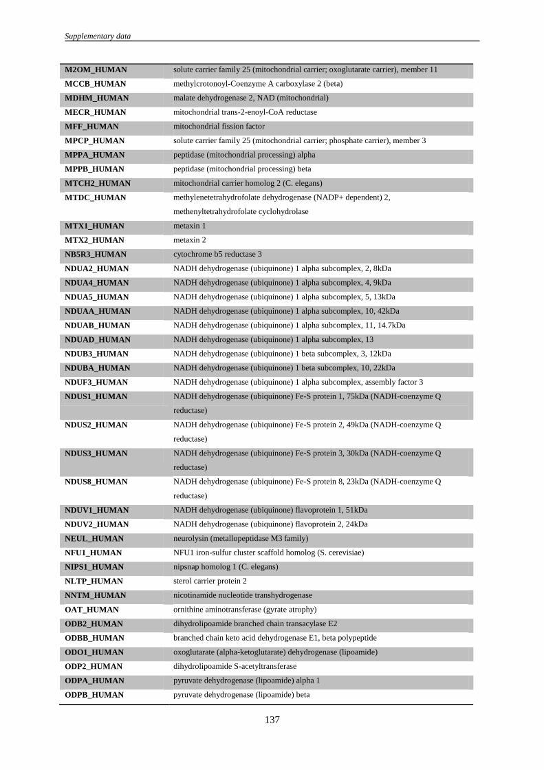
Supplementary data
137
M2OM_HUMAN solute carrier family 25 (mitochondrial carrier; oxoglutarate carrier), member 11
MCCB_HUMAN methylcrotonoyl-Coenzyme A carboxylase 2 (beta)
MDHM_HUMAN malate dehydrogenase 2, NAD (mitochondrial)
MECR_HUMAN mitochondrial trans-2-enoyl-CoA reductase
MFF_HUMAN mitochondrial fission factor
MPCP_HUMAN solute carrier family 25 (mitochondrial carrier; phosphate carrier), member 3
MPPA_HUMAN peptidase (mitochondrial processing) alpha
MPPB_HUMAN peptidase (mitochondrial processing) beta
MTCH2_HUMAN mitochondrial carrier homolog 2 (C. elegans)
MTDC_HUMAN methylenetetrahydrofolate dehydrogenase (NADP+ dependent) 2,
methenyltetrahydrofolate cyclohydrolase
MTX1_HUMAN metaxin 1
MTX2_HUMAN metaxin 2
NB5R3_HUMAN cytochrome b5 reductase 3
NDUA2_HUMAN NADH dehydrogenase (ubiquinone) 1 alpha subcomplex, 2, 8kDa
NDUA4_HUMAN NADH dehydrogenase (ubiquinone) 1 alpha subcomplex, 4, 9kDa
NDUA5_HUMAN NADH dehydrogenase (ubiquinone) 1 alpha subcomplex, 5, 13kDa
NDUAA_HUMAN NADH dehydrogenase (ubiquinone) 1 alpha subcomplex, 10, 42kDa
NDUAB_HUMAN NADH dehydrogenase (ubiquinone) 1 alpha subcomplex, 11, 14.7kDa
NDUAD_HUMAN NADH dehydrogenase (ubiquinone) 1 alpha subcomplex, 13
NDUB3_HUMAN NADH dehydrogenase (ubiquinone) 1 beta subcomplex, 3, 12kDa
NDUBA_HUMAN NADH dehydrogenase (ubiquinone) 1 beta subcomplex, 10, 22kDa
NDUF3_HUMAN NADH dehydrogenase (ubiquinone) 1 alpha subcomplex, assembly factor 3
NDUS1_HUMAN NADH dehydrogenase (ubiquinone) Fe-S protein 1, 75kDa (NADH-coenzyme Q
reductase)
NDUS2_HUMAN NADH dehydrogenase (ubiquinone) Fe-S protein 2, 49kDa (NADH-coenzyme Q
reductase)
NDUS3_HUMAN NADH dehydrogenase (ubiquinone) Fe-S protein 3, 30kDa (NADH-coenzyme Q
reductase)
NDUS8_HUMAN NADH dehydrogenase (ubiquinone) Fe-S protein 8, 23kDa (NADH-coenzyme Q
reductase)
NDUV1_HUMAN NADH dehydrogenase (ubiquinone) flavoprotein 1, 51kDa
NDUV2_HUMAN NADH dehydrogenase (ubiquinone) flavoprotein 2, 24kDa
NEUL_HUMAN neurolysin (metallopeptidase M3 family)
NFU1_HUMAN NFU1 iron-sulfur cluster scaffold homolog (S. cerevisiae)
NIPS1_HUMAN nipsnap homolog 1 (C. elegans)
NLTP_HUMAN sterol carrier protein 2
NNTM_HUMAN nicotinamide nucleotide transhydrogenase
OAT_HUMAN ornithine aminotransferase (gyrate atrophy)
ODB2_HUMAN dihydrolipoamide branched chain transacylase E2
ODBB_HUMAN branched chain keto acid dehydrogenase E1, beta polypeptide
ODO1_HUMAN oxoglutarate (alpha-ketoglutarate) dehydrogenase (lipoamide)
ODP2_HUMAN dihydrolipoamide S-acetyltransferase
ODPA_HUMAN pyruvate dehydrogenase (lipoamide) alpha 1
ODPB_HUMAN pyruvate dehydrogenase (lipoamide) beta

Supplementary data
138
OPA1_HUMAN optic atrophy 1 (autosomal dominant)
P4HA1_HUMAN prolyl 4-hydroxylase, alpha polypeptide I
P5CR1_HUMAN pyrroline-5-carboxylate reductase 1
P5CR1_HUMAN sirtuin (silent mating type information regulation 2 homolog) 7 (S. cerevisiae)
P5CS_HUMAN aldehyde dehydrogenase 18 family, member A1
PCKGM_HUMAN phosphoenolpyruvate carboxykinase 2 (mitochondrial)
PDIP2_HUMAN polymerase (DNA-directed), delta interacting protein 2
PGES2_HUMAN prostaglandin E synthase 2
PHB_HUMAN prohibitin
PHB2_HUMAN prohibitin 2
PNPT1_HUMAN polyribonucleotide nucleotidyltransferase 1
PP1G_HUMAN protein phosphatase 1, catalytic subunit, gamma isoform
PPIF_HUMAN peptidylprolyl isomerase F
PRDX3_HUMAN peroxiredoxin 3
PRDX4_HUMAN peroxiredoxin 4
PRS6B_HUMAN similar to 26S protease regulatory subunit 6B
PTCD3_HUMAN Pentatricopeptide repeat domain 3
PTH2_HUMAN peptidyl-tRNA hydrolase 2
PYC_HUMAN pyruvate carboxylase
QCR1_HUMAN ubiquinol-cytochrome c reductase core protein I
QCR2_HUMAN ubiquinol-cytochrome c reductase core protein II
QCR7_HUMAN similar to ubiquinol-cytochrome c reductase binding protein; ubiquinol-cytochrome c
reductase binding protein pseudogene; ubiquinol-cytochrome c reductase binding protein
RAB32_HUMAN RAB32, member RAS oncogene family
RM12_HUMAN mitochondrial ribosomal protein L12
RM19_HUMAN mitochondrial ribosomal protein L19
RM37_HUMAN mitochondrial ribosomal protein L37
RM39_HUMAN mitochondrial ribosomal protein L39
RM44_HUMAN mitochondrial ribosomal protein L44
RM46_HUMAN mitochondrial ribosomal protein L46
RM49_HUMAN mitochondrial ribosomal protein L49
RMD3_HUMAN family with sequence similarity 82, member A2
RRFM_HUMAN mitochondrial ribosome recycling factor
RT10_HUMAN mitochondrial ribosomal protein S10
RT22_HUMAN mitochondrial ribosomal protein S22
RT23_HUMAN mitochondrial ribosomal protein S23
RT27_HUMAN mitochondrial ribosomal protein S27
RT28_HUMAN mitochondrial ribosomal protein S28
RT29_HUMAN death associated protein 3
SCMC1_HUMAN solute carrier family 25 (mitochondrial carrier; phosphate carrier), member 24
SCOT1_HUMAN 3-oxoacid CoA transferase 1
SDHF2_HUMAN chromosome 11 open reading frame 79
SFXN3_HUMAN sideroflexin 3
SLIRP_HUMAN chromosome 14 open reading frame 156
SPNS1_HUMAN spinster homolog 1 (Drosophila)

Supplementary data
139
SSBP_HUMAN single-stranded DNA binding protein 1
STML2_HUMAN stomatin (EPB72)-like 2
STOM_HUMAN phosphatidylethanolamine binding protein 1
SUCB1_HUMAN succinate-CoA ligase, ADP-forming, beta subunit
SUCB2_HUMAN similar to sucb; succinate-CoA ligase, GDP-forming, beta subunit
SYDM_HUMAN aspartyl-tRNA synthetase 2, mitochondrial
SYJ2B_HUMAN synaptojanin 2 binding protein
SYLM_HUMAN leucyl-tRNA synthetase 2, mitochondrial
SYTM_HUMAN threonyl-tRNA synthetase 2, mitochondrial (putative)
THIL_HUMAN acetyl-Coenzyme A acetyltransferase 1
THIM_HUMAN hypothetical LOC648603; acetyl-Coenzyme A acyltransferase 2
TIM44_HUMAN translocase of inner mitochondrial membrane 44 homolog (yeast)
TIM50_HUMAN translocase of inner mitochondrial membrane 50 homolog (S. cerevisiae)
TOM22_HUMAN translocase of outer mitochondrial membrane 22 homolog (yeast)
TOM40_HUMAN translocase of outer mitochondrial membrane 40 homolog (yeast)
TOM70_HUMAN translocase of outer mitochondrial membrane 70 homolog A (S. cerevisiae)
TRAP1_HUMAN TNF receptor-associated protein 1
TXTP_HUMAN solute carrier family 25 (mitochondrial carrier; citrate transporter), member 1
UCRI_HUMAN ubiquinol-cytochrome c reductase, Rieske iron-sulfur polypeptide-like 1; ubiquinol-
cytochrome c reductase, Rieske iron-sulfur polypeptide 1
USMG5_HUMAN up-regulated during skeletal muscle growth 5 homolog (mouse)
VATA_HUMAN ATPase, H+ transporting, lysosomal 70kDa, V1 subunit A
VATE1_HUMAN ATPase, H+ transporting, lysosomal 31kDa, V1 subunit E1
VDAC1_HUMAN voltage-dependent anion channel 1; similar to voltage-dependent anion channel 1
VDAC2_HUMAN voltage-dependent anion channel 2
VDAC3_HUMAN voltage-dependent anion channel 3
Supplementary Table S6: DAVID ontology analysis of the remaining proteins that have been identified.
Enrichment Score: 3.53 Count
Annotation Cluster 1
GOTERM_CC_FAT cytoskeletal part 21
GOTERM_CC_FAT cytoskeleton 26
GOTERM_CC_FAT actin cytoskeleton 11
GOTERM_CC_FAT non-membrane-bounded organelle 32
GOTERM_CC_FAT intracellular non-membrane-bounded organelle 32
Annotation Cluster 2 Enrichment Score: 2.12
GOTERM_CC_FAT nuclear pore 6
GOTERM_CC_FAT pore complex 6
GOTERM_CC_FAT endomembrane system 16
GOTERM_CC_FAT nuclear envelope 7
GOTERM_CC_FAT organelle envelope 7
GOTERM_CC_FAT envelope 7
Annotation Cluster 3 Enrichment Score: 1.81
GOTERM_CC_FAT endomembrane system 16
GOTERM_CC_FAT endoplasmic reticulum 18

Supplementary data
140
GOTERM_CC_FAT endoplasmic reticulum membrane 7
GOTERM_CC_FAT nuclear envelope-endoplasmic reticulum network 7
GOTERM_CC_FAT endoplasmic reticulum part 7
GOTERM_CC_FAT organelle membrane 9
Annotation Cluster 4 Enrichment Score: 1.77
GOTERM_CC_FAT microtubule cytoskeleton 12
GOTERM_CC_FAT centrosome 6
GOTERM_CC_FAT microtubule organizing center 6
Annotation Cluster 5 Enrichment Score: 1.34
GOTERM_CC_FAT keratin filament 4
GOTERM_CC_FAT intermediate filament 5
GOTERM_CC_FAT intermediate filament cytoskeleton 5
Annotation Cluster 6 Enrichment Score: 1.32
GOTERM_CC_FAT ribonucleoprotein complex 10
GOTERM_CC_FAT cytosolic ribosome 4
GOTERM_CC_FAT cytosolic part 5
GOTERM_CC_FAT ribosomal subunit 4
GOTERM_CC_FAT ribosome 4
Annotation Cluster 7 Enrichment Score: 1.16
GOTERM_CC_FAT dynein complex 3
GOTERM_CC_FAT microtubule 6
GOTERM_CC_FAT microtubule associated complex 3
Annotation Cluster 8 Enrichment Score: 0.52
GOTERM_CC_FAT kinetochore 3
GOTERM_CC_FAT chromosome, centromeric region 3
GOTERM_CC_FAT chromosome 5
GOTERM_CC_FAT chromosomal part 4