drug-induced movement disorders · • drug-induced movement disorders are varied ... • zolpidem...
TRANSCRIPT

Drug-induced
Movement Disorders
Mishal Abu Al-Melh, MD, FRCPC
19 May 16

Introduction
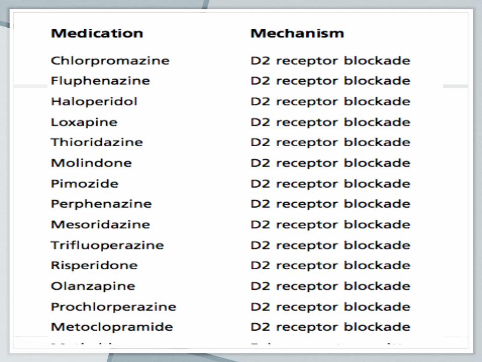
• Drug-induced movement disorders are varied and can be caused by a number of medications that alter central nervous system neurochemistry (D2 receptor blockade)
• Include:
• Parkinsonism
• Tardive phenomena
• Chorea

• Dystonia
• Tremor
• Akathisia
• Myoclonus
• Tics, and
• Neuroleptic malignant syndrome
• The movements can be acute or chronic
phenomena.
• Can be focal, hemi-, or generalized in nature.
• With the exception of the tardive phenomenon,
can usually be treated by elimination of the
offending medication.

• 1. Acute:
• Akathisia
• Dystonia
• 2. Overdosage:
• Drug-induced Parkinsonism
• 3. Neuroleptic malignant syndrome

• Chronic: Tardive Syndromes
• Dyskinesia
• Akathisia
• Dystonia

• Tremors
• Tics
• Myoclonus

Acute Dystonia
• Are sustained muscle contractions,
frequently causing twisting and repetitive
movements, or abnormal postures.
• Earliest abnormal involuntary movement to
appear after initiation of dopamine receptor
antagonist therapy.
• Can be associated with oculogyric crisis.

• The reaction may occur after the first dose.
• In 50%, occurs within 48 hours.
• In 90%, it occurs by 5 days after starting
the therapy.
• High incidence rate ( >50%) with highly
potent dopamine receptor blockers such as
haloperidol.

• Acute dystonia was significantly higher in
males than in females (77.8% vs. 28.6%, P
< 0.05).
• Younger males (≤30 years) had an
extremely high incidence (91.7%).
• The incidence is much lower with the
atypical antipsychotics.

• Second generation antipsychotics were associated with a significantly lower risk of acute dystonia (relative risk = 0.19) and acute akathisia (relative risk = 0.25), compared with haloperidol alone.
• Serotonergic agents have also been reported to induce acute dystonic reactions.


• Tetrabenazine (TBZ), has been reported to
induce acute dystonic reactions.
• In addition to depleting dopamine, TBZ
blocks dopamine receptors.
• Acute dystonia mostly affect the:
• Ocular muscles (oculogyric crisis),
• Face, jaw, tongue, neck, and trunk, and
• Less often the limbs.
• A typical acute dystonic reaction may consist of :
• head tilt backward or sideways with tongue protrusion and
• forced opening of the mouth,
• often with arching of trunk and
• ocular deviation upward or laterally
• Hypothesis:
• After neuroleptic dose:
• 1- Surge of dopamine release
• 2- Denervation supersensitivity of postsynaptic dopamine receptors
• 3- Result in markedly increased striatal dopaminergic activity at about 20–40 hours
• Symptoms can be relieved within minutes
after parenteral anticholinergics or
antihistaminics. D/c offending drug.
• Diphenhydramine 50 mg, benztropine
mesylate or biperiden 1–2 mg is given
intravenously.
• Intravenous diazepam

• If untreated, the majority of cases still resolve
spontaneously in 12–48 hours.
• Dopamine receptor antagonists with high
anticholinergic activities have low incidence rates
of acute dystonic reactions.
• Prophylactic use of anticholinergics especially in
the young and on high potency drugs.

Acute Akathisia
• A hyperkinetic (sensorimotor) movement disorder characterized by restlessness and the irresistible urge to move.
• May be an acute, subacute, or tardive phenomenon.
• Might reflect an alteration of the dopaminergic mesolimbic system.
• Occurs with medications that alter central nervous system dopamine levels ( 20%-30%)
• Including: typical > atypical neuroleptics, antiemetic agents, reserpine, and tetrabenazine.
• The motor aspect of akathisia (akathitic
movements):
• Excessive movements that are complex,
semipurposeful, stereotypic, and repetitive,
suppressible and decrease with distraction.
• Akathisia is also seen in patients with Parkinson
disease, cocaine abuse, and SSRI use. (Daras et
al., 1994), ands (Poyurovsky et al., 1995)

• Acute akathisia is self-limited, disappearing on discontinuation of neuroleptics.
• First line therapies include:
• Propranolol : Below 80 mg/day
• Mirtazipine : A literature search revealed mirtazapine to be effective and superior to propranolol (43.3% vs. 30.0%) (Hieber et al., 2008)
• Trazadone

• Clonidine
• Mianserin : 5 HT2 antagonist
• Nicotine patch
• Amantadine
• Tetrabenazine/reserpine
• Gabapentin

• Zolpidem : GABA-mimetic drug & selective
agonist of the BZD receptor.
• Fluvoxamine, a sigma-1 agonist
• Potential future class of medications that may be
effective in treating akathisia is the adenosine A2a
antagonist group.
• DBS +/-
• One study compared the efficacy of B6, mianserin and placebo in the treatment of acute akathisia
• Sixty schizophrenia and schizoaffective in-patients with akathisia were randomized to receive vitamin B6 1200 mg/d, mianserin 15 mg/d, or placebo for 5 days, in a double-blind design
• Compared with the placebo group, the vitamin B6-treated and mianserin-treated patients showed a significant improvement in the subjective symptoms (P < 0.0001)
Drug-induced
Parkinsonism
• Clinically indistinguishable from idiopathic
Parkinson’s disease.
• May occur as a symmetric or asymmetric
phenomenon.
• Tremor, rigidity, bradykinesia, and less commonly
postural instability are common features.
• Due to medications that affect presynaptic,
synaptic, or postsynaptic dopamine levels.

• Women are affected almost twice as frequently as
men.
• Occurs increasingly with advanced age in parallel with
the incidence of idiopathic PD.
• All DRBAs can induce parkinsonism, except clozapine
(there are only rare reports with clozapine) Factor and
Friedman, 1997)

• It can be either a subacute or chronic condition.
• In patients at risk, 50% to 70% will develop symptoms within 1 month of starting therapy and 90% within 3 months (Ayd, 1961).
• The most effective treatment for drug-induced parkinsonism is elimination of the offending medication.
• If symptoms persist, the patient most likely has subclinical parkinsonism that was unmasked.
• Neuroimaging with [18F]fluoro- dopa positron emission tomography :
• Patients with pure drug- induced parkinsonism will have normalization of their radioactive dopa uptake in the basal ganglia after elimination of precipitating drugs.
• While patients with unmasking of an underlying parkinsonism will have persistent diminished uptake after elimination of the precipitating medication (Burn and Brook, 1993)

• Parkinsonism from neuroleptics is typically
reversible when the medication is reduced or
discontinued.
• Sometimes, the reversal can take many months;
an interval of up to 18 months has been noted in
the literature (Fleming et al., 1970).

• SSRIs can sometimes worsen parkinsonism in patients with PD (Meco et al., 1994).
• Occasionally can induce parkinsonism in patients who never had symptoms of PD (Coulter and Pillans, 1995; DiRocco et al., 1998)
• In an intensive monitoring program in New Zealand of the SSRI drug fluoxetine over a 4-year period, there were 15 reports of parkinsonism in 5555 patients who were exposed to the drug (Coulter and Pillans, 1995)

• The explanation for inducing or enhancing
parkinsonism is that:
• Increased serotonergic activity in the substantia
nigra will inhibit dopamine-containing neurons,
• Thus causing functional dopamine deficiency in
the nigrostriatal pathway (Baldessarini and Marsh,
1992

• Treatment:
• Stop the offending drug
• Usually initiated with anticholinergics or
amantadine.
• Levodopa & dopamine agonist not effective
except in cases who were on dopamine depletors
( reserpine).


Neuroleptic Malignant
Syndrome
• NMS is an idiosyncratic reaction that can sometimes be life-threatening.
• Clinical triad consists of:
• (1) Hyperthermia, usually with other autonomic dysfunctions such as tachycardia, diaphoresis, and labile blood pressure
• (2) Extrapyramidal signs, usually increased muscle tone of rigidity or dystonia, often with accompanying elevation of muscle enzymes; and
• (3) Alteration of mental status, such as agitation, inattention, and confusion.
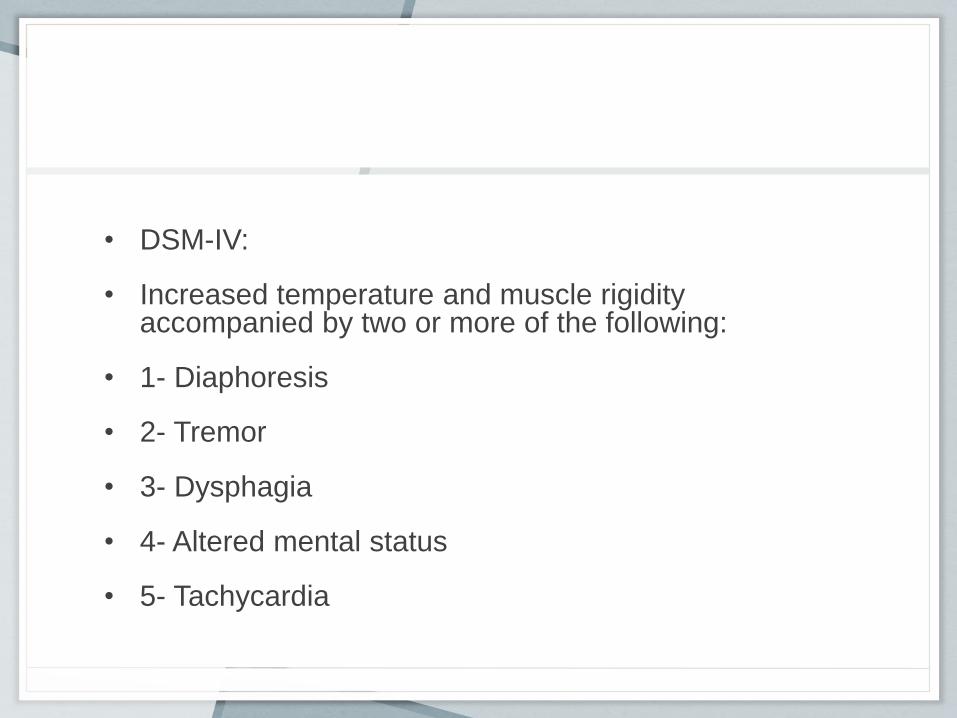
• DSM-IV:
• Increased temperature and muscle rigidity accompanied by two or more of the following:
• 1- Diaphoresis
• 2- Tremor
• 3- Dysphagia
• 4- Altered mental status
• 5- Tachycardia

• Incontinence
• Dysregulation of blood pressure
• Leukocytosis, and elevated creatine kinase.

• Fever is not an essential symptom and it can be delayed.
• The syndrome begins abruptly while the patient is on therapeutic, not toxic, dosages of medication.
• All the symptoms are fully manifest within 24 hours and reach a maximum within 72 hours. ( 3-9 days of tx)
• NMS can develop soon after the first dose or at any time after prolonged treatment.
• Recovery usually occurs within 1 to several weeks, but can be fatal in 20–30% of cases.

• The incidence of NMS is between 0.5% and 2.4%.
• Mortality rates have been reported as low as 4% and as high as 20% (Bertorini, 1997).
• Pathophysiologic basis: dopaminergic dysregulation and blockade of dopamine in the basal ganglia and hypothalamus.

• All agents that block D2 receptors can induce
NMS, including :
• Risperidone
• Amisulpride
• Olanzapine and
• Phenothiazines with antihistaminic activity, such
as alimemazine

• A case of NMS associated with bupropion has
been reported (Kasantikul and Kanchanatawan,
2006)
• Tetrabenazine has been reported to cause NMS;
this seems likely to be due to its D2-blocking
activity (Reches et al., 1983)
• Reserpine has not been reported to cause NMS.
• Abrupt withdrawal of levodopa & dopamine
agonists.
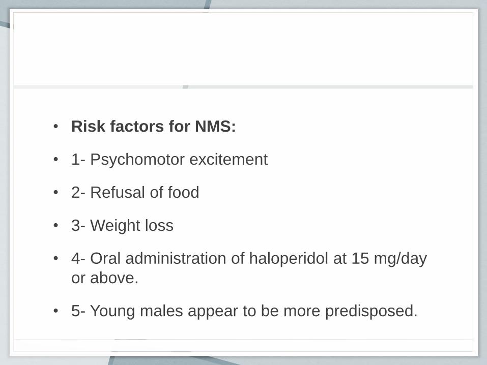
• Risk factors for NMS:
• 1- Psychomotor excitement
• 2- Refusal of food
• 3- Weight loss
• 4- Oral administration of haloperidol at 15 mg/day
or above.
• 5- Young males appear to be more predisposed.

• Reports of an NMS-like syndrome following the sudden withdrawal of:
• Amantadine
• Baclofen
• Combination of a long-acting neuroleptic and an anticholinergic agent.
• The idiosyncratic nature and rarity of the
syndrome remain unexplained.
• TaqI A polymorphism of the dopamine D2 receptor
gene appears to occur more commonly in patients
who developed NMS.
• Kishida et al. (2004) found that patients with NMS
had a higher association with a polymorphism in
the D2 receptor gene.
• Treatment of NMS consists of discontinuing the
antipsychotic drugs and providing supportive
measures.
• Rapid relief of symptoms has been reported with the
use of dantrolene 3mg/kg-5mg/kg IV tds-qid,
bromocriptine 5mgqid, or levodopa.
• Nisijima and colleagues (1997) found levodopa to be
more effective than dantrolene.
• Tsujimoto and colleagues (1998) found intravenous
dantrolene plus hemodialysis to be effective.
• Subcutaneous apomorphine has been found to be
effective as a solo treatment (Wang and Hsieh,
2001)
• Gratz and Simpson (1994) recommended using
anticholinergics in an attempt to reverse rigidity
prior to utilizing bromocriptine.
• Carbamazepine was dramatically effective in two
patients (with recurrence on withdrawal of the
drug) (Thomas et al., 1998)

• Steroids added to standard therapy have been
reported to speed recovery time (Sato et al.,
2003).
• Re-exposure to dopamine receptor antagonists
does not necessarily lead to recurrence of NMS
(Singh and Albaranzanchi, 1995; Singh and
Hambidge, 1998).
• Residual catatonia that can last weeks to months
has been reported, with some patients responding
to ECT (Caroff et al., 2000).

• Hyponatremia can sometimes occur due to:
• 1- SIADH
• 2- Cerebral salt wasting syndrome
• Managed by salt replacement

• Other modalities :
• Benzodiazepines
• Amantadine, and
• Electroconvulsive therapy

Tardive Syndromes
• Refers to a group of disorders that fit all of the following essential criteria:
• 1- Abnormal involuntary movements or a sensation of restlessness that often causes “unvoluntary” movements
• 2- Exposure to at least one DRBA within 6 months of the onset of symptoms (in exceptional cases, exposure could be up to 12 months)
• 3- The disorder persists for at least 1 month after stopping the offending drug(Fahn, 1984a; Stacy and Jankovic, 1991)
Classic Tardive Dyskinesia
• Tardive dyskinesia, a hyperkinetic movement
disorder that causes choreic movements.
• Chorea is characterized by involuntary, rapid,
nonrepetitive, random, small-amplitude
movements that may be symmetric or asymmetric.

• Risk factors:
• Old age
• Certain predisposition of women
• Mental retardation & affective disorders
• History of substance abuse and
• Traumatic head injury
• Associated with chronic use, greater than 3
months, of dopaminergic blocking medications
including:
• Typical and atypical neuroleptic medications.
• Antiemetic agents: prochlorperazine and
metoclopramide.
• Except quetiapine and clozapine

• The tardive dyskinesia syndromes tend to persist and
can remain permanently.
• Can occur when the patient is taking these drugs or
within a period of time after stopping the drugs.
• Withdrawing the offending drugs often exacerbates the
severity of the movements because of removal of
dopamine receptor blockade.
• Increasing the dosage of these drugs often
ameliorates the movements because of increasing the
blockade.
• Most common is the pattern of repetitive, almost
rhythmic, movements that can be labeled as
stereotypic.
• This pattern often occurs in the oral-buccal-lingual (O-
B-L) region.
• Other parts of the body may also express rhythmic
movements, such as the hands, feet, and trunk (less
often).
• Respirations may also be affected with an altered
rhythmical pattern.

• TD is often accompanied by a feeling of inner
restlessness (akathisia): focal vs generalized.
• Focal akathisia is extremely uncomfortable and is
often expressed by the patient as a burning
sensation.
• Generalized akathisia is often accompanied by a
pattern of movement that appears to be executed
in an attempt to relieve the abnormal
uncomfortable sensations.

• The forehead and eyebrows are seldom involved
unless tardive dystonia is also present.
• Contrast to Huntington disease, in which chorea of
the forehead and eyebrows is more common than
choreic movements of the oral musculature.
• In TD, the mouth tends to show a pattern of
repetitive, complex chewing motions.

• Video dyskinesia

• The involuntary movements of the mouth in
classic TD are readily suppressed.
• Movements cease as the patient is putting food in
the mouth, when talking, or when a finger is
placed on the lips.
• the tongue tends to assume a continual writhing
motion of athetoid side-to-side and coiling
movements.

• Constant lingual movements might lead to tongue
hypertrophy, and macroglossia is a common
clinical sign.
• Tongue protrusion without darting back into the
mouth for more than half a minute is common.
• Contrast to chorea in Huntington’s disease, where
motor impersistence is common.
• Video OBL dyskinesia

Pathophysiology of TD
• The biochemical basis of TD is still unclear.
• The explanation involving dopamine receptor supersensitivity alone does not seem to be sufficient.
• Gerlach (1991) suggested that TD might be due to an increased ratio of D1/D2 receptor activity.
• The typical neuroleptics block pre- and postsynaptic D2 receptors, leaving D1 receptors spared.
• Thus, it is proposed that an increased D1 receptor activation would lead to the dyskinesias.

• The effect of DRBAs on other interconnected
systems is suggested.
• Altered synaptic patterns between subsets of
dopaminergic neurons and other interconnected
neurons.
• Decreased GABA activity in the subthalamic
nucleus lend support to involvement of this
nucleus in TD.

• Oxyradicals have been implicated in the
pathogenetic mechanism for the tardive
syndromes (Lohr, 1991).
• DRBAs cause an increase in dopamine turnover,
resulting in an increased synthesis of hydrogen
peroxide.
• Hydrogen peroxide, if not rapidly metabolized, will
form oxyradicals, which can damage cell
membranes.

• Steen and colleagues (1997) reported that a specific allelic variation of the dopamine D3 receptor (DRD3) gene is found at a higher frequency (22–24%) of homozygosity
• A serine to glycine polymorphism in the first exon of the DRD3 gene appears to be a risk factor for developing TD (Liao et al., 2001, Ozdemir et al., 2001).
• Polymorphisms in DRD3 have also been associated with TD (Zai et al., 2009).
Tardive Dystonia
• These movements are involuntary, repetitive, and twisting.
• They may be intermittent or sustained and are often painful.
• The dystonic movements are described by location as focal, segmental, hemi-, or generalized.

• Persistent dystonic movement as a complication
of DRBA therapy has long been noted (Druckman
et al., 1962)
• Has a different epidemiology and pharmacologic
response from those of classic tardive dyskinesia.
• Prevalence of tardive dystonia in chronic
psychiatric inpatients has been estimated to be
1.5–2% (Friedman et al., 1986; Yassa et al.,
1986).

• Occurs after prolonged use (ie, greater than 3
months) of dopamine blocking agents.
• Same medications that induces classic tardive
dyskinesia also triggers tardive dystonia.

• Focal tardive dystonias are usually cranial in
location.
• Affecting the jaw, tongue, and facial muscles.
• Tardive dystonia may also be accompanied by
tardive akathisia and by classic TD.
• Tardive dystonia can occur at all ages, whereas
classic TD is more common in the elderly.

• In primary dystonia, patients at younger age of onset tend to develop generalized dystonia.
• Those with onset in adulthood are more likely to have craniocervical focal or segmental dystonia.
• Regardless of age at onset, tardive dystonia usually progresses over months or years from a focal onset to become more widespread.
• Only 17% remain focal at the time of maximum severity (Kiriakakis et al., 1998).
• Tardive dystonia in adults tends to remain focal or
segmental and tends to involve the craniocervical
region.
• The onset can be from days to years after
exposure to a DRBA.
• The range to extend from 4 days to 23 years of
exposure ( Kiriakakis and colleagues (1998).

• Men are significantly younger than women at
onset of dystonia.
• It develops after shorter exposure in men.
• Severe tardive dystonia was more common in
young men.
• Severe classic tardive dyskinesia was more
common in older women.

• Tardive dystonia tends to occur in all ages without
predilection for any particular age range.
• The mean age of onset in the literature is about 40
years.
• Idiopathic dystonia, shows a bimodal distribution
with one early peak in childhood and another later
peak in adulthood.
• Improvement with sensory tricks (geste
antagoniste) occurs both in idiopathic torsion
dystonia and tardive dystonia.
• Focal dystonias, such as tardive cervical dystonia,
tardive blepharospasm can resemble idiopathic
form.
• Retrocollis is more with tardive dystonia.
• A comparison of tardive and primary
oromandibular dystonia (OMD) showed :
• 1- Similar demographics
• 2- Both occurring predominantly in women
• 3- With jaw-closing dystonia being the most
common form (Tan and Jankovic, 2000).

• Primary OMD patients were more likely to have
coexistent cervical dystonia.
• Limb stereotypies, akathisia, and respiratory
dyskinesia were seen only in the tardive OMD.

• More characteristic of tardive dystonia:
• Retrocollis
• Trunk arching backward
• Internal rotation of the arms, extension of the elbows and flexion of the wrists
• The presence of myoclonic movements in association with dystonia.
• Reduction of dystonic movements with voluntary action such as walking

• Video
• Patients with idiopathic dystonia more often have lateral torticollis and twisting of the trunk laterally.
• The dystonic movements are usually exacerbated by voluntary action.
• It can be severe enough to jeopardize patients by causing life-threatening dysphagia.

• Video

Tardive Akathisia
• The clinical phenomenology of tardive akathisia is thought to be the same as that of acute akathisia.
• Moaning & focal pain are more common in tardive akathisia than in acute akathisia.
• Mean age at onset of tardive akathisia was 58 years with a range from 21 to 82 years.
• The mean duration of dopamine receptor antagonist exposure before the onset was 4.5 years with a range from 2 weeks to 22 years.
Withdrawal Emergent
Syndrome
• First described in children who had been on
antipsychotic drugs for a long period of time and
then were withdrawn abruptly from their
medication (Polizos et al., 1973).
• The abnormal movements are brief and flow from
one muscle to another in a seemingly random
way.
• They differ from the movements of classic tardive
dyskinesia, which are brief, but stereotypical and
repetitive.

• Involve mainly the limbs, trunk, and neck, and
rarely the oral region, which is the most
prevalent site in classic tardive dyskinesia.
• The dyskinetic movements disappear
spontaneously within several weeks after
withdrawal of the DRBA.
• For immediate suppression of movements,
dopamine receptor antagonists can be reinstituted
and withdrawn gradually without recurrence of the
withdrawal emergent syndrome (Fahn, 1984a).
• A withdrawal reaction from melatonin with O-B-L
dyskinesia and akathisia was reported by Giladi
and Shabtai (1999)
• Withdrawal emergent syndrome is analogous to
the classic tardive dyskinesia seen in adults.
• Course is more benign and movements are more
generalized, resembling the choreic movements of
Sydenham disease.

• Video wes
Treatment
• In classic tardive dyskinesia, prospective data show 33% remission in 2 years following elimination of the DRBA (Kane et al., 1986).
• In retrospective studies, the remission rates were 12% for tardive dystonia and 8% for tardive akathisia (Kang et al., 1986; Burke et al., 1989).
• Younger age is associated with better chance of remission. (Smith and Baldessarini, 1980)
• Earlier detection and discontinuation of dopamine receptor antagonists were more favorable for remission (Quitkin et al., 1977).

Treatment of Classic
Tardive Dyskinesia Pharmacological property Tetrabenazine Reserpine
Mechanism of action Selectively binds
hVMAT2
Reversibly binds
VMAT2
Binds intravesicular
site
Binds hVMAT1 and
hVMAT2
Irreversibly binds
VMAT
Binds cytoplasmic site
Peripheral monoamine depletion No Yes
Duration of action in humans Short (approx. 12
hours)
Several days
Hypotension No Yes
Gastrointestinal effects No Yes
Dopamine receptor blocking activity Yes No
• Open-Label Extension of KINECT: A Phase 2
Study of Valbenazine (NBI-98854) for Tardive
Dyskinesia (S27.001)
• Mohammed Bari, Raj Shiwach, Roland Jimenez,
Scott Siegert, and Christopher O'Brien
• April 5, 2016, 86:16 Supplement S27.001;
published ahead of print April 8, 2015, 1526-632X

• Data from KINECT 2, a phase 2 randomized,
double-blind, placebo-controlled dose-
escalating trial in subjects (n=102) with
schizophrenia, schizoaffective disorder, mood
disorder or gastrointestinal (GI) disorder with
moderate or severe TD.
• The goal of the trial was to evaluate the safety
and efficacy of once-daily administered
valbenazine for TD.

• Valbenazine or placebo was administered once daily for six weeks.
• All subjects randomized to valbenazine received 25 mg through week two, at which point the dose could be increased to 50 mg or maintained.
• At week four, the dose could be increased to 75 mg, maintained, or reduced to the previous dose.
• Participants who took the placebo capsule also underwent similar, blinded dose adjustments.
• Valbenazine was associated with a marked reduction in the abnormal movements associated with TD as assessed by the Abnormal Involuntary Movement Scale (AIMS) and clinical global impression of change (CGI-TD) score.
• The CGI-TD showed significant improvement with treatment (67 percent versus 16 percent were “much improved” or “very much improved.”
• AIMS scores improved by 2.6 points in the valbenazine group versus a decrease of 0.2 points in the placebo arm.

• KINECT 3: A Randomized, Double-Blind,
Placebo-Controlled Phase 3 Trial of
Valbenazine (NBI-98854) for Tardive
Dyskinesia (PL02.003)
• Robert Hauser6, Stewart Factor2,
Stephen Marder4, Mary Ann Knesevich5, Paul
Michael Ramirez1, Roland Jimenez3, Joshua
Burke3, Grace Liang3 and Christopher O'Brien3

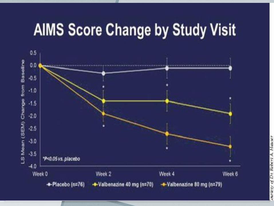
• In the phase 3, KINECT 3 study, 234 patients with moderate to severe neuroleptic-induced TD were randomized to receive a once-daily dose of 80 mg valbenazine, 40 mg valbenazine, or placebo, for six weeks.
• The study's primary endpoint was a change from baseline in the Abnormal Involuntary Movement Scale (AIM) total dyskinesia score, as assessed by blinded video raters.
• Safety was determined by adverse event (AE) rates together with laboratory, electrocardiography, and psychiatric assessments.

• Among the patients, 66 percent had
schizophrenia and 86 percent were receiving
stable doses of concomitant neuroleptics,
including 77 percent who were taking atypical
antipsychotic medication
• Valbenazine 80mg resulted in a significant
improvement in AIMS score vs. placebo (mean
change from baseline -3.2 vs. -0.1; p<0.0001).
The AIMS score was also reduced in the 40mg
group vs. placebo (mean change from baseline
-1.9 vs. -0.1; p=0.0021).

• Valbenazine was for the most part well tolerated, with the frequency of adverse events similar in all treatment groups and consistent with those of prior studies.
• The most commonly reported adverse event was somnolence, which occurred in 5 percent of subjects in the 80 mg treatment cohort, 4 percent in the 40 mg group, and 4 percent in the placebo group.
• Three percent of subjects in the 40mg and placebo groups discontinued treatment due to adverse
events, as did 4 percent in the 80 mg group.
• SD-809, or deutetrabenazine, was
granted breakthrough status by
the FDA last November.

• Atypical antipsychotics:
• Clozapine: upto 400 mg/day ( 50% response)
• Quetiapine: upto 600 mg/day
• Olanzapine
• Effect due to further D2 receptor blockade.

• Dopamine agonists:
• Activate the presynaptic dopamine receptors by using low doses of a dopamine agonist.
• Which in turn would reduce the biosynthesis and release of dopamine.
• Use of levodopa in an attempt to desensitize the postsynaptic dopamine receptors.
• This can cause initial worsening of symptoms before eventual improvement is expected after discontinuation of levodopa.
• Dopaminergic drugs can also lead to overt recurrence
of underlying psychosis.
• This approach has theoretical merit, but not carried
out.
• Amantadine has been reported to have some benefit,
mostly due to its glutamate receptor blocking effect
rather than its dopaminergic effect.

• Nondopaminergic medications:
• Clonazepam
• Propranolol, fusaric acid, and clonidine
• Anticholinergics, pyridoxine, tryptophan, cyproheptadine, vasopressin, naloxone, morphine, and estrogen were reported to be of no benefit.
• Pyridoxine was found to reduce the severity of TD (Lerner et al., 2001).

• Buspirone has been reported to be beneficial (Moss et al., 1993)
• Calcium channel blockers have been reported to reduce the severity of tardive dyskinesia (Kushnir and Ratner, 1989
• A combination of acetazolamide and thiamine was found to reduce both TD and drug-induced parkinsonism (Cowen et al., 1997)
• Lithium
• Meta-analysis showed that baclofen, deanol, and
diazepam were no more effective than a placebo.
• Meta-analysis found that five interventions were
effective: levodopa, oxypertine, sodium valproate,
tiapride and vitamin E.
• Data from single randomized clinical trials
revealed that insulin, α-methyldopa, and reserpine
were more effective than a placebo.

• Treatment with vitamin E has been found to
reduce the severity of tardive dyskinesia (Elkashef
et al., 1990
• There continue to be reports of TD responding to
open-label trials. Gabapentin, pyridoxine,
branched amino acids.
• Levetiracetam was helpful in a small trial
(Konitsiotis et al., 2006).

• Injections of botulinum toxin into the muscles
causing oral dyskinesia have been reported to be
effective in reducing the movements (Rapaport et
al., 2000).
• Sporadic reports noted efficacy of
electroconvulsive therapy in refractory cases of
TD (Price and Levin, 1978)
Treatment of Tardive
Dystonia
• The most effective medications for tardive
dystonia are also antidopaminergic drugs (Kang et
al., 1986)
• Reserpine and TBZ each produce improvement in
about 50% of patients.
• DRBAs are more effective in suppressing the
movements (77%).
• The atypical antipsychotic clozapine has been
helpful in some patients with tardive dystonia.
• There are reports of quetiapine’s effectiveness as well (Gourzis et al., 2005).
• Combination of clozapine and clonazepam has been effective in some patients (Shapleske et al., 1996).
• Antimuscarinics are almost as effective as antidopaminergic drugs.
• This is different from classic tardive dyskinesia, which may get worse with antimuscarinics (Yassa, 1988).

• Improvement rate ( 46 % ) on antimuscarinics
such as trihexyphenidyl and ethopropazine.
• Benzodiazepines are mainly helpful as adjunctive
therapy with dopamine-depleting or anticholinergic
drugs.
• Minimal success with propranolol, levodopa,
carbamazepine, and baclofen has been noted.

• Bromocriptine, deanol, clonidine, lisuride, amantadine,
and valproate were reported with mixed results.
• Verapamil was reported to be effective in one patient
(Abad and Ovsiew, 1993)
• Opioids do not have lasting value in suppressing
tardive dystonia (Berg et al., 2001)
• One study found the combination of naltrexone and
clonazepam to offer some benefit (Wonodi et al.,
2004b).

• Botulinum toxin injection into the affected parts might be helpful.
• Electroconvulsive therapy (ECT) might be effective in intractable cases (Yoshida et al., 1996)
• Deep brain stimulation in the globus pallidus interna is often effective (Franzini et al., 2005)
• Deep brain stimulation in the pallidum can be safe and effective (Capelle et al., 2010; Chang et al., 2010)
• Intrathecal baclofen (Dressler et al., 1997)


Treatment of Tardive
Akathisia
• Tardive akathisia is difficult to treat and does not respond to anticholinergics, which have been reported to help acute akathisia.
• Patients improved on reserpine up to 5 mg/day (87%) and 58% on TBZ up to 175 mg/day.
• Opioids were reported to be beneficial (Walters et al., 1986) but the effect has not been persistent.
• ECT can be effective in refractory cases (Hermesh et al., 1992).

Treatment Summary
• Taper and slowly eliminate causative agents if clinically possible.
• Avoid sudden cessation of these drugs.
• Avoid drugs, if possible (i.e., wait for spontaneous recovery).
• If necessary to treat the symptoms, first use dopamine depleting drugs.
• Consider melatonin on the basis of one report (Shamir et al., 2001).

• Consider the true atypical antipsychotic agents,
clozapine and quetiapine.
• If these fail, consider tiny doses of a dopamine
receptor agonist to activate only the presynaptic
dopamine receptor and reduce the biosynthesis of
dopamine.
• For tardive dystonia, consider antimuscarinics.
• For intractable tardive akathisia, consider ECT

• Typical antipsychotic agents can be used when all
fails.
• Combining this with a dopamine depletor may:
• 1- Increase the potency of the antidyskinetic
effect.
• 2- Protect against a worsening of the underlying
tardive pathology.

• Thalamotomy, pallidotomy, and deep brain
stimulation of the thalamus and pallidum have
been performed for tardive dystonia with success.
• DBS-Gpi appears to be the preferred surgical
procedure if the symptoms remain severe and all
medication trials fail.


DRUG-INDUCED TREMOR
• A rhythmic, involuntary, oscillatory movement of
any body part and is named for the position of
great- est prominence.
• Typically begin shortly after institution of the
offending medication.
• May be postural, rest, or intention.
• The most common form is the enhanced physi-
ologic tremor.

• Elimination of the offending medication often ameliorates drug-induced tremor.
• If the offending agent cannot be discontinued:
• Propanolol up to 240 mg a day and
• Primidone up to 250 mg a day, and benzodiazepines such as clonazepam.


DRUG-INDUCED
MYOCLONUS
• An involuntary ( jerky-lightning) hyperkinetic
movement disorder.
• Classified as either positive or negative.
• Positive myoclonus: sudden brief muscular
contractions.
• Negative myoclonus involves pauses in muscular
activity.
• Pathophysiologic mechanism :
• Enhancement of serotonin and g-amino- butyric
acid GABA.
• The best therapy for drug-induced myoclonus is
identification and elimination of the offending
agent.


DRUG-INDUCED TICS
• A hyperkinetic movement disorder consisting of :
• Repetitive, stereotyped motor or vocal movements
that may be simple or complex.
• An urge to perform the movement.
• Relief after performing the movement.
• Some ability to suppress the movement for short
amounts of time.

• Tics have a predilection for cranial and cervical
musculature but may occur in any body location.
• Caused by enhanced dopamine levels
• Associated with multiple drugs including:
• Methylphenidate, dextroamphetamine, pemoline,
cocaine, and, lamotrigine.
• Tx: eliminate the drug.

Conclusion
• Drug-induced movement disorders can be commonly seen in outpatient and inpatient clinical practice.
• Take a thorough past and present medication history.
• Any recently added medications or changes in dosages.
• The best therapy for treating a drug-induced movement disorder is elimination of the offending agent.
• If elimination is not possible due to underlying illness, then attempting to decrease the dosage or change to a less-offensive agent.

• If all of the above measures are impossible or a
tardive syndrome exists, then medical therapy can
be considered.
• The choice of therapeutic agent is guided by:
• 1- Type of movement disorder.
• 2- Coexistent medical problems and medications.

Thank you
• References:
• " Adler CH. Differential diagnosis of Parkinson’s disease. Med Clin North Am 1999;83:349 – 367.
• Review article with an excellent differential diagnosis of parkinsonism.
• " Armon C, Shin C, Miller P, et al. Reversible parkinsonism and cognitive impairment with chronic valproate use. Neurology 1996;47:626–635.
• Early description of valproate-induced parkinsonism.
• " Ayd FJ Jr. A survey of drug-induced extrapyramidal reactions. JAMA 1961; 175:1054 – 1060.

• Abad V., Ovsiew F.: Treatment of persistent myoclonic tardive dystonia with verapamil. Br J Psychiatry. 162:554-556 1993 8097657
• Abbasian C., Power P.: A case of aripiprazole and tardive dyskinesia. J Psychopharmacol. 23 (2):214-215 2009 18515468
• Abu-Kishk I., Toledano M., Reis A., et al.: Neuroleptic malignant syndrome in a child treated with an atypical antipsychotic. J Toxicol Clin Toxicol. 42 (6):921-925 2004 15533033
• Achiron A., Zoldan Y., Melamed E.: Tardive dyskinesia induced by sulpiride. Clin Neuropharmacol. 13:248-252 1990 2357706

• Adler C.M., Malhotra A.K., Elman I., et al.: Amphetamine-induced dopamine release and post-synaptic specific binding in patients with mild tardive dyskinesia. Neuropsychopharmacology. 26 (3):295-300 2002 11850144
• Adler L., Angrist B., Peselow E., et al.: A controlled assessment of propranolol in the treatment of neuroleptic-induced akathisia. Br J Psychiatry. 149:42-45 1986 2877708
• Adler L., Angrist B., Peselow E., et al.: Clonidine in neuroleptic-induced akathisia. Am J Psychiatry. 144:235-236 1987 2880516