dt glaukoma
DESCRIPTION
mataTRANSCRIPT
Acute Glaucoma NI’MATUL MUTHMAINNAH I11111054
Aqueous humor circulation
•The aqueous humor is formed by the ciliary processes and secreted into the posterior chamber of the eye
•Produce 2–6 μl per minute and a total anterior and posterior chamber volume of about 0.2–0.4ml, about 1–2% of the aqueous humor is replaced each minute.
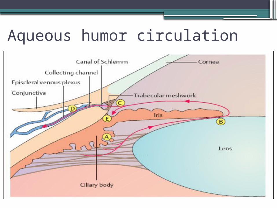
Aqueous humor circulation
Definition
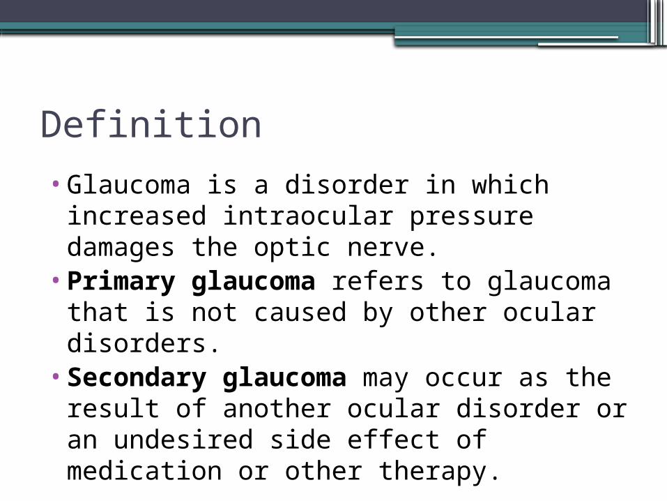
•Glaucoma is a disorder in which increased intraocular pressure damages the optic nerve.
•Primary glaucoma refers to glaucoma that is not caused by other ocular disorders.
•Secondary glaucoma may occur as the result of another ocular disorder or an undesired side effect of medication or other therapy.
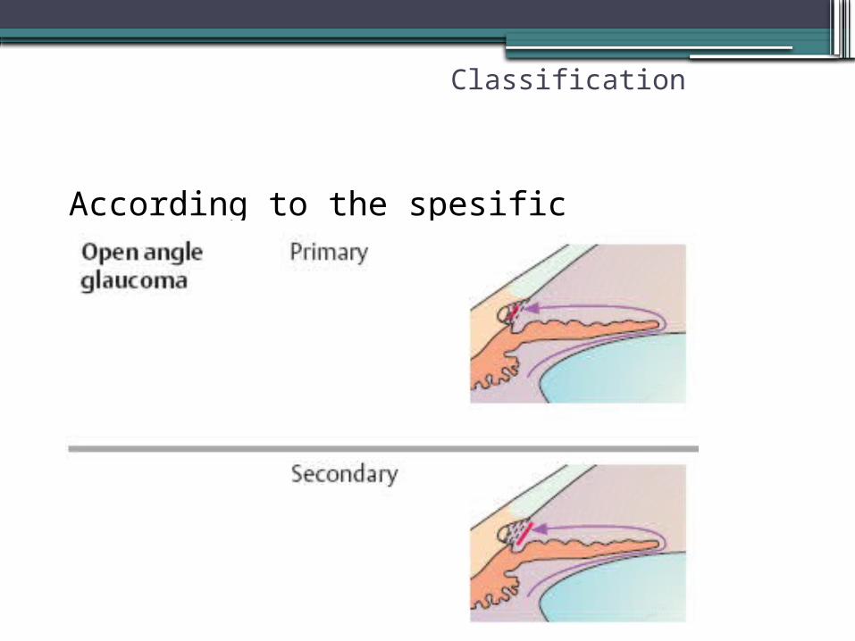
Classification
According to the spesific pathofisiology
Primary open angle glaucoma
•structure of the trabecular look normal but an increase in flow out of aqueous resistance which causes increased ocular pressure
•Etiology : drainage of aqueous humor is impeded.
Classification
Epidemiology
•The most common form of glaucoma•90% of adult glaucomas. •The incidence of the disorder significantly
increases beyond the age of 40, reaching a peak between the ages of 60 and 70.
Symtomps
•Unspesific symptoms •Headache•Burning sensation in the eyes•Blurred or decreased vision•Halo
Secondary open angle glaucoma •The anatomic relationships between the
root of the iris, the trabecular meshwork, and peripheral cornea are not disturbed.
•The trabecular meshwork is congested and the resistance to drainage is increased.
Classification
Secondary open angle glaucoma • Inflammatory glaucoma. Two mechanisms contribute to the increase in intraocular
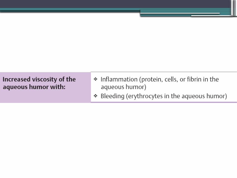
pressure:1. The viscosity of the aqueous humor increases as a result of
the influx of protein from inflamed iris vessels.2. The trabecular meshwork becomes congested with
inflammatory cells and cellular debris.• Phacolytic glaucoma. This is acute glaucoma in eyes with hypermature cataracts.
Denatured lens protein passes through the intact lens capsule into the anterior chamber and is phagocytized. The trabecular meshwork becomes congested with protein-binding macrophages and the protein itself.
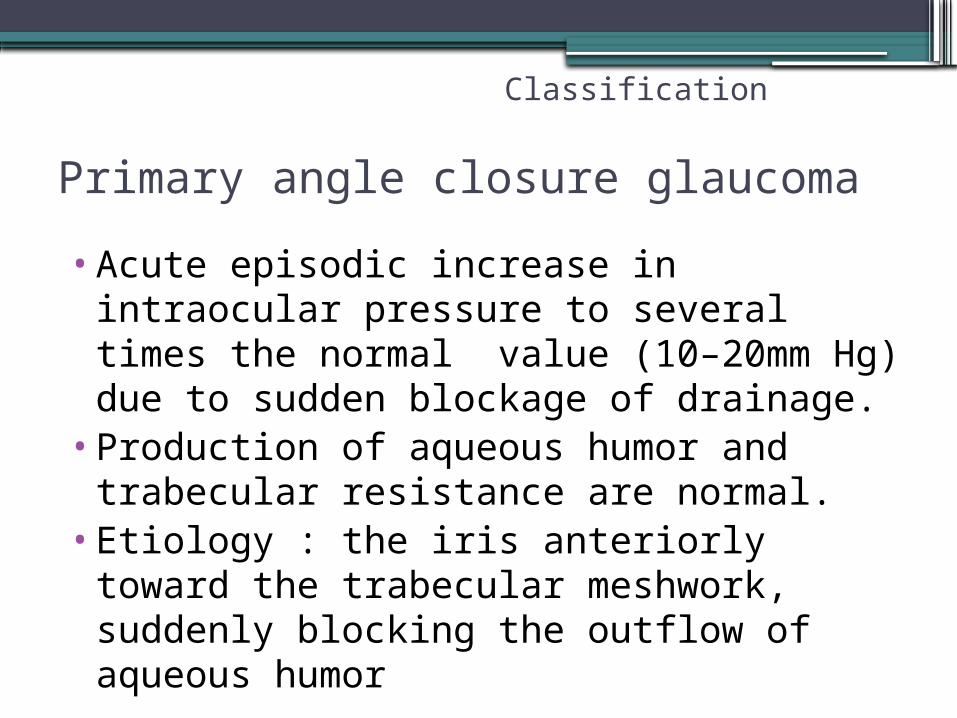
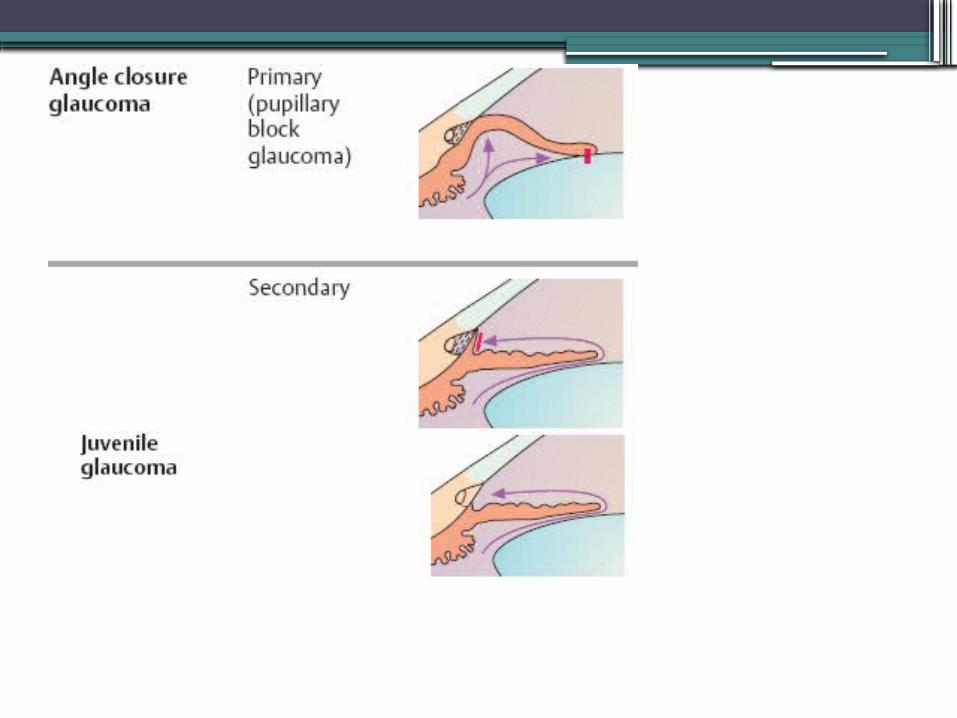
Primary angle closure glaucoma
•Acute episodic increase in intraocular pressure to several times the normal value (10–20mm Hg) due to sudden blockage of drainage.
•Production of aqueous humor and trabecular resistance are normal.
•Etiology : the iris anteriorly toward the trabecular meshwork, suddenly blocking the outflow of aqueous humor
Classification

Symtomps
•Acute onset of intense pain. The elevated intraocular pressure acts on the corneal nerves to cause dull pain.
•Nausea and vomiting•↓visual acuity•Prodromal symptoms--blurred vision or
the appearance of colored halos around lights
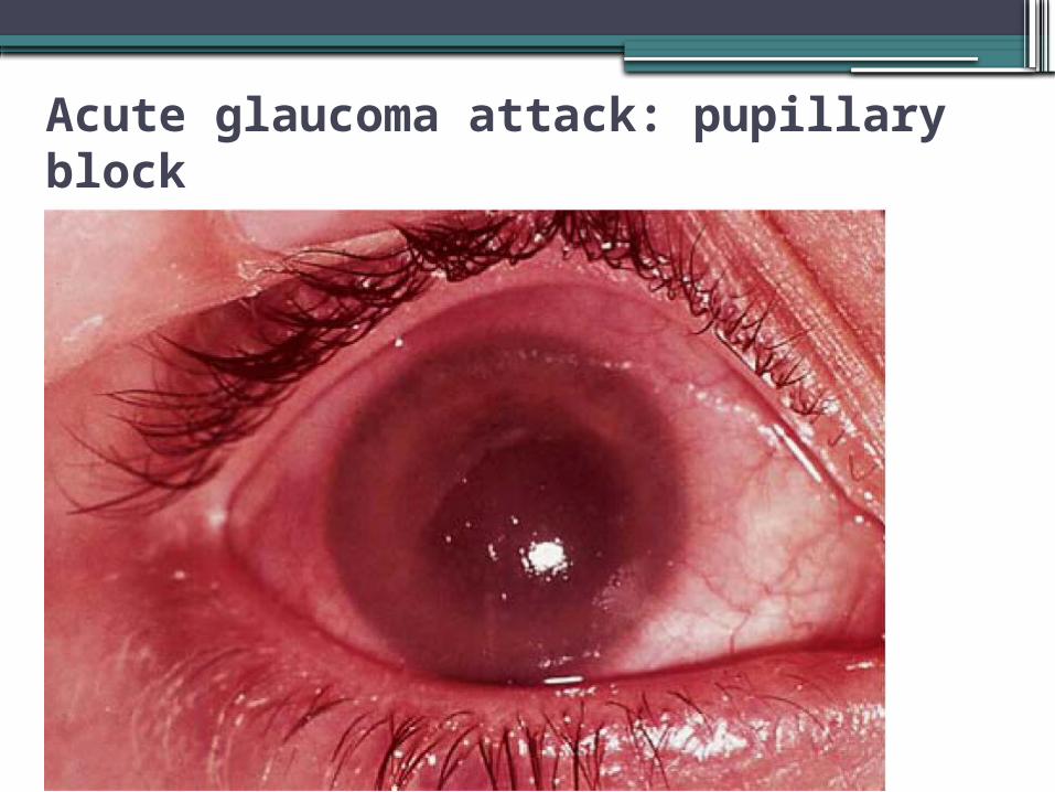
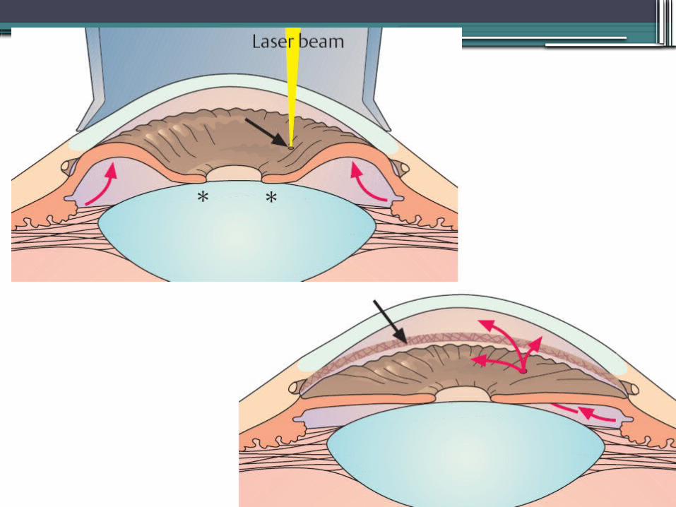
Acute glaucoma attack: pupillary block
•Conjunctival and ciliary injection (red eye).•Corneal edema.•Opacification of the corneal•Anterior chamber is shallow.•The pupil is oval instead of round and
dilated.• Intraocular pressure is elevated; the eye is
rock hard to palpation.•Severe headache and gastrointestinal
symptoms are present.
Secondary angle closure glaucoma
•In secondary angle closure glaucoma as in primary angle closure glaucoma,the increase in intraocular pressure is due to blockage of the trabecular meshwork.
•However, the primary configuration of the anterior chamber is not the decisive factor.
Classification
Secondary angle closure glaucoma •Trauma.
Post-traumatic presence of blood or exudate in the angle of the anterior chamber and prolonged contact between the iris and trabecular meshwork in a collapsed anterior chamber can lead to anterior synechiae and angle closure
Juvenile glaucoma
•Any abnormal increase in intraocular pressure during the first years of life will cause dilatation of the wall of the globe, and especially of the cornea.
•The result is a characteristic, abnormally large eye (buphthalmos) with a progressive increase in corneal diameter.
Classification
Symptoms
•Photophobia•Epiphora•corneal opacification,•Unilateral or bilateral enlargement of the
cornea. •These changes may be present from birth (in
congenital glaucoma) or may develop shortly after birth or during the first few years of life.
•Children with this disorder are irritable, poor eaters, and rub their eyes often.
Pathogenesis
•The major mechanism of visual loss in glaucoma is retinal ganglion cell apoptosis, leading to thinning of the inner nuclear and nerve fiber layers of the retina and axonal loss in the optic nerve. The optic disk becomes atrophic, with enlargement of the optic cup
Pathogenesis
•Increase of TIO induced mechanic damage in akson optic nerve
•Increase of TIO also induced ischemic of nerve akson due to decreased blood flow in papil optic nerve.

Examination • Oblique illumination of the anterior chamber---
The anterior chamber is illuminated by a beam of light tangential. a shallow anterior chamber an angle that is partially or completely closed
• Slit lamp examination-- To evaluate the depth of the anterior chamber
• Gonioscopy --- evaluae the angle of the anterior chamber
• Measuring intraocular pressure--- Palpation, Schiøtz,
• Optic disk ophtalmoscopy --- increase in the size of the optic cup and to pale discoloration of the optic disk
• Visual field testing
Treatment
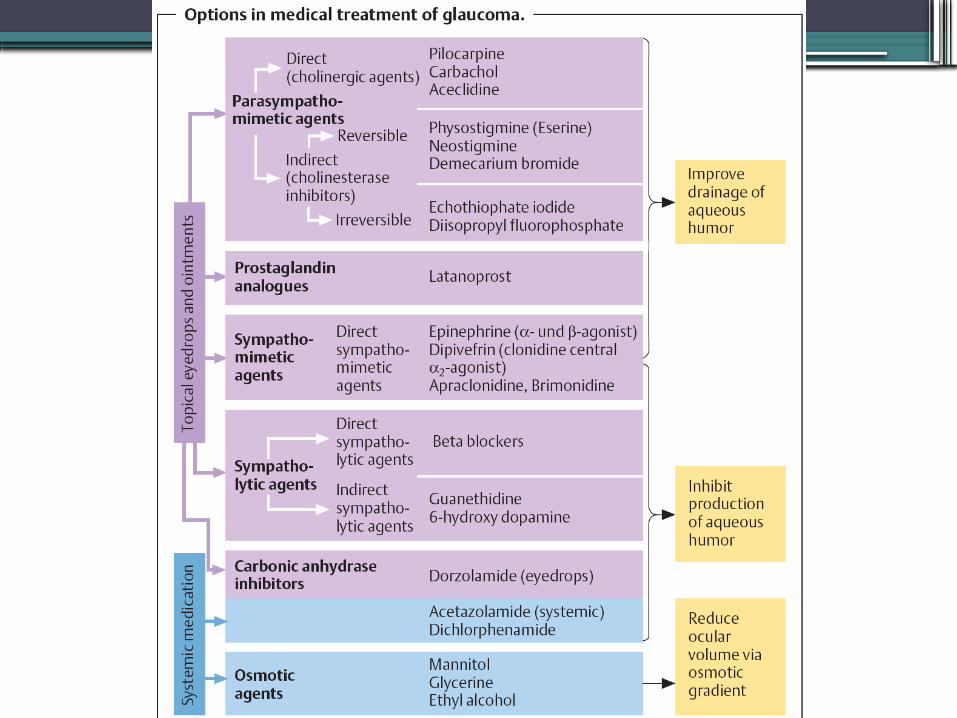
•Inhibit aqueous humor prodduction•Increase trabecular outflow•Increase uveoscleral outflow
Treatment
•Pilokarpin 2% a drop/min in 5 min, after that every 1 hour
•Asetazolamid 500mg IV (TIO > 50mmHg) or oral (TIO < 50mmHg)
•alternative : mannitol 20% 1-2g/KgBB, gliserol 50% 1-1,5g/kgBB (KI: DM).
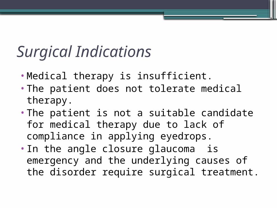
Surgical Indications
•Medical therapy is insufficient.•The patient does not tolerate medical
therapy.•The patient is not a suitable candidate for
medical therapy due to lack of compliance in applying eyedrops.
•In the angle closure glaucoma is emergency and the underlying causes of the disorder require surgical treatment.

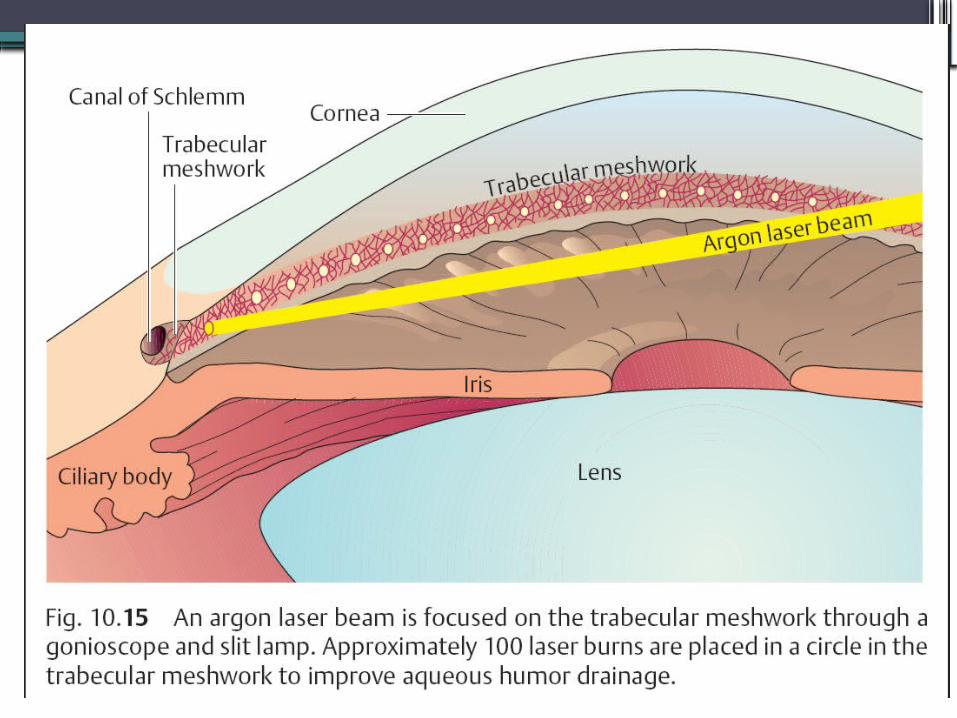
Trabeculoplasty
•Laser burns in the trabecular meshwork cause tissue contraction that widens the intervening spaces and improves outflowthrough the trabecular meshwork.
•Laser surgery in the angle of anterior chamber is possible only if the angle is open.
Peripheral iridectomy
•A limbal incision is made at 12 o’clock under topical anesthesia or general anesthesia,

THANKS…..
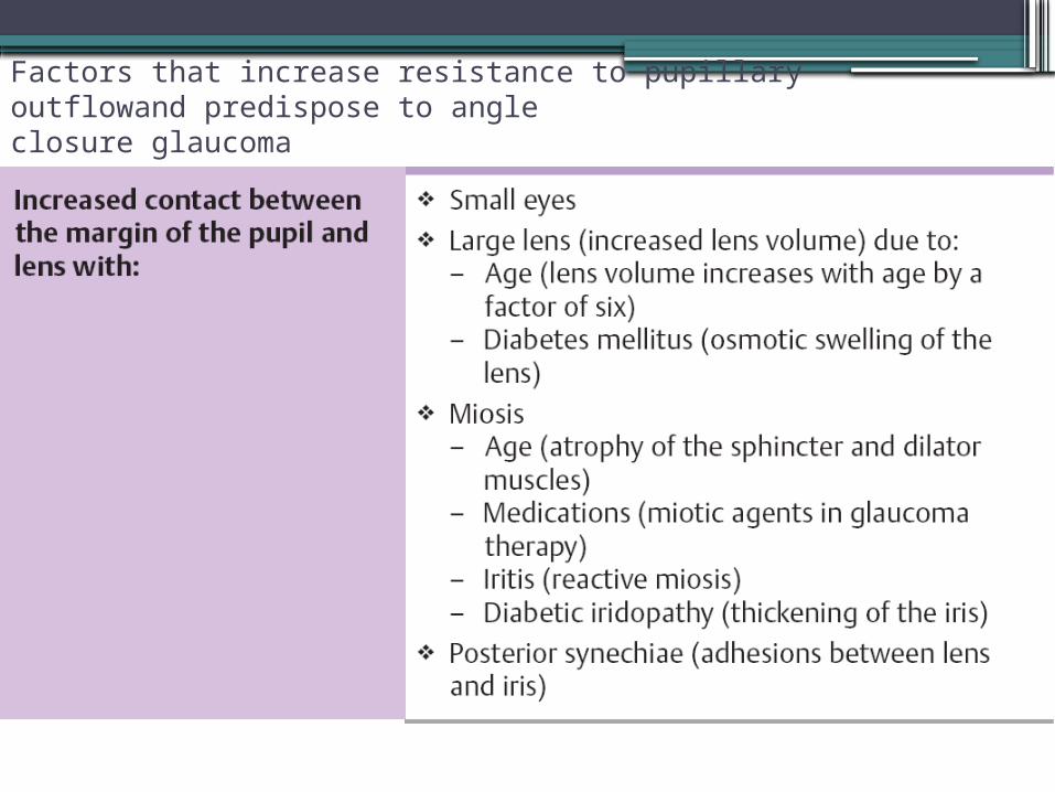
Factors that increase resistance to pupillary outflowand predispose to angleclosure glaucoma