duced in the adult mouse brain by single graded doses of
TRANSCRIPT
EFFECTS OF NITROGEN MUSTARD ON THE MOUSE BRAIN
T. P. MCDONALD, MS., AND M. ASANO, M.D.
From the Biology Division, Oak Ridge National Laboratory,* Oak Ridge, Tenn.
Mustargen [methyl-bis (t]-chloroethyl) amine hydrochloride orHN2-HCI], a radiomimetic alkylating agent of the nitrogen mustardgroup, has been widely used in the treatment of intracranial neoplasticdisorders since its first clinical use in 1942 by Gilman and Phillips.' Thebasis for its use was the presumption that normal nervous tissue wasrelatively resistant to HN2-HCI and that the drug exerted its greatesteffects on rapidly growing neoplastic tissue.2 Many investigators,2 how-ever, have described functional aberrations in nitrogen mustard-treatedanimals that might result from damage to the central nervous system,and French, West, von Amerongen and Magoun6 reported histologicchanges in the brains of cats and monkeys as a result of the intracarotidinjection of 0.25 to I mg. of HN2 per kg. of body weight. The pathologiceffects of nitrogen mustard on the developing nervous system of themouse and rat have been reported by Hicks.7 The principal object ofthis investigation was to assay by histologic methods the effects pro-duced in the adult mouse brain by single graded doses of intravenouslyinjected nitrogen mustard.
MATERIAL AND METHODSFemale RF mice, io weeks of age and weighing from 2I to 26 gin., were used in
this study. Four hundred and forty mice were used to determine the LD50/3o days ofHN2-HC1, and the 30-day survivors were kept to investigate delayed somatic effects.These will be reported later. The brains of an additional I89 mice were examinedmicroscopically.
Mustargen hydrochloride (HN2-HCI) dissolved in o.9 per cent saline with a pHof 4.2 to 4.6 was injected into the dorsal caudal vein in doses of o.o0, o.o6, o.o8, o.i,0.12,0.I5, 0.2 and 04 mg. per mouse.
Mice for histologic study were sacrificed by ether anesthesia at regular intervalsbetween i and 28 days after injection of the drug, and brain tissues were taken formination. The craniuims, with skin, lower jaw, and neck removed, were allowed
to fix overnight in Halmi's fixative. Brains were then cut into 4 parts: (a) themedulla oblongata and posterior part of the cerebellum; (b) the mid-brain at thelevel of the superior quadfigeminal bodies; (c) the brain just inside the opticcbiama; and (d) the forebrain, including olfactory lobes. The parts were placed inZenker-formol solution for 24 hours. The sections were stained with hematoxylinand eosin or with cresyl echt violet. In addition, brains of some mice were fixed inio per cent formalin, mordanted for 4 days (3 per cent potassium dichromate), de-
Accepted for publication, January 20, I96I.* Operated by Union Carbide Corporation for the United States Atomic Energy Com-
mission.
695

6MC DONALD AND ASANO
hydrated, embedded in tissue mat (56 to 580 C), and stained with Lillie's variantof the Weil-Weigert method or Luxol-fast-blue-and-Nissl stain for myelin sheath.
RESULTSSurvival
A straight line was fitted to the survival data by the method of leastsquares, and the LDso/3o days was determined to be 0.127 mg. per
0%.90-
70-
cnw 50-
w30-
0wa
30-
l0.
-lW
-75
-50 Zw
crw-25 a.
0
QO05 QIO 0.15 0.20DOSE (fmg/mouse)
TExT-FiGuRz i. Thirty-day mortality in relation to dose of HN2*HCI (Arc SineTransformation: 0 = arc sine VP, where P = per cent mortality).
mouse with a 95 per cent confidence interval of 0.I03 to O.I62 (Text-fig.i). The survival time varied inversely in relation to dose (Text-fig. 2).At doses of 0.4 mg. per mouse, all mice died within 4 days, 50 per centof the deaths occurring within the first day after treatment. About 83per cent of the mice injected with 0.2 to 0.15 mg. per mouse died withinthe first week, most of them between the fourth and seventh days. Afterdoses of O.I and O.J2 mg. per mouse, mortality at 30 days was 39 and55 per cent respectively, 4I per cent of the deaths occurring by theseventh day. After doses of o.o8 to o.o5 mg. per mouse, 5 of 6o mice died.
Macroscopic Observations
Animals injected with nitrogen mustard showed the following symp-
toms: extreme emaciation, ruffling of fur, inactivity, diarrhea, lacri-mation, lack of muscular coordination, hyperirritability, and spontane-ous convulsions. With doses of o.o5 mg. per mouse, a moderate lack ofcoordination, with weakness and ruffling of fur, was present betweenthe third and sixth days. In general, mice given o. i mg. were unaffecteduntil the second or third day, when depression and ruffling of fur or
0 0
0 I
Ai 1i II I
I i I
I I
I II I I
696 Vol. 38, No. 6

NITROGEN MUSTARD
general unkemptness appeared. This condition persisted on the thirdand fourth days with the onset of diarrhea, weakness, lacrimation, andanorexia. Emaciation, increased irritability, and depression were com-
mon on the fifth day. Within the next day or so, the animals tendedeither to die or to start to recover. Hyperirritability and loss of coordi-nation, however, remained for 2 to 4 weeks. In a few instances perma-
nent abnormalities of posture and movement were noticed among 3o-daysurvivors.
Doses of O.12 and O.I5 mg. per mouse produced essentially the same
toxicity symptoms as 0. I mg. per mouse. Symptoms were, however, morepronounced, and weakness and ruffling of fur were apparent the dayafter treatment. Mice given these doses did not live long enough to de-velop fully the neuropathy found at the o.i mg. dose level, but all micegiven HN2-HCl showed edema and congestion of the brain, occasionallywith subarachnoid hemorrhage between the fourth and I4th days.
Microscopic Observations
In Table I data for later sacrifice periods in the higher dose groups
are lacking because of earlier mortality at these doses. Brains of micereceiving doses at or above the LD5o/3o days showed multiple areas of
INTERVAL AFTER TREATMENT(days)0-2 3-5 6-8 9-1 112-415-17 - - 2-24mg/MOUSE
I I I I I I I I I
mmI I s
30-20-
- 10.-J
> 30-w 20-Z 10-wa. 30-o20.L"
tO-
.
20-1O-
I I.* _
TFxT-FIGUJRE 2. Mortality of mice in relation to dose of HN2'HCl and time afterinjection.
hemorrhage and necrosis. Little necrosis was present in brains of micegiven lower doses. Sagittally, the sites of predilection for brain damagein all groups were the neocortex, pyriform cortex, hippocampus, cere-
bellum, and medulla oblongata (Fig. i). Selective neuronal shrinkagepreceded hemorrhage in all treated animals. In the group that received
0.05
0.10
0.15
NO.OF
MICE
1o
170
160
20
10.0
38.8
55.0
85.0
697June., I96I
TOTAL DEAD30 DAY %

MC DONALD AND ASANO
+ 0+
I .+ + +
I +4 +
I + +
4. 4. 4. +
4. 4. 4. t
0 0 1Y )CI U) Go
0 eq
6 6 6
Vol. 38, No. 6
Vbo
0sa 2ea
+0
t 1 11++oo +++
a
to.0
1111 11AVc)xZ
698
U)
el0el'It
0eq
0,c
21
Wlv
00
10
20
-wco
A
V
(..V
V
0
P0
IM3
V
E-
V
10
0V
V-
to
z
z
U)
cs---
0o _
904) c -":- -V"CC 4) te0 > E 1=0
I:z -a "-,0E
NITROGEN MUSTARD
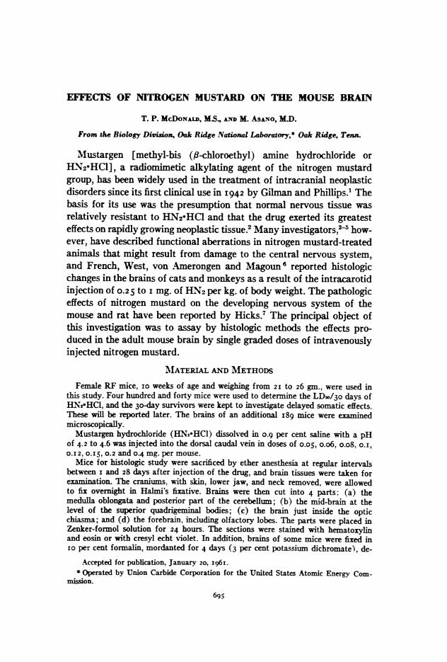
the highest dose there was observed a neuronal necrosis that resembledSpielmeyer's "homogenisierende Ganglienzellnekrose" (homogenizingganglion cell necrosis), an important ischemic cell change appearingchiefly in Purkinje cells, the olivary nucleus, and the fascia dentata.8Hemorrhage and Focal Necrosis. As illustrated in Figure 2, hemor-
rhage was localized in the dorso-lateral neocortex and the pyriformcortex, especially in the deeper cortical laminas, in Sommer's sector ofthe hippocampus, in the ventro-lateral part of the medulla oblongata,and in both white matter and granular layer of the cerebellum (Figs.3 and 4). Focal necrosis occurred only in the deeper cortical laminas andSommer's sector of the hippocampus from the fourth day in the O.I5mg. group to the sixth day for lower doses. In some mice of the O.12 mg.and O.I mg. groups, hemorrhage occurred at the hypothalamic region ofthe midbrain and forebrain on the sixth or seventh day. The hemorrhagiclesions were somewhat similar in appearance in all groups and variedfrom extravasation of red cells, accompanied by a perivascular gather-ing of phagocytes, to multiple hemorrhages of varied sizes. A slightedema was present in the areas of hemorrhage. The regions of necrosiswere usually accompanied by hemorrhage and disappearance of nervecells, with only a few neutrophils in the area. When focal necrosis andhemorrhage occurred in the white matter, myelinated fibers were intactin the hemorrhagic region; in the regions of necrosis, however, myelinand axis cylinders were destroyed and the myelin was phagocytized.Neuronophagia was not noted, nor was fibrovascular organization pres-ent. Cysts were noted on the sixth to twelfth days in the O. I 5 mg. group,on the twelfth to I4th days in the O.12 mg. group, and on the tenth dayin the o.i mg. group. Numerous fat-laden macrophages were present atthe peripheries of the cysts, and many newly proliferated capillariesand proliferating oligodendroglia and microglia appeared in the areasurrounding the cysts. The endothelium of capillaries was swollen andshowed proliferation (Fig. 5), and thrombi were evident in somecapillaries in the pericystic tissues (Fig. 6). In areas surrounding cystsin animals that survived for 12 days in the O.12 and O.I5 mg. groups,microglia and astrocytes were hyperplastic, newly formed vessels wereproliferating, and perivascular lymphocytic infiltration was pronounced(Fig. 7).Patchy swelling of myelinated fibers was seen on the medial lemniscus,
the posterior spino-cerebellar tract of the medulla oblongata, and thewhite matter of the cerebellum on the first and second days in the 0.I5mg. group.No hemorrhage or focal necrosis were noted in any of the controls.Neuronal "Homogenisierende" Change. The neuronal "homogeni-
699June., I96i

MC DONALD AND ASANO
sierende" change was noted only in the 0.I 5 mg. group and occurred onthe fourth to sixth day after treatment (Fig. 8). The cytoplasm of nervecells on the ventro-lateral and lateral regions of the medulla oblongataand superior olivary nuclei had a glassy appearance and was swollen,and nuclei were irregularly shrunken or the nuclear membrane showedmembranolysis. Chromatin granules also appeared denser. No neuro-nophagy was seen.
Selective Neuronal Shrinkage. The selective neuronal shrinkage wasvery constant in the pyramidal layer of the hippocampus, especially inSommer's sector, on the first to fourth days in the O.I5 mg. group andthen again after the twelfth day, on the second to i7th days in the O.Img. group, and within 6 hours to the 2 Ist day in the 0.05 mg. group. Theselective neuronal shrinkage often appeared in the olivary nucleus ofthe medulla oblongata by the tenth or twelfth day in the O.I5 mg. and0.12 mg. groups, on the seventh to I7th days in the O.I mg. group, andon the i4th to 2Ist day in the 0.05 mg. group (Fig. 9). The cytoplasmof shrunken nerve cells became sharply eosinophilic, the nucleus small,homogeneous, and poorly defined. Whereas all treated animals exhibitedneuronal shrinkage, which usually affected many neurons in a given area(Fig. 9), this change was noted in but two controls, in which only oc-casional neurons were affected.
DISCUSSION
Many investigators2-5'll have reported physiologic changes thatappeared to have arisen from neurologic damage caused by nitrogenmustard intoxication. The symptoms recorded include convulsive run-ning movements, salivation, urination, defecation, lacrimation, depres-sion, and kinetic tremors. Only French and co-workers6 describedpathologic lesions in the adult brain attributable to HN2-HCl intoxica-tion. Their observations were: edema, hemorrhage, neuronal and glialdegeneration, and demyelination.The above-listed physiologic and neurologic alterations are, in gen-
eral, similar to those reported here except that convulsive running move-ments were not observed by us. We did, however, observe spontaneousconvulsions. Another symptom seen in our mice and previously reportedby Anslow, Karnofsky, Jager and Smith,9 and Sternberg, Philips andScholler5 was marked hyperirritability, as evidenced by an abnormalresponsiveness to sound and handling.
Goldin, Noe, Landing, Shapiro and Goldberg 2 reported on the neuro-logic syndrome in mice intoxicated with various chlorinated tertiaryamines. Although their work did not deal specifically with methyl-bis(13-chloroethyl) amine, they did investigate the effects of several bis
Vol. 38, No. 6700

NITROGEN MUSTARD
(,-chloroethyl) amines and concluded that these compounds did not pro-duce the "waltzing syndrome," which they described as "hyperactivity,retropulsion, choreic head movement, running in circles, incoordination,poor balance, poor righting reflex, and uncoordinated swimming pat-tern." Our results are not in complete agreement with these findingssince we did observe a lack of muscular coordination.
Demyelination, as described by French and co-workers,6 was not ap-parent in this investigation, and the thrombus formation they noted asan early lesion occurred only after development of brain necrosis in ourmice. The localized pathologic alterations in the mouse brain weresimilar to those seen in anoxic brains (Fig. I; Table I). These includedhemorrhage, necrosis, "homogenisierende" change, and shrinkage of thenerve cells of the neocortex, pyriform cortex, hippocampus, cerebellum,and medulla oblongata. The reasons for this localized distribution ofinjury remain obscure, but may have resulted from local variation intissue vulnerability or vascular factors.13The lethal doses of nitrogen mustard found here (Text-fig. 2) are
similar in magnitude to those previously reported.5910'14 The extent towhich brain damage contributed to death is being investigated in a com-parative study of damage to all tissues and organs. Appearance of severebrain damage at the time of most frequent deaths suggests a causal con-nection.
Su&ARYWhen various doses of methyl-bis ( -chloroethyl) amine hydrochlo-
ride (HN2.HCI) were injected intravenously into albino mice, injury tothe brain was severe and proportional to dose. The LD5o/3o days ofnitrogen mustard was found to be O.127 mg. per mouse (95 per centconfidence interval of 0.I03 to O.I62). Brains of mice receiving doseslarger than the LD5o/30 days (O.I5 mg. per mouse) showed neuronalshrinkage, localized neuronal "homogenisierende" changes, severe hem-orrhage, and multiple foci of necrosis; doses near the LDso/3o days (o.iand O.12 mg. per mouse) produced selective neuronal shrinkage in theearly stages and hemorrhage and focal necrosis later. At lower doses(o.os mg. per mouse), the most significant damage was shrinkage ofthe nerve cells. In a sagittal plane, the sites of predilection for braindamage in all groups were the neocortex, pyriform cortex, hippocampus,cerebellum, and medulla oblongata. Brain damage and death rate forthe high-dose groups (above o.os mg. per mouse) reached maximumon days 6 to 8. The following signs and symptoms were also observedin intoxicated animals: extreme emaciation, ruffling of fur, inactivity,decreased muscular coordination, hyperirritability, spontaneous con-vulsions, diarrhea, and lacrimation.
JuneC, I96z 70I

702 MC DONALD AND ASANO Vol. 38, No. 6
REFERENCES
i. GILMAN. A., and PHILps, F. S. The biological actions and therapeutic appli-cations of the ,-chloroethyl amines and sulfides. Science, I946, 103, 409-415.
2. KARNOFSEY, D. A. Suxmmary of results obtained with nitrogen mustard in thetreatment of neoplastic disease. Ann. New York Acad. Sc., I958, 68, 899-914.
3. GRAEF, I.; KARNOFSKY, D. A.; JAGER, V. B.; KRICSKY, B., and SMiTH, H. W.The clinical and pathologic effects of the nitrogen and sulfur mustards inlaboratory animals. Am. J. Path., I948, 24, I-47.
4. KINDRED, J. E. Histologic changes occurring in the hemopoietic organs ofalbino rats after single injections of 2-chloroethyl vesicants; a quantitativestudy. Arch. Path., 1947, 43, 253-295.
5. STERNBERG, S. S.; PHILIPs, F. S., and SCHOLLER, J. Pharmacological andpathological effects of alkylating agents. Ann. New York Acad. Sc., I958, 68,8i I-825.
6. FRENCH, J. D.; WEST, P. M.; VON AMERONGEN, F. K., and MAGOUN, H. W.Effects of intracarotid administration of nitrogen mustard on normal brainand brain tumors. I. Neurosurg., I952, 9, 378-389.
7. HICKS, S. P. The effects of ionizing radiation, certain hormones and radiomi-metic drugs on the developing nervous system. J. CeM. & Comp. Physiol.,I954, 43, Suppl. I, I51-I 78-
8. ScHozL, W. Die nicht zur Erweichung fiihrenden unvolistindigen Gewebs-nekrosen. (Elektive Parenchymnekrose.) In: Handbuch der speziellen pathol-ogischen Anatomie umd Eistologie. Vol. XIIIIi, Part B, Nervensystem,Erkrankungen der zentralen Nervensystems I. LUBARsCH, 0.; HENKE, F., andR6ssiu, R. (eds.). Springer-Verlag, Berlin, 1957, P. I304.
9. ANsLow, W. P., JR; KARNOFSKY, D. A.; JAGER, B. V., and SITH, H. W. Thetoxicity and pharmacological action of the nitrogen mustards and certain re-lated compounds. J. Pharmacol. & Exper. Therap., I947, 91, 224-235.
io. BOYLAND, E. The toxicity of alkyl-bis (P-chloroethyl) amines and of theproducts of their reaction with water. Brit. J. Pharmacol., I946. I, 247-254.
i i. BRA%-D E. D.; HARRis, T. D.; GOODMAN, L. S., and BoRISoN, H. L. Locus ofemetic action of nitrogen mustard (HN2). (Abstract) Fed. Proc., 1953, 12,303.
I2. GOLDIN, A.; NOE, H. A.; LANDING, B. H.; SHAPRo, D. M., and GOLDBERG, B.A neurological syndrome induced by administration of some chlorinatedtertiary amines. J. Pharmacol. & Exper. Therap., I948. 94, 249-26i.
I3. SPIELMEYER, W. The significance of local factors for electivity in centralnervous system disease processes. Medicine, I93I, I0, 243-256.
I4. WarrE, L. P. Influence of pH on the toxicity of nitrogen mustard. Science,I960, 131, I04I-I043.
The authors are grateful to Dr. A. C. Upton and Dr. T. T. Odell, Jr., for technicaladvice and consultation, and to Mr. W. D. Gude for histologic assistance. It is also a pleasureto thank Dr. M. A. Kastenbaum of the Mathematics Panel, Oak Ridge National Laboratory,for statistical analysis of data.

NITROGEN MUSTARD
[Ilustration folow ]
703Jcs, zq6

MC DONALD AND ASANO
LEGENDS FOR FIGURES
Except where indicated, photographs were prepared from sections stained withhemtoxylin and eosin.
FIG. I. Cross sections of a control brain, showing sites of predilection of:
[I] shrinkage of nerve cells;
z hemorrhage;
En necrosis;
hemorrhage and necrosis.
(a) Cross section of forebrain; (b) a section through the optic chiasma at thediencephalon; (c) a cross section through the level of the rostral pole at the habenularcomplex; and (d) a cross section of medulla oblongata and cerebellum. X io.
Vol. 38, No. 6704

June, I96I NITROGEN MUSTARD 705
-a
u

706 MC DONALD AND ASANO
FIG. 2. Hemorrhage and necrosis in the hippocampus. X I4.
FIG. 3. Hemorrhage and necrosis in Sommer's sector of the hippocampus. X 360.
FIG. 4. Hemorrhage in the medulla oblongata. X I40.
FIG. 5. Malacic cysts in the hippocampus. x I4O.
V'ol. 38, No. 6
June, I96I NITROGEN MUSTARD 707
3
5
. 'sisa4
D ' . '.
2
4
T5 ; -,
W.*:. q\e
t;
"...- IC -
z "P -.w 4.. V.W -0...Mz ..,: 0 -'. isr.,,; .-.7 -7" 'N I .-
,R. I .4,
708 MC DONALD AND ASANO 'ol. 38, No. 6
FIG. 6. Thrombus and surrounding erythrocytes in a malacic cyst of the hippo-campus. X 380.
FIG. 7. Proliferation of microglia and astrocytes in the hippocampus. X 380.
FIG. 8. Spielmeyer's "/omogenisierende" necrosis in the medulla oblongata. X 560.FIG. 9. Shrinkage of nerve cells in the medulla oblongata. Nissl stain. X 56o.

June, Ig6I NITROGEN MUSTARD 709
6
oI 1
'.'
,...4
01
.aS
8 9
-.. '111,10.",". -.- "a,