duchenne muscular dystrophy: an historical treatment review
TRANSCRIPT
579
https://doi.org/10.1590/0004-282X20190088
VIEW AND REVIEW
Duchenne muscular dystrophy: an historical treatment reviewDistrofia muscular de Duchenne: revisão histórica do tratamentoLineu Cesar WERNECK1, Paulo José LORENZONI1, Renata Dal-Prá DUCCI1, Otto Hernández FUSTES1, Cláudia Suemi Kamoi KAY1, Rosana Herminia SCOLA1
Duchenne muscular dystrophy (DMD) is a progressive hereditary muscular disease with X-linked recessive inheri-tance, occurring mainly in males. Muscular impairment is ini-tially in the proximal muscles of the lower limbs with reduced muscle strength and progressive contractures with gait impair-ment. Subsequently, with reduced muscle strength in the upper limbs, the patients develop contractures in the arms, and impairment of the respiratory and heart muscles. Although there is no effective therapy, most patients currently walk until age 13, some even until age 20, and some survive for more than 30 years due to ventilatory and cardiac support1.
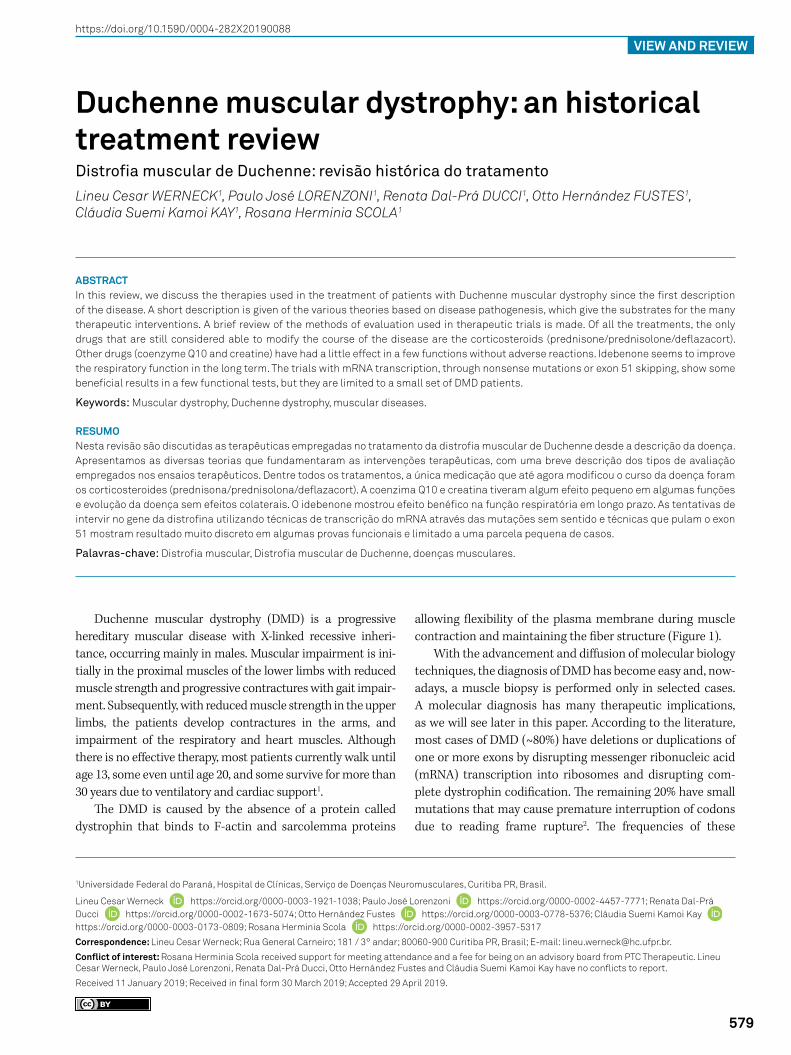
The DMD is caused by the absence of a protein called dystrophin that binds to F-actin and sarcolemma proteins
allowing flexibility of the plasma membrane during muscle contraction and maintaining the fiber structure (Figure 1).
With the advancement and diffusion of molecular biology techniques, the diagnosis of DMD has become easy and, now-adays, a muscle biopsy is performed only in selected cases. A molecular diagnosis has many therapeutic implications, as we will see later in this paper. According to the literature, most cases of DMD (~80%) have deletions or duplications of one or more exons by disrupting messenger ribonucleic acid (mRNA) transcription into ribosomes and disrupting com-plete dystrophin codification. The remaining 20% have small mutations that may cause premature interruption of codons due to reading frame rupture2. The frequencies of these
1Universidade Federal do Paraná, Hospital de Clínicas, Serviço de Doenças Neuromusculares, Curitiba PR, Brasil.
Lineu Cesar Werneck https://orcid.org/0000-0003-1921-1038; Paulo José Lorenzoni https://orcid.org/0000-0002-4457-7771; Renata Dal-Prá Ducci https://orcid.org/0000-0002-1673-5074; Otto Hernández Fustes https://orcid.org/0000-0003-0778-5376; Cláudia Suemi Kamoi Kay https://orcid.org/0000-0003-0173-0809; Rosana Herminia Scola https://orcid.org/0000-0002-3957-5317
Correspondence: Lineu Cesar Werneck; Rua General Carneiro; 181 / 3° andar; 80060-900 Curitiba PR, Brasil; E-mail: [email protected].
Conflict of interest: Rosana Herminia Scola received support for meeting attendance and a fee for being on an advisory board from PTC Therapeutic. Lineu Cesar Werneck, Paulo José Lorenzoni, Renata Dal-Prá Ducci, Otto Hernández Fustes and Cláudia Suemi Kamoi Kay have no conflicts to report.
Received 11 January 2019; Received in final form 30 March 2019; Accepted 29 April 2019.
ABSTRACTIn this review, we discuss the therapies used in the treatment of patients with Duchenne muscular dystrophy since the first description of the disease. A short description is given of the various theories based on disease pathogenesis, which give the substrates for the many therapeutic interventions. A brief review of the methods of evaluation used in therapeutic trials is made. Of all the treatments, the only drugs that are still considered able to modify the course of the disease are the corticosteroids (prednisone/prednisolone/deflazacort). Other drugs (coenzyme Q10 and creatine) have had a little effect in a few functions without adverse reactions. Idebenone seems to improve the respiratory function in the long term. The trials with mRNA transcription, through nonsense mutations or exon 51 skipping, show some beneficial results in a few functional tests, but they are limited to a small set of DMD patients.
Keywords: Muscular dystrophy, Duchenne dystrophy, muscular diseases.
RESUMONesta revisão são discutidas as terapêuticas empregadas no tratamento da distrofia muscular de Duchenne desde a descrição da doença. Apresentamos as diversas teorias que fundamentaram as intervenções terapêuticas, com uma breve descrição dos tipos de avaliação empregados nos ensaios terapêuticos. Dentre todos os tratamentos, a única medicação que até agora modificou o curso da doença foram os corticosteroides (prednisona/prednisolona/deflazacort). A coenzima Q10 e creatina tiveram algum efeito pequeno em algumas funções e evolução da doença sem efeitos colaterais. O idebenone mostrou efeito benéfico na função respiratória em longo prazo. As tentativas de intervir no gene da distrofina utilizando técnicas de transcrição do mRNA através das mutações sem sentido e técnicas que pulam o exon 51 mostram resultado muito discreto em algumas provas funcionais e limitado a uma parcela pequena de casos.
Palavras-chave: Distrofia muscular, Distrofia muscular de Duchenne, doenças musculares.
580 Arq Neuropsiquiatr 2019;77(8):579-589
changes vary depending on the location of the study and are important for the modern therapeutic approach.
In this review, an ‘historical analysis’ on the published therapies for DMD patients briefly reports the main results of the completed trials and their backgrounds. Experimental research that is ongoing, but not yet applied to DMD patients (e.g. CRISPR-Cas9) has not been included. This review details the few therapies that have had a beneficial response in the DMD patients. In addition, this review may also help physi-cians understand why some therapies were abandoned while others will be emerging on the horizon in the near future.
METHODS FOR EVALUATING TREATMENT RESULTS
Since the earliest descriptions of DMD, several thera-peutic modalities have been employed, some empirically and others based on reports of biochemical or metabolic abnormalities from human and animal studies. Some papers have reported promising results, but the meth-ods of evaluation varied widely and generally did not have a placebo group. This led some researchers to study the natural history of DMD. One of the first parameters used was the age of gait loss resulting from reduction of
muscular strength. To obtain a quantitative evaluation of the muscle strength reduction, the Medical Research Council scale (quantitative manual muscle testing force) was used, with small modifications. Several muscles were examined, given a score and then summed3. Functional tests have also been developed, where certain activities are requested and the time to execution measured and correlated to the age for that function, or the baseline values compared with the use of the drug being studied. Among the functions evaluated are: time to get up from the floor and stand up; Gowers’ maneuver; keeping the arms elevated; chair lift; climbing four steps, going down four steps; time to walk 10 meters; the 6-Minute Walk Test (6MWT – distance walked in six minutes), among others. These tests can be analyzed in isolation or grouped as in the North Star Ambulatory Assessment4.
TREATMENTS AND THEIR RESULTS
Several treatment modalities have been grouped accord-ing to the knowledge of the disease at the time they were evaluated (Table).
ElectrostimulationSince 1861, Duchenne had been applying electrical stimu-
lation in patients with paralysis of any nature but there are no reliable descriptions of the response. The reports of chronic low-frequency electrical stimulation in DMD patients have found some benefit, but only in a few patients and the fol-low-up was not reported5. In 1997, it was experimentally demonstrated that electrical stimulation caused greater accumulation of intracellular calcium, leading to premature degeneration of the fiber by possible activation of the pro-teases, and could accelerate the degeneration of the muscle fiber. After this discovery, enthusiasm for electrical stimula-tion was lost and it was no longer studied6.
Figure 1. Immunohistochemical stain for dystrophin. A. Normal muscle with dystrophin in the sarcolemma. B. Absence of dystrophin in the sarcolemma of muscle fibers of Duchenne muscular dystrophy.
A B
Table. Therapies used for Duchenne muscular dystrophy.
Main mechanism Therapy
Electrostimulation Electricity
Cholinesterase Inhibitors Galantamine
Drugs acting on biochemical, metabolic and oxidative stress Laevadosin, Allopurinol, vitamin D and Calcium, Creatine and Glutamine, Coenzyme Q10, Idebenone
Drugs acting on growth, height and muscular function Mazindol, Growth Hormone, Isaxonine, Anabolics (Sinestrol and Oxandrolone), Albuterol
Drugs producing changes in sarcolemma and calcium aggregate Verapamil, Flunarizine, Nifedipine, Diltiazem
Drugs interfering in the muscle blood flow Methysergide, Tadalafil
Conjunctive tissue proliferation Penicillamine, Pentoxifylline
Inflammatory reaction Azathioprine, Cyclosporine
Corticosteroids Prednisone, Prednisolone, Deflazacort
Restoration of premature termination codon Gentamicin, Ataluren
Exon Skipping Drisapersen, Eteplirsen
581Werneck LC et al. Treatment of Duchenne muscular dystrophy over time
CHOLINESTERASE INHIBITORS
GalantamineGalantamine is an anticholinesterase drug with action
in the central and peripheral nervous systems. It prolongs the action of the acetylcholine in the receptor located in the myoneural junction. An attempt to use galantamine in DMD patients was made, with no apparent response7.
ANTIOXIDANT DRUGS ACTING IN BIOCHEMICAL AND METABOLIC PATHWAYS
Muscle injury in muscular dystrophies of animals and humans induces several metabolic abnormalities, with increases in free radical levels, and subsequent oxidative stress and changes in others muscle fiber components. This raised the possibility that antioxidant therapy or the replace-ment of abnormally low substances found in DMD may have some effect on the disease progression.
LaevadosinLaevadosin is a mixture of nucleotides and nucleosides
that supposedly could have some effect on DMD patients, as reported by Thomson and Guest in 19638. However, a study with 20 DMD patients, of whom 10 were controls, over a period of 42 days, did not show any benefit with respect to muscle strength compared with placebo, although it reduced creatine kinase in some patients9.
AllopurinolConsidering the hypothesis of a defect in purine metab-
olism, allopurinol was tried to facilitate the transport of purines. The first study, by Thomson and Smith in 197810 had 16 DMD patients who used allopurinol for 18 weeks. Some patients improved strength for more than six months compared with placebo. These data have subsequently not been confirmed11.
Vitamin D and calciumThe role of calcium in the physiology and function of
muscle fiber has been known for long time. It was found that DMD patients had a vitamin D deficiency compared with controls. This deficiency may be the cause of osteo-penia and a higher incidence of fractures found in patients with DMD, and which is aggravated by the use of corticoste-roids. A correlation was found between muscle strength and reduction of bone mass, as well as reduction of bone mass and the cumulative dose of corticosteroids. Administration of vitamin D with or without alendronate has shown an increase in bone mass but the effect on fracture reduction was uncertain and there was no change in the functional progression of the disease12.
Creatine and glutamineAfter the demonstration of creatine reduction in DMD
muscle and the possible beneficial effect, supplementation with creatine 0.10 g/kg/day was tested in a double-blind study with 30 DMD patients over a period of four months, where half of the patients were using corticosteroids. There was an increase in grip strength (p < 0.05) in the dominant hand and strength in general (p = 0.056), with no improvement in activi-ties of daily living13. Another attempt with creatine was made in 50 DMD patients (16 on placebo, 15 taking creatine 5 g/day and 19 taking glutamine 0.3 mg/kg/day). The creatine and glutamine groups had minor deterioration in manual muscle strength tests. In the functional stair-climbing test, the group that used creatine had a better performance (p = 0.015). These drugs were safe and well tolerated in the prescribed doses, but the impact on long term survival was not studied14.
Coenzyme Q10Based on studies in animals, a therapeutic approach was
taken by Folkers and Simonsen in 199515, using coenzyme Q10 in one DMD patient, and other neuromuscular disor-ders. The patients become more active and could walk and run better. After this initial study, coenzyme Q10 was used in 13 DMD patients using corticosteroids, with an initial dose of 400 mg, with escalations of 100 mg/day. Improvements on several muscular and functional tests during the study period (six months) were observed16. These data were corroborated in patients using corticosteroids and various vitamin and dietary supplements, through multivariate analysis. In these patients, the duration of walking increased in 155 patients with coen-zyme Q10 (p = 0.007) and in 246 patients who used vitamin D (p = 0.004)17. The long-term survival was not established.
IdebenoneIdebenone is a synthetic analogue of coenzyme Q10 with
similar effects and could be useful in respiratory function. It was studied over 52 weeks in 31 DMD patients com-pared with 33 participants on placebo, aged between 10 and 18 years, in a dose of 900 mg/day, testing several parameters of respiratory function. The patients who used the idebenone improved their peak inspiratory flow (L/min), forced expira-tory volume, forced expiratory volume in one second, and had a smaller decline in peak inspiratory flow (2.5% vs 6.27%, p = 0.031). These data suggest some preservation of pulmo-nary muscle function in the long term18.
DRUGS ACTING ON GROWTH, HEIGHT AND MUSCULAR FUNCTION
In 1981, Zatz et al. described a DMD patient with slower progression of the disease associated with growth hormone deficiency19. This opened up the possibility of using growth hormone inhibitors as a therapeutic option.
582 Arq Neuropsiquiatr 2019;77(8):579-589
MazindolMazindol is an inhibitor of growth hormone. In 1988,
Coakley et al., used mazindol in DMD patients20. They did not find any significant changes in muscle force or func-tional tests. This study was interrupted due to the side effects. Another study with 83 DMD patients (3 mg/day for 12 months against placebo) was done and no benefit was observed in relation to muscle strength, contractures or functional tests21.
Growth hormoneOver the years, it has been found that one of the side
effects of corticosteroids was the reduction of the growth of patients with DMD. Growth hormone was administered for one year in patients using corticosteroids. This increased the rate of growth, improving some motor functions without side effects or change in cardiac function22. However, as the patient grows, the muscle mass and weight increases, with-out improvement of muscle strength. This may impair the gait in some patients.
IsaxonineIsaxonine is a synthetic compound that, in animals,
accelerates nerve regeneration and increases the percent-age of plasma membrane proteins in muscle fibers. A double-blind study was performed over a period of two years, using 20 patients with ambulatory DMD, who were aged 5.5 to 10 years. There was no significant difference in disease pro-gression compared with placebo23.
SinestrolThe fact that anabolic steroids accelerate muscle growth
led a Russian author to try sinestrol in 15 DMD patients, compared with 14 controls over three weeks. Six months after withdrawal of sinestrol, they found that the disease con-tinued to progress, although less so in the treated group24.
OxandroloneOxandrolone is an anabolic steroid that can promote
growth with minimal toxicity. It facilitates weight gain in chronic debilitation and promotes growth in boys with con-stitutional delay and may increase muscle protein synthesis. Oxandrolone was tested in DMD patients in a double-blind, six-month study, at a dose of 0.1 mg/kg/day in 26 patients, and 25 controls receiving placebo before the use of corticoste-roids. Those who used oxandrolone had a better performance, but this was not statistically significant (p = 0.13). When only strength in the upper limbs was analyzed, the treated group showed stabilization (p = 0.005). In this study, the authors thought it may be useful before starting corticosteroids25.
AlbuterolAlbuterol is a beta-2a adrenergic receptor agonist that,
theoretically, would maintain the protein structure and
indirectly act as an anabolic effect. Albuterol was used in 14 DMD patients (seven albuterol vs seven placebo) at a dose of 12 mg/day for 12 weeks. For comparison, muscle strength was tested manually and using several functional tests. In the short term, the patients who used albuterol increased lean body mass, and improved running time (p = 0.025), but there was no change in muscle strength observed26. This small impact on the long-term survival of DMD patients was also responsible for there being no cur-rent use of this drug in DMD.
DRUGS PRODUCING CHANGES IN SARCOLEMMA AND CALCIUM ACCUMULATION
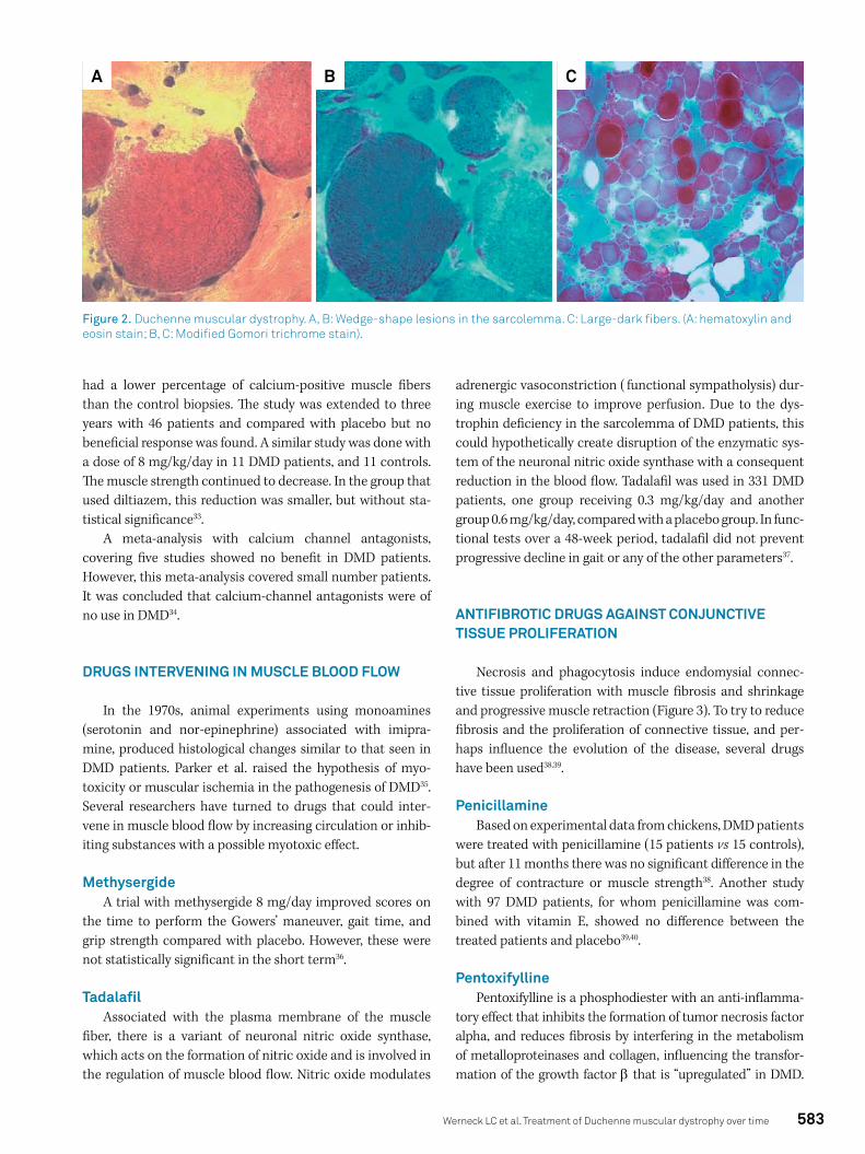
In 1975, Mokri and Engel described wedge-shape lesions with failure in the plasma membrane of muscle fibers in patients with DMD27. This disruption of the membrane allowed the output or the ingress of material, creating abnor-malities in the underlying cellular organelles and myofibrils. One of these substances accumulated in large-dark fibers was calcium, which can activate the proteolytic enzymes leading to necrosis28 (Figure 2). This finding led to the investi-gation of calcium antagonists in DMD.
VerapamilVerapamil was used in DMD patients (three patients
on verapamil vs five on placebo) using electronic ergom-eters as a method of evaluation. The three patients who used verapamil improved in strength, but it was not a “dra-matic” effect. Over time, the DMD patients increased their PR interval on an electrocardiogram and, as a precaution, the drug was discontinued29.
FlunarizineThe effect of flunarizine on DMD patients was evaluated
in a double-blind controlled study of 27 patients (13 on med-ication) for one year. No difference was found between the treated patients when compared with the placebo group. Some patients in the flunarizine group had a faster func-tional decline in their lower limbs30.
NifedipineNifedipine was administered in 105 DMD patients, at a
dosage of 0.75-1.0 mg/kg/day for the first six months and then 1.5-2.0 mg/kg/day for another 12 months, which was then compared with a placebo group. No statistically significant difference was found in any of the analyzed parameters31.
DiltiazemIn 1988, Pernice et al. studied the therapy with diltiazem
at a dose of 5 mg/kg in DMD patients (13 patients vs 13 con-trols)32. The initial study lasted one year and no difference was found in all analyzed parameters, but the diltiazem group
583Werneck LC et al. Treatment of Duchenne muscular dystrophy over time
had a lower percentage of calcium-positive muscle fibers than the control biopsies. The study was extended to three years with 46 patients and compared with placebo but no beneficial response was found. A similar study was done with a dose of 8 mg/kg/day in 11 DMD patients, and 11 controls. The muscle strength continued to decrease. In the group that used diltiazem, this reduction was smaller, but without sta-tistical significance33.
A meta-analysis with calcium channel antagonists, covering five studies showed no benefit in DMD patients. However, this meta-analysis covered small number patients. It was concluded that calcium-channel antagonists were of no use in DMD34.
DRUGS INTERVENING IN MUSCLE BLOOD FLOW
In the 1970s, animal experiments using monoamines (serotonin and nor-epinephrine) associated with imipra-mine, produced histological changes similar to that seen in DMD patients. Parker et al. raised the hypothesis of myo-toxicity or muscular ischemia in the pathogenesis of DMD35. Several researchers have turned to drugs that could inter-vene in muscle blood flow by increasing circulation or inhib-iting substances with a possible myotoxic effect.
MethysergideA trial with methysergide 8 mg/day improved scores on
the time to perform the Gowers’ maneuver, gait time, and grip strength compared with placebo. However, these were not statistically significant in the short term36.
TadalafilAssociated with the plasma membrane of the muscle
fiber, there is a variant of neuronal nitric oxide synthase, which acts on the formation of nitric oxide and is involved in the regulation of muscle blood flow. Nitric oxide modulates
adrenergic vasoconstriction ( functional sympatholysis) dur-ing muscle exercise to improve perfusion. Due to the dys-trophin deficiency in the sarcolemma of DMD patients, this could hypothetically create disruption of the enzymatic sys-tem of the neuronal nitric oxide synthase with a consequent reduction in the blood flow. Tadalafil was used in 331 DMD patients, one group receiving 0.3 mg/kg/day and another group 0.6 mg/kg/day, compared with a placebo group. In func-tional tests over a 48-week period, tadalafil did not prevent progressive decline in gait or any of the other parameters37.
ANTIFIBROTIC DRUGS AGAINST CONJUNCTIVE TISSUE PROLIFERATION
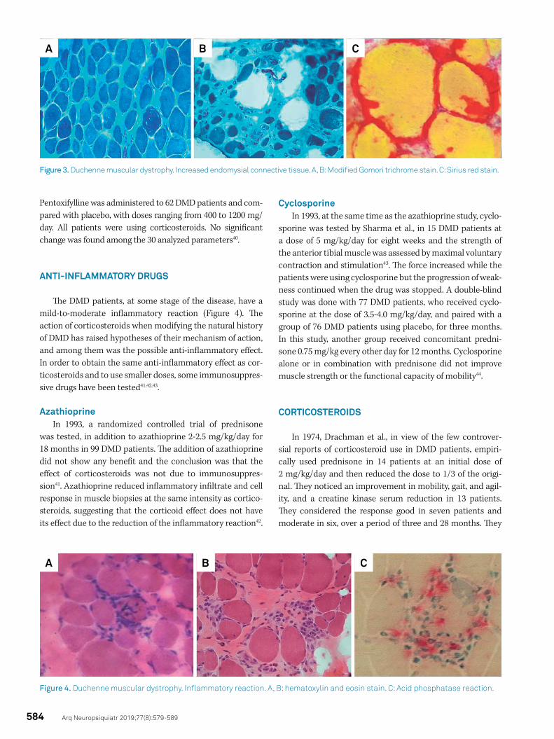
Necrosis and phagocytosis induce endomysial connec-tive tissue proliferation with muscle fibrosis and shrinkage and progressive muscle retraction (Figure 3). To try to reduce fibrosis and the proliferation of connective tissue, and per-haps influence the evolution of the disease, several drugs have been used38,39.
PenicillamineBased on experimental data from chickens, DMD patients
were treated with penicillamine (15 patients vs 15 controls), but after 11 months there was no significant difference in the degree of contracture or muscle strength38. Another study with 97 DMD patients, for whom penicillamine was com-bined with vitamin E, showed no difference between the treated patients and placebo39,40.
PentoxifyllinePentoxifylline is a phosphodiester with an anti-inflamma-
tory effect that inhibits the formation of tumor necrosis factor alpha, and reduces fibrosis by interfering in the metabolism of metalloproteinases and collagen, influencing the transfor-mation of the growth factor β that is “upregulated” in DMD.
Figure 2. Duchenne muscular dystrophy. A, B: Wedge-shape lesions in the sarcolemma. C: Large-dark fibers. (A: hematoxylin and eosin stain; B, C: Modified Gomori trichrome stain).
A B C
584 Arq Neuropsiquiatr 2019;77(8):579-589
Pentoxifylline was administered to 62 DMD patients and com-pared with placebo, with doses ranging from 400 to 1200 mg/day. All patients were using corticosteroids. No significant change was found among the 30 analyzed parameters40.
ANTI-INFLAMMATORY DRUGS
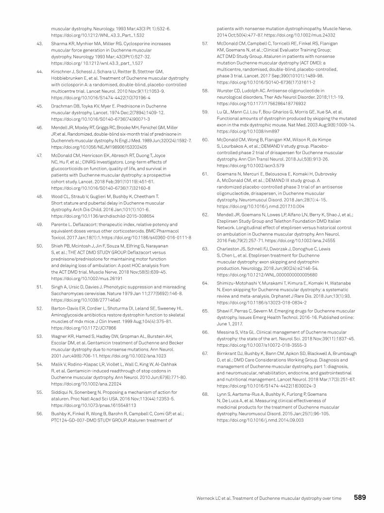
The DMD patients, at some stage of the disease, have a mild-to-moderate inflammatory reaction (Figure 4). The action of corticosteroids when modifying the natural history of DMD has raised hypotheses of their mechanism of action, and among them was the possible anti-inflammatory effect. In order to obtain the same anti-inflammatory effect as cor-ticosteroids and to use smaller doses, some immunosuppres-sive drugs have been tested41,42,43.
AzathioprineIn 1993, a randomized controlled trial of prednisone
was tested, in addition to azathioprine 2-2.5 mg/kg/day for 18 months in 99 DMD patients. The addition of azathioprine did not show any benefit and the conclusion was that the effect of corticosteroids was not due to immunosuppres-sion41. Azathioprine reduced inflammatory infiltrate and cell response in muscle biopsies at the same intensity as cortico-steroids, suggesting that the corticoid effect does not have its effect due to the reduction of the inflammatory reaction42.
CyclosporineIn 1993, at the same time as the azathioprine study, cyclo-
sporine was tested by Sharma et al., in 15 DMD patients at a dose of 5 mg/kg/day for eight weeks and the strength of the anterior tibial muscle was assessed by maximal voluntary contraction and stimulation43. The force increased while the patients were using cyclosporine but the progression of weak-ness continued when the drug was stopped. A double-blind study was done with 77 DMD patients, who received cyclo-sporine at the dose of 3.5-4.0 mg/kg/day, and paired with a group of 76 DMD patients using placebo, for three months. In this study, another group received concomitant predni-sone 0.75 mg/kg every other day for 12 months. Cyclosporine alone or in combination with prednisone did not improve muscle strength or the functional capacity of mobility44.
CORTICOSTEROIDS
In 1974, Drachman et al., in view of the few controver-sial reports of corticosteroid use in DMD patients, empiri-cally used prednisone in 14 patients at an initial dose of 2 mg/kg/day and then reduced the dose to 1/3 of the origi-nal. They noticed an improvement in mobility, gait, and agil-ity, and a creatine kinase serum reduction in 13 patients. They considered the response good in seven patients and moderate in six, over a period of three and 28 months. They
Figure 3. Duchenne muscular dystrophy. Increased endomysial connective tissue. A, B: Modified Gomori trichrome stain. C: Sirius red stain.
A B C
Figure 4. Duchenne muscular dystrophy. Inflammatory reaction. A, B: hematoxylin and eosin stain. C: Acid phosphatase reaction.
A B C

585Werneck LC et al. Treatment of Duchenne muscular dystrophy over time
concluded that prednisone prolonged the period of ambula-tion and improved the patients’ quality of life45.
Even without controlled studies, the prescription of prednisone in DMD patients was diffused, corroborated by the clinical practice of apparent improvement. In 1989, a placebo-controlled trial of 103 patients using prednisone 0.75 and 1.5 mg/kg/day for six months, showed improvement in strength and functional activities in DMD patients46. Since then, prednisone has routinely been prescribed because the benefits are greater than the side effects.
Cooperative studies with a greater number of patients (440) confirmed that corticosteroid administration prolongs muscle function in the upper and lower limbs, maintaining the strength for longer, helping in day-to-day functions, walk-ing for a longer time, improving quality of life and survival. In addition, it has beneficial effects on pulmonary ventilation and cardiomyopathy47.
The largest study on the action of corticosteroids enrolled 5,345 DMD patients, 2,658 of whom were continuously using corticosteroids (49.7%), 2,015 had never used (37.7%) and 522 had used corticosteroids in the past (9.8%). Those who used corticosteroids continuously walked for a longer time (three years) when compared with those who did not. The use of corticosteroids significantly reduced the number of scoliosis surgeries, delayed the use of assisted ventilation, but did not influence the incidence of myocardiopathy after 20 years. The single deletion of exon 45 treated with cortico-steroids delayed the loss of gait and increased the survival time compared with the other types of deletions1.
Benefits vs adverse effectsCorticosteroid therapy improves muscle strength by pro-
longing gait time, preserves upper limb function, helps pre-vent scoliosis, reduces cardiomyopathy progression, and delays the need for invasive ventilation, with some patients surviving longer than 30 years. However, chronic use of cor-ticosteroids causes delayed growth and puberty, weight gain, skin changes, a Cushingoid appearance, behavioral disorders, adrenal suppression, reduction of bone mineralization, cata-racts and metabolic changes48. Each of these complications should be monitored and, when they arise, they should be appropriately treated.
Deflazacort versus PrednisoneAlthough there are several corticosteroid drugs, only
prednisone and deflazacort have trials in DMD patients that have been published.
Deflazacort is a synthetic heterocyclic corticosteroid obtained by the fusion of methyloxazoline in the predni-sone structure with great effectiveness and good tolerability. It causes less retention of sodium, has a strong anti-inflam-matory action with immunosuppressive activity, low interfer-ence in carbohydrate metabolism and in the metabolism of calcium and phosphorus49.
Deflazacort may replace prednisone, and a study has shown that the time to loss of the gait in DMD patients was slightly longer, but with increased side effects such as a Cushingoid appearance, erythema, hirsutism, increased weight, nasopharyngitis, delayed puberty, cataracts, and ver-tebral fractures. Even with more side effects, it seems that the deflazacort group had a better functional result, but this war-rants a head-to-head comparison study50.
Despite these studies of side effects, both corticosteroid drugs (prednisone and deflazacort) remain the most impor-tant therapy for DMD patients, extending the duration of gait and delaying the appearance of scoliosis when the patients become wheelchair bound.
RESTORATION OF DYSTROPHIN
Transcription through nonsense mutations
GentamicinIn 1979, Singh et al., found that the addition of aminoglyco-
side antibiotics enabled the researchers to ignore some non-sense mutations in fungal and yeast cultures51. In the following years, it was demonstrated that gentamicin addition promoted the codons of premature termination readthrough in the mdx mouse with a point mutation (nonsense) in the DMD gene, which led to the production of dystrophin and reduction of the serum creatine kinase levels in this animal model52.
Gentamicin was initially tested in two DMD patients and two patients with Becker muscular dystrophy. This study resulted in production of an incomplete truncated dystrophin in these patients, with no change in muscle strength. However, these patients reduced their serum creatine kinase levels and had no side effects53. Gentamicin 7.5 mg/kg/day was given intravenously for 14 days to 10 patients with stop codon in the DMD gene (DMD group) and 10 patients with other frame-shift abnormalities in the DMD gene (control group). The stop codon group had a significant reduction of serum creatine kinase up to day 28. After six months, they showed stabiliza-tion of the force, functional tests without alteration, discrete increase in forced vital capacity (p = 0.06), reduction of serum creatine kinase level (p = 0.04) and an increase of 15% in the dystrophin levels in muscle (p < 0.001)54.
In the following years, with the results of aminoglycosides studies achieving transcription of nonsense mutations in DMD, motivated trials to develop biological molecules with the same properties (e.g. ataluren), to overcome a premature termination codon55.
AtalurenAtaluren is a biological molecule that allows different pre-
mature termination codons readthrough, such as UAG, UAA, and UGA during mRNA transcription. This drug promotes the incorporation of specific amino acid units in premature

586 Arq Neuropsiquiatr 2019;77(8):579-589
termination codons (Glc, Lys, Tyr in the codons UAA and UAG; Trp, Arg and Cys in the UGA codon)55.
One study evaluated therapy with ataluren for 48 weeks in 174 DMD patients. In this trial, DMD patients were divided into three groups: 57 patients in the group using ataluren at a dose of 40 mg/kg/day, 60 patients in the group using atal-uren at a dose of 80 mg/kg/day and 57 patients in the placebo group. Most of the DMD patients were using corticosteroids (71%). The main parameter of evaluation was the 6MWT. The 40 mg/kg/day group reduced the distance by 12.8 meters and the placebo group by 44.1 meters (p = 0.056). The differ-ence in the 80 mg/kg/day group was negligible. However, in the 40 mg/kg/day group, there was a subgroup of patients (less than nine years old, receiving corticosteroids and with a 6MWT baseline less than 350 meters in the beginning) in whom the difference in distance covered (68.2 meters lon-ger than the placebo group) was statistically significant (p = 0.0053). Previous studies had found that DMD patients who walked more than 350 meters in the six minutes did not change significantly in 48 weeks. In the functional tests (run-ning or walking 10 meters, climbing four stairs, descending four stairs, and supine-to-stand), the group that used ata-luren 40 mg/kg/day was better than placebo, although not statistically significant56.
Another study evaluated therapy with ataluren in 230 DMD patients using corticosteroids for longer than six months (115 received ataluren 40 mg/kg/day vs 115 receiv-ing placebo). In this trial, the DMD patients were divided into groups (younger and older than nine years of age) that were analyzed using functional tests and the North Star Ambulatory Assessment (NSAA). In the 6MWT, no group showed a signifi-cant difference. In the subgroup with a baseline greater than 300 meters and less than 400 meters, DMD patients had a sig-nificant improvement after 32 weeks, which increased up to 48 weeks (p = 0.007). In the groups with a baseline walk less than 300 meters and greater than 400 meters, the results were not significant. The beneficial effect of ataluren in relation to gait occurred in 50% (47/114) of the patients. In the NSAA, although not significant, the ataluren group had a better per-formance. In addition, in the subgroup with a baseline walk between 300 and 400 meters, the proportion was 14.3% vs 25.3% in the placebo group (p = 0.010). There was also no dif-ference in quality of life between groups57.
Exon skippingAntisense nucleotides (ASO) are nucleic acid chains of
8-50 bases (oligos). These oligos have a corresponding base in the pre-mRNA and mRNA sequence modulating their func-tion and are additionally ligated (antisense). Antisense nucle-otides bind at specific mRNA sites mimicking DNA-RNA pairing. The sequences skipping the exons induce the inclu-sion of spliced exons. Administration of an ASO that antago-nizes the “natural antisense transcripts” in the mRNA allows the translation of the corresponding protein58.
In 2003, ASO use, in mdx mice with a mutation in exon 23 of the dystrophin gene, induced persistent production of this protein in a large number of muscle fibers, which resulted in the functional improvement of the animals with-out inducing immune responses59. This, and other experi-ments, were the basis for therapeutic attempts to restore the reading frame of the dystrophin interrupted by the pre-mature stop of the translation. The ASOs intend to replace the deleted exons, promoting the exon skipping and restor-ing the reading frame of the mRNA. This can produce a func-tional dystrophin, even internally deleted, resulting in a more benign phenotype2.
DrisapersenDrisapersen is an ASO (2’-O-methyl-phosphorothioate)
that induces exon skipping of exon 51 from the dystrophin, restoring the interruption of the reading frame and allow-ing continuity of mRNA reading. A phase 2 study was under-taken, comparing three groups of DMD patients with exon 50 deletion, over 24 weeks and followed up for 48 weeks: 17 patients received doses of 3 mg/kg/week, 18 patients received 6 mg/kg/week and 16 patients received a pla-cebo. The 6 mg/kg/week group showed a discrete benefit in the 6MWT test at 24 weeks (p = 0.051), which persisted at 48 weeks (p = 0.154). However, no difference was found in the muscle force and functional tests60.
In the follow-up phase 3 study, 186 DMD patients (125 receiving 6 mg/kg/week and 61 receiving placebo) were tested for 48 weeks using the same criteria as the phase 2 study. The 6MWT test showed better performance but was not statistically significant (p = 0.415). There was no statisti-cal difference in the NSAA (p = 0.757), in the speed of climbing four steps (p = 0.718) and descending four steps (p = 0.513) or the velocity of running 10 meters (p = 0.881). In a later analysis, when separating the patients who covered between 300 and 400 meters in the 6MWT at basal evaluation, dris-apersen was found to be favorable. In the overall evaluation of all parameters from the patients who improved and wors-ened, the clinical impression was significant (p = 0.002) and drisapersen may be useful in less-affected patients61. Despite potential promising results, the study with drisapersen was terminated by the sponsors and has not been marketed.
EteplirsenEteplirsen is a morpholino phosphorodiamidate oligo-
mer formed from 30 nucleotides that induces exon 51 skip-ping in the pre-mRNA of DMD. It was tested in 12 patients randomized among 186 DMD patients eligible for exon 51 skipping (deletions of exons 45-50; 48-50; 49-50; 50; 52) who were older than seven years of age, and using cortico-steroids. They were compared with 13 controls with the same criteria of the randomization. The historical controls who did not received eteplirsen had a distance in the 6MWD 151 meters less (p < 0.01) than the treated group during the

587Werneck LC et al. Treatment of Duchenne muscular dystrophy over time
study period (36 months). Pulmonary function was stabilized in the treated patients, when compared with the natural his-tory over that period62.
Eteplirsen increased muscle dystrophin after 180 weeks of use in 11 treated DMD patients, compared with 13 controls. Dystrophin increased 100% when measured by real-time PCR techniques, 11.6 times by Western blot techniques (p < 0.007) and 7.4 to 15.5 times by immunohistochemistry (p < 0.001)63.
Despite the results obtained with drugs that allow tran-scription of premature termination codons and exon skip-ping, there are doubts about the real benefit of their clini-cal application and the modification of the natural history of the disease. Eteplirsen was approved, based on the study of only 12 DMD patients, where the main objective was the dis-tance covered in six minutes in a subgroup of DMD patients. A joint analysis of the patients using eteplirsen and dris-apersen found that the latter showed a favorable response in the 6MWT, but neither drug was statistically significant in the NSAA. The change in the amount of dystrophin was very small and the long-term significance is unknown64.
In addition, a major problem at this time is whether the high cost of these medications (eteplirsen $300,000/year and ataluren $385,440) is worth the small benefit observed65.
FINAL COMMENTS
Despite the great effort of several researchers, most drugs, except the corticosteroids, did not substantially change the course of DMD and most of them showed disappointing or controversial results.
Research for new therapeutic alternatives contin-ues and there are several clinical studies in progress, and some in phase 2 or 3 have been registered in clinical trials (https://clinicaltrials.gov/). These trials are retesting old
medications as well new drugs, exploring different metabolic pathways to minimize the collateral effects of the available therapies: 1). Antioxidants for prevention of muscle fiber injury (idebenone, metformin, L-arginine, L-citrulline, cre-atine with and without glutamine). 2) Antifibrotic (Givinostat, FG-3019, pamrevlumab). 3) Antimyostatin (BM5-986089, MYO-029, domagrozumab). 4) Utrophin modulator (ezutro-mid). 5) Granulocyte stimulating factor ( filgrastim analog). 6) Anti-inflammatory (doxycycline, edasalonexent, vamoro-lone, HT-100, MA-0211). 7) Cell therapy (cardiac stem cells). 8) Exon 53 skipping (golodirsen). 9) Dystrophin substitution (viral-microdystrophin vectors)66.
CONCLUSION
The only medications that altered the natural history of DMD were the corticosteroids, associated with physiotherapy, orthoses, controlled exercises, surgical orthopedic measures, nutrition, family counseling, psychological family aid when necessary, pulmonary and cardiologic support. Coenzyme Q10, idebenone and creatine may possibly have a small impact on the long-term survival of DMD patients and their use is war-ranted due to lack of collateral effects. All these measures have increased the survival of DMD patients to around 27 years of age and some could survive up to 40 years67,68.
The newer drugs that have some action on dystrophin production have had a very small impact (ataluren and eteplirsen) and may be used in a selective group of patients to improve some functions but so far have not shown any change in evolution or quality of life.
Possibly in the near future, one or more medications in the early stages of research, added to those actually in use, may provide better control and perhaps a cure for Duchenne muscular dystrophy.
References
1. Koeks Z, Bladen CL, Salgado D, Zwett E, Pogoryelova O, McMacken G, et al. Clinical outcomes in Duchenne muscular dystrophy: a study of 5345 patients from TRET-NMD global database. J Neuromusc Dis. 2017;4(4):293-306. https://doi.org/10.3233/JND-170280
2. Echevarría L, Aupy P, Goyenvalle A. Echevarría L, Aupy P, Goyenvally A. Exon-skipping advances for Duchenne muscular dystrophy. Hum Mol Genet. 2018;27 R2:R163-72. https://doi.org/10.1093/hmg/ddy171
3. Mendell JR, Province MA, Moxley RT 3rd, Griggs RC, Brooke MH, Fenichel GM, et al. Clinical investigation of Duchenne muscular dystrophy. A methodology for therapeutic trials based on natural history controls. Arch Neurol. 1987 Aug;44(8):808-11. https://doi.org/10.1001/archneur.1987.00520200012009
4. Mayhew AG, Cano SF, Scott E, Eagle M, Bushby K, Manzur A, et al. Detecting meaningful changes using the North Star Ambulatory Assessment in Duchenne muscular dystrophy. Dev Med Child Neurol 2013;55:1046:52. https://doi.org/10.1111/dmcn.12220
5. Zupan A. Long-term electrical stimulation of muscles in children with Duchenne and Becker muscular dystrophy. Muscle Nerve. 1992 Mar;15(3):362-7. https://doi.org/10.1002/mus.880150316
6. Yoshida M, Matsuzaki T, Date M, Wada K. Skeletal muscle fiber degeneration in mdx mice induced by electrical stimulation. Muscle Nerve. 1997 Nov;20(11):1422-32. https://doi.org/10.1002/(SICI)1097-4598(199711)20:11<1422::AID-MUS10>3.0.CO;2-3
7. Serafini L, Bonvini E. Therapeutic trials with galantamine in Duchenne-Griesinger-type progressive muscular dystrophy]. Rass Clin Sci. 1961 Jan;37:20-4. Italian.
8. Thomson WHS, Guest KE. A trial of therapy by nucleosides and nucleotides in muscular dystrophy. J Neurol Neurosurg Psychiatry 1963 Apr;26(2):111-22. https://doi.org/10.1136/jnnp.26.2.111
9. Pearce JM, Gubbay SS, Hardy J, Pennington RJ, Newell DJ, Walton JN. Laevadosin in treatment of the Duchenne type of muscular dystrophy: preliminary results of a double-blind controlled trial. BMJ. 1964 Oct;2(5414):915-7. https://doi.org/10.1136/bmj.2.5414.915

588 Arq Neuropsiquiatr 2019;77(8):579-589
10. Thomson WHS, Smith I, X-linked recessive (Duchenne) muscular dystrophy (DMD) and purine metabolism: Effects of oral allopurinol and adenylate. Metabolism 1978 Feb.27(2):151-63.
11. Bertorini TE, Palmieri GM, Griffin J, Chesney C, Pifer D, Verling L, et al. Chronic allopurinol and adenine therapy in Duchenne muscular dystrophy: effects on muscle function, nucleotide degradation, and muscle ATP and ADP content. Neurology. 1985 Jan;35(1):61-5. https://doi.org/10.1212/WNL.35.1.61
12. Bianchi ML, Morandi L, Andreucci E, Vai S, Frasunkiewicz J, Cottafava R. Low bone density and bone metabolism alterations in Duchenne muscular dystrophy: response to calcium and vitamin D treatment. Osteoporos Int. 2011 Feb;22(2):529-39. https://doi.org/10.1007/s00198-010-1275-5
13. Tarnopolsky MA, Mahoney DJ, Vajsar J, Rodriguez C, Doherty TJ, Roy BD, et al. Creatine monohydrate enhances strength and body composition in Duchenne muscular dystrophy. Neurology. 2004 May;62(10):1771-7. https://doi.org/10.1212/01.WNL.0000125178.18862.9D
14. Escolar DM, Buyse G, Henricson E, Leshner R, Florence J, Mayhew J, et al.; CINRG Group. CINRG randomized controlled trial of creatine and glutamine in Duchenne muscular dystrophy. Ann Neurol. 2005 Jul;58(1):151-5. https://doi.org/10.1002/ana.20523
15. Folkers K, Simonsen. Two successful double-blind trials with coenzyme Q10 (vitamin Q10) on muscular dystrophies and nerogenic atrophies. Biochim Biophys Acta 1995 May 24;1271 (1) 281-6. DOI 10.1016/0925-4439(95)00040-b
16. Spurney CF, Rocha CT, Henricson E, Florence J, Mayhew J, Gorni K, et al. CINRG pilot trial of coenzyme Q10 in steroid-treated Duchenne muscular dystrophy. Muscle Nerve. 2011 Aug;44(2):174-8. https://doi.org/10.1002/mus.22047
17. Wang RT, Silverstein Fadlon CA, Ulm JW, Jankovic I, Eskin A, Lu A, et al. Online self-report data for Duchenne muscular dystrophy confirms natural history and can be used to assess for therapeutic benefits. PLoS Curr. 2014 Oct;6:6. https://doi.org/10.1371/currents.md.e1e8f2be7c949f9ffe81ec6fca1cce6a
18. Buyse GM, Voit T, Schara U, Straathof CS, D’Angelo MG, Bernert G, et al. DELOS Study Group. Efficacy of idebenone on respiratory function in patients with Duchenne muscular dystrophy not using glucocorticoids (DELOS): a double-blind randomised placebo-controlled phase 3 trial. Lancet. 2015 May;385(9979):1748-57. https://doi.org/10.1016/S0140-6736(15)60025-3
19. Zatz M, Betti RT, Levy JA. Benigns Duchenne muscular dystrophy in a patient with growth hormone deficiency. Am J Med Genet 1981;10(3):301-4. https;//doi.org/10.1002/ajmg.1320100313
20. Coakley JH, Moorcraft J, Hipkin LJ, Smith CS, Griffiths RD, Edwards RHT. The effect of mazindol on growth hormone secretion in boys with Duchenne muscular dystrophy. J Neurol Neurosurg Psychiatry 1988 Dec;51(12):1551-7. https://doi.org/10.1136/jnnp.51.12.1551
21. Griggs RC, Moxley RT 3rd, Mendell JR, Fenichel GM, Brooke MH, Miller PJ, et al. Randomized, double-blind trial of mazindol in Duchenne dystrophy. Muscle Nerve. 1990 Dec;13(12):1169-73. https://doi.org/10.1002/mus.880131212
22. Rutter MM, Collins J, Rose SR, Woo JG, Sucharew H, Sawnani H, et al. Growth hormone treatment in boys with Duchenne muscular dystrophy and glucocorticoid-induced growth failure. Neuromuscul Disord. 2012 Dec;22(12):1046-56. https://doi.org/10.1016/j.nmd.2012.07.009
23. Heckmatt JZ, Hyde SA, Gabain A, Dubowitz V. Therapeutic trial of isaxonine in Duchenne muscular dystrophy. Muscle Nerve. 1988 Aug;11(8):836-47. https://doi.org/10.1002/mus.880110807
24. Zavadenko NN, Kamennykh LN. [Effect of sinestrol on the course of the myodystrophic process in progressive Duchenne muscular dystrophy]. Zh Nevropatol Psikhiatr Im S S Korsakova. 1989;89(8):41-5. Russian.
25. Fenichel GM, Griggs RC, Kissel J, Kramer TI, Mendell JR, Moxley RT, et al. A randomized efficacy and safety trial of oxandrolone in the
treatment of Duchenne dystrophy. Neurology. 2001 Apr;56(8):1075-9. https://doi.org/10.1212/WNL.56.8.1075
26. Skura CL, Fowler EG, Wetzel GT, Graves M, Spencer MJ. Albuterol increases lean body mass in ambulatory boys with Duchenne or Becker muscular dystrophy. Neurology. 2008 Jan;70(2):137-43. https://doi.org/10.1212/01.WNL.0000287070.00149.a9
27. Mokri B, Engel A. Duchenne dystrophy: electron microscopic findings pointing to a basic or early abnormality in the plasma membrane of the muscle fiber. Neurology 1975 Dec;25(12):1111-20. https://doi.org/10.1212/wnl.25.12.1111
28. Bodensteiner JB, Engel AG. Intracellular calcium accumulation in Duchenne dystrophy and other myopathies: a study of 567,000 muscle fibers in 114 biopsies. Neurology. 1978 May;28(5):439-46. https://doi.org/10.1212/WNL.28.5.439
29. Emery AE, Skinner R, Howden LC, Matthews MB. Verapamil in Duchenne muscular dystrophy. Lancet. 1982 Mar;1(8271):559. https://doi.org/10.1016/S0140-6736(82)92063-3
30. Dick DJ, Gardner-Medwin D, Gates PG, Gibson M, Simpson JM, Walls TJ. A trial of flunarizine in the treatment of Duchenne muscular dystrophy. Muscle Nerve. 1986 May;9(4):349-54. https://doi.org/10.1002/mus.880090412
31. Moxley RT 3rd, Brooke MH, Fenichel GM, Mendell JR, Griggs RC, Miller JP, et al. Clinical investigation in Duchenne dystrophy. VI. Double-blind controlled trial of nifedipine. Muscle Nerve. 1987 Jan;10(1):22-33. https://doi.org/10.1002/mus.880100106
32. Pernice W, Beckmann R, Ketelsen UP, Frey M, Schmidt-Redermann B, Haap KP et al. A double-blind placebo controlled trial of diltiazem in Duchenne dystrophy. Klin Wochenschr. 1988 Jul 1;66(13):565-70. https://doi.org/10.1007/bf01720830
33. Bertorini TE, Palmieri GM, Griffin JW, Igarashi M, McGee J, Brown R, et al. Effect of chronic treatment with the calcium antagonist diltiazem in Duchenne muscular dystrophy. Neurology. 1988 Apr;38(4):609-13. https://doi.org/10.1212/WNL.38.4.609
34. Phillips MF, Quinlivan R. Calcium antagonists for Duchenne muscular dystrophy. Cochrane Database Syst Rev. 2008 Oct;(4):CD004571. https://doi.org/ 00.1002/14651858.CD004571.pub2
35. Parker JM, Mendell JR. Proximal myopathy induced by 5-HT-imipramine simulates Duchenne dystrophy. Nature. 1974 Jan;247(5436):103-4. https://doi.org/10.1038/247103b0
36. Patten BM, Zeller RS. Clinical trials of vasoactive and antiserotonin drugs in Duchenne muscular dystrophy. Ann Clin Res. 1983 Aug;15(4):164-6.
37. Victor RG, Sweeney HL, Finkel R, McDonald CM, Byrne B, Eagle M, et al.; Tadalafil DMD Study Group. A phase 3 randomized placebo-controlled trial of tadalafil for Duchenne muscular dystrophy. Neurology. 2017 Oct;89(17):1811-20. https://doi.org/10.1212/WNL.0000000000004570
38. Bradley WG, Enomoto A, Gardner-Medwin D. A double-blind controlled trial of Penicillamine therapy in Duchenne muscular dystrophy: interim comments. Proc R Soc Med. 1977;70 Suppl 3:94.
39. Fenichel GM, Brooke MH, Griggs RC, Mendell JR, Miller JP, Moxley RT 3rd, et al. Clinical investigation in Duchenne muscular dystrophy: penicillamine and vitamin E. Muscle Nerve. 1988 Nov;11(11):1164-8. https://doi.org/10.1002/mus.880111110
40. Escolar DM, Zimmerman A, Bertorini T, Clemens PR, Connolly AM, Mesa L, et al. Pentoxifylline as a rescue treatment for DMD: a randomized double-blind clinical trial. Neurology. 2012 Mar;78(12):904-13. https://doi.org/10.1212/WNL.0b013e31824c46be
41. Griggs RC, Moxley RT 3rd, Mendell JR, Fenichel GM, Brooke MH, Pestronk A, et al. Duchenne dystrophy: randomized, controlled trial of prednisone (18 months) and azathioprine (12 months). Neurology. 1993 Mar;43(3 Pt 1):520-7. https://doi.org/10.1212/WNL.43.3_Part_1.520
42. Kissel JT, Lynn DJ, Rammohan KW, Klein JP, Griggs RC, Moxley RT 3rd, et al. Mononuclear cell analysis of muscle biopsies in prednisone- and azathioprine-treated Duchenne
589Werneck LC et al. Treatment of Duchenne muscular dystrophy over time
muscular dystrophy. Neurology. 1993 Mar;43(3 Pt 1):532-6. https://doi.org/10.1212/WNL.43.3_Part_1.532
43. Sharma KR, Mynhier MA, Miller RG. Cyclosporine increases muscular force generation in Duchenne muscular dystrophy. Neurology 1993 Mar; 43(3Pt1):527-32. https://doi.org/ 10.1212/wnl.43.3_part_1.527
44. Kirschner J, Schessl J, Schara U, Reitter B, Stettner GM, Hobbiebrunken E, et al. Treatment of Duchenne muscular dystrophy with ciclosporin A: a randomised, double-blind, placebo-controlled multicentre trial. Lancet Neurol. 2010 Nov;9(11):1053-9. https://doi.org/10.1016/S1474-4422(10)70196-4
45. Drachman DB, Toyka KV, Myer E. Prednisone in Duchenne muscular dystrophy. Lancet. 1974 Dec;2(7894):1409-12. https://doi.org/10.1016/S0140-6736(74)90071-3
46. Mendell JR, Moxley RT, Griggs RC, Brooke MH, Fenichel GM, Miller JP, et al. Randomized, double-blind six-month trial of prednisone in Duchenne’s muscular dystrophy. N Engl J Med. 1989 Jun;320(24):1592-7. https://doi.org/10.1056/NEJM198906153202405
47. McDonald CM, Henricson EK, Abresch RT, Duong T, Joyce NC, Hu F, et al.; CINRG Investigators. Long-term effects of glucocorticoids on function, quality of life, and survival in patients with Duchenne muscular dystrophy: a prospective cohort study. Lancet. 2018 Feb;391(10119):451-61. https://doi.org/10.1016/S0140-6736(17)32160-8
48. Wood CL, Straub V, Guglieri M, Bushby K, Cheetham T. Short stature and pubertal delay in Duchenne muscular dystrophy. Arch Dis Child. 2016 Jan;101(1):101-6. https://doi.org/10.1136/archdischild-2015-308654
49. Parente L. Deflazacort: therapeutic index, relative potency and equivalent doses versus other corticosteroids. BMC Pharmacol Toxicol. 2017 Jan;18(1):1. https://doi.org/10.1186/s40360-016-0111-8
50. Shieh PB, Mcintosh J, Jin F, Souza M, Elfring G, Narayanan S, et al.; THE ACT DMD STUDY GROUP. Deflazacort versus prednisone/prednisolone for maintaining motor function and delaying loss of ambulation: A post HOC analysis from the ACT DMD trial. Muscle Nerve. 2018 Nov;58(5):639-45. https://doi.org/10.1002/mus.26191
51. Singh A, Ursic D, Davies J. Phenotypic suppression and misreading Saccharomyces cerevisiae. Nature 1979 Jan 11;277(5692):146-8. https://doi.org/10.1038/277146a0
52. Barton-Davis ER, Cordier L, Shoturma DI, Leland SE, Sweeney HL. Aminoglycoside antibiotics restore dystrophin function to skeletal muscles of mdx mice. J Clin Invest. 1999 Aug;104(4):375-81. https://doi.org/10.1172/JCI7866
53. Wagner KR, Hamed S, Hadley DW, Gropman AL, Burstein AH, Escolar DM, et al. Gentamicin treatment of Duchenne and Becker muscular dystrophy due to nonsense mutations. Ann Neurol. 2001 Jun;49(6):706-11. https://doi.org/10.1002/ana.1023
54. Malik V, Rodino-Klapac LR, Viollet L, Wall C, King W, Al-Dahhak R, et al. Gentamicin-induced readthrough of stop codons in Duchenne muscular dystrophy. Ann Neurol. 2010 Jun;67(6):771-80. https://doi.org/10.1002/ana.22024
55. Siddiqui N, Sonenberg N. Proposing a mechanism of action for ataluren. Proc Natl Acad Sci USA. 2016 Nov;113(44):12353-5. https://doi.org/10.1073/pnas.1615548113
56. Bushby K, Finkel R, Wong B, Barohn R, Campbell C, Comi GP, et al.; PTC124-GD-007-DMD STUDY GROUP. Ataluren treatment of
patients with nonsense mutation dystrophinopathy. Muscle Nerve. 2014 Oct;50(4):477-87. https://doi.org/10.1002/mus.24332
57. McDonald CM, Campbell C, Torricelli RE, Finkel RS, Flanigan KM, Goemans N, et al.; Clinical Evaluator Training Group; ACT DMD Study Group. Ataluren in patients with nonsense mutation Duchenne muscular dystrophy (ACT DMD): a multicentre, randomised, double-blind, placebo-controlled, phase 3 trial. Lancet. 2017 Sep;390(10101):1489-98. https://doi.org/10.1016/S0140-6736(17)31611-2
58. Wurster CD, Ludolph AC. Antisense oligonucleotide in neurological disorders. Ther Adv Neurol Disorder. 2018;11:1-19. https://doi.org/10.1177/1756286418776932
59. Lu QL, Mann CJ, Lou F, Bou-Gharios G, Morris GE, Xue SA, et al. Functional amounts of dystrophin produced by skipping the mutated exon in the mdx dystrophic mouse. Nat Med. 2003 Aug;9(8):1009-14. https://doi.org/10.1038/nm897
60. McDonald CM, Wong B, Flanigan KM, Wilson R, de Kimpe S, Lourbakos A, et al.; DEMAND V study group. Placebo-controlled phase 2 trial of drisapersen for Duchenne muscular dystrophy. Ann Clin Transl Neurol. 2018 Jul;5(8):913-26. https://doi.org/10.1002/acn3.579
61. Goemans N, Mercuri E, Belousova E, Komaki H, Dubrovsky A, McDonald CM, et al.; DEMAND III study group. A randomized placebo-controlled phase 3 trial of an antisense oligonucleotide, drisapersen, in Duchenne muscular dystrophy. Neuromuscul Disord. 2018 Jan;28(1):4-15. https://doi.org/10.1016/j.nmd.2017.10.004
62. Mendell JR, Goemans N, Lowes LP, Alfano LN, Berry K, Shao J, et al.; Eteplirsen Study Group and Telethon Foundation DMD Italian Network. Longitudinal effect of eteplirsen versus historical control on ambulation in Duchenne muscular dystrophy. Ann Neurol. 2016 Feb;79(2):257-71. https://doi.org/10.1002/ana.24555
63. Charleston JS, Schnell FJ, Dworzak J, Donoghue C, Lewis S, Chen L, et al. Eteplirsen treatment for Duchenne muscular dystrophy: exon skipping and dystrophin production. Neurology. 2018 Jun;90(24):e2146-54. https://doi.org/10.1212/WNL.0000000000005680
64. Shimizu-Motohashi Y, Murakami T, Kimura E, Komaki H, Watanabe N. Exon skipping for Duchenne muscular dystrophy: a systematic review and meta-analysis. Orphanet J Rare Dis. 2018 Jun;13(1):93. https://doi.org/10.1186/s13023-018-0834-2
65. Shawi F, Perras C, Severn M. Emerging drugs for Duchenne muscular dystrophy. Issues Emerg Health Technol. 2016-16. Published online: June 1, 2017.
66. Messina S, Vita GL. Clinical management of Duchenne muscular dystrophy: the state of the art. Neurol Sci. 2018 Nov;39(11):1837-45. https://doi.org/10.1007/s10072-018-3555-3
67. Birnkrant DJ, Bushby K, Bann CM, Apkon SD, Blackwell A, Brumbaugh D, et al.; DMD Care Considerations Working Group. Diagnosis and management of Duchenne muscular dystrophy, part 1: diagnosis, and neuromuscular, rehabilitation, endocrine, and gastrointestinal and nutritional management. Lancet Neurol. 2018 Mar;17(3):251-67. https://doi.org/10.1016/S1474-4422(18)30024-3
68. Lynn S, Aartsma-Rus A, Bushby K, Furlong P, Goemans N, De Luca A, et al. Measuring clinical effectiveness of medicinal products for the treatment of Duchenne muscular dystrophy. Neuromuscul Disord. 2015 Jan;25(1):96-105. https://doi.org/10.1016/j.nmd.2014.09.003