duodenal duplication cyst in children: case report · portion of the duodenum. the duplicated...
TRANSCRIPT
Remedy Publications LLC.
Journal of Surgical Techniques and Procedures
2017 | Volume 1 | Issue 2 | Article 10081
Duodenal Duplication Cyst in Children: Case Report
OPEN ACCESS
*Correspondence:Noviello Carmine, Department of
Pediatric Surgery Unit, Salesi Acedemic Hospital, Via Corridoni, 11, 60100
Ancona, Italy,E-mail: [email protected]
Received Date: 06 Jul 2017Accepted Date: 26 Sep 2017Published Date: 13 Oct 2017
Citation: Carmine N, Mercedes R, Ascanio M,
Giovanni C. Duodenal Duplication Cyst in Children: Case Report. J Surg Tech
Proced. 2017; 1(2): 1007.
Copyright © 2017 Noviello Carmine. This is an open access article distributed under the Creative
Commons Attribution License, which permits unrestricted use, distribution,
and reproduction in any medium, provided the original work is properly
cited.
Case ReportPublished: 13 Oct, 2017
AbstractThe gastrointestinal duplication cysts are rare congenital malformations that give some difficulties in the diagnosis because of the specific clinical presentation. The surgical treatment must be carefully performed to avoid some damage to the papilla. The Authors report a case of periampullary duodenal duplication cyst treated laparoscopically.
IntroductionThe Gastrointestinal Duplication Cysts (GDCs) are rare congenital anomalies that occurred
during embryonic development, anywhere along the alimentary tract. The most common location is distal ileum, followed by esophagus and ileocecal region, while Duodenal Duplication Cyst (DDC) is extremely rare and account for 2% to 12% of digestive tract duplications, with an incidence of less than 1 per 100,000 live births [1]. These conditions are diagnosed in infancy and childhood, when they cause obstructive or bleeding symptoms; in rare cases, it can be asymptomatic until adulthood [2]. In extremely rare instances, DDC can communicate with the biliopancreatic duct and cause acute pancreatitis or biliary obstruction [3]. Treatment has classically involved surgical resection, using either laparoscopic or open transduodenal approach, despite of the complexity for the close proximity of the cyst to the papilla and the biliopancreatic confluence. We report a case of periampullary DDC approached laparoscopically.
Case PresentationA 3-year-old child was admitted to the hospital for recurrent acute pancreatitis. When the
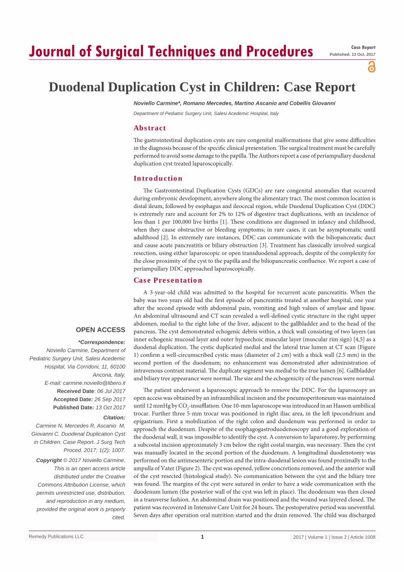
baby was two years old had the first episode of pancreatitis treated at another hospital, one year after the second episode with abdominal pain, vomiting and high values of amylase and lipase. An abdominal ultrasound and CT scan revealed a well-defined cystic structure in the right upper abdomen, medial to the right lobe of the liver, adjacent to the gallbladder and to the head of the pancreas. The cyst demonstrated echogenic debris within, a thick wall consisting of two layers (an inner echogenic mucosal layer and outer hypoechoic muscular layer (muscular rim sign) [4,5] as a duodenal duplication. The cystic duplicated medial and the lateral true lumen at CT scan (Figure 1) confirm a well-circumscribed cystic mass (diameter of 2 cm) with a thick wall (2.5 mm) in the second portion of the duodenum; no enhancement was demonstrated after administration of intravenous contrast material. The duplicate segment was medial to the true lumen [6]. Gallbladder and biliary tree appearance were normal. The size and the echogenicity of the pancreas were normal.
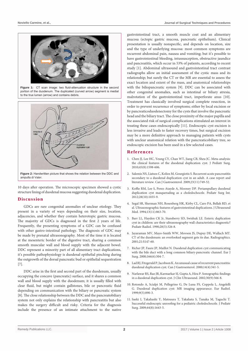
The patient underwent a laparoscopic approach to remove the DDC. For the laparoscopy an open access was obtained by an infraumbilical incision and the pneumoperitoneum was maintained until 12 mmHg by CO2-insufflation. One 10-mm laparoscope was introduced in an Hasson umbilical trocar. Further three 5-mm trocar was positioned in right iliac area, in the left ipocondrium and epigastrium. First a mobilization of the right colon and duodenum was performed in order to approach the duodenum. Despite of the esophagogastroduodenoscopy and a good exploration of the duodenal wall, it was impossible to identify the cyst. A conversion to laparotomy, by performing a subcostal incision approximately 3 cm below the right costal margin, was necessary. Then the cyst was manually located in the second portion of the duodenum. A longitudinal duodenotomy was performed on the antimesenteric portion and the intra-duodenal lesion was found proximally to the ampulla of Vater (Figure 2). The cyst was opened, yellow concretions removed, and the anterior wall of the cyst resected (histological study). No communication between the cyst and the biliary tree was found. The margins of the cyst were sutured in order to have a wide communication with the duodenum lumen (the posterior wall of the cyst was left in place). The duodenum was then closed in a transverse fashion. An abdominal drain was positioned and the wound was layered closed. The patient was recovered in Intensive Care Unit for 24 hours. The postoperative period was uneventful. Seven days after operation oral nutrition started and the drain removed. The child was discharged
Noviello Carmine*, Romano Mercedes, Martino Ascanio and Cobellis Giovanni
Department of Pediatric Surgery Unit, Salesi Acedemic Hospital, Italy
Noviello Carmine, et al., Journal of Surgical Techniques and Procedures
Remedy Publications LLC. 2017 | Volume 1 | Issue 2 | Article 10082
10 days after operation. The microscopic specimen showed a cystic structure lining of duodenal mucosa suggesting duodenal duplication.
DiscussionGDCs are rare congenital anomalies of unclear etiology. They
present in a variety of ways depending on their size, location, adjacencies, and whether they contain heterotopic gastric mucosa. The majority of GDCs is diagnosed in the first 2 years of life. Frequently, the presenting symptoms of a GDC can be confused with other gastro-intestinal pathology. The diagnosis of GDC may be made by prenatal ultrasonography. Most of the time it is located at the mesenteric border of the digestive tract, sharing a common smooth muscular wall and blood supply with the adjacent bowel. DDC represent a minority part of all alimentary tract duplications, it’s possible pathophysiology is duodenal epithelial pinching during the outgrowth of the dorsal pancreatic bud or epithelial sequestration [7].
DDC arise in the first and second part of the duodenum, usually occupying the concave (pancreatic) surface, and it shares a common wall and blood supply with the duodenum; it is usually filled with clear fluid, but might contain gallstones, bile or pancreatic fluid depending on communication with the biliary or pancreatic system [8]. The close relationship between the DDC and the pancreatobiliary system not only explains the relationship with pancreatitis but also makes the surgery difficult and risky. Criteria for the diagnosis include the presence of an intimate attachment to the native
gastrointestinal tract, a smooth muscle coat and an alimentary mucosa (ectopic gastric mucosa, pancreatic epithelium). Clinical presentation is usually nonspecific, and depends on location, size and the type of underlying mucosa: most common symptoms are recurrent abdominal pain, nausea and vomiting; but it’s possible to have gastrointestinal bleeding, intussusception, obstructive jaundice and pancreatitis, which occur in 53% of patients, according to recent study [1]. Abdominal ultrasound and gastrointestinal tract contrast radiographs allow an initial assessment of the cystic mass and its relationship; but surely the CT or the MR are essential to assess the exact location and extent of the mass, and anatomical relationships with the biliopancreatic system [9]. DDC can be associated with other congenital anomalies, such as intestinal or biliary atresia, malrotation of the gastrointestinal tract, imperforate anus [10]. Treatment has classically involved surgical complete resection, in order to prevent recurrence of symptoms; either by local excision or by pancreaticoduodenectomy for the cysts that involve the pancreatic head and the biliary tract. The close proximity of the major papilla and the associated risk of surgical complications stimulated an interest in treating these cases endoscopically [11]. Endoscopic cyst excision is less invasive and leads to faster recovery times, but surgical excision may be a more definitive approach to managing patients with cysts with unclear anatomical relation with the pancreaticobiliary tree, so endoscopic excision has been used in a few selected cases.
References1. Chen JJ, Lee HC, Yeung CY, Chan WT, Jiang CB, Sheu JC. Meta-analysis:
the clinical features of the duodenal duplication cyst. J Pediatr Surg. 2010;45(8):1598-606.
2. Salemis NS, Liatsos C, Kolios M, Gourgiotis S. Recurrent acute pancreatitis secondary to a duodenal duplication cyst in an adult. A case report and literature review. Can J Gastroenterol. 2009;23(11):749-52.
3. Koffie RM, Lee S, Perez-Atayde A, Mooney DP. Periampullary duodenal duplication cyst masquerading as a choledochocele. Pediatr Surg Int. 2012;28(10):1035-9.
4. Segal SR, Sherman NH, Rosenberg HK, Kirby CL, Caro PA, Bellah RD, et al. Ultrasonographic features of gastrointestinal duplications. J Ultrasound Med. 1994;13(11):863-70.
5. Barr LL, Hayden CK Jr, Stansberry SD, Swishuk LE. Enteric duplication cyst in children: are their ultrasonography wall characteristics diagnostic? Pediatr Radiol. 1990;20(5):326-8.
6. Jayaraman MV, Mayo-Smith WW, Movson JS, Dupuy DE, Wallach MT. CT of the duodenum: an overlooked segment gets its due. Radiographics. 2001;21:S147-60.
7. Richer JP, Faure JP, Maillot N. Duodenal duplication cyst communicating with the bile duct with a long common biliary-pancreatic channel. Eur J Surg. 2000;166(6):504-7.
8. Lad RJ, Fitzgerald P, Jacobson K. An unusual cause of recurrent pancreatitis: duodenal duplication cyst. Can J Gastroenterol. 2000;14(4):341-5.
9. Narlawar RS, Rao JR, Karmarkar SJ, Gupta A, Hira P. Sonographic findings in a duodenal duplication cyst. J Clin Ultrasound. 2002;30(9):566-8.
10. Rotondo A, Scialpi M, Pellegrino G, De Luna FS, Coppola L, Angelelli G. Duodenal duplication cyst: MR imaging appearance. Eur Radiol. 1999;9(5):890-3.
11. Saeki I, Takahashi Y, Matsuura T, Takahata S, Tanaka M, Taguchi T. Successful endoscopic unroofing for a pediatric choledochocele. J Pediatr Surg. 2009;44(8):1643-5.
Figure 1: CT scan image: two fluid-attenuation structure in the second portion of the duodenum. The duplicated (curved arrow) segment is medial to the true lumen (arrow) and contains debris.
Figure 2: Handwritten picture that shows the relation between the DDC and ampulla di Vater.